Review

Genetic Basis of the Epidemiological Features and Clinical Significance of Renal Hypouricemia

by
Masayuki Hakoda
1,* and
Kimiyoshi Ichida
2
1
Department of Nutritional Sciences, Faculty of Human Ecology, Yasuda Women’s University, 6-13-1 Yasuhigashi, Asaminami-ku, Hiroshima 731-0153, Japan
2
Department of Pathophysiology, Tokyo University of Pharmacy and Life Sciences, 1432-1 Horinouchi, Tokyo 192-0392, Japan
*
Author to whom correspondence should be addressed.
Biomedicines 2022, 10(7), 1696; https://doi.org/10.3390/biomedicines10071696
Submission received: 25 May 2022 / Revised: 8 July 2022 / Accepted: 11 July 2022 / Published: 13 July 2022
(This article belongs to the Special Issue Hypouricemia)

Abstract

:
A genetic defect in urate transporter 1 (URAT1) is the major cause of renal hypouricemia (RHUC). Although RHUC is detected using a serum uric acid (UA) concentration <2.0 mg/dL, the relationship between the genetic state of URAT1 and serum UA concentration is not clear. Homozygosity and compound heterozygosity with respect to mutant URAT1 alleles are associated with a serum UA concentration of <1.0 mg/dL and are present at a prevalence of ~0.1% in Japan. In heterozygous individuals, the prevalence of a serum UA of 1.1–2.0 mg/dL is much higher in women than in men. The frequency of mutant URAT1 alleles is as high as 3% in the general Japanese population. The expansion of a specific mutant URAT1 allele derived from a single mutant gene that occurred in ancient times is reflected in modern Japan at a high frequency. Similar findings were reported in Roma populations in Europe. These phenomena are thought to reflect the ancient migration history of each ethnic group (founder effects). Exercise-induced acute kidney injury (EI-AKI) is mostly observed in individuals with homozygous/compound heterozygous URAT1 mutation, and laboratory experiments suggested that a high UA load on the renal tubules is a plausible mechanism for EI-AKI.

1. Introduction

Uric acid (UA) is an end product of purine metabolic pathways and is poorly soluble in body fluids [1]. This characteristic causes the accumulation of UA in patients with hyperuricemia, causing gout, urinary calculi, and renal damage [1]. UA was also implicated in the development of cardiovascular diseases in some ways [2], the scavenging of reactive oxygen species [3], and as an endogenous danger signal, alerting the immune system to the presence of dying cells [4]. However, the clinical significance of hypouricemia is unclear, except with respect to exercise-induced acute kidney injury (EI-AKI) and renal calculi [5]. The former occurs in patients with renal hypouricemia (RHUC), which is caused by reduced reabsorption of UA by the proximal tubules, resulting in greater excretion of UA in the urine [5], while the latter occurs in the presence of hypouricemia owing to RHUC or xanthinuria. Xanthinuria is much less common, and for most patients with hypouricemia in the general population, RHUC is the etiology [5]. Since the identification of urate transporter 1 (URAT1) [6] and glucose transporter 9 (GLUT9) [7], our understanding of the epidemiology and pathophysiology of RHUC has substantially improved.
For example, the prevalence of RHUC is different between men and women when the definition of hypouricemia is set to be a serum UA (SUA) concentration of ≤2.0 mg/dL, and this difference can be explained by different levels of SUA concentration in individuals that are heterozygous for mutant URAT1 alleles between men and women. Further, a relatively high prevalence of RHUC in Japan is based on a high frequency of a specific mutant URAT1 allele among the general population. This prevalent mutant URAT1 allele was derived from a single mutant gene that occurred in ancient times and then expanded during the population history. A similar phenomenon was observed in Roma populations in Europe. The pathophysiology of EI-AKI was investigated using a new animal model with a defect of URAT1 function, where the importance of high UA load on renal tubules for the development of EI-AKI was demonstrated. The importance of a high UA load on renal tubules for the development of decreased kidney function was also demonstrated by clinical studies of potent inhibitors of URAT1. Individuals that are heterozygous for mutant URAT1 alleles are associated with potential risks of kidney damage through a high UA load on renal tubules and are present at high frequencies among Japanese, Korean, and Romani people.

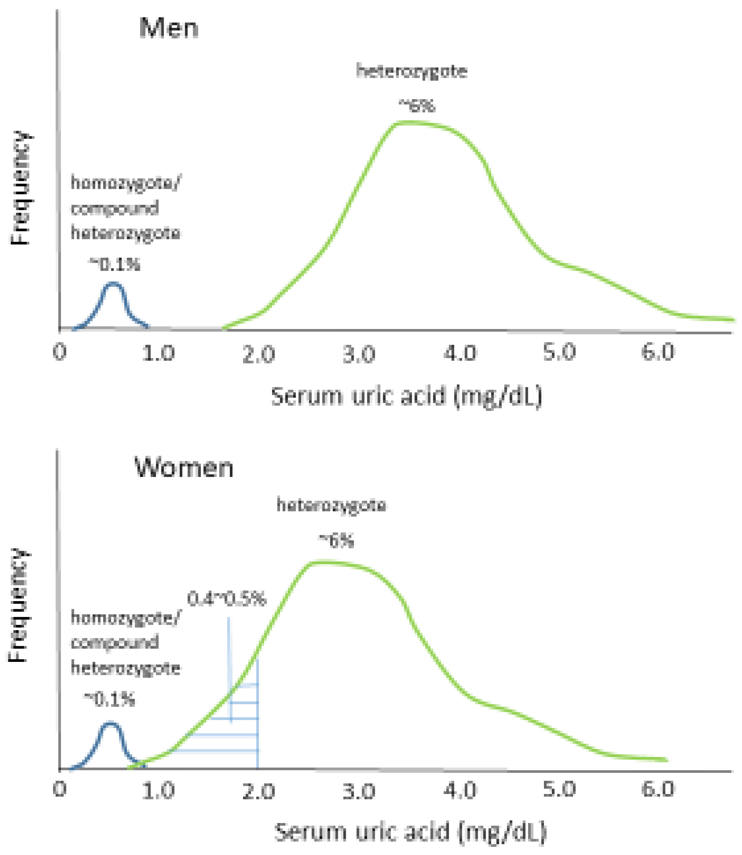
2. Prevalence of Hypouricemia

Hypouricemia is frequently defined using an SUA concentration of ≤2.0 mg/dL [5]. Using this reference value, the prevalence of hypouricemia in the general population was reported for Japan [8,9,10,11] and Korea [12]. These two countries have similar prevalences of hypouricemia at between 0.2 and 0.4%. The prevalence of hypouricemia is higher in women than in men in both countries, ranging from 0.4% to 0.6% in women and from 0.1% to 0.2% in men. The prevalence tends to be higher in younger women than in older women in both these countries [8,12]. However, the prevalence of hypouricemia in the general population of other countries has not been reported recently, probably because this abnormality is more common in Japan and Korea than elsewhere. Indeed, an assessment of 3200 blood donors (2097 men and 1103 women) in four studies conducted since 1962 in Germany identified only three individuals with SUA concentrations ≤2.0 mg/dL [13]. These three individuals were all women; no men with an SUA concentration <2.0 mg/dL were identified, even though a larger number of men were included.
In Japan, the prevalence of hypouricemia, with an SUA concentration ≤1.0 mg/dL, was shown to be similar for men and women at ~0.1% in a large study of >10,000 individuals of each sex recruited from the general population [10,11]. This is in contrast to the result of the studies described above, which showed that the prevalence of an SUA concentration ≤2.0 mg/dL in women is two or three times higher than that in men in Japan and Korea. In addition, in individuals with an SUA concentration ≤1.0 mg/dL, there are small but distinct groups of both men and women with SUA concentrations of 0.6–0.7 mg/dL [9,10]. This may suggest that distinct pathological mechanisms operate in individuals with hypouricemia with ≤1.0 mg/dL and 1.1–2.0 mg/dL.

3. Genetic Basis for the Epidemiological Features of Hypouricemia

UA is filtered through the glomeruli in the kidneys and approximately 90% of the filtered UA is reabsorbed in the renal proximal tubules. Thus, only 10% of the filtered UA is excreted in the urine. A deficiency in the renal reabsorption of UA causes a low SUA concentration (RHUC), which is characterized by a high UA-to-creatinine clearance ratio (the fractional excretion of uric acid (FEUA)). The urate transporter URAT1, located on the apical membrane of renal proximal tubular cells, and GLUT9, located on the basolateral membrane, are the principal mediators of the reabsorption of UA because homozygous or compound heterozygous deficiency in these transporters causes the SUA concentration to be low at ≤1.0 mg/dL [6,14,15], whereas that of healthy individuals is generally between 3.0 and 7.0 mg/dL.
The RHUC caused by URAT1 deficiency is called RHUC type 1 (OMIM: 220150), and more than 30 mutant URAT1 genes that cause a functional deficiency of URAT1 were reported (HGMD® 2022. 4). However, in Japan, two mutant URAT1 alleles—W258X (rs121907892) and R90H (rs121907896)—predominate [14,15,16]. A study of 1875 individuals randomly selected from among the residents of a Japanese city showed that the allele frequency of W258X was as high as 2.37% (89 alleles), that of R90H was 0.40% (15 alleles), and only one other mutant URAT1 allele (D313A) was identified [16]. Similar prevalences of the URAT1 W258X allele were also reported in other Japanese populations [17,18,19] (Table 1). Therefore, the total frequency of mutant URAT1 alleles seems to be high at nearly 3% in Japan.
The genotype frequencies measured previously did not deviate from a Hardy–Weinberg equilibrium [16]. Therefore, the prevalence of the two mutant URAT1 alleles (homozygote or compound heterozygote) can be estimated to be ~0.09% (square of the mutant allele frequency of ~3%) in Japan, which is similar to the reported prevalence of ~0.1% for individuals with an SUA concentration ≤1.0 mg/dL in the general population [10,11]. Indeed, Kawamura et al. reported that 11 of 17 men and 13 of 19 women from the general population whose SUA concentration was ≤1.0 mg/L were homozygous or compound heterozygous for the W258X and the R90H alleles [11] (Table 2), and only two with an SUA concentration ≤ 1.0 mg/dL did not have either the W258X or R90H allele (Table 2). Hamajima et al. studied a different sample of the general Japanese population and found that two out of three men and all of the three women whose SUA concentration was <1.0 mg/L were homozygous for the W258X allele [19]. Almost all of the individuals (48 out of 52) who did not have chronic renal failure and had two mutant URAT1 alleles (homozygotes or compound heterozygotes) had SUA concentrations of <1.0 mg/dL [15]. These results suggest that most people with a low SUA concentration of ≤1.0 mg/dL are homozygous or compound heterozygous for mutant URAT1 alleles in Japan. This implies that hypouricemia with an SUA concentration of ≤1.0 mg/dL is, in essence, a genetic disorder.
Kawamura et al. reported that the prevalence of an SUA concentration of 1.1–2.0 mg/dL is low in men (0.03%), approximately one-fifth of that of an SUA concentration of ≤1.0 mg/L [11] (Table 3). In contrast, the prevalence of an SUA concentration of 1.1–2.0 mg/dL in women (0.41%) is approximately three times higher than that of an SUA concentration of ≤1.0 mg/L (Table 3). Therefore, the prevalence of an SUA concentration of 1.1–2.0 mg/dL in women is more than 10 times higher than that of men. In this study, approximately two-thirds of women with an SUA concentration of 1.1–2.0 mg/dL were heterozygous for one of the two common mutant URAT1 alleles (W258X and R90H) [11] (Table 2). Therefore, the higher prevalence of hypouricemia (SUA concentration ≤ 2.0 mg/dL) in women than in men can be explained by a higher prevalence of heterozygosity for a mutant URAT1 allele, causing an SUA concentration of 1.1–2.0 mg/dL, in women than in men (Figure 1). The SUA concentrations of men who are heterozygous for a URAT1 mutation were most frequently within the range of 3.0–4.9 mg/dL [19]. The lower SUA concentrations in women than in men who are heterozygous for a URAT1 mutation are most probably explained by the effect of female sex hormones to increase the renal excretion of uric acid [20]. Consistent with this, a higher prevalence of hypouricemia (SUA concentration ≤ 2.0 mg/dL) was found in younger (<50 years old) than in older (≥50 years old) women in both Japan and Korea [8,12]. Women of <50 years of age are typically premenopausal, with higher concentrations of female sex hormones.
Another cause of RUHC is a genetic defect in GLUT9, which encodes a protein that transports reabsorbed UA from the proximal tubular cells into circulation (RHUC type 2, OMIM:612076) [21]. GLUT9 mutations seem to cause a more severe defect in UA reabsorption in the kidney than URAT1 mutations because the FEUA is sometimes >100% in patients with the former [21]. Matsuo et al. screened the genomic sequence of the GLUT9 gene in 70 Japanese individuals with an SUA concentration ≤3.0 mg/dL and found two mutant GLUT9 alleles (the URAT1 W258X allele was identified in 47 out of 70 individuals) [7]. These GLUT9 mutant alleles were not identified in 130 randomly selected healthy individuals (260 alleles). Anzai et al. identified a different mutant GLUT9 allele (P412R) in patients with RHUC who did not have a URAT1 mutation, but could not find the same mutation in 50 other randomly selected healthy Japanese people (100 alleles) [22]. Therefore, mutant GLUT9 alleles appear to be rarer than mutant URAT1 alleles in Japan given that the allele frequency of the latter was found to be close to 3% in the general population, as described above. One study of 17 patients with renal hypouricemia and exercise-induced acute kidney injury (EI-AKI), for whom both the URAT1 and GLUT9 genes were sequenced, showed that 13 were homozygous or compound heterozygous for mutant URAT1 alleles, two were heterozygous for mutant URAT1 alleles, and only one was compound heterozygous for mutant GLUT9 alleles (both of the two mutant GLUT9 alleles differed from those reported by Matsuo et al. [7] or Anzai et al. [22,23]).
Among Roma populations and in Korea, where the same URAT1 mutant alleles were repeatedly identified, lower frequencies of GLUT9 mutant alleles than of URAT1 mutant alleles were reported [24,25,26]. In China, screening of the URAT1 and GLUT9 genes for mutations was performed in 31 individuals with hypouricemia (SUA concentration ≤2.0 mg/dL) selected from the general population [27], and this showed that the number of people with GLUT9 mutations was lower than that of people with URAT1 mutations (two vs. four, no mutations in the URAT1 or GLUT9 genes were found in the other 25 individuals). In this study, neither the URAT1 W258X nor R90H mutant alleles, which are the predominant genetic causes of renal hypouricemia in Japan and Korea, were identified.

4. Genetic Basis for the Differing Prevalence of Renal Hypouricemia between Different Ethnic Groups

As described above, the same mutations of the URAT1 gene were identified in multiple unrelated patients with RHUC in Japan. An analysis of 13 genetic markers flanking the most prevalent URAT1 mutation (W258X) showed that all of the mutant alleles were derived from one mutant haplotype that developed approximately 6820 years ago [15]. Because the genotype frequencies do not deviate from Hardy–Weinberg equilibrium [16], it seems unlikely that a survival advantage was conferred by the mutant URAT1 allele. Interestingly, the same mutant URAT1 allele (W258X) was found in studies of individuals with hypouricemia in Korea and this was also the most prevalent (57%) mutant URAT1 allele in the Korean population [26]. Haplotype analysis of the W258X mutant allele was also performed in the Korean population [28], and this showed that the mutant W258X allele was associated with the same haplotype in Korea as in Japan. However, it cannot be concluded that the origin of the W258X allele is common to Japan and Korea because different sets of genetic markers were used in the studies in Japan and Korea.
Immigrants from the Korean peninsula or China helped the recovery of the population in Japan approximately 2300 years ago [29]. Therefore, founder effects are the most likely mechanisms for the spread of the URAT1 mutant alleles in Japan (Figure 2). The mutant URAT1 allele R90H is also found in multiple individuals and it is the second most prevalent mutant allele in both Japan and Korea [14,15,16,26]. The expansion of single mutant URAT1 alleles may be the basis for the high prevalence of hypouricemia in Japan and Korea, but a similar phenomenon has not been identified in other ethnic groups, except in Roma populations in Europe.
Stiburkova et al. reported that the allele frequencies of T467M (1400C > T) and L415-G417 del (1245_1253 del) were 5.56% and 1.92%, respectively, in 1016 Roma who had been randomly selected from five European regions [30]. Both of these mutations differed from those found in Japan and Korea. The authors speculated that the high frequency of URAT1 mutant alleles in these populations might be explained by the migration of a fairly small group of around 1000 individuals from India approximately 1000 years ago [31,32,33]. Thus, founder effects likely contributed to the high frequency of URAT1 mutant alleles among the Roma. Although the prevalence of hypouricemia has not been assessed in Roma populations, it may be higher than those in Japan or Korea because the frequencies of the most and the second most prevalent alleles (5.56% and 1.92%, respectively) are both higher than those in Japan (2.37% and 0.40%, respectively).

5. Clinical Manifestation of Renal Hypouricemia

EI-AKI and renal stone formation are the two major clinical manifestations of RHUC. Although both were found in ~10% of patients with RHUC who consulted or were referred to clinicians at university hospitals [14], the general prevalence of the symptoms in patients with RHUC has not been published.
Most patients with RHUC and EI-AKI are homozygous or compound heterozygous for mutant URAT1 or GLUT9 alleles and exhibit high FEUAs exceeding 30% (it is usually <10% in healthy individuals) [14,15]. Recently, Hosoyamada’s group developed a mouse model of EI-AKI associated with RHUC by knocking out both the URAT1 and UOX (urate oxidase) genes [34,35] and making a genetic modification that increased their hypoxanthine phosphoribosyl transferase activity. After exercising the mice to exhaustion through forced swimming, they showed increases in their serum creatinine concentrations (EI-AKI) and in their urinary excretion of UA, whereas the control mice did not. In addition, the administration of a xanthine oxidoreductase inhibitor (XOI) (allopurinol or topiroxostat) prevented the development of EI-AKI and the high urinary excretion of UA [34,35]. Therefore, the EI-AKI associated with RHUC appears to be the result of renal damage induced by the high concentration of UA in the renal tubules following the exercise.
Yeun et al. showed for the first time that it is possible to reproduce EI-AKI in a patient with RHUC who had experienced episodes of EI-AKI by getting the patient to perform physical exercise to exhaustion in a laboratory [36]. Although it is unknown whether the patient had mutations in their URAT1 or GLUT9 genes because the experiment was performed before these genes had been identified, the patient had typical features of RHUC, including a low SUA concentration (0.5 mg/dL) and a high FEUA (55.2–69.4%). After the exercise to exhaustion, the creatinine clearance of the patient decreased (EI-AKI) and their urinary excretion of UA increased. In contrast, neither the creatinine clearance nor the urinary excretion of UA of the four control participants changed following the exercise. Furthermore, the administration of allopurinol at a dose of 300 mg/day for 5 days before the exercise inhibited both the development of EI-AKI and the increase in the urinary excretion of UA in the patient following the exercise. On the basis of these results, the authors concluded that the EI-AKI was induced by an increase in the urinary excretion of UA following the exercise and that the administration of allopurinol could prevent EI-AKI [36] (Figure 3). A further case report showed the effectiveness of allopurinol administration for the prevention of EI-AKI (300 mg for 3 days before a 400 m run) [37].
XOIs, including allopurinol, inhibit the production of oxygen radicals derived from xanthine oxidase [38], and therefore, their effect on preventing EI-AKI does not eliminate another hypothesis that EI-AKI in RUHC patients is the result of vasoconstriction induced by the oxidative stress that develops during exercise to exhaustion, which would be largely prevented by a normal concentration of UA, which is a powerful antioxidant [39].
Although studies have not shown whether the prevalence of renal calculi is high in patients with RHUC, greater urinary excretion of UA logically would represent a risk factor for the formation of renal stones because the administration of 300 mg allopurinol was shown to prevent the recurrence of calculi in patients with hyperuricosuria, but not RHUC, in a randomized trial [40].

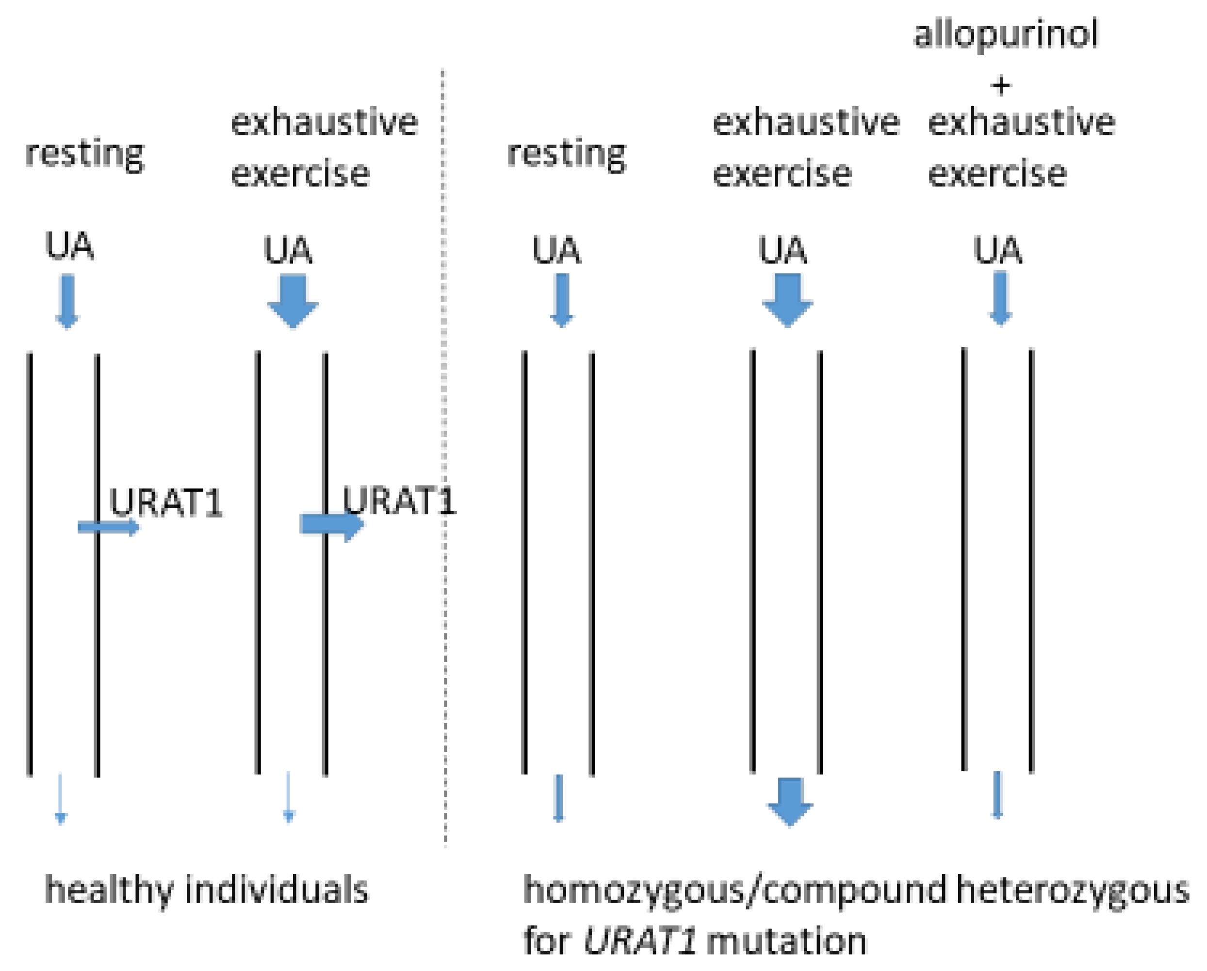
6. Relationships between URAT1 Mutations and Reduced Renal Function

Tabara et al. found that the URAT1 W258X allele, which is the most common mutant URAT1 allele in Japan, was independently associated with a low estimated glomerular filtration rate (eGFR) after multiple adjustments for clinical factors, including hypertension, SUA concentration, and body mass index (BMI), in two cohorts recruited from different Japanese cities [18]. Wakasugi et al. evaluated the relationship between a low SUA concentration and the prevalence of impaired kidney function (eGFR < 60 mL/min/1.73 m2) in a large general population recruited from multiple Japanese cities, and found that the prevalence was higher in men with an SUA concentration ≤ 2.0 mg/dL [8]. Furthermore, Kanda et al. performed a prospective study of the contribution of low SUA to a decline in eGFR in a Japanese cohort [41] and found that low SUA (men, <5.0 mg/dL; women, <3.6 mg/dL) was associated with the decrease in eGFR over time. In a different Japanese cohort study, Mori et al. found that low SUA (≤3.5 mg/dL) was a risk factor for the development of chronic kidney disease (CKD) in women, but not in men [42]. Conversely, Koto et al. found no relationship between renal dysfunction and low SUA concentration [10].
In patients with RHUC, EI-AKI usually resolves within a short period (~2 weeks) with only conservative renal support; however, the long-term effects on the kidney are not known [43]. Low-level pathology may remain and lead to chronic renal dysfunction, especially if recurrent episodes of AKI occur. EI-AKI is usually diagnosed in patients who exhibit overt symptoms, such as vomiting and lower back pain, and who attend a hospital [39,43], and it is unknown whether all patients with impaired kidney function following exercise to exhaustion experience such symptoms.
A potent URAT1 inhibitor, namely, lesinurad, increases the FEUA to >30%, which is a value similar to that of patients with homozygous or compound heterozygous loss-of-function mutations in URAT1 [44]. This drug causes renal toxicity and reduces creatinine clearance [45], and in some patients, the impaired renal function does not recover until 3 months after the use of the drug is discontinued [45]. The mechanism for the nephrotoxicity of the drug is considered to be the high UA load in the renal tubules because the toxicity is more common in patients with high baseline SUA concentrations who are not taking an XOI [45]. Another potent URAT1 inhibitor, namely, verinurad, also increases the FEUA to a level similar to that of patients with homozygous or compound heterozygous loss-of-function URAT1 mutations [46] and causes renal dysfunction [47]. These drug studies indicate that a tubular load of UA similar to those of patients with homozygous or compound heterozygous loss-of-function URAT1 mutations may be capable of inducing renal dysfunction. In contrast, another selective inhibitor of URAT1, namely, dotinurad, does not cause renal toxicity and is now being used clinically in Japan [48]. This drug does increase the FEUA, but only by <10% [49]. Benzbromarone, which has long been used as a uricosuric in Japan, also does not cause renal toxicity, and also increases the FEUA by <10% [50].

7. Clinical Significance of Heterozygous States for URAT1 Function

The frequency of mutant URAT1 alleles is close to 3% in Japan, and therefore, the prevalence of heterozygosity is approximately 6% in the population. In Korea, the frequency of mutant URAT1 alleles and the prevalence of heterozygosity may be similarly high. In Roma populations, the frequency of mutant URAT1 alleles and the prevalence of heterozygosity is even higher, probably twice those in Japan. Because heterozygous individuals tend to have a higher FEUA than wild-type individuals [11], they may be at higher risk of a high UA load in the renal tubules when the filtration of UA is increased. In fact, EI-AKI was identified in some heterozygous individuals, although almost all of the reported patients with EI-AKI were homozygous or compound heterozygous [23]. Okabayashi et al. reported an impressive case of renal transplantation in which the donor was heterozygous for a URAT1 mutant allele (a nonsense mutation in exon 5) [51] and the recipient was a son of the donor and also heterozygous for the same URAT1 mutant allele. There were no clinical complications following the transplantation and a biopsy of the transplanted kidney was performed after 3 months, according to the protocol. Histological analysis of the biopsy showed scattered UA crystals and calcium-based calculi in the distal tubules. The SUA concentrations in the recipient were 5.2 mg/dL and 1.9 mg/dL, before and 3 days after the transplantation, respectively, and his FEUA was 29% after the transplantation. The authors speculated that UA had accumulated in the body of the recipient before the transplantation because of renal insufficiency, but that this was filtered very swiftly and accumulated in the renal tubules after the transplantation [51]. One year later, the UA crystals and calcium-based calculi had disappeared without the use of an XOI. Thus, the kidney tubules of heterozygous individuals are at risk of a high UA load when the filtration of UA is increased.
The identification of heterozygous individuals by means of standard blood and urine tests is rather difficult because the SUA concentration and FEUA of heterozygous individuals are highly variable and overlap with the normal ranges [11,19]. In addition to lifestyle factors, the high prevalence of ABCG2 mutations, which cause renal UA overload, can affect both the SUA concentration and FEUA of individuals who are heterozygous for URAT1 [52,53]. Thus, clinicians should bear in mind that heterozygosity for URAT1 is relatively prevalent in Japan at >5%. Although the prevalence of heterozygosity may be much lower in countries other than Japan and Korea, and in the Roma, it is much higher than in those with homozygosity or compound heterozygosity.

8. Conclusions

Patients with RHUC who are homozygous or compound heterozygous for loss-of-function mutations of the URAT1 gene have SUA concentrations ≤ 1.0 mg/dL and are present at a prevalence of ~0.1% in both men and women. However, many more women than men who are heterozygous for loss-of-function URAT1 mutations have SUA concentrations of 1.1 to 2.0 mg/dL, and this is the principal contributor to the difference in the prevalence of SUA concentrations ≤ 2.0 mg/dL between the sexes in the general population. The frequency of URAT1 mutant alleles is nearly 3% in Japan and the spread of a mutant allele from a single individual in ancient times is sufficient to explain this. A similar phenomenon was speculated in Roma populations in which the frequency of a loss-of-function mutant allele is also high.
Impaired reabsorption of filtered UA, resulting in a high UA load on renal tubules following exercise to exhaustion, may be the cause of EI-AKI in patients with RHUC, and this can be prevented by the administration of an XOI. Thus, one of the physiological roles of URAT1 may be the protection of renal tubular cells against the damage induced by a high UA load after exercise to exhaustion, which was presumably more frequent in ancient times, when food was obtained mainly through hunting and gathering. In addition to EI-AKI, chronic renal damage may be caused by mutations in URAT1. Because heterozygosity for URAT1 mutants is much more common than homozygous or compound heterozygous loss-of-function states in the population and is associated with potential risks of kidney damage and renal calculi, clinicians should bear in mind that such individuals are relatively common, especially in Japanese, Korean, and Romani people.

Author Contributions

Writing—Original Draft Preparation, M.H.; Writing—Review and Editing, K.I. All authors have read and agreed to the published version of the manuscript.

Funding

This research received no external funding.

Institutional Review Board Statement

Not applicable.

Informed Consent Statement

Not applicable.

Data Availability Statement

Not applicable.

Acknowledgments

We thank Mark Cleasby from Edanz (https://jp.edanz.com/ac, accessed on 19 May 2022) for editing a draft of this manuscript.

Conflicts of Interest

The authors declare no conflict of interest.

References

  1. Dalbeth, N.; Merriman, T.R.; Stamp, L.K. Gout. Lancet 2016, 388, 2039–2052. [Google Scholar] [CrossRef]
  2. Kim, S.Y.; Guevara, J.P.; Kim, K.M.; Choi, H.K.; Heitjan, D.F.; Albert, D.A. Hyperuricemia and coronary heart disease: A systematic review and meta-analysis. Arthritis Care Res. 2010, 62, 170–180. [Google Scholar] [CrossRef] [PubMed] [Green Version]
  3. Waring, W.S.; Webb, D.J.; Maxwell, S.R. Systemic uric acid administration increases serum antioxidant capacity in healthy volunteers. J. Cardiovasc. Pharmacol. 2001, 38, 365–371. [Google Scholar] [CrossRef] [PubMed]
  4. Shi, Y.; Evans, J.E.; Rock, K.L. Molecular identification of a danger signal that alerts the immune system to dying cells. Nature 2003, 425, 516–521. [Google Scholar] [CrossRef] [PubMed]
  5. Nakayama, A.; Matsuo, H.; Ohtahara, A.; Ogino, K.; Hakoda, M.; Hamada, T.; Hosoyamada, M.; Yamaguchi, S.; Hisatome, I.; Ichida, K.; et al. Clinical practice guideline for renal hypouricemia (1st edition). Hum. Cell 2019, 32, 83–87. [Google Scholar] [CrossRef] [Green Version]
  6. Enomoto, A.; Kimura, H.; Chairoungdua, A.; Shigeta, Y.; Jutabha, P.; Cha, S.H.; Hosoyamada, M.; Takeda, M.; Sekine, T.; Igarashi, T.; et al. Molecular identification of a renal urate anion exchanger that regulates blood urate levels. Nature 2002, 417, 447–452. [Google Scholar] [CrossRef]
  7. Matsuo, H.; Chiba, T.; Nagamori, S.; Nakayama, A.; Domoto, H.; Phetdee, K.; Wiriyasermkul, P.; KikuchI, Y.; Oda, T.; Nishiyama, J.; et al. Mutations in glucose transporter 9 gene SLC2A9 cause renal hypouricemia. Am. J. Hum. Genet. 2008, 83, 44–51. [Google Scholar] [CrossRef] [Green Version]
  8. Wakasugi, M.; Kazama, J.J.; Narita, I.; Konta, T.; Fujimoto, S.; Iseki, K.; Moriyama, T.; Yamagata, K.; Tsuruya, K.; Asahi, K.; et al. Association between hypouricemia and reduced kidney function: A cross-sectional population-based study in Japan. Am. J. Nephrol. 2015, 41, 138–146. [Google Scholar] [CrossRef]
  9. Kawasoe, S.; Ide, K.; Usui, T.; Kubozono, T.; Yoshifuku, S.; Miyahara, H.; Maenohara, S.; Ohishi, M.; Kawakami, K. Distribution and Characteristics of Hypouricemia within the Japanese General Population: A Cross-Sectional Study. Medicina 2019, 55, 61. [Google Scholar] [CrossRef] [Green Version]
  10. Koto, R.; Sato, I.; Kuwabara, M.; Seki, T.; Kawakami, K. Temporal trends in the prevalence and characteristics of hypouricaemia: A descriptive study of medical check-up and administrative claims data. Clin. Rheumatol. 2022, 41, 2113–2119. [Google Scholar] [CrossRef]
  11. Kawamura, Y.; Nakayama, A.; Shimizu, S.; Toyoda, Y.; Nishida, Y.; Hishida, A.; Katsuura-Kamano, S.; Shibuya, K.; Tamura, T.; Kawaguchi, M.; et al. A Proposal for Practical Diagnosis of Renal Hypouricemia: Evidenced from Genetic Studies of Nonfunctional Variants of URAT1/SLC22A12 among 30,685 Japanese Individuals. Biomedicines 2021, 9, 1012. [Google Scholar] [CrossRef] [PubMed]
  12. Koo, B.S.; Jeong, H.J.; Son, C.N.; Kim, S.H.; Kim, H.J.; Kim, G.H.; Jun, J.B. Distribution of serum uric acid levels and prevalence of hyper- and hypouricemia in a Korean general population of 172,970. Korean J. Intern. Med. 2021, 36 (Suppl. S1), S264–S272. [Google Scholar] [CrossRef] [PubMed]
  13. Gresser, U.; Gathof, B.; Zöllner, N. Uric acid levels in southern Germany in 1989. A comparison with studies from 1962, 1971, and 1984. Klin. Wochenschr. 1990, 68, 1222–1228. [Google Scholar] [CrossRef]
  14. Ichida, K.; Hosoyamada, M.; Hisatome, I.; Enomoto, A.; Hikita, M.; Endou, H.; Hosoya, T. Clinical and molecular analysis of patients with renal hypouricemia in Japan-influence of URAT1 gene on urinary urate excretion. J. Am. Soc. Nephrol 2004, 15, 164–173. [Google Scholar] [CrossRef]
  15. Ichida, K.; Hosoyamada, M.; Kamatani, N.; Kamitsuji, S.; Hisatome, I.; Shibasaki, T.; Hosoya, T. Age and origin of the G774A mutation in SLC22A12 causing renal hypouricemia in Japanese. Clin. Genet. 2008, 74, 243–251. [Google Scholar] [CrossRef] [PubMed]
  16. Iwai, N.; Mino, Y.; Hosoyamada, M.; Tago, N.; Kokubo, Y.; Endou, H. A high prevalence of renal hypouricemia caused by inactive SLC22A12 in Japanese. Kidney Int. 2004, 66, 935–944. [Google Scholar] [CrossRef] [PubMed] [Green Version]
  17. Taniguchi, A.; Urano, W.; Yamanaka, M.; Yamanaka, H.; Hosoyamada, M.; Endou, H.; Kamatani, N. A common mutation in an organic anion transporter gene, SLC22A12, is a suppressing factor for the development of gout. Arthritis Rheum. 2005, 52, 2576–2577. [Google Scholar] [CrossRef] [PubMed]
  18. Tabara, Y.; Kohara, K.; Kawamoto, R.; Hiura, Y.; Nishimura, K.; Morisaki, T.; Kokubo, Y.; Okamura, T.; Tomoike, H.; Iwai, N.; et al. Association of four genetic loci with uric acid levels and reduced renal function: The J-SHIPP Suita study. Am. J. Nephrol. 2010, 32, 279–286. [Google Scholar] [CrossRef]
  19. Hamajima, N.; Naito, M.; Hishida, A.; Okada, R.; Asai, Y.; Wakai, K. Serum uric acid distribution according to SLC22A12 W258X genotype in a cross-sectional study of a general Japanese population. BMC Med. Genet. 2011, 12, 33. [Google Scholar] [CrossRef] [Green Version]
  20. Yahyaoui, R.; Esteva, I.; Haro-Mora, J.J.; Almaraz, M.C.; Morcillo, S.; Rojo-Martínez, G.; Martínez, J.; Gómez-Zumaquero, J.M.; González, I.; Hernando, V.; et al. Effect of long-term administration of cross-sex hormone therapy on serum and urinary uric acid in transsexual persons. J. Clin. Endocrinol. Metab. 2008, 93, 2230–2233. [Google Scholar] [CrossRef] [Green Version]
  21. Dinour, D.; Gray, N.K.; Ganon, L.; Knox, A.J.; Shalev, H.; Sela, B.A.; Campbell, S.; Sawyer, L.; Shu, X.; Valsamidou, E.; et al. Two novel homozygous SLC2A9 mutations cause renal hypouricemia type 2. Nephrol. Dial. Transplant. 2012, 27, 1035–1041. [Google Scholar] [CrossRef] [PubMed] [Green Version]
  22. Anzai, N.; Ichida, K.; Jutabha, P.; Kimura, T.; Babu, E.; Jin, C.J.; Srivastava, S.; Kitamura, K.; Hisatome, I.; Endou, H.; et al. Plasma urate level is directly regulated by a voltage-driven urate efflux transporter URATv1 (SLC2A9) in humans. J. Biol. Chem. 2008, 283, 26834–26838. [Google Scholar] [CrossRef] [PubMed] [Green Version]
  23. Kaito, H.; Ishimori, S.; Nozu, K.; Shima, Y.; Nakanishi, K.; Yoshikawa, N.; Iijima, K. Molecular background of urate transporter genes in patients with exercise-induced acute kidney injury. Am. J. Nephrol. 2013, 38, 316–320. [Google Scholar] [CrossRef]
  24. Stiburkova, B.; Bohatá, J.; Pavelcová, K.; Tasic, V.; Plaseska-Karanfilska, D.; Cho, S.K.; Potočnaková, L.; Šaligová, J. Renal Hypouricemia 1: Rare Disorder as Common Disease in Eastern Slovakia Roma Population. Biomedicines 2021, 9, 1607. [Google Scholar] [CrossRef]
  25. Claverie-Martin, F.; Trujillo-Suarez, J.; Gonzalez-Acosta, H.; Aparicio, C.; Roldan, M.L.J.; Stiburkova, B.; Ichida, K.; Martín-Gomez, M.A.; Herrero Goñi, M.; Hidalgo-Barquero, M.C.; et al. URAT1 and GLUT9 mutations in Spanish patients with renal hypouricemia. Clin. Chim. Acta 2018, 481, 83–89. [Google Scholar] [CrossRef]
  26. Cha, D.H.; Gee, H.Y.; Cachau, R.; Choi, J.M.; Park, D.; Jee, S.H.; Ryu, S.; Kim, K.K.; Won, H.H.; Limou, S.; et al. Contribution of SLC22A12 on hypouricemia and its clinical significance for screening purposes. Sci. Rep. 2019, 9, 14360. [Google Scholar] [CrossRef] [PubMed] [Green Version]
  27. Zhou, Z.; Wang, K.; Zhou, J.; Wang, C.; Li, X.; Cui, L.; Han, L.; Liu, Z.; Ren, W.; Wang, X.; et al. Amplicon targeted resequencing for SLC2A9 and SLC22A12 identified novel mutations in hypouricemia subjects. Mol. Genet. Genom. Med. 2019, 7, e00722. [Google Scholar] [CrossRef] [PubMed] [Green Version]
  28. Im, S.W.; Chae, J.; Son, H.Y.; Cho, B.; Kim, J.I.; Park, J.H. A population-specific low-frequency variant of SLC22A12 (p.W258*) explains nearby genome-wide association signals for serum uric acid concentrations among Koreans. PLoS ONE 2020, 15, e0231336. [Google Scholar]
  29. Watanabe, Y.; Naka, I.; Khor, S.S.; Sawai, H.; Hitomi, Y.; Tokunaga, K.; Ohashi, J. Analysis of whole Y-chromosome sequences reveals the Japanese population history in the Jomon period. Sci. Rep. 2019, 9, 8556. [Google Scholar] [CrossRef] [PubMed] [Green Version]
  30. Stiburkova, B.; Gabrikova, D.; Čepek, P.; Šimek, P.; Kristian, P.; Cordoba-Lanus, E.; Claverie-Martin, F. Prevalence of URAT1 allelic variants in the Roma population. Nucleosides Nucleotides Nucleic Acids 2016, 35, 529–535. [Google Scholar] [CrossRef]
  31. Gresham, D.; Morar, B.; Underhill, P.A.; Passarino, G.; Lin, A.A.; Wise, C.; Angelicheva, D.; Calafell, F.; Oefner, P.J.; Shen, P.; et al. Origins and divergence of the Roma (gypsies). Am. J. Hum. Genet. 2001, 69, 1314–1331. [Google Scholar] [CrossRef] [Green Version]
  32. Morar, B.; Gresham, D.; Angelicheva, D.; Tournev, I.; Gooding, R.; Guergueltcheva, V.; Schmidt, C.; Abicht, A.; Lochmuller, H.; Tordai, A.; et al. Mutation history of the roma/gypsies. Am. J. Hum. Genet. 2004, 75, 596–609. [Google Scholar] [CrossRef] [PubMed] [Green Version]
  33. Pamjav, H.; Zalán, A.; Béres, J.; Nagy, M.; Chang, Y.M. Genetic structure of the paternal lineage of the Roma people. Am. J. Phys. Anthropol. 2011, 145, 21–29. [Google Scholar] [CrossRef] [PubMed]
  34. Hosoya, T.; Uchida, S.; Shibata, S.; Tomioka, N.H.; Matsumoto, K.; Hosoyamada, M. Xanthine Oxidoreductase Inhibitors Suppress the Onset of Exercise-Induced AKI in High HPRT Activity Urat1-Uox Double Knockout Mice. J. Am. Soc. Nephrol. 2022, 33, 326–341. [Google Scholar] [CrossRef] [PubMed]
  35. Hosoyamada, M. Hypothetical Mechanism of Exercise-Induced Acute Kidney Injury Associated with Renal Hypouricemia. Biomedicines 2021, 9, 1847. [Google Scholar] [CrossRef]
  36. Yeun, J.Y.; Hasbargen, J.A. Renal hypouricemia: Prevention of exercise-induced acute renal failure and a review of the literature. Am. J. Kidney Dis. 1995, 25, 937–946. [Google Scholar] [CrossRef]
  37. Bhasin, B.; Stiburkova, B.; De Castro-Pretelt, M.; Beck, N.; Bodurtha, J.N.; Atta, M.G. Hereditary renal hypouricemia: A new role for allopurinol? Am. J. Med. 2014, 127, e3–e4. [Google Scholar] [CrossRef]
  38. George, J.; Carr, E.; Davies, J.; Belch, J.J.; Struthers, A. High-dose allopurinol improves endothelial function by profoundly reducing vascular oxidative stress and not by lowering uric acid. Circulation 2006, 114, 2508–2516. [Google Scholar] [CrossRef] [Green Version]
  39. Ishikawa, I. Acute renal failure with severe loin pain and patchy renal ischemia after anaerobic exercise in patients with or without renal hypouricemia. Nephron 2002, 91, 559–570. [Google Scholar] [CrossRef]
  40. Ettinger, B.; Tang, A.; Citron, J.T.; Livermore, B.; Williams, T. Randomized trial of allopurinol in the prevention of calcium oxalate calculi. N. Engl. J. Med. 1986, 315, 1386–1389. [Google Scholar] [CrossRef]
  41. Kanda, E.; Muneyuki, T.; Kanno, Y.; Suwa, K.; Nakajima, K. Uric acid level has a U-shaped association with loss of kidney function in healthy people: A prospective cohort study. PLoS ONE 2015, 10, e0118031. [Google Scholar] [CrossRef] [PubMed]
  42. Mori, K.; Furuhashi, M.; Tanaka, M.; Numata, K.; Hisasue, T.; Hanawa, N.; Koyama, M.; Osanami, A.; Higashiura, Y.; Inyaku, M.; et al. U-shaped relationship between serum uric acid level and decline in renal function during a 10-year period in female subjects: BOREAS-CKD2. Hypertens. Res. 2021, 44, 107–116. [Google Scholar] [CrossRef] [PubMed]
  43. Ohta, T.; Sakano, T.; Igarashi, T.; Itami, N.; Ogawa, T. ARF Assocoated with Renal Hypouricemia Research Group. Exercise-induced acute renal failure associated with renal hypouricaemia: Results of a questionnaire-based survey in Japan. Nephrol. Dial. Transplant. 2004, 19, 1447–1453. [Google Scholar] [CrossRef] [PubMed]
  44. Miner, J.N.; Tan, P.K.; Hyndman, D.; Liu, S.; Iverson, C.; Nanavati, P.; Hagerty, D.T.; Manhard, K.; Shen, Z.; Girardet, J.-L.; et al. Lesinurad, a novel, oral compound for gout, acts to decrease serum uric acid through inhibition of urate transporters in the kidney. Arthritis Res. Ther. 2016, 18, 214. [Google Scholar] [CrossRef] [Green Version]
  45. Sanchez-Niño, M.D.; Zheng-Lin, B.; Valiño-Rivas, L.; Sanz, A.B.; Ramos, A.M.; Luño, J.; Goicoechea, M.; Ortiz, A. Lesinurad: What the nephrologist should know. Clin. Kidney J. 2017, 10, 679–687. [Google Scholar] [CrossRef]
  46. Shen, Z.; Gillen, M.; Miner, J.N.; Bucci, G.; Wilson, D.M.; Hall, J.W. Pharmacokinetics, pharmacodynamics, and tolerability of verinurad, a selective uric acid reabsorption inhibitor, in healthy Japanese and non-Asian male subjects. Drug. Des. Dev. Ther. 2017, 11, 2077–2086. [Google Scholar] [CrossRef] [Green Version]
  47. Fitz-Patrick, D.; Roberson, K.; Niwa, K.; Fujimura, T.; Mori, K.; Hall, J.; Yan, X.; Shen, Z.; Liu, S.; Ito, Y.; et al. Safety and efficacy of verinurad, a selective URAT1 inhibitor, for the treatment of patients with gout and/or asymptomatic hyperuricemia in the United States and Japan: Findings from two phase II trials. Mod. Rheumatol. 2019, 29, 1042–1052. [Google Scholar] [CrossRef] [Green Version]
  48. Hosoya, T.; Fushimi, M.; Okui, D.; Sasaki, T.; Ohashi, T. Open-label study of long-term administration of dotinurad in Japanese hyperuricemic patients with or without gout. Clin. Exp. Nephrol. 2020, 24 (Suppl. S1), 80–91. [Google Scholar] [CrossRef] [Green Version]
  49. Taniguchi, T.; Ashizawa, N.; Matsumoto, K.; Saito, R.; Motoki, K.; Sakai, M.; Chikamatsu, N.; Hagihara, C.; Hashiba, M.; Iwanaga, T. Pharmacological Evaluation of Dotinurad.; a Selective Urate Reabsorption Inhibitor. J. Pharmacol. Exp. Ther. 2019, 371, 162–170. [Google Scholar] [CrossRef]
  50. Yamamoto, T.; Moriwaki, Y.; Takahashi, S.; Hada, T.; Higashino, K. Renal excretion of purine bases. Effects of probenecid, benzbromarone and pyrazinamide. Nephron 1988, 48, 116–120. [Google Scholar] [CrossRef]
  51. Okabayashi, Y.; Yamamoto, I.; Komatsuzaki, Y.; Niikura, T.; Yamakawa, T.; Katsumata, H.; Kawabe, M.; Katsuma, A.; Nakada, Y.; Kobayashi, A.; et al. Rare case of nephrocalcinosis in the distal tubules caused by hereditary renal hypouricaemia 3 months after kidney transplantation. Nephrology 2016, 21 (Suppl. S1), 67–71. [Google Scholar] [CrossRef] [PubMed] [Green Version]
  52. Ichida, K.; Matsuo, H.; Takada, T.; Nakayama, A.; Murakami, K.; Shimizu, T.; Yamanashi, Y.; Kasuga, H.; Nakashima, H.; Nakamura, T.; et al. Decreased extra-renal urate excretion is a common cause of hyperuricemia. Nat. Commun. 2012, 3, 764. [Google Scholar] [CrossRef] [PubMed] [Green Version]
  53. Matsuo, H.; Takada, T.; Ichida, K.; Nakamura, T.; Nakayama, A.; Ikebuchi, Y.; Ito, K.; Kusanagi, Y.; Chiba, T.; Tadokoro, S.; et al. Common Defects of ABCG2, a High-Capacity Urate Exporter, Cause Gout: A Function-Based Genetic Analysis in a Japanese Population. Sci. Transl. Med. 2009, 1, 5ra11. [Google Scholar] [CrossRef] [PubMed]
Figure 1. Distribution of serum uric acid concentrations in men and women with a homozygous/compound heterozygous URAT1 mutation and those with a heterozygous URAT1 mutation.
Biomedicines 10 01696 g001
Figure 2. Founder effects. A small number of individuals, including one with URAT1 mutation (red circle), immigrated to a new area and expanded. As the population expanded in the new area, the number of individuals with the URAT1 mutation increased.
Biomedicines 10 01696 g002
Figure 3. Increased UA load on renal tubules in individuals with homozygous/compound heterozygous URAT1 mutation after exhaustive exercise. A large amount of UA is produced by exhaustive exercise and then filtered through the glomeruli of the kidneys. In healthy individuals, an increased amount of filtered UA is reabsorbed by URAT1 and the amount of UA excreted into the urine does not increase [36]. On the other hand, in individuals with a homozygous/compound heterozygous URAT1 mutation, the filtered UA cannot be reabsorbed, resulting in increased excretion of UA in the urine [36]. In this case, it was suggested that the UA load on renal tubules increased after exhaustive exercise. Administration of allopurinol decreased the production of UA by exhaustive exercise and thus inhibited the increase of UA excretion in the urine [36]. EI-AKI did not develop after allopurinol administration [36]. A similar phenomenon will be observed in individuals with homozygous/compound heterozygous GLUT9 mutation.
Biomedicines 10 01696 g003
Table 1. Allele frequencies of URAT1 W258X in Japanese populations.
ReportsNumber of Alleles ExaminedNumber of W258X AllelesPercentage of W258X AllelesArea (Japan)
Iwai, N et al. [16]3750892.37Suita
Taniguchi, A et al. [17]1960452.3Tokyo
Tabara, Y et al. [18]103302632.55Suita + Ehime
Hamajima, N et al. [19]95862352.34Hamamatsu
Table 2. Frequency of mutant URAT1 alleles (W258X and R90H) in individuals with low SUA concentrations [11].
Men (n = 108)Women (n = 932)
SUA (mg/dL)Number of Deficient
URAT1 Alleles
Number of Deficient
URAT1 Alleles
012Total012Total
0.0–1.0241117061319
1.1–2.013042037057
2.1–3.029580875702860856
Table 3. Prevalence of low SUA concentration in individuals undergoing a general health examination in Japan [11].
Men (n = 13,607)Women (n = 17,078)
SUA (mg/dL)Number Frequency (%)NumberFrequency (%)
0.0–1.0200.15230.13
1.1–2.040.03700.41
2.1–3.01070.7910936.40
Publisher’s Note: MDPI stays neutral with regard to jurisdictional claims in published maps and institutional affiliations.

