Article

Novel 5-Nitrofuran-Tagged Imidazo-Fused Azines and Azoles Amenable by the Groebke–Blackburn–Bienaymé Multicomponent Reaction: Activity Profile against ESKAPE Pathogens and Mycobacteria

1
Institute of Chemistry, Saint Petersburg State University, Saint Petersburg 199034, Russia
2
Pasteur Institute of Epidemiology and Microbiology, 14 Mira Street, Saint Petersburg 197101, Russia
3
Laboratory of Chemoinformatics and Bioinformatics, Sechenov First Moscow State Medical University, Moscow 119435, Russia
4
Saint Petersburg Research Institute of Phthisiopulmonology, 2-4 Ligovsky Prospekt, Saint Petersburg 191036, Russia
5
Peptide Chemistry Research Center, K. N. Toosi University of Technology, Tehran 19697, Iran
6
Medical Biology Research Center, Kermanshah University of Medical Sciences Kermanshah, Kermanshah 67155, Iran
7
School for Living Systems, Immanuel Kant Baltic Federal University, Kaliningrad 236041, Russia
*
Authors to whom correspondence should be addressed.
Biomedicines 2022, 10(9), 2203; https://doi.org/10.3390/biomedicines10092203
Submission received: 5 August 2022 / Revised: 26 August 2022 / Accepted: 31 August 2022 / Published: 6 September 2022
(This article belongs to the Special Issue Antibiotic Resistance in Biomedicines)

Abstract

:
A chemically diverse set of 13 5-nitrofuran-tagged heterocyclic compounds has been prepared via the Groebke–Blackburn–Bienaymé multicomponent reaction. The testing of these compounds against the so-called ESKAPE panel of pathogens identified an apparent lead compound—N-cyclohexyl-2-(5-nitrofuran-2-yl)imidazo[1,2-a]pyridine-3-amine (4a)—which showed an excellent profile against Enterobacter cloacae, Staphylococcus aureus, Klebsiella pneumoniae, and Enterococcus faecalis (MIC 0.25, 0.06, 0.25 and 0.25 µg/mL, respectively). Its antibacterial profile and practically convenient synthesis warrant further pre-clinical development. Certain structure-activity relationships were established in the course of this study which were rationalized by the flexible docking experiments in silico. The assessment of antitubercular potential of the compounds synthesized against drug sensitive H37v strain of Mycobacterium tuberculosis revealed little potential of the imidazo-fused products of the Groebke–Blackburn–Bienaymé multicomponent reaction as chemotherapeutic agents against this pathogen.

Graphical Abstract

1. Introduction

Modern drug discovery entails the screening of chemically diverse sets of small-molecule compounds in order to discover suitable leads for subsequent medicinal chemistry optimization into drug candidates [1]. This approach requires that a large portion of druglike chemical space be sampled by the screening set in question and annotated against relevant target-based [2] or phenotypical [3] assays. The need for accessing as large a chemical diversity as possible within the confines of tangible, reasonably sized screening collections mandated that the library synthesis is planned and conducted in such a way that each library member is representative of a unique portion of the chemical space both at the scaffold and the periphery appendage level [4]. This goal is essentially the ideological core of the diversity-oriented synthesis [5] where highly efficient synthetic strategies based on atom-economical and multicomponent processes play the central role [6].
In 1998, a new isocyanide-based multicomponent reaction was described by three independent research groups (Blackburn [7], Bienaymé [8], and Groebke [9]) which has been dubbed the Groeble–Blackburn–Bienaymé (GBB) reaction [10]. This reaction involves the reaction of amidine-like 2-aminoazines and azoles 1 with aldehydes and isocyanides to render N-bridgehead heterobicyclic compounds 2 in a one-pot format. Undoubtedly, it does allow to control both the scaffold and the periphery group diversity within a single chemical event. Considering the undisputed biological relevance of the imidazo-fused scaffolds 2 and the practical simplicity of the reaction directly catering to the needs of the diversity-oriented synthesis, the Groeble–Blackburn–Bienaymé reaction quickly became one of the gems in the toolbox for modern medicinally-spirited compound library development [11]. The productivity of the reaction in delivering valuable leads for drug discovery is illustrated by numerous success stories in such therapeutic and target areas as kinase inhibition [12], M. tuberculosis glutamine synthetase inhibition [13], antimicrobial and antifungal drug discovery [14], anticancer agents [15], dual soluble epoxide hydrolase (sEH)/ 5-lipoxygenase (5-LOX) inhibition [16], and, particularly relevant to the field of the present study, antibacterial lead generation resulting in the discovery lead compound 3 which showed efficacy against drug susceptible and methicillin-resistant Staphylococcus aureus [17] (Figure 1).
Although numerous various nitro heterocyclic moieties have manifested themselves as a source of antibacterial leads [18], the conjugation of classical nitrofurans to various heterocyclic motifs not only can render these compounds generally non-cytotoxic [19] but also can direct their activity towards a specific type of antibacterial property, that does not affect the microbiome at large [20]. Moreover, nitrofurans proved to be versatile synthetic intermediates en route to other furan derivatives that were accessed via nucleophilic substitution of the nitro group [21]. A few years ago, we [22,23] and others [24] have already explored this strategy successfully by conjugating privileged [25] aminoalkylimidazoles, 1,2,4-oxadiazoles, and pyrimidines to 5-nitrofuranoyl moiety, which resulted in compounds that were found to be antibacterial. Considering the intrinsic association of the GBB reaction-derived imidazo-fused scaffold with antibacterial activity, as demonstrated by the biological profile of compound 3, combining the cluster of imidazo-fused scaffolds amenable by the GBB reactions with the known antibacterial pharmacophores such as 5-nitrofuryl moiety, surprisingly, has not been exploited to-date and clearly presents itself as a viable and well substantiated research opportunity. Herein, we report on the successful realization of this antibacterial drug design idea.

2. Materials and Methods

2.1. Organic Synthesis

2.1.1. General Considerations

All commercial reagents and solvents were used without further purification, unless otherwise noted. THF for the synthesis was distilled over Na and stored under nitrogen over freshly activated molecular sieves 4 Å. The NMR spectra were recorded on a Bruker Avance III 400 spectrometer (1H: 400.13 MHz; 13C: 100.61 MHz; chemical shifts are reported as parts per million (δ, ppm)) (Bruker, Billerica, MA, USA); the residual solvent peaks were used as internal standards: 7.28 and 2.50 ppm for 1H in CDCl3 and DMSO-d6, respectively, 40.01 and 77.02 ppm for 13C in DMSO-d6 and CDCl3, respectively. Mass spectra were recorded on a Bruker Maxis HRMS-ESI-qT spectrometer (ESI ionization) (Bruker, Billerica, MA, USA). The melting points were determined in open capillary tubes on a Stuart SMP30 Melting Point Apparatus (Staffordshire, UK). Analytical thin-layer chromatography was carried out on Silufol UV-254 silica gel plates (Chemapol, Czech Republic) using appropriate mixtures of ethyl acetate and hexane. The compounds were visualized with short-wavelength UV light. Column chromatography was performed on silica gel 60 (230–400 mesh).

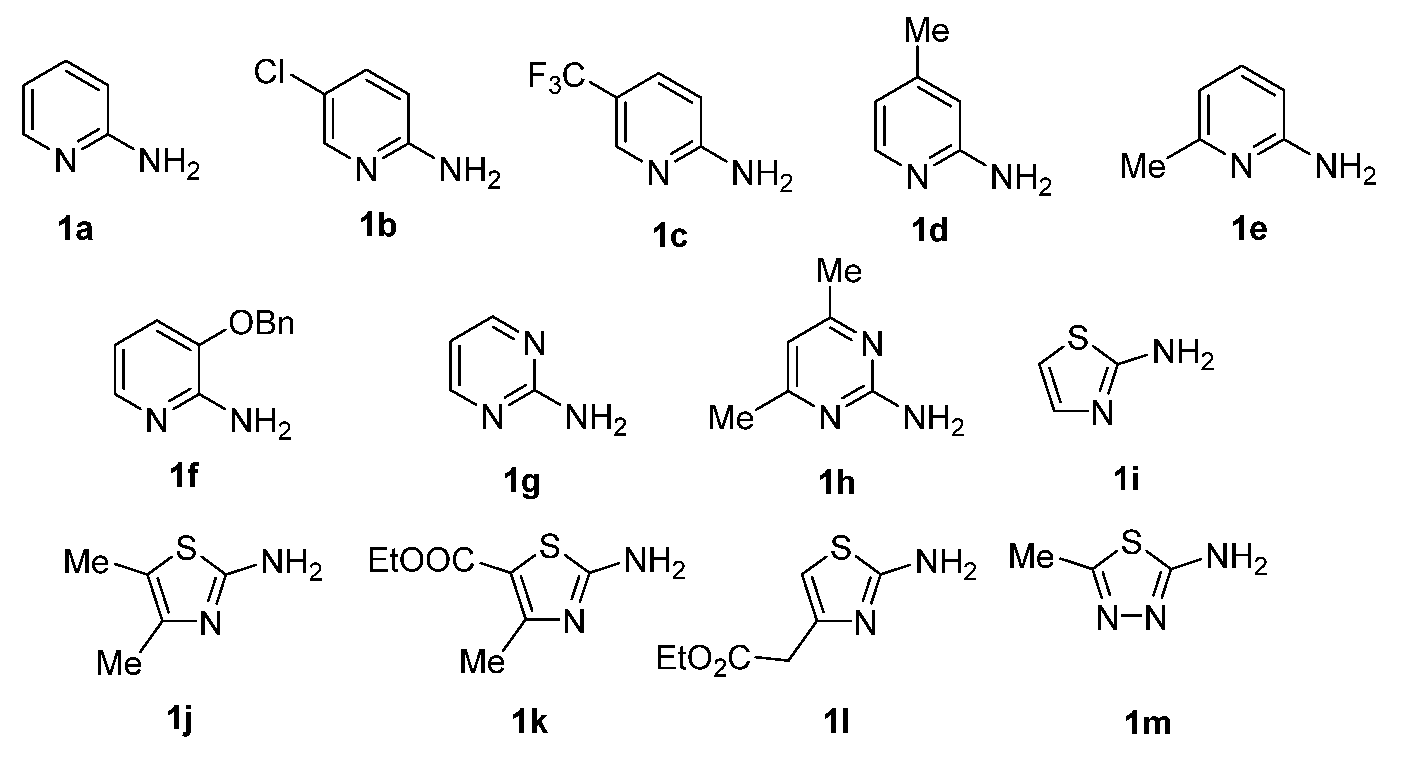
2.1.2. General Procedure for the Synthesis of GBB Reaction Products 4am

To the solution of the 2-aminoheteroarene 1a–m (0.50 mmol) and 5-nitrofuran-2-carbaldehyde (71 mg, 0.6 mmol) in methanol (1 mL) was added isocyanide (0.6 mmol) and p-toluenesulfonic acid (17 mg, 0.1 mmol). After stirring at room temperature for 24 h, the solvent was removed by rotary evaporation under vacuum. The residue was taken up in water (10 mL) and extracted with ethyl acetate (10 mL). The organic phase was washed with water (2 × 10 mL), dried over anhydrous Na2SO4, filtered, and concentrated by rotary evaporation under vacuum. Column chromatography on SiO2 using 0→5% EtOAc in CH2Cl2 as eluent afforded the title compound.

N-Cyclohexyl-2-(5-nitrofuran-2-yl)imidazo[1,2-a]pyridin-3-amine (4a)

Yield 73%; brown solid; mp 191–193 °C; 1H NMR (400 MHz, DMSO-d6): δ 8.32 (d, J = 6.9 Hz, 1H), 7.85 (d, J = 3.9 Hz, 1H), 7.50 (d, J = 9.1 Hz, 1H), 7.29–7.22 (m, 1H), 7.17 (d, J = 3.9 Hz, 1H), 6.95 (t, J = 6.7 Hz, 1H), 5.04 (d, J = 7.4 Hz, 1H), 3.08 (tdt, J = 11.0, 7.6, 3.8 Hz, 1H), 1.94–1.77 (m, 2H), 1.78–1.61 (m, 2H), 1.59–1.46 (m, 1H), 1.44–1.28 (m, 2H), 1.26–1.08 (m, 3H) ppm; 13C{1H} NMR (101 MHz, CDCl3): δ 153.8, 151.2, 141.7, 130.5, 125.8, 124.4, 124.1, 117.7, 116.3, 112.8, 110.2, 57.2, 34.2 (2C), 25.8, 25.0 (2C) ppm; HRMS (ESI), m/z calcd for C17H18N4O3 [M+H]+ 327.1452, found 327.1456.

6-Chloro-N-cyclohexyl-2-(5-nitrofuran-2-yl)imidazo[1,2-a]pyridin-3-amine (4b)

Yield 81%; dark orange solid; mp 247–250 °C; 1H NMR (400 MHz, DMSO-d6): δ 8.54 (d, J = 2.0 Hz, 1H), 7.86 (d, J = 4.0 Hz, 1H), 7.56 (d, J = 9.6 Hz, 1H), 7.29 (dd, J = 9.5, 2.0 Hz, 1H), 7.16 (d, J = 4.0 Hz, 1H), 5.16 (d, J = 7.8 Hz, 1H), 3.21–3.01 (m, 1H), 1.92–1.79 (m, 3H), 1.79–1.67 (m, 2H), 1.60–1.48 (m, 1H), 1.46–1.27 (m, 2H), 1.27–1.09 (m, 4H) ppm; 13C{1H} NMR (101 MHz, CDCl3): δ 153.1, 139.9, 131.1, 126.5, 124.8 (2C), 121.8, 120.1, 118.7, 116.2, 110.5, 57.2, 34.1 (2C), 25.7, 25.1 (2C) ppm; HRMS (ESI), m/z calcd for C17H17ClN4O3 [M+H]+ 361.1062, found 361.1065.

N-Cyclohexyl-2-(5-nitrofuran-2-yl)-6-(trifluoromethyl)imidazo[1,2-a]pyridin-3-amine (4c)

Yield 84%; orange solid; mp 240–243 °C; 1H NMR (400 MHz, DMSO-d6): δ 8.90 (d, J = 2.2 Hz, 1H), 7.86 (d, J = 3.9 Hz, 1H), 7.70 (d, J = 9.5 Hz, 1H), 7.45 (dd, J = 9.6, 1.8 Hz, 1H), 7.17 (d, J = 4.0 Hz, 1H), 5.42 (d, J = 8.0 Hz, 1H), 3.26–3.14 (m, 1H), 1.92–1.80 (m, 2H), 1.76–1.62 (m, 2H), 1.59–1.50 (m, 1H), 1.42–1.28 (m, 2H), 1.27–1.10 (m, 3H) ppm; 13C{1H} NMR (101 MHz, CDCl3): δ 152.8, 151.5, 140.9, 132.1, 129.7 (q, J = 262.5 Hz), 124.6 (q, J = 6.0 Hz), 123.7, 120.7 (q, J = 2.5 Hz), 118.9 (2C), 116.24 (d, J = 34.1 Hz), 110.9, 57.5, 34.1 (2C), 25.7, 25.1 (2C) ppm; HRMS (ESI), m/z calcd for C18H17F3N4O3 [M+H]+ 395.1326, found 395.1329.

N-Cyclohexyl-7-methyl-2-(5-nitrofuran-2-yl)imidazo[1,2-a]pyridin-3-amine (4d)

Yield 77%; dark brown solid; mp 203–206 °C; 1H NMR (400 MHz, CDCl3): δ 7.96 (d, J = 7.1 Hz, 1H), 7.48 (d, J = 3.9 Hz, 1H), 7.26 (d, J = 1.8 Hz, 1H), 7.03 (d, J = 3.8 Hz, 1H), 6.68 (dd, J = 7.1, 1.6 Hz, 1H), 3.86 (d, J = 7.7 Hz, 1H), 3.12–2.98 (m, 1H), 2.42 (s, 3H), 2.01–1.92 (m, 2H), 1.83–1.75 (m, 2H), 1.46–1.34 (m, 2H), 1.31–1.19 (m, 3H) ppm; 13C{1H} NMR (101 MHz, CDCl3): δ 154.1, 143.0, 136.4, 129.1, 124.3, 122.4, 115.9, 115.2, 114.4, 108.4, 77.2, 57.5, 34.1 (2C), 25.5, 25.0 (2C), 21.4 ppm; HRMS (ESI), m/z calcd for C18H20N4O3 [M+H]+ 341.1608, found 341.1605.

N-(tert-Butyl)-5-methyl-2-(5-nitrofuran-2-yl)imidazo[1,2-a]pyridin-3-amine (4e)

Yield 80%; reddish solid; mp 164–167 °C; 1H NMR (400 MHz, DMSO-d6): δ 7.85 (d, J = 4.0 Hz, 1H), 7.42–7.32 (m, 2H), 7.19 (dd, J = 9.0, 6.7 Hz, 1H), 6.67 (dt, J = 6.8, 1.2 Hz, 1H), 4.50 (s, 1H), 2.93 (s, 3H), 0.96 (s, 9H) ppm; 13C{1H} NMR (101 MHz, DMSO-d6): δ 153.8, 151.2, 143.8, 137.9, 130.3, 129.7, 126.3, 116.0, 115.7, 115.0, 112.2, 56.4, 29.5, 20.3 (3C) ppm; HRMS (ESI), m/z calcd for C16H18N4O3 [M+H]+ 315.1452, found 315.1457.

8-(Benzyloxy)-N-(tert-butyl)-2-(5-nitrofuran-2-yl)imidazo[1,2-a]pyridin-3-amine (4f)

Yield 69%; reddish solid; mp 157–160 °C; 1H NMR (400 MHz, CDCl3): δ 7.91 (dd, J = 6.9, 0.9 Hz, 1H), 7.53–7.47 (m, 2H), 7.44 (d, J = 3.9 Hz, 1H), 7.41–7.29 (m, 4H), 7.24 (d, J = 4.0 Hz, 1H), 6.64 (t, J = 7.2 Hz, 1H), 6.47 (d, J = 7.5 Hz, 1H), 5.36 (s, 2H), 3.79–3.60 (m, 1H), 1.21 (s, 9H) ppm; 13C{1H} NMR (101 MHz, CDCl3): δ 146.1, 142.8, 132.7, 131.2, 123.9 (2C), 123.6, 123.4, 122.5 (2C), 112.5 (2C), 109.4, 107.3, 105.4, 98.9, 66.2, 52.4, 25.2 (3C). ppm; HRMS (ESI), m/z calcd for C22H22N4O4 [M+H]+ 407.1714, found 407.1717.

N-Cyclohexyl-2-(5-nitrofuran-2-yl)imidazo[1,2-a]pyrimidin-3-amine (4g)

Yield 63%; dark brown solid; mp 230–233 °C; 1H NMR (400 MHz, CDCl3): δ 8.56 (dd, J = 4.0, 2.0 Hz, 1H), 8.39 (dd, J = 6.9, 2.1 Hz, 1H), 7.49 (d, J = 3.8 Hz, 1H), 7.19 (d, J = 3.9 Hz, 1H), 6.91 (dd, J = 6.9, 4.1 Hz, 1H), 3.92 (d, J = 7.5 Hz, 1H), 3.15–2.99 (m, 1H), 2.03–1.91 (m, 2H), 1.86–1.71 (m, 2H), 1.51–1.35 (m, 2H), 1.35–1.21 (m, 4H) ppm; 13C{1H} NMR (101 MHz, CDCl3): δ 153.1, 150.8, 145.0, 130.8, 127.4 (2C), 125.8, 114.1, 110.2, 108.7, 57.85 34.1 (2C), 25.4 (2C), 24.8 ppm; HRMS (ESI), m/z calcd for C16H17N5O3 [M+H]+ 328.1404, found 328.1406.

N-Cyclohexyl-5,7-dimethyl-2-(5-nitrofuran-2-yl)imidazo[1,2-a]pyrimidin-3-amine (4h)

Yield 72%; dark brown solid; mp > 250 °C; 1H NMR (400 MHz, CDCl3): δ 7.46 (d, J = 3.9 Hz, 1H), 7.14 (d, J = 3.9 Hz, 1H), 6.45 (s, 1H), 3.62 (d, J = 7.8 Hz, 1H), 2.95 (s, 3H), 2.93–2.82 (m, 1H), 2.53 (s, 3H), 1.92 (d, J = 12.3 Hz, 2H), 1.75 (q, J = 4.3 Hz, 2H), 1.42–1.07 (m, 6H) ppm; 13C{1H} NMR (101 MHz, CDCl3): δ 161.2, 153.6, 151.5, 146.5, 145.7, 128.6, 127.3, 114.2, 111.3, 110.2, 61.0, 33.2 (2C), 25.6, 25.3 (2C), 24.55, 18.6 ppm; HRMS (ESI), m/z calcd for C18H21N5O3 [M+H]+ 356.1717, found 356.1719.

N-Cyclohexyl-6-(5-nitrofuran-2-yl)imidazo[2,1-b]thiazol-5-amine (4i)

Yield 51%; dark brown solid; mp 117–120 °C; 1H NMR (400 MHz, CDCl3): δ 7.47 (d, J = 3.9 Hz, 1H), 7.37 (d, J = 4.6 Hz, 1H), 6.86 (d, J = 4.6 Hz, 1H), 6.83 (d, J = 3.9 Hz, 1H), 4.26–4.17 (m, 1H), 3.21–3.09 (m, 1H), 2.05–1.95 (m, 2H), 1.88–1.75 (m, 2H), 1.43–1.23 (m, 6H) ppm; 13C{1H} NMR (101 MHz, CDCl3): δ 154.32, 146.2, 142.0, 136.4, 132.5, 116.98, 114.95, 113.58, 106.85, 57.2, 33.99 (2C), 25.50 (2C) ppm; HRMS (ESI), m/z calcd for C15H16N4O3S [M+H]+ 333.1016, found 333.1014.

N-(tert-Butyl)-2,3-dimethyl-6-(5-nitrofuran-2-yl)imidazo[2,1-b]thiazol-5-amine (4j)

Yield 53%; dark orange solid; mp 132–235 °C; 1H NMR (400 MHz, CDCl3): δ 7.44 (d, J = 3.9 Hz, 1H), 6.87 (d, J = 3.9 Hz, 1H), 3.38 (s, 1H), 2.60 (d, J = 1.2 Hz, 3H), 2.31 (q, J = 1.1 Hz, 3H), 1.15 (s, 9H) ppm; 13C{1H} NMR (101 MHz, CDCl3): δ 154.1, 146.6, 145.9, 130.7, 129.1, 125.1, 120.0, 114.5, 108.2, 56.3, 29.5 (3C), 12.7, 11.5 ppm; HRMS (ESI), m/z calcd for C15H18N4O3S [M+H]+ 335.1172, found 335.1176.

Ethyl 5-(cyclohexylamino)-3-methyl-6-(5-nitrofuran-2-yl)imidazo[2,1-b]thiazole-2-carboxylate (4k)

Yield 60%; dark orange solid; mp 188–191 °C; 1H NMR (400 MHz, CDCl3): δ 7.45 (d, J = 3.9 Hz, 1H), 6.90 (d, J = 3.8 Hz, 1H), 4.45–4.32 (m, 2H), 3.60 (d, J = 8.6 Hz, 1H), 3.05 (s, 3H), 2.98–2.85 (m, 1H), 2.05–1.94 (m, 2H), 1.88–1.73 (m, 2H), 1.49–1.15 (m, 8H) ppm; 13C{1H} NMR (101 MHz, CDCl3): δ 161.8, 153.0, 146.6, 138.5, 132.9, 128.2, 118.2, 115.3, 114.3, 108.4, 61.8, 60.5, 33.4 (2C), 25.4 (3C), 14.2, 12.4 ppm; HRMS (ESI), m/z calcd for C19H22N4O5S [M+H]+ 419.1384, found 419.1387.

Ethyl 2-(5-(tert-butylamino)-6-(5-nitrofuran-2-yl)imidazo[2,1-b]thiazol-3-yl)acetate (4l)

Yield 58%; dark oil; 1H NMR (400 MHz, CDCl3): δ 7.44 (d, J = 3.9 Hz, 1H), 6.91 (d, J = 3.9 Hz, 1H), 6.70 (s, 1H), 4.19–4.10 (m, 3H), 3.32 (s, 1H), 1.31 (t, J = 7.0 Hz, 3H), 1.14 (s, 9H) ppm; 13C{1H} NMR (101 MHz, CDCl3): δ 154.1, 146.6, 130.7, 129.1, 125.1, 120.0, 116.4, 114.5, 112.8, 108.2, 56.37 (2C), 29.5 (3C), 12.7, 11.5 ppm; HRMS (ESI), m/z calcd for C17H20N4O5S [M+H]+ 393.1227, found 393.1230.

N-(tert-Butyl)-2-methyl-6-(5-nitrofuran-2-yl)imidazo[2,1-b][1,3,4]thiadiazol-5-amine (4m)

Yield 32%; brown solid; mp 136–138 °C; 1H NMR (400 MHz, CDCl3): δ 7.44 (d, J = 3.9 Hz, 1H), 6.85 (d, J = 3.9 Hz, 1H), 4.02 (s, 1H), 2.73 (s, 3H), 1.35 (s, 9H) ppm; 13C{1H} NMR (101 MHz, CDCl3): δ 160.03, 153.73, 150.43, 143.24, 131.10, 125.48, 114.51, 107.50, 56.56, 30.18 (3C), 18.11 ppm; HRMS (ESI), m/z calcd for C13H15N5O5S [M+H]+ 322.0968, found 322.0965.

2.2. ESKAPE Pathogen Susceptibility Testing

Testing was conducted against the following microorganisms: Enterococcus faecalis (ATCC 29812), Staphylococcus aureus (ATCC 25912), Klebsiella pneumoniae (ATCC 19882), Acinetobacter baumannii (948®, patient-derived strain from the Pasteur Institute own collection), Pseudomonas aeruginosa (ATCC 27853), and Enterobacter cloacae (ATCC 13047) for compounds 4a–m, nitrofurans nitrofurantoin [26] and furazidine [27] as well as ciprofloxacin (employed as a positive control) using the Kirby–Bauer disk diffusion test [28] under the Standard Operating Procedure of The European Committee on Antimicrobial Susceptibility Testing (EUCAST) [29]. Paper disks bearing 5 mg of the nitrofuran compounds and ciprofloxacin were used. Solutions of compounds 4a–m made up in DMSO (1 mg/10 mL) were prepared and diluted to a total volume of 1 mL with deionized water. Aliquots of the resulting solutions (5 µL each) were added to a Petri dish containing Muller–Hilton agar that was inoculated with a bacterial suspension (McFarland OD ¼ 0.5). After the compound solution has dried off, the Petri dish was incubated at 37 °C for 18 h. The bacterial growth inhibition zone diameter around the disc with ciprofloxacin or the compounds’ dried solution circular spot indicated the general susceptibility to a drug being assessed. Thereupon, minimum inhibitory concentrations (MIC, µg/mL) were determined using serial broth dilutions [30]. All measurements were done in triplicate.

2.3. Testing against Mycobacterium tuberculosis

The testing was performed as described previously [31]. All the measurements were done in triplicate.

2.4. Molecular Modeling

2.4.1. Target Selection

As the potential targets for observed nitrofuran derivatives 4a–m the following two proteins were selected: Escherichia coli nitroreductase NfsB (PDP 1YKI) and oxygen-insensitive NADPH nitroreductase NfsA (PDP 7NB9). These proteins have been investigated as molecular targets for nitrofuran-based drugs [32]. The main reference interactants in this case are nitrofurazone [33] or nitrofurantoin [26], respectively.

2.4.2. Protein and Ligand Structure Preparation

All protein structures were downloaded from the RCSB protein data bank [34] and preprocessed with use of Schrodinger Protein prepwizard. Invalid bond orders, protonation, missing residues, or residue sidechains were corrected in this step [35]. The geometry of ligands was generated with the use of LigPrep module. All of the manipulations with molecules performed in OPLS4 forcefield [36]. For calculations Schrodinger Suite version 2021-4 was used.

2.4.3. Molecular Docking

Ligand binding poses were calculated with the use of the induced-fit docking method [37]. Unlike molecular docking, this method implies the flexibility of the protein structure. The docking grid box was calculated in accordance with the reference ligand positioning and the ligand size (grid positioning on complexed ligand centroid, maximal grid side size was 12 Å). The flexibility of the amino acids was taken into account within the 5 Å radius from the ligand atoms (not centroids). The best-fitting pose was selected manually by evaluating the reproducibility of interactions of the nitrofuran moiety in the reference ligand that was present in the protein active site (as in PDB file).

2.4.4. Molecular Mechanics with Generalized Born and Surface Area Solvation (MM-GBSA)

For each ligand’s binding pose, the Gibbs free energy (ΔG) was calculated in the presence of implicit solvent. The calculations were performed with the use of MM-GBSA (Molecular mechanics with generalized Born and surface area solvation) method [38]. The point of interest was the strain energy value distribution which indicated the most strained protein-ligand interactions. This parameter was seen as able to explain the absence of the correct interaction of the ligand with the protein and, consequently, the diminished biological activity.

3. Results

3.1. Compound Synthesis

In order to explore the maximum chemical diversity within a limited compound set—and yet to be able to make pairwise comparison of the structure-activity relationships, we chose to place the 5-nitrofuran moiety within the aldehyde component, use only two fairly generic isocyanides (cyclohexyl and tert-butyl), and put a particular emphasis on the diversity of substituted 2-aminoazine and -azole inputs 1, employing a new substrate out of 13 (1a–m) to create each of the GBB reaction products (Figure 2).
A unique substrate input (1a–m) was employed in the preparation of each of the GBB products (4a–m) by mixing equimolar amounts of 1, 5-nitrofurancarboxaldehyde and an isocyanide (c-HexNC or t-BuNC) in methanol in the presence of 20 mol% of p-toluenesulfonic acid catalyst [39]. The reactions were complete within 24 h at room temperature. Following a brief aqueous workup, the target imidazo-fused GBB reaction products 4a–m were isolated chromatographically in moderate to excellent yield (Scheme 1). The yields of the GBB reaction products that were derived from 2-aminoazoles were markedly lower compared to their 2-azine-derived counterparts.

3.2. Activity against ESKAPE Pathogens

Nitrofurans 4a–m were tested against Gram-positive (S. aureus and E. faecium) or Gram-negative (E. cloacae, P. aeruginosa, A. baumannii, K. pneumoniae) pathogens belonging to the so-called ESKAPE panel [40] of highly virulent and antibiotic resistant bacterial pathogens which are, throughout the world, the major cause of life-threatening nosocomial or hospital-acquired infections in immunocompromised and critically ill patients who are most at risk [41]. There were two clinically used nitrofuran antibiotics—nitrofurantoin [26] and furazidine [27] as well as broad-spectrum fluoroquinoline antibiotic ciprofloxacin [42] that were used as positive controls and comparators. The compounds were initially screened at a single concentration to determine the presence and the diameter of the bacterial growth inhibition zone around the drug-treated disk. Those compounds that displayed growth inhibition were tested in serial dilution mode to determine the minimum inhibitory concentration (MIC) (Table 1).

3.3. Activity against Mycobacterium tuberculosis

All 13 compounds 4a–m were tested against Mycobacterium tuberculosis H37Rv drug-sensitive strain relative to isoniazid that was employed as a positive control. As it follows from the minimum inhibitory concentration (MIC) data that are summarized in Table 2, most of the compounds were either inactive or only weakly active against this pathogen except for N-cyclohexyl-6-(5-nitrofuran-2-yl)imidazo[2,1-b]thiazol-5-amine (4i) which showed MIC value of 6.2 µg/mL.

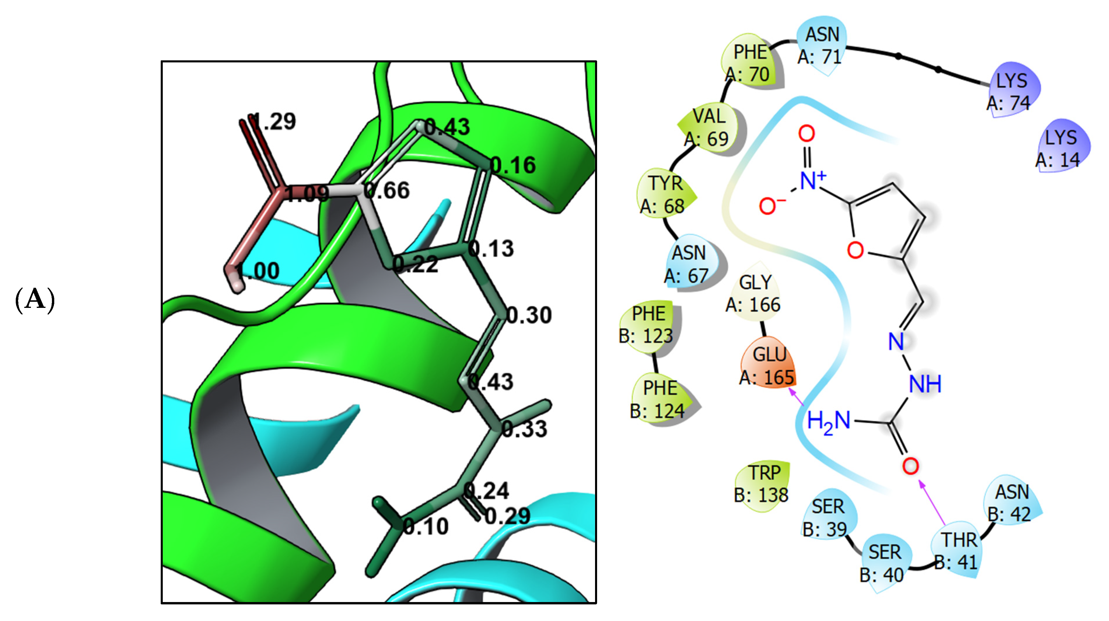
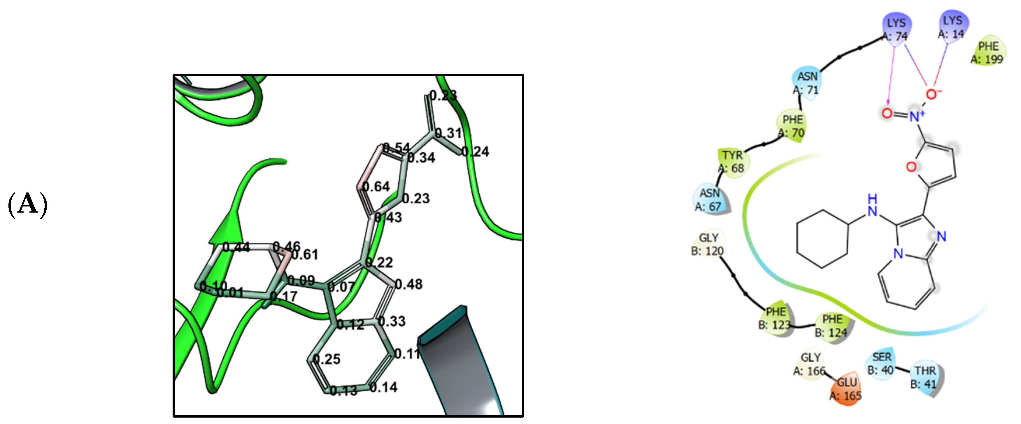
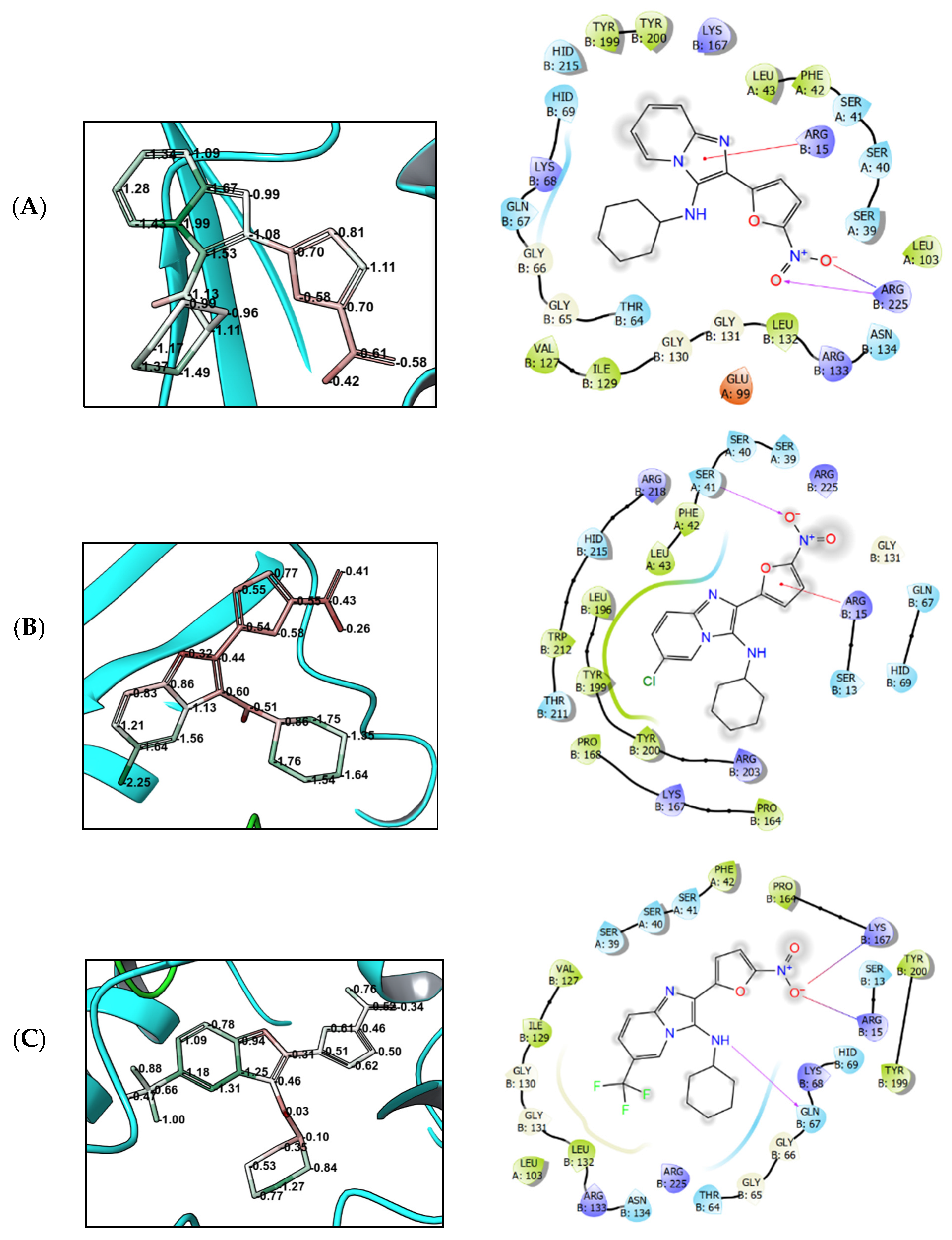
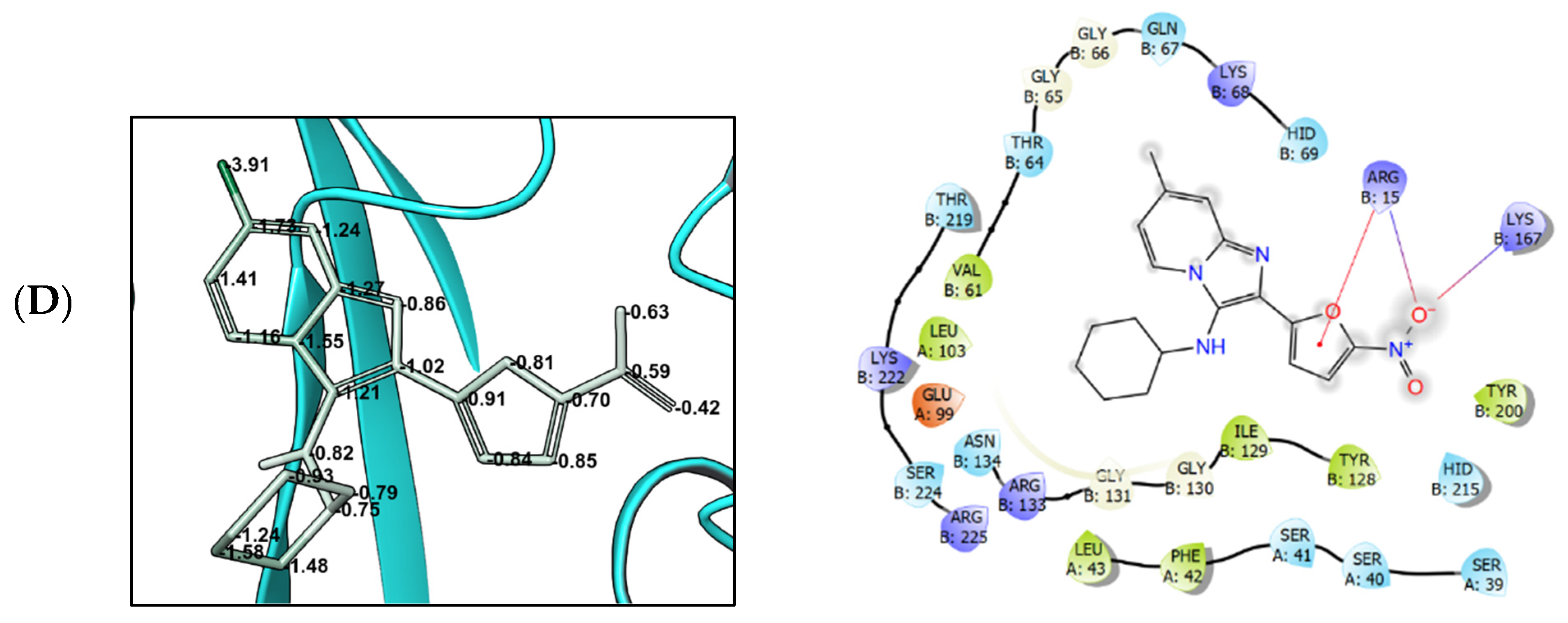
3.4. Molecular Modeling

The crystal structures of the two targets stipulated for nitrofurans—E. coli nitroreductase NfsB (PDB 1YKI) and oxygen-insensitive NADPH nitroreductase NfsA (PDB 7NB9) contain nitrofurazone and nitrofurantoin as bound ligands, respectively. In order to validate the docking protocol, these ligands were re-docked into the binding site. The docking poses that were thus obtained (allowing for determination of ligand strain components as well as key interactions with the target) reproduced the position of the ligand in the PDB structures with RMSD < 1.5 Å (Figure 3).
Having validated the docking protocol, we proceeded to perform docking of four selected GBB reaction-derived nitrofuran compounds (4a–d) two of which (4b and 4c), surprisingly, showed no activity against the ESKAPE panel of pathogens while the other two (4a and 4d) were quite active (except for no activity observed against A. baumannii and P. aeruginosa for all nitrofurans including the reference compounds). As all of the docked compounds showed good binding affinity according to the initially determined docking score values which did not allow differentiating between the active and inactive pair. Hence, we proceeded to switch to the induced-fit docking as described in the Materials and Methods section. With this approach, we observed significant differences between the active compounds (4a and 4d) as well the nitrofuran reference ligands and inactive compounds (4b and 4c) in their total calculated system energy as well as MM-GBSA Gibbs free energy (ΔG) values which were markedly higher for inactive protein-ligand complexes with both E. coli nitroreductase NfsB and oxygen-insensitive NADPH nitroreductase NfsA (Table 3).
The binding poses of ligands 4a–d in E. coli nitroreductase NfsB (PDB 1YKI) (Figure 4) and oxygen-insensitive NADPH nitroreductase NsfA (PDB 7NB9) (Figure 5) allowed analyzing the free energy distribution—in particular, the energies of strained ligand-protein contacts. This also allowed us to understand the components that contributed most to the differences between the active (4a and 4d) and inactive (4b and 4c) compounds.
In the case of E. coli nitroreductase NfsB, the addition of the chloro (4b) or the trifluoromethyl (4c) substituents induced the rearrangement of the ligand binding mode due to the increased hydrophobic interactions with Phe123 and Phe124. This also leads to a noticeable increase in the strain energy per atom of the imidazo[1,2-a]pyridine scaffold as well as the molecular periphery (Figure 4B,C increased strain indicated by the numbers as well as color change from mostly green in Figure 4A,D).
In the case of the oxygen-insensitive NADPH nitroreductase NsfA, the difference in the strain energy is less pronounced. However, compounds 4b and 4c still stand out in terms of the per-atom strain energy. The presence of the chloro of the trifluoromethyl substituents acts as a powerful hydrophobic anchor which stimulates the hydrophobic contacts with multiple hydrophobic residues in the enzyme’s binding pocket: aromatic amino acids such as Trp212, Phe42, Tyr199, and Tyr200 as well as non-polar Leu43 and Leu196. This exceedingly large number of lipophilic contacts induces the increase in the strain energy which eventually causes the ligand to migrate and bind in an unfavorable pose. This is likely what leads to the loss of the antibacterial activity due to the exclusion of the nitrofuran moiety from the enzyme’s catalytic center.

4. Discussion

The synthesis of compounds 4a–m (see Supplementary Materials) is based on the GBB multicomponent reaction and commercially available starting materials. This makes any leads that surface from the present study particularly attractive from the development perspective as bringing up larger quantities of these compounds for in vivo efficacy and other preclinical profiling will not present a problem. Although currently nitrofurans such as furazidine, nitrofurantoin, and nitrofurazone are mostly used as topical medicines, their unfavorable activity profile and conjugation of nitrofuran moiety to druglike cores in the absence of apparent toxophores has been shown to deliver candidate molecules that are devoid of systemic toxicity [43]. This can potentially render the lead nitrofurans that were identified through this study as candidates for the development as orally acting anti-infectives. In this regard, compounds 4a–m hold a great promise as they are entirely devoid of any violations of the rules for drug-likeness as defined by Lipinski [44] (Table 4).
As it can be seen from the antibacterial data that are summarized in Table 1, all of the compounds 4a–m displayed a distinctly ‘nitrofuran-like’ profile in the sense that, similar to nitrofurantoin and furazidine, none of these compounds showed any activity towards A. baumannii and P. aeruginosa. The other apparent—and somewhat surprising—result that is shown in Table 1 is the total absence of antibacterial activity of compounds 4b and 4c across the entire ESKAPE panel. Besides these gashing voids in the activity profile—and a few other instances of inactivity (cf. 4c–d, 4h–I, and 4k against E. cloacae; 4f against K. pneumoniae; 4d–e and 4l–m against E. faecalis)—compounds 4, in general, showed a superior activity profile relative to the two nitrofuran comparators and a comparable activity profile to that of broad-spectrum fluoroquinoline antibiotic ciprofloxacin. In terms of the overall activity profile against all four pathogens that wereaffected by nitrofurans—E. cloacae (G−), S. aureus (G+), K. pneumonia (G−), and E. faecalis (G+)—compound 4a presented itself as a convincing lead displaying low MIC values against these bacteria. On top of the distinctly simple, one-step synthesis of this compound from readily available commercial precursors, this compound is apparently attractive from the further development perspective considering the low-cost and convenience of bringing up larger quantities of this candidate compound for subsequent in vivo efficacy and other preclinical profiling.
At the same time, it became clear that substitution of the imidazo[1,2-a]pyridine core with lipophilic groups such as chloro (4b) or trifluoromethyl (4c) negatively impacts on the antibacterial activity of the GBB reaction-derived nitrofurans. As demonstrated by the flexible docking experiments, these compounds differ from their active counterparts (4a and 4d) in the increased per-atom ligand strain when bound to either E. coli nitroreductase NfsB or oxygen-insensitive NADPH nitroreductase NsfA. This interferes with the correct binding of the nitrofuran ligand in the enzymes’ active site and can even lead to a complete change of the binding mode which is the likely reason for the observed loss of the antibacterial activity.
Compounds 4a–m showed little potential as representatives of an antitubercular chemotype with the majority of compounds having no or only a weak activity. One of these compounds (4i), however, displayed an MIC of 6.2 µg/mL. Considering that its thiadiazole congeners 4j–l and thiadiazole 4m all displayed much weaker activities, 4i clearly stands alone as a potential antitubercular lead whose activity might be optimized further by the manipulation of the isocyanide-derived periphery.

5. Conclusions

We have reported synthesis, via the Groebke–Blackburn–Bienaymé multicomponent reaction, of a chemically diverse set of imidazo-fused azines and azoles containing a 5-nitrofuran moiety. These compounds were profiled for the antibacterial activity against Gram-positive (S. aureus and E. faecalis) and Gram-negative (E. cloacae, P. aeruginosa, A. baumannii, K. pneumoniae) pathogens belonging to the so-called ESKAPE panel, the highly virulent and antibiotic-resistant bacterial pathogens. As the other two nitrofuran comparator compounds (furazidine and nitrofurantoin), the newly synthesized 5-nitrofuran-tagged GBB reaction products did not show any activity towards A. baumannii and P. aeruginosa. However, towards the other four pathogens, the activity level significantly exceeded that of furazidine and nitrofurantoin and was en par with the antibacterial potency of ciprofloxacin, a broad-spectrum fluoroquinolone antibiotic. The only notable exception was presented by the two compounds containing lipophilic (chloro or trifluoromethyl) substituents in position 6 of the imidazo[1,2-a]pyridine core. These two compounds showed no activity whatsoever against any of the ESKAPE pathogens. As we hypothesized that the observed activity was mediated by nitroreductase enzymes, we performed flexible docking of these two inactive compounds as well as two closely related active ones into the active site of E. coli nitroreductase NfsB and oxygen-insensitive NADPH nitroreductase NsfA. The calculation of the total system energy for the ligands that were bound to the two enzymes showed a significant increase in the energy for the compounds which were inactive compared to the active ones as well as the enzymes’ native nitrofuran ligands. Detailed analysis of the ligand-protein interactions allowed us to conclude that this increase in energy is likely due to an increase in the ligand strain that is bound to the protein. This affects the correct binding of the nitrofuran moiety and can even cause the ligands to completely change its pose within the enzyme’s active site, which is the likely reason for the observed loss of activity. Altogether, a new antibacterial lead—N-cyclohexyl-2-(5-nitrofuran-2-yl)imidazo[1,2-a]pyridin-3-amine (compound 4a) was identified which showed an excellent profile against E. cloacae, S. aureus, K. pneumoniae, and E. faecalis (MIC 0.25, 0.06, 0.25, and 0.25 µg/mL, respectively). This profile as well as its distinctly practical synthesis from commercially available precursors clearly warrants its further pre-clinical investigation.
The testing of the same set of compounds against the drug-sensitive H37v strain of Mycobacterial tuberculosis revealed the compounds’ low potential as antitubercular agents, compared to their outstanding activity against ESKAPE pathogens. Only one of the compounds (4i) displayed an MIC value of 6.2 µg/mL signifying itself as a starting point for further optimization.

Supplementary Materials

The following supporting information can be downloaded at: https://www.mdpi.com/article/10.3390/biomedicines10092203/s1. Copies of 1H and 13C NMR spectra.

Author Contributions

Conceptualization, M.K. and S.V.B.; Formal analysis, M.K.; Funding acquisition, M.K. and S.B.; Investigation, A.S., E.R., L.K., M.D., T.V. and M.G.; Methodology, M.K., A.S., L.K., T.V. and S.B.; Project administration, M.K.; Resources, M.K., L.K., T.V. and M.G.; Supervision, M.K., P.Y., L.K. and T.V.; Validation, A.S., E.R. and M.D.; Writing—original draft, M.K.; Writing—review and editing, M.K. and S.B. All authors have read and agreed to the published version of the manuscript.

Funding

This research was funded by the Russian Foundation of Basic Research (grant 20-53-56002 to M.K.) and the Iranian National Science Foundation (grant 98029575 to S.B.).

Institutional Review Board Statement

Not applicable.

Informed Consent Statement

Not applicable.

Data Availability Statement

Not applicable.

Conflicts of Interest

The authors declare no conflict of interest. The funders had no role in the design of the study; in the collection, analyses, or interpretation of data; in the writing of the manuscript, or in the decision to publish the results.

References

  1. Swinney, D.C.; Anthony, J. How were new medicines discovered? Nat. Rev. Drug Discov. 2011, 10, 507–519. [Google Scholar] [CrossRef] [PubMed]
  2. Swinney, D.C. Phenotypic vs. Target-Based Drug Discovery for First-in-Class Medicines. Clin. Pharmacol. Ther. 2013, 93, 299–301. [Google Scholar] [CrossRef] [PubMed]
  3. Moffat, J.G.; Vincent, F.; Lee, J.A.; Eder, J.; Prunotto, M. Opportunities and challenges in phenotypic drug discovery: An industry perspective. Nat. Rev. Drug Discov. 2017, 16, 531–543. [Google Scholar] [CrossRef] [PubMed]
  4. Langdon, S.R.; Brown, N.; Blagg, J. Scaffold Diversity of Exemplified Medicinal Chemistry Space. J. Chem. Inf. Model. 2011, 51, 2174–2185. [Google Scholar] [CrossRef] [PubMed]
  5. Galloway, W.R.J.D.; Isidro-Llobet, A.; Spring, D.R. Diversity-oriented synthesis as a tool for the discovery of novel biologically active small molecules. Nat. Commun. 2010, 1, 80. [Google Scholar] [CrossRef]
  6. Ganem, B. Strategies for Innovation in Multicomponent Reaction Design. Acc. Chem. Res. 2009, 42, 463–472. [Google Scholar] [CrossRef]
  7. Blackburn, C.; Guan, B.; Fleming, P.; Shiosaki, K.; Tsai, S. Parallel synthesis of 3-aminoimidazo[1,2-a]pyridines and pyrazines by a new three-component condensation. Tetrahedron Lett. 1998, 39, 3635–3638. [Google Scholar] [CrossRef]
  8. Bienaymé, H.; Bouzid, K. A new heterocyclic multicomponent reaction for the combinatorial synthesis of fused 3-aminoimidazoles. Angew. Chem. Int. Ed. 1998, 37, 2234–2237. [Google Scholar] [CrossRef]
  9. Groebke, K.; Weber, L.; Mehlin, F. Synthesis of imidazo[1,2-a] annulated pyridines, pyrazines and pyrimidines by a novel threecomponent condensation. Synlett 1998, 6, 661–663. [Google Scholar] [CrossRef]
  10. Boltjes, A.; Dömling, A. The Groebke-Blackburn-Bienaymé Reaction. Eur. J. Org. Chem. 2019, 2019, 7007–7049. [Google Scholar] [CrossRef]
  11. Shaaban, S.; Abdel-Wahab, B.F. Groebke-Blackburn-Bienaymé multicomponent reaction: Emerging chemistry for drug discovery. Mol. Divers. 2016, 20, 233–254. [Google Scholar] [CrossRef] [PubMed]
  12. Akritopoulou-Zanze, I.; Wakefield, B.D.; Gasiecki, A.; Kalvin, D.; Johnson, E.F.; Kovar, P.; Djuric, S.W. Scaffold oriented synthesis. Part 4: Design, synthesis and biological evaluation of novel 5-substituted indazoles as potent and selective kinase inhibitors employing heterocycle forming and multicomponent reactions. Bioorg. Med. Chem. Lett. 2011, 21, 1480–1483. [Google Scholar] [CrossRef] [PubMed]
  13. Odell, L.R.; Nilsson, M.T.; Gising, J.; Lagerlund, O.; Muthas, D.; Nordqvist, A.; Karlen, A.; Larhed, M. Functionalized 3-amino-imidazo[1,2-a]pyridines: A novel class of drug-like Mycobacterium Tuberculosis glutamine synthetase inhibitors. Bioorg. Med. Chem. Lett. 2009, 19, 4790–4793. [Google Scholar] [CrossRef] [PubMed]
  14. Al-Tel, T.H.; Al-Qawasmeh, R.A.; Zaarour, R. Design, synthesis and in vitro antimicrobial evaluation of novel Imidazo[1,2-a]pyridine and imidazo[2,1-b][1,3]benzothiazole motifs. Eur. J. Med. Chem. 2011, 46, 1874–1881. [Google Scholar] [CrossRef] [PubMed]
  15. Akbarzadeh, T.; Noushini, S.; Taban, S.; Mahdavi, M.; Khoshneviszadeh, M.; Saeedi, M.; Emami, S.; Eghtedari, M.; Sarrafi, Y.; Khoshneviszadeh, M.; et al. Synthesis and cytotoxic activity of novel poly-substituted imidazo[2,1-c][1,2,4]triazin-6-amines. Mol. Divers. 2015, 19, 273–281. [Google Scholar] [CrossRef] [PubMed]
  16. Meirer, K.; Roedl, C.B.; Wisniewska, J.M.; George, S.; Haefner, A.-K.; Buscato, E.I.; Klingler, F.-M.; Hahn, S.; Berressem, D.; Wittmann, S.K.; et al. Synthesis and Structure—Activity Relationship Studies of Novel Dual Inhibitors of Soluble Epoxide Hydrolase and 5-Lipoxygenase. J. Med. Chem. 2013, 56, 1777–1781. [Google Scholar] [CrossRef]
  17. Shukla, N.M.; Salunke, D.B.; Yoo, E.; Mutz, C.A.; Balakrishna, R.; David, S.A. Antibacterial activities of Groebke-Blackburn-Bienaymé-derived imidazo[1,2-a]pyridin-3-amines. Bioorg. Med. Chem. 2012, 20, 5850–5863. [Google Scholar] [CrossRef]
  18. Kalinin, S.; Vedekhina, T.; Paramonova, P.; Krasavin, M. Antimicrobial activity of 5-membered nitroheteroaromatic compounds beyond nitrofurans and nitroimidazoles: Recent progress. Curr. Med. Chem. 2021, 28, 5926–5982. [Google Scholar] [CrossRef]
  19. Zorzi, R.R.; Jorge, S.D.; Palace-Berl, F.; Pascualoto, K.F.M.; de Sá Bortolozo, L.; de Castro Siqueira, A.M.; Tavares, L.C. Exploring 5-nitrofuran derivatives against nosocomial pathogens: Synthesis, antimicrobial activity and chemometric analysis. Bioorg. Med. Chem. 2014, 22, 2844–2854. [Google Scholar] [CrossRef]
  20. Murugasu-Oei, B.; Dick, T. Bactericidal activity of nitrofurans against growing and dormant Mycobacterium bovis BCG. J. Antimicrob. Chemother. 2000, 46, 917–919. [Google Scholar] [CrossRef] [Green Version]
  21. Verbitskiy, E.V.; Baskakova, S.A.; Rusinov, G.L.; Charushin, V.N. New approach to 5-arylamino-4-(5-aryloxyfuran-2-yl)pyrimidines: Synthesis and antibacterial activity. Russ. Chem. Bull. 2021, 70, 937–942. [Google Scholar] [CrossRef]
  22. Krasavin, M.; Lukin, A.; Vedekhina, T.; Manicheva, O.; Dogonadze, M.; Vinogradova, T.; Zabolotnykh, N.; Rogacheva, E.; Kraeva, L.; Yablonsky, P. Conjugation of a 5-nitrofuran-2-oyl moiety to aminoalkylimidazoles produces non-toxic nitrofurans that are efficacious in vitro and in vivo against multidrug-resistant Mycobacterium tuberculosis. Eur. J. Med. Chem. 2018, 157, 1115–1126. [Google Scholar] [CrossRef] [PubMed]
  23. Krasavin, M.; Shetnev, A.; Panova, V.; Ivanovskyi, S.; Kalinin, S.; Vinogradova, T.; Sharoyko, V.; Yablonsky, P. Hetaryl- and heteroarylvinyl-substituted nitrofurans identified as non-cytotoxic selective antitubercular agents. Mendeleev Commun. 2022, 32, 452–453. [Google Scholar] [CrossRef]
  24. Verbitskiy, E.V.; Baskakova, S.A.; Belyaev, D.V.; Vakhrusheva, D.V.; Eremeeva, N.I.; Rusinov, G.L.; Charushin, V.N. Renaissance of 4-(5-nitrofuran-2-yl)-5-arylamino substituted pyrimidines: Microwave-assisted synthesis and antitubercular activity. Mendeleev Commun. 2021, 31, 210–212. [Google Scholar] [CrossRef]
  25. Schneider, P.; Schneider, G. Privileged Structures Revisited. Angew. Chem. Int. Ed. 2017, 56, 7971–7974. [Google Scholar] [CrossRef]
  26. Huttner, A.; Verhaegh, E.M.; Harbarth, S.; Muller, A.E.; Theuretzbacher, U.; Mouton, J.W. Nitrofurantoin revisited: A systematic review and meta-analysis of controlled trials. J. Antimicrob. Chemother. 2015, 70, 2456–2464. [Google Scholar] [CrossRef]
  27. Männistö, P.; Karttunen, P. Pharmacokinetics of furagin, a new nitrofurantoin congener, on human volunteers. Int. J. Clin. Pharmacol. Biopharm. 1979, 17, 264–270. [Google Scholar] [PubMed]
  28. Bauer, A.W.; Kirby, W.M.; Sherris, J.C.; Turck, M. Antibiotic Susceptibility Testing by a Standardized Single Disk Method. Am. J. Clin. Pathol. 1966, 45, 493–496. [Google Scholar] [CrossRef]
  29. EUCAST SOP 9.2; Procedure for Establishing Zone Diameter Breakpoints and Quality Control Criteria. EUCAST: Växjö, Sweden, 2020. Available online: https://www.eucast.org/fileadmin/src/media/PDFs/EUCAST_files/EUCAST_SOPs/2020/EUCAST_SOP_9.2_Disk_diffusion_breakpoints_and_QC_ranges_final.pdf (accessed on 28 July 2022).
  30. Wiegand, I.; Hilpert, K.; Hancock, R.E. Agar and broth dilution methods to determine the minimal inhibitory concentration (MIC) of antimicrobial substances. Nat. Protoc. 2008, 3, 163–175. [Google Scholar] [CrossRef]
  31. Chuprun, S.; Dar’in, D.; Rogacheva, E.; Kraeva, L.; Levin, O.; Manicheva, O.; Dogonadze, M.; Vinogradova, T.; Bakulina, O.; Krasavin, M. Mutually isomeric 2- and 4-(3-nitro-1,2,4-triazol-1-yl)pyrimidines inspired by an antimycobacterial screening hit: Synthesis and biological activity against the ESKAPE panel of pathogens. Antibiotics 2020, 9, 666. [Google Scholar] [CrossRef]
  32. Stevens, M.; Howe, C.; Ray, A.-M.; Washburn, A.; Chitre, S.; Sivinski, J.; Park, Y.; Hoang, Q.Q.; Chapman, E.; Johnson, S.M. Analogs of nitrofuran antibiotics are potent GroEL/ES inhibitor pro-drugs. Bioorg. Med. Chem. 2020, 28, 115710. [Google Scholar] [CrossRef] [PubMed]
  33. Ryan, A.; Kaplan, E.; Laurieri, N.; Lowe, E.; Sim, E. Activation of nitrofurazone by azoreductases: Multiple activities in one enzyme. Sci. Rep. 2011, 1, 63. [Google Scholar] [CrossRef] [PubMed]
  34. Berman, H.M.; Westbrook, J.; Feng, Z.; Gilliland, G.; Bhat, T.N.; Weissig, H.; Shindyalov, I.N.; Bourne, P.E. The Protein Data Bank. Nucleic Acids Res. 2000, 28, 235–242. [Google Scholar] [CrossRef] [PubMed]
  35. Sastry, G.M.; Adzhigirey, M.; Day, T.; Annabhimoju, R.; Sherman, W. Protein and ligand preparation: Parameters, protocols and influence on virtual screening enrichments. J. Comp. Aided Mol. Des. 2013, 27, 221–234. [Google Scholar] [CrossRef]
  36. Lu, C.; Wu, C.; Ghoreishi, D.; Chen, W.; Wang, L.; Damm, W.; Ross, G.A.; Dahlgren, M.K.; Russell, E.; Von Bargen, C.D.; et al. OPLS4: Improving Force Field Accuracy on Challenging Regimes of Chemical Space. J. Chem. Theory Comput. 2021, 17, 4291–4300. [Google Scholar] [CrossRef]
  37. Sherman, W.; Day, T.; Jacobson, M.P.; Friesner, R.A.; Farid, R. Novel procedure for modeling ligand/receptor induced fit effects. J. Med. Chem. 2006, 49, 534–553. [Google Scholar] [CrossRef]
  38. Suenaga, A.; Okimoto, N.; Hirano, Y.; Fukui, K. An efficient computational method for calculating ligand binding affinities. PLoS ONE 2012, 7, e42846. [Google Scholar] [CrossRef]
  39. Shao, T.; Gong, Z.; Su, T.; Hao, W.; Che, C. A practical and efficient approach to imidazo[1,2-a]pyridine-fused isoquinolines through the post-GBB transformation strategy. Beilstein J. Org. Chem. 2017, 13, 817–824. [Google Scholar] [CrossRef]
  40. Pendleton, J.N.; Gorman, S.P.; Gilmore, B.F. Clinical relevance of the ESKAPE pathogens. Expert Rev. Anti Infect. Ther. 2013, 11, 297–308. [Google Scholar] [CrossRef]
  41. Rice, L.B. Federal funding for the study of antimicrobial resistance in nosocomial pathogens: No ESKAPE. J. Infect. Dis. 2008, 197, 1079–1081. [Google Scholar] [CrossRef]
  42. Donaldson, P.M.; Pallett, A.P.; Carroll, M.P. Ciprofloxacin in general practice. Brit. Med. J. 1994, 308, 1437. [Google Scholar] [CrossRef] [PubMed]
  43. Krasavin, M.; Lukin, A.; Vedekhina, T.; Manicheva, O.; Dogonadze, M.; Vinogradova, T.; Zabolotnykh, N.; Rogacheva, E.; Kraeva, L.; Sharoyko, V.; et al. Attachment of a 5-nitrofuroyl moiety to spirocyclic piperidines produces non-toxic nitrofurans that are efficacious in vitro against multidrug-resistant Mycobacterium tuberculosis. Eur. J. Med. Chem. 2019, 166, 125–135. [Google Scholar] [CrossRef] [PubMed]
  44. Lipinski, C.A.; Lombardo, F.; Dominy, B.W.; Feeney, P.J. Experimental and computational approaches to estimate solubility and permeability in drug discovery and development settings. Adv. Drug Deliv. Rev. 2001, 46, 3–26. [Google Scholar] [CrossRef]
  45. Available online: https://www.molinspiration.com/ (accessed on 7 August 2022).
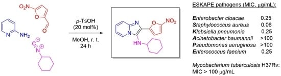
Figure 1. The Groeble–Blackburn–Bienaymé (GBB) reaction and relevant GBB reaction-derived antibacterial lead compound 3.
Biomedicines 10 02203 g001
Figure 2. The diversity of 2-aminoazines and -azoles 1a–m that were employed in the GBB synthesis of 5- nitrofuran-tagged compounds that were investigated in this work.
Biomedicines 10 02203 g002
Scheme 1. Synthesis of imidazo-fused products of the GBB reaction with 1a–m showing the structure and percentage yield of each compound.
Biomedicines 10 02203 sch001
Figure 3. Strain energies and interaction diagrams of (A) nitrofurazone re-docked into the structure of E. coli nitroreductase NfsB (PDB 1YKI) and (B) nitrofurantoin re-docked into the structure of oxygen-insensitive NADPH nitroreductase NsfA (PDB 7NB9); (C) ligand interactions diagram legend.
Biomedicines 10 02203 g003aBiomedicines 10 02203 g003b
Figure 4. Strain energies and interaction diagrams of compounds 4a (A), 4b (B), 4c (C), and 4d (D) that were docked into the structure of E. coli nitroreductase NfsB (PDB 1YKI) using the flexible docking protocol.
Biomedicines 10 02203 g004aBiomedicines 10 02203 g004b
Figure 5. Strain energies and interaction diagrams of compounds 4a (A), 4b (B), 4c (C), and 4d (D) that were docked into the structure of oxygen-insensitive NADPH nitroreductase NsfA (PDB 7NB9) using the flexible docking protocol.
Biomedicines 10 02203 g005aBiomedicines 10 02203 g005b
Table 1. Antibacterial activity (disk diffusion method inhibition zone (IZ, mm) and minimal inhibitory concentration (MIC, µg/mL) of compounds 4a–m, nitrofurans furazidine and nitrofurantoin as well as ciprofloxacin (positive controls) against the ESKAPE panel of pathogens (E1 = Enterobacter cloacae (G−), S = Staphylococcus aureus (G+), K = Klebsiella pneumoniae (G−), A = Acinetobacter baumannii (G−), P = Pseudomonas aeruginosa (G−), E2 = Enterococcus faecalis (G+)); NT—not tested. The compounds that were not tested or having no activity are marked red. The MIC values are mean from three different assays (errors were in the range of ±5–10% of the reported values).
CompoundE1SKAPE2
IZMICIZMICIZMICIZMICIZMICIZMIC
4a170.25170.0670.250NT 0NT 90.25
4b0NT 0NT 0NT 0NT 0NT 0NT
4c0NT0NT0NT0NT 0NT 0NT
4d0NT100.4361.750NT 0NT 0NT
4e140.45150.2370.450NT 0NT 0NT
4f110.78111.550NT0NT 0NT 131.55
4g101170.580.50NT 0NT 130.5
4h0NT90.4370.430NT 0NT 0NT
4i0NT160.3960.780NT 0NT 91.55
4j122.1190.5362.10NT 0NT 131.05
4k0NT180.6690.830NT 0NT 150.66
4l150.9390.9351.850NT 0NT 0NT
4m70.61110.6192.450NT 0NT 0NT
Furazidine133223814320NT 0NT 212
Nitrofurantoin15821412640NT 0NT 244
CiprofloxacinNT1.25NT1.25NT0.6NT2.5NT0.6NT0.3
Table 2. Activity of compounds 4a–m against Mycobacterium tuberculosis H37Rv drug-sensitive strain. The most active compound (4i) is marked green.
CompoundMIC (µg/mL)CompoundMIC (µg/mL)
4a>1004h100
4b>1004i6.2
4c>1004j50
4d>1004k50
4e504l100
4f>1004m100
4g100Isoniazid0.10
Table 3. Total system energy as well as MM-GBSA free Gibbs energy of compounds 4a–d that were docked into E. coli nitroreductase NfsB (PDB 1YKI) and oxygen-insensitive NADPH nitroreductase NsfA (PDB 7NB9) using flexible docking protocol [native ligand = nitrofurazone (1YKI) or nitrofurantoin (7NB9)]. Comopounds with good binding energy parameters are marked green; compounds with poor binding energy parameters are marked orange.
CompoundTotal System Energy (kcal/mol)MM-GBSA ΔG (kcal/mol)
1YKI7NB91YKI7NB9
native-ligand−17,665.90−19,712.29−75.40−47.54
4a−17,677.63−19,737.57−62.04−51.57
4b−15,897.41−17,646.36−11.97−27.15
4c−15,842.04−17,638.79−15.63−34.85
4d−17,641.98−19,842.16−58.44−53.58
Table 4. Molecular parameters of compounds 4a–m defining drug-likeness according to Lipinski [44] [MW—molecular weight (<500), cLogP—calculated lipophilicity characteristic defined as 1-octanol/water partitioning coefficient (<5.0), HBA—number of hydrogen bond acceptors (<10), HBD—number of hydrogen bond donors (<5), NRotB—number of rotatable bonds (<10)]; calculated using Molinspiration Cheminformatics tools [45].
CompoundMWcLogPHBAHBDNRotB
4a326.44.08714
4b360.84.71714
4c394.44.93715
4d340.44.49714
4e314.43.20714
4f406.44.97817
4g327.33.16814
4h355.43.82814
4i332.43.82714
4j334.43.54714
4k392.43.77917
4l392.43.76918
4m321.42.48814
Publisher’s Note: MDPI stays neutral with regard to jurisdictional claims in published maps and institutional affiliations.

