Article

Donor-Derived Cell-Free DNA at 1 Month after Kidney Transplantation Relates to HLA Class II Eplet Mismatch Load

by
Elena González-López
1,
Javier Gonzalo Ocejo-Vinyals
2,
Mónica Renuncio-García
1,
Adriel Roa-Bautista
1,
David San Segundo Arribas
1,
Clara Escagedo
3,
María del Mar García-Saiz
4,
Rosalía Valero
3,
Pilar García-Berbel
5,
Juan Carlos Ruíz San Millán
3 and
Emilio Rodrigo
3,*
1
Immunology Department, Immunopathology Group, Marqués de Valdecilla University Hospital-IDIVAL, University of Cantabria, 39008 Santander, Spain
2
Immunology Department, Infectious Diseases and Clinical Microbiology Group, Marqués de Valdecilla University Hospital-IDIVAL, University of Cantabria, 39008 Santander, Spain
3
Nephrology Department, Immunopathology Group, Marqués de Valdecilla University Hospital-IDIVAL, University of Cantabria, 39008 Santander, Spain
4
Clinical Pharmacology Department, Marqués de Valdecilla University Hospital-IDIVAL, University of Cantabria, 39008 Santander, Spain
5
Pathological Anatomy Department, Marqués de Valdecilla University Hospital-IDIVAL, University of Cantabria, 39008 Santander, Spain
*
Author to whom correspondence should be addressed.
Biomedicines 2023, 11(10), 2741; https://doi.org/10.3390/biomedicines11102741
Submission received: 16 August 2023 / Revised: 3 October 2023 / Accepted: 9 October 2023 / Published: 10 October 2023
(This article belongs to the Section Immunology and Immunotherapy)

Abstract

:
Kidney transplantation is the preferred therapeutic option for end-stage renal disease; however, the alloimmune response is still the leading cause of renal allograft failure. To better identify immunologic disparities in order to evaluate HLA compatibility between the donor and the recipient, the concept of eplet load has arisen. Regular kidney function monitoring is essential for the accurate and timely diagnosis of allograft rejection and the appropriate treatment. Donor-derived cell-free DNA (dd-cfDNA) has been proposed as a potential biomarker of acute rejection and graft failure in kidney transplantation. The proportion of plasma dd-cfDNA was determined in forty-two kidney patients at 1 month after transplantation. A total of eleven (26.2%) patients had a dd-cfDNA proportion of ≥1.0%. The only pretransplant variable related to dd-cfDNA > 1.0% was the HLA class II eplet mismatch load, mainly the HLA-DQB1 eplet mismatch load. Furthermore, dd-cfDNA was able to discriminate the patients with antibody-mediated rejection (AbMR) (AUC 87.3%), acute rejection (AUC 78.2%), and troubled graft (AUC 81.4%). Increased dd-cfDNA levels were associated with kidney allograft deterioration, particularly rejection, as well as a greater HLA class II eplet mismatch load. Consequently, combining dd-cfDNA determination and HLA eplet mismatch load calculation should improve the assessment of the risk of short- and long-term allograft damage.

1. Introduction

Kidney transplantation is the preferred therapeutic option for end-stage renal disease. In this research field, a marked improvement in one-year graft survival rates has been shown [1], although alloimmune response is still the leading cause of renal allograft failure [2]. The most important non-self-antigens implicated in the alloimmune response are the human leukocyte antigens (HLA) [3]. In kidney transplantation, HLA mismatch between the donor and the recipient has been associated with early acute rejection and poor graft outcome [4], leading to T-cell-mediated rejection (TCMR) and antibody-mediated rejection (AbMR) [5,6].
The concept of eplets has emerged as a novel method to fine-tune the HLA risk and better identify immunologic disparities in a more detailed way in order to evaluate HLA compatibility between the donor and the recipient, which may lower the risk of allograft rejection [7,8,9,10]. Several studies have demonstrated a relationship between donor–recipient eplet disparity and graft outcome in renal transplantation [11,12,13].
Effective immunosuppressive treatments are another important strategy for ensuring long-term transplant survival [14]. Nonetheless, maintaining an appropriate immunosuppressive balance is crucial in order to avoid severe side effects, such as infection and cancer [15]. Consequently, regular kidney function monitoring is essential for the accurate and timely diagnosis of allograft rejection and the appropriate treatment [16].
Several biomarkers, including serum creatinine levels and estimated glomerular filtration rate (eGFR), are used to monitor graft function and rejection, but their capacity to identify early damage is limited and they may not discriminate acute from chronic graft damage [17,18]. The gold standard for diagnosing rejection and graft decline is the histologic analysis of the allograft biopsy [19]. However, biopsies are unsuitable for routine monitoring since they are invasive, costly, and risk consequences.
As a result, non-invasive and new indicators of early and late graft failure in renal transplantation are required to enhance complication management and increase patient and graft survival [20,21]. To achieve this, donor-derived cell-free DNA (dd-cfDNA) has been proposed as a potential biomarker of acute rejection and graft failure in kidney transplants [21,22]. With a size of 150–180 base pairs, the majority of circulating cfDNA is released into the bloodstream via cell apoptosis [23], and its levels are altered in chronic inflammation, malignancies, and injury, which can be clinically useful [16,24]. Several studies have shown that dd-cfDNA levels rise immediately after a kidney transplant and subsequently decline within 10 days [25]. Moreover, higher initial levels of circulating dd-cfDNA have been observed in deceased donors, rejection, or graft failure and in situations of insufficient immunosuppression [26].

2. Materials and Methods

2.1. Study Design and Patients’ Information

We conducted this prospective study in accordance with the principles outlined in the Declaration of Helsinki and with approval from our institution’s Regional Ethics Committee (reference number: PI20/01710; 22 December 2020). We enrolled a total of 42 consecutive recipients of kidney transplants from deceased donors, spanning from January 2021 to April 2022. All the participants provided written consent before undergoing kidney transplantation. We excluded recipients who had received non-kidney solid organ transplants, those from non-controlled cardiac death donations, individuals with preformed donor-specific antibodies, and those with a panel-reactive antibody level exceeding 98%.
We gathered relevant information regarding the recipients, donors, and transplant characteristics. Biopsy confirmation was required for all acute rejection episodes. Indication biopsies were performed when creatinine levels increased by 25% or more compared to the previous value, or when there was persistent proteinuria exceeding 1 g per day. We defined a “troubled” graft as one undergoing acute rejection or other issues, such as obstruction or graft infection. Maintenance immunosuppressive therapy consisted of twice-daily doses of tacrolimus, mycophenolate mofetil, and prednisone. Mycophenolate mofetil was started at 500 mg twice daily, whereas the initial tacrolimus dose was 0.1 mg/kg twice daily. The target tacrolimus blood levels at our institution for the first 3 months ranged from 8 to 12 ng/mL. Recipients of organs from expanded criteria donors and those at risk of delayed graft function received basiliximab induction therapy. Thymoglobulin was used as induction therapy for patients at a higher risk of rejection due to hypersensitization or previous graft loss caused by acute rejection. All patients received trimethoprim–sulfamethoxazole prophylaxis for 6 months post-transplant, and valganciclovir was given for 3 months to CMV IgG-negative recipients of a CMV IgG-positive donor and in patients receiving thymoglobulin induction.

2.2. dd-cfDNA Determination

Peripheral blood samples were collected from kidney transplant recipients at 1 month post-transplant into Streck Cell-Free DNA BCT tubes. Then, after centrifugation steps of 1,600× g for 20 min and 16,000× g for 10 min, plasma was obtained. cfDNA was extracted using the QIAamp Circulating Nucleic Acid Kit (Qiagen, Redwood City, CA, USA) and to determine the relative amount of dd-cfDNA, a targeted NGS assay employing 202 single nucleotide polymorphisms (SNPs) was then utilized (AlloSure®, CareDx, Inc., Brisbane, CA, USA) (Table S1). First, a multiplexed polymerase chain reaction (PCR) was performed with primers to amplify the targeted SNPs and the joining of indexes to identify the samples, as well as adapters required for the sequencing step. The amplified product was then pooled and purified in preparation for sequencing. The result of PCR was sequenced on Illumina (Illumina, Inc. Brisbane, CA, USA) MiSeq equipment. Finally, dd-cfDNA% was calculated using CareDx AlloSeq cfDNA software 1.0 (“Allosoft”) by using informative SNPs between donor and recipient. The kit amplified 202 SNPs that were consciously selected along the 22 autosomal chromosomes. After reading the amplified SNPs using NGS, the number of reads for each nucleotide within each of the SNPs could be identified, making it feasible to identify the cfDNA of the donor and the recipient. Because only post-transplant samples were used, and there was no prior donor–recipient genotyping, the program only used homozygous informative SNPs to complete the analysis. When the percentage of two-nucleotide readings at an SNP site was outside the range of 30–70%, a homozygous SNP was regarded as informative. For each SNP, the nucleotide with the highest number of reads related to the recipient, while the one with the lowest proportion corresponded to the donor (e.g., if there are 2% of “A” reads and 98% of “G” reads, the donor is AA and the recipient is GG for that SNP, and this was used to calculate the % dd-cfDNA). Finally, the computer calculated an average of the percentage of nucleotide readings from the informative SNPs that correspond to the donor. Furthermore, AlloSoft considers several quality parameters to ensure the reliability of the calculated results, such as the uniformity of readings obtained between different SNPs, the possibility of contamination of the sample through the detection of outliers and total readings. The reporting of % of dd-cfDNA in the literature allows for the assumption that the free DNA of a lower quantity comes from the donor in the post-transplant sample. It can be shown that the percentage of dd-cfDNA in lung [27], kidney [16], and heart [28] transplants is always lower than the percentage of free DNA in the patient. A cutoff of ≥1.0% was used, in accordance with the manufacturer’s instructions, as an anomalous dd-cfDNA result.

2.3. Immunosuppressive Drugs Monitoring

We quantified the concentration of mycophenolic acid (MPA) in human plasma (mg/L) using a homogeneous enzyme immunoassay (Emit 2000 Mycophenolic Acid Assay; Siemens, Saint Paul, MN, USA) at the one-month mark. Whole blood concentrations (µg/L) of tacrolimus were determined using chemiluminescent microparticle immunoassay (CMIA; Abbott Laboratories, Abbott Park, IL, USA) on the Architect iSystem. We collected tacrolimus levels up to day 30. We assessed the variability of tacrolimus blood levels using the coefficient of variation (CV), calculated as CV (%) = (σ/μ) × 100, where σ represents the standard deviation, and μ denotes the mean tacrolimus concentration of all available samples [26]. We determined the percentage of time within the therapeutic range (8 to 12 ng/mL) and above 12 ng/mL using the Rosendaal method [27]. Tacrolimus C/D (concentration-to-dose) ratios were calculated at the one-month and three-month marks, with fast metabolizers defined as those having a tacrolimus level–dose ratio below 1.05 [28]. “Any tacrolimus level < 5 ng/mL at month 1” and “any tacrolimus level < 6 ng/mL at month 1” were defined if the patients had at least one trough tacrolimus blood level below the threshold of 5 ng/mL or 6 ng/mL at any time up to month 1.

2.4. Eplet Mismatch Examination

Antigen HLA and eplet mismatch load were calculated to establish donor and recipient compatibility. The HLAMatchmaker 3.1 software (available at http://www.epitopes.net/downloads.html accessed on 15 April 2023) was used to assess eplet matching.

2.5. Statistical Analysis

The median and interquartile range (IQR) were used to express continuous variables, and relative frequencies were used to characterize categorical variables. To compare continuous variables among dichotomous data, the Mann–Whitney U test was used, and the chi-square test was used to investigate the relationship between two qualitative variables. The ability of dd-cfDNA% to distinguish between rejection and troubled graft was examined via receiver operating characteristic (ROC) curves. Statistical significance was defined as a p-value less than 0.05. Statistical analyses were performed with SPSS, version 15.0 (SPSS, Inc., Chicago, IL, USA).

3. Results

3.1. Patient Characteristics

The main patient characteristics are shown in Table 1. The median dd-cfDNA value in kidney transplant recipients was 0.61, with an interquartile range from 0.34 to 1.06. A total of 11 patients (26.2%) had a percentage of dd-cfDNA ≥ 1.0%. Differences in variables between patients with dd-cfDNA below and above 1.0% are shown in Table 1. The only pretransplant variable related to dd-cfDNA percentage was HLA class II eplet mismatch load, predominantly the HLA-DQB1 eplet mismatch load (p = 0.022 and p = 0.041, respectively) (Table 2). Interestingly, neither antigen HLA mismatch number nor HLA class I eplet mismatch load was associated with dd-cfDNA. Moreover, immunosuppressive exposure was not related to dd-cfDNA.

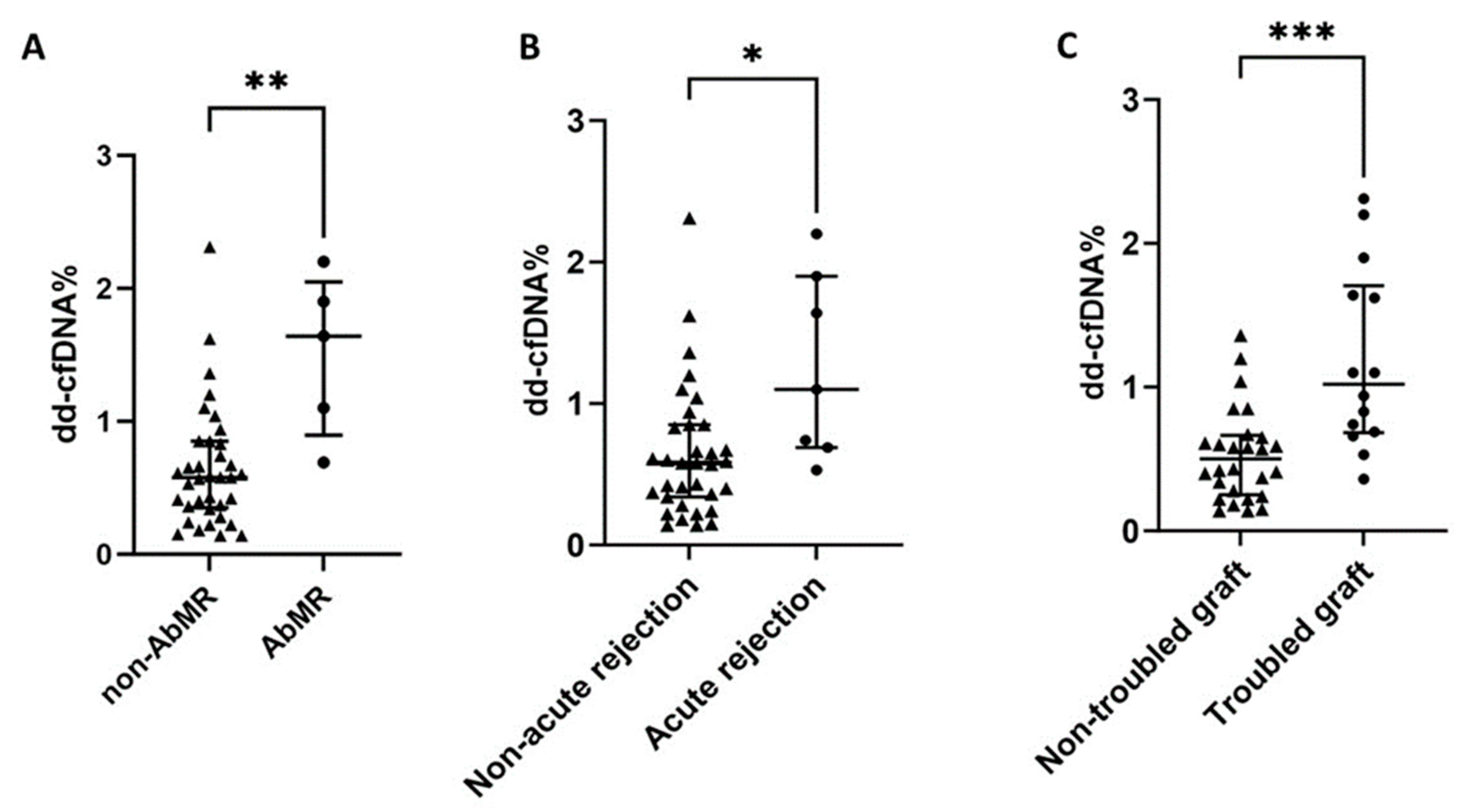
3.2. dd-cfDNA% and Troubled Graft Patients

As presented in Table 1, patients with acute rejection, AbMR, and troubled grafts in the first month post-transplant had significantly higher levels of dd-cfDNA%. Seven patients developed acute rejection within the first month; the median time was 14 days after transplantation (range 9–30 days). Among the 11 patients with a dd-cfDNA ≥ 1.0%, 4 patients developed acute rejection, and 3 of them were suffering different problems that could justify the elevation of dd-cfDNA% (2 patients needed a nephrostomy tube due to graft hydronephrosis, 1 of them developed a urinary sepsis due to Klebsiella, and 1 required a bladder catheter due to urinary obstruction). Moreover, 4 patients had a dd-cfDNA ≥ 1.0% without a clear cause: 1 patient developed cytomegalovirus infection in the first month, and 1 patient needed a percutaneous tube to drain a peri-graft collection without hydronephrosis. Among 5 patients with AbMR, only 1 of them had a dd-cfDNA < 1.0% (0.69%), but this sample was obtained after finishing AbMR therapy with plasmapheresis plus intravenous immunoglobulins. Two patients with isolated borderline TCMR had dd-cfDNA values below 1.0% (0.74 and 0.53).

3.3. dd-cfDNA% in AbMR Patients

dd-cfDNA was able to discriminate those patients with AbMR (AUC-ROC 87.3%, 95%CI 74.8–99.8, p = 0.007) (Figure 1A), with acute rejection (AUC-ROC 78.2, 95%CI 61.7–94.6, p = 0.020) (Figure 1B), and with troubled kidney graft (AUC-ROC 81.4, 95%CI 67.9–94.8, p = 0.001) (Figure 1C). Patients with AbMR (1.64, IQR 1.15 vs. 0.58, IQR 0.50, p = 0.004) (Figure 2A), acute rejection (1.10, IQR 1.21 vs. 0.58, IQR 0.51, p = 0.017) (Figure 2B), and troubled graft (1.02, IQR 1.02 vs. 0.50, IQR 0.42, p = 0.001) (Figure 2C) had significantly higher levels of dd-cfDNA. Moreover, patients with a dd-cfDNA ≥ 1.0% had a significantly higher risk of AbMR (HR 27.03, 95%CI 1.27–577.01, p = 0.035), independent of renal function. Notably, AbMR did not relate to a higher eplet mismatch load.

4. Discussion

Regular graft function monitoring in kidney transplant recipients is critical for the accurate detection of graft deterioration and starting appropriate therapy to avert graft loss. dd-cfDNA has emerged as a new and non-invasive biomarker for graft injury in kidney transplants, with evidence that it overcomes the limitations of traditional approaches. It has been demonstrated that a dd-cfDNA level of >1.0% is associated with active rejection (AbMR or TCMR), and that those patients with levels of dd-cfDNA of 1.0% or below do not show active rejection [16]. Accordingly, when using 1% as a cutoff point in kidney transplants, the negative predictive value of dd-cfDNA is 84% for active rejection and 96% for antibody-mediated rejection [16]. The median dd-cfDNA levels found in different kidney transplant groups with rejection ranged from 1.0% to 3.0% [16,24,29,30]. Our results show that the majority of the patients who developed rejection (AbMR) at 1-month post-transplant (36.4%) had levels of dd-cfDNA ≥ 1.0%.
Similarly, we demonstrated that dd-cfDNA levels ≥ 1.0% can differentiate patients with acute rejection (AUC–ROC 78.2%, 95%CI 61.7–94.6, p = 0.020), especially AbMR (AUC–ROC 87.3%, 95%CI 74.8–99.8, p = 0.007), from those without rejection. This ability to discriminate those patients with rejection has been proved in a previously reported study. Sigdel et al. [24] and Zhang et al. [30] reported that the relative dd-cfDNA determination could differentiate between patients with and without acute rejection (AUC–ROC of 87% and 90%, respectively), whereas Gielis et al. [31] reported an AUC–ROC of 64% with a cutoff of 0.88%. There are two approaches for quantifying dd-cfDNA: relative and absolute [32]. Whitlam et al. [33] demonstrated that absolute dd-cdDNA (cp/mL) determination has a 91% AUC–ROC in the diagnosis of AbMR. Furthermore, they assessed the sensitivity and specificity for relative and absolute dd-cfDNA measurements, revealing 75% and 79% specificity, respectively, and the same sensitivity (85%) for the two approaches. The researchers concluded that both might be equally useful in detecting rejection in kidney transplant recipients.
Previous studies have demonstrated that dd-cfDNA is not effective in distinguishing TCMR from non-rejection or stable patients with lower levels than patients with AbMR [34], and similar findings have been reported in heart [35] and lung [36] transplantation. Because of the small number of pure TCMR in our data limits, we could not perform a non-AbMR-specific analysis.
In this study, dd-cfDNA% was assessed one month after transplant. The long-term effects of solid organ transplantation are still far from desirable. Studies have examined the potential of serial dd-cfDNA for long-term monitoring in kidney transplant recipients with acute rejection and intravenous steroid therapy. Shen et al. [37] observed a decline in dd-cfDNA% at 3 days and at 1, 3, and 6 months, correlating with estimated glomerular filtration, which indicated a response to rejection treatment. In addition, when combined with other clinically relevant findings (infections, malignancies), dd-cfDNA long-term monitoring might aid in the adaptation of immunosuppressive therapy [38].
Furthermore, we investigated the ability of dd-cfDNA to distinguish various sources of allograft damage or loss of function. Patients were classified into two groups: troubled graft, if the graft was suffering acute rejection or any other problem such as obstruction or graft infection, and non-troubled graft. dd-cfDNA levels of more than 1.0% were able to distinguish between these two groups (AUC–ROC 81.4%, 95%CI 67.9–94.8, p = 0.001). In line with this, Goussouss et al. [39] reported seven cases in which transplant infection was linked with an increase in plasma dd-cfDNA. In terms of obstruction, ureteral blockage occurs in 2–10% of renal transplant patients within the first few weeks or the first year [40], and to the best of our knowledge, this is the first study to incorporate obstruction as a post-renal transplantation complication in the analysis of dd-cfDNA.
In histocompatibility laboratories, antigen HLA mismatch is a common examination factor in kidney transplant candidates. However, the idea of epitope “load” has recently emerged as a novel technique to better characterize donor–recipient immunologic compatibility in solid organ transplantation. Matching recipients and donors according to the level of epitopes, as compared with traditional antigen HLA typing, may improve risk prediction for the development of donor-specific antibodies implicated in AbMR [41]. Thus, we found that patients with dd-cfDNA levels of more than 1.0% had a higher HLA class II eplet mismatch load (46 vs. 21, p = 0.022). Moreover, in recent years, the importance of antigen HLA-DQ matching for transplant outcome has been highlighted [42]. Several studies have shown that HLA-DQ eplet mismatch load is associated with an increased risk of developing de novo donor-specific antibodies, which are implicated in kidney transplant rejection, graft function decline, and graft failure [43,44,45], which may influence dd-cfDNA levels. In addition, urinary CXCL10, a non-invasive biomarker associated with inflammation and graft survival, has been linked to HLA-DBQ1 eplet mismatch load [46]. In this context, we found that elevated HLA-DQB1 eplet mismatch load was associated with higher levels of dd-cfDNA (p = 0.041). HLA matching between potential donors and recipients is assessed in kidney allocation by comparing their HLA antigens in order to have a successful transplant outcome, limiting the possibility of sensitization and subsequent rejection [47]. Unexpectedly, we found no relationship between conventional antigen HLA class I and class II mismatch and the percentage of dd-cfDNA. To our knowledge, this work is the first to show an association between the presence of HLA class II eplet mismatch with elevated dd-cfDNA levels (≥1.0%).
The main limitation of this single-center study was the sample size; we could only assess dd-cfDNA% in forty-two kidney transplant recipients one month after transplant. Because of the small sample size, the number of events, especially TCMR, was limited, restricting the capacity to demonstrate the relationship between dd-cfDNA and this type of rejection. Our study, on the other hand, has certain advantages. We conducted a prospective analysis on a cohort of kidney transplant patients who were followed in a single center, and all rejections were biopsy-proven. Furthermore, we conducted an in-depth study of the eplet mismatch load, which has not previously been associated with the percentage of dd-cfDNA.

5. Conclusions

Our findings support the value of measuring dd-cfDNA to detect alloimmune damage, particularly AbMR, in kidney transplantation. Only recipients with a high eplet mismatch load with their donors and an elevated percentage of dd-cfDNA were at risk of acute rejection and AbMR. Furthermore, an optimum assessment of the risk of short- and long-term allograft injury should involve the quantification of both the eplet mismatch load and post-transplant dd-cfDNA percentage.

Supplementary Materials

The following supporting information can be downloaded at: https://www.mdpi.com/article/10.3390/biomedicines11102741/s1, Table S1: Data from the SNPs studied.

Author Contributions

Conceptualization, E.R. and J.G.O.-V.; methodology, E.G.-L., M.R.-G., A.R.-B., D.S.S.A. and P.G.-B.; software, E.G.-L., E.R. and P.G.-B.; validation, C.E., M.d.M.G.-S. and R.V.; formal analysis, E.R.; investigation, E.R., E.G.-L. and J.G.O.-V.; resources, E.R.; data curation, E.R. and E.G.-L.; writing—original draft preparation, E.R., E.G.-L. and J.G.O.-V.; writing—review and editing, C.E., M.d.M.G.-S., R.V. and J.C.R.S.M.; visualization, E.R., E.G.-L., J.G.O.-V. and D.S.S.A.; supervision, E.R.; project administration, E.R. and J.G.O.-V.; funding acquisition, E.R. and J.C.R.S.M. All authors have read and agreed to the published version of the manuscript.

Funding

This work was supported by the Instituto de Salud Carlos III through grant PI20/01710, co-funded by the European Regional Development Fund, “A way to make Europe”.

Institutional Review Board Statement

The study was conducted in accordance with the Declaration of Helsinki, and approved by the Regional Ethics Committee of Cantabria (reference number: PI20/01710; 22 December 2020).

Informed Consent Statement

Informed consent was obtained from all subjects involved in the study.

Data Availability Statement

The data presented in this study are available upon request from the corresponding author.

Acknowledgments

The authors are indebted to the nurses of the Nephrology Laboratory (Hospital Marqués de Valdecilla-IDIVAL) for their continuous support.

Conflicts of Interest

The authors declare no conflict of interest.

References

  1. Lamb, K.E.; Lodhi, S.; Meier-Kriesche, H.U. Long-term renal allograft survival in the United States: A critical reappraisal. Am. J. Transpl. 2011, 11, 450–462. [Google Scholar] [CrossRef]
  2. Filippone, E.J.; Gulati, R.; Farber, J.L. Noninvasive Assessment of the Alloimmune Response in Kidney Transplantation. Adv. Chronic Kidney Dis. 2021, 28, 548–560. [Google Scholar] [CrossRef] [PubMed]
  3. Petrányi, G.G. The complexity of immune and alloimmune response. Transpl. Immunol. 2002, 10, 91–100. [Google Scholar] [CrossRef]
  4. Lim, W.H.; Chadban, S.J.; Clayton, P.; Budgeon, C.A.; Murray, K.; Campbell, S.B.; Cohney, S.; Russ, G.R.; McDonald, S.P. Human leukocyte antigen mismatches associated with increased risk of rejection, graft failure, and death independent of initial immunosuppression in renal transplant recipients. Clin. Transpl. 2012, 26, E428–E437. [Google Scholar] [CrossRef] [PubMed]
  5. Sellarés, J.; de Freitas, D.G.; Mengel, M.; Reeve, J.; Einecke, G.; Sis, B.; Hidalgo, L.G.; Famulski, K.; Matas, A.; Halloran, P.F. Understanding the causes of kidney transplant failure: The dominant role of antibody-mediated rejection and nonadherence. Am. J. Transpl. 2012, 12, 388–399. [Google Scholar] [CrossRef]
  6. Parajuli, S.; Aziz, F.; Garg, N.; Panzer, S.E.; Joachim, E.; Muth, B.; Mohamed, M.; Blazel, J.; Zhong, W.; Astor, B.C.; et al. Histopathological characteristics and causes of kidney graft failure in the current era of immunosuppression. World J. Transpl. 2019, 9, 123–133. [Google Scholar] [CrossRef]
  7. Duquesnoy, R.J. HLAMatchmaker: A molecularly based algorithm for histocompatibility determination. I. Description of the algorithm. Hum. Immunol. 2002, 63, 339–352. [Google Scholar] [CrossRef]
  8. Duquesnoy, R.J.; Marrari, M. HLAMatchmaker: A molecularly based algorithm for histocompatibility determination. II. Verification of the algorithm and determination of the relative immunogenicity of amino acid triplet-defined epitopes. Hum. Immunol. 2002, 63, 353–363. [Google Scholar] [CrossRef] [PubMed]
  9. Wiebe, C.; Nickerson, P. Human leukocyte antigen mismatch and precision medicine in transplantation. Curr. Opin. Organ. Transpl. 2018, 23, 500–505. [Google Scholar] [CrossRef]
  10. Ehlayel, A.; Simms, K.J.A.; Ashoor, I.F. Emerging monitoring technologies in kidney transplantation. Pediatr. Nephrol. 2021, 36, 3077–3087. [Google Scholar] [CrossRef]
  11. Mohammadhassanzadeh, H.; Oualkacha, K.; Zhang, W.; Klement, W.; Bourdiec, A.; Lamsatfi, J.; Yi, Y.; Foster, B.; Keown, P.; Gebel, H.M.; et al. On Path to Informing Hierarchy of Eplet Mismatches as Determinants of Kidney Transplant Loss. Kidney Int. Rep. 2021, 6, 1567–1579. [Google Scholar] [CrossRef]
  12. Daniëls, L.; Naesens, M.; Bosmans, J.L.; Abramowicz, D.; Nagler, E.; Van Laecke, S.; Peeters, P.; Kuypers, D.; Emonds, M.P. The clinical significance of epitope mismatch load in kidney transplantation: A multicentre study. Transpl. Immunol. 2018, 50, 55–59. [Google Scholar] [CrossRef] [PubMed]
  13. Lachmann, N.; Niemann, M.; Reinke, P.; Budde, K.; Schmidt, D.; Halleck, F.; Pruß, A.; Schönemann, C.; Spierings, E.; Staeck, O. Donor-Recipient Matching Based on Predicted Indirectly Recognizable HLA Epitopes Independently Predicts the Incidence of De Novo Donor-Specific HLA Antibodies Following Renal Transplantation. Am. J. Transpl. 2017, 17, 3076–3086. [Google Scholar] [CrossRef]
  14. Bauer, A.C.; Franco, R.F.; Manfro, R.C. Immunosuppression in Kidney Transplantation: State of the Art and Current Protocols. Curr. Pharm. Des. 2020, 26, 3440–3450. [Google Scholar] [CrossRef] [PubMed]
  15. Briggs, J.D. Causes of death after renal transplantation. Nephrol. Dial. Transpl. 2001, 16, 1545–1549. [Google Scholar] [CrossRef] [PubMed]
  16. Bloom, R.D.; Bromberg, J.S.; Poggio, E.D.; Bunnapradist, S.; Langone, A.J.; Sood, P.; Matas, A.J.; Mehta, S.; Mannon, R.B.; Sharfuddin, A.; et al. Cell-Free DNA and Active Rejection in Kidney Allografts. J. Am. Soc. Nephrol. 2017, 28, 2221–2232. [Google Scholar] [CrossRef] [PubMed]
  17. Josephson, M.A. Monitoring and managing graft health in the kidney transplant recipient. Clin. J. Am. Soc. Nephrol. 2011, 6, 1774–1780. [Google Scholar] [CrossRef] [PubMed]
  18. Quaglia, M.; Merlotti, G.; Guglielmetti, G.; Castellano, G.; Cantaluppi, V. Recent Advances on Biomarkers of Early and Late Kidney Graft Dysfunction. Int. J. Mol. Sci. 2020, 21, 5404. [Google Scholar] [CrossRef]
  19. Loupy, A.; Haas, M.; Roufosse, C.; Naesens, M.; Adam, B.; Afrouzian, M.; Akalin, E.; Alachkar, N.; Bagnasco, S.; Becker, J.U.; et al. The Banff 2019 Kidney Meeting Report (I): Updates on and clarification of criteria for T cell- and antibody-mediated rejection. Am. J. Transpl. 2020, 20, 2318–2331. [Google Scholar] [CrossRef]
  20. Naesens, M.; Friedewald, J.; Mas, V.; Kaplan, B.; Abecassis, M.M. A Practical Guide to the Clinical Implementation of Biomarkers for Subclinical Rejection Following Kidney Transplantation. Transplantation 2020, 104, 700–707. [Google Scholar] [CrossRef]
  21. Edwards, R.L.; Menteer, J.; Lestz, R.M.; Baxter-Lowe, L.A. Cell-free DNA as a solid-organ transplant biomarker: Technologies and approaches. Biomark. Med. 2022, 16, 401–415. [Google Scholar] [CrossRef]
  22. Thongprayoon, C.; Vaitla, P.; Craici, I.M.; Leeaphorn, N.; Hansrivijit, P.; Salim, S.A.; Bathini, T.; Rivera, F.H.C.; Cheungpasitporn, W. The Use of Donor-Derived Cell-Free DNA for Assessment of Allograft Rejection and Injury Status. J. Clin. Med. 2020, 9, 1480. [Google Scholar] [CrossRef] [PubMed]
  23. Heitzer, E.; Auinger, L.; Speicher, M.R. Cell-Free DNA and Apoptosis: How Dead Cells Inform About the Living. Trends Mol. Med. 2020, 26, 519–528. [Google Scholar] [CrossRef] [PubMed]
  24. Sigdel, T.K.; Archila, F.A.; Constantin, T.; Prins, S.A.; Liberto, J.; Damm, I.; Towfighi, P.; Navarro, S.; Kirkizlar, E.; Demko, Z.P.; et al. Optimizing Detection of Kidney Transplant Injury by Assessment of Donor-Derived Cell-Free DNA via Massively Multiplex PCR. J. Clin. Med. 2018, 8, 19. [Google Scholar] [CrossRef] [PubMed]
  25. Shen, J.; Zhou, Y.; Chen, Y.; Li, X.; Lei, W.; Ge, J.; Peng, W.; Wu, J.; Liu, G.; Yang, G.; et al. Dynamics of early post-operative plasma ddcfDNA levels in kidney transplantation: A single-center pilot study. Transpl. Int. 2019, 32, 184–192. [Google Scholar] [CrossRef] [PubMed]
  26. Oellerich, M.; Shipkova, M.; Asendorf, T.; Walson, P.D.; Schauerte, V.; Mettenmeyer, N.; Kabakchiev, M.; Hasche, G.; Gröne, H.J.; Friede, T.; et al. Absolute quantification of donor-derived cell-free DNA as a marker of rejection and graft injury in kidney transplantation: Results from a prospective observational study. Am. J. Transpl. 2019, 19, 3087–3099. [Google Scholar] [CrossRef] [PubMed]
  27. Khush, K.K.; De Vlaminck, I.; Luikart, H.; Ross, D.J.; Nicolls, M.R. Donor-derived, cell-free DNA levels by next-generation targeted sequencing are elevated in allograft rejection after lung transplantation. ERJ Open Res. 2021, 7, 00462–2020. [Google Scholar] [CrossRef]
  28. Khush, K.K.; Patel, J.; Pinney, S.; Kao, A.; Alharethi, R.; DePasquale, E.; Ewald, G.; Berman, P.; Kanwar, M.; Hiller, D.; et al. Noninvasive detection of graft injury after heart transplant using donor-derived cell-free DNA: A prospective multicenter study. Am. J. Transpl. 2019, 19, 2889–2899. [Google Scholar] [CrossRef]
  29. Huang, E.; Sethi, S.; Peng, A.; Najjar, R.; Mirocha, J.; Haas, M.; Vo, A.; Jordan, S.C. Early clinical experience using donor-derived cell-free DNA to detect rejection in kidney transplant recipients. Am. J. Transpl. 2019, 19, 1663–1670. [Google Scholar] [CrossRef]
  30. Zhang, H.; Zheng, C.; Li, X.; Fu, Q.; Li, J.; Su, Q.; Zeng, L.; Liu, Z.; Wang, J.; Huang, H.; et al. Diagnostic Performance of Donor-Derived Plasma Cell-Free DNA Fraction for Antibody-Mediated Rejection in Post Renal Transplant Recipients: A Prospective Observational Study. Front. Immunol. 2020, 11, 342. [Google Scholar] [CrossRef]
  31. Gielis, E.M.; Ledeganck, K.J.; Dendooven, A.; Meysman, P.; Beirnaert, C.; Laukens, K.; De Schrijver, J.; Van Laecke, S.; Van Biesen, W.; Emonds, M.P.; et al. The use of plasma donor-derived, cell-free DNA to monitor acute rejection after kidney transplantation. Nephrol. Dial. Transpl. 2020, 35, 714–721. [Google Scholar] [CrossRef] [PubMed]
  32. Osmanodja, B.; Akifova, A.; Budde, K.; Choi, M.; Oellerich, M.; Schütz, E.; Beck, J. Absolute or Relative Quantification of Donor-derived Cell-free DNA in Kidney Transplant Recipients: Case Series. Transpl. Direct 2021, 7, e778. [Google Scholar] [CrossRef] [PubMed]
  33. Whitlam, J.B.; Ling, L.; Skene, A.; Kanellis, J.; Ierino, F.L.; Slater, H.R.; Bruno, D.L.; Power, D.A. Diagnostic application of kidney allograft-derived absolute cell-free DNA levels during transplant dysfunction. Am. J. Transplant. 2019, 19, 1037–1049. [Google Scholar] [CrossRef] [PubMed]
  34. Wijtvliet, V.; Plaeke, P.; Abrams, S.; Hens, N.; Gielis, E.M.; Hellemans, R.; Massart, A.; Hesselink, D.A.; De Winter, B.Y.; Abramowicz, D.; et al. Donor-derived cell-free DNA as a biomarker for rejection after kidney transplantation: A systematic review and meta-analysis. Transpl. Int. 2020, 33, 1626–1642. [Google Scholar] [CrossRef]
  35. Agbor-Enoh, S.; Tunc, I.; De Vlaminck, I.; Fideli, U.; Davis, A.; Cuttin, K.; Bhatti, K.; Marishta, A.; Solomon, M.A.; Jackson, A.; et al. Applying rigor and reproducibility standards to assay donor-derived cell-free DNA as a non-invasive method for detection of acute rejection and graft injury after heart transplantation. J. Heart Lung Transpl. 2017, 36, 1004–1012. [Google Scholar] [CrossRef]
  36. Agbor-Enoh, S. 2018 ATS BEAR Cage Winning Proposal: Cell-Free DNA to Improve Lung Transplant Outcomes. Am. J. Respir. Crit. Care Med. 2019, 199, 1058–1060. [Google Scholar] [CrossRef]
  37. Shen, J.; Guo, L.; Yan, P.; Zhou, J.; Zhou, Q.; Lei, W.; Liu, H.; Liu, G.; Lv, J.; Liu, F.; et al. Prognostic value of the donor-derived cell-free DNA assay in acute renal rejection therapy: A prospective cohort study. Clin. Transpl. 2020, 34, e14053. [Google Scholar] [CrossRef]
  38. Oellerich, M.; Budde, K.; Osmanodja, B.; Bornemann-Kolatzki, K.; Beck, J.; Schütz, E.; Walson, P.D. Donor-Derived Cell-free DNA for Personalized Immunosuppression in Renal Transplantation. Ther. Drug Monit. 2023, 45, 20–25. [Google Scholar] [CrossRef]
  39. Goussous, N.; Xie, W.; Dawany, N.; Scalea, J.R.; Bartosic, A.; Haririan, A.; Kalil, R.; Drachenberg, C.; Costa, N.; Weir, M.R.; et al. Donor-derived Cell-free DNA in Infections in Kidney Transplant Recipients: Case Series. Transpl. Direct 2020, 6, e568. [Google Scholar] [CrossRef]
  40. Berger, P.M.; Diamond, J.R. Ureteral obstruction as a complication of renal transplantation: A review. J. Nephrol. 1998, 11, 20–23. [Google Scholar]
  41. Larkins, N.G.; Wong, G.; Taverniti, A.; Lim, W.H. Epitope matching in kidney transplantation: Recent advances and current limitations. Curr. Opin. Organ. Transpl. 2019, 24, 370–377. [Google Scholar] [CrossRef] [PubMed]
  42. Leeaphorn, N.; Pena, J.R.A.; Thamcharoen, N.; Khankin, E.V.; Pavlakis, M.; Cardarelli, F. HLA-DQ Mismatching and Kidney Transplant Outcomes. Clin. J. Am. Soc. Nephrol. 2018, 13, 763–771. [Google Scholar] [CrossRef] [PubMed]
  43. Senev, A.; Coemans, M.; Lerut, E.; Van Sandt, V.; Kerkhofs, J.; Daniëls, L.; Driessche, M.V.; Compernolle, V.; Sprangers, B.; Van Loon, E.; et al. Eplet Mismatch Load and De Novo Occurrence of Donor-Specific Anti-HLA Antibodies, Rejection, and Graft Failure after Kidney Transplantation: An Observational Cohort Study. J. Am. Soc. Nephrol. 2020, 31, 2193–2204. [Google Scholar] [CrossRef] [PubMed]
  44. Kishikawa, H.; Kinoshita, T.; Hashimoto, M.; Fukae, S.; Taniguchi, A.; Yamanaka, K.; Nakagawa, M.; Nishimura, K. Class II HLA Eplet Mismatch Is a Risk Factor for De Novo Donor-Specific Antibody Development and Antibody-mediated Rejection in Kidney Transplantation Recipients. Transpl. Proc. 2018, 50, 2388–2391. [Google Scholar] [CrossRef] [PubMed]
  45. Tafulo, S.; Malheiro, J.; Santos, S.; Dias, L.; Almeida, M.; Martins, S.; Pedroso, S.; Mendes, C.; Lobato, L.; Castro-Henriques, A. Degree of HLA class II eplet mismatch load improves prediction of antibody-mediated rejection in living donor kidney transplantation. Hum. Immunol. 2019, 80, 966–975. [Google Scholar] [CrossRef]
  46. San Segundo, D.; Guiral-Foz, S.A.; Benito-Hernández, A.; Fernández, A.R.; Arnau, A.; Valero, R.; Ramos-Barrón, M.A.; García-Unzueta, M.T.; Gómez-Ortega, J.M.; López-Hoyos, M.; et al. Urinary CXCL10 specifically relates to HLA-DQ eplet mismatch load in kidney transplant recipients. Transpl. Immunol. 2022, 70, 101494. [Google Scholar] [CrossRef]
  47. Montgomery, R.A.; Tatapudi, V.S.; Leffell, M.S.; Zachary, A.A. HLA in transplantation. Nat. Rev. Nephrol. 2018, 14, 558–570. [Google Scholar] [CrossRef]
Figure 1. AUC–ROC curve of dd-cfDNA% for discriminating AbMR (A), acute rejection (B), and troubled graft (C). Abbreviations: AbMR—antibody-mediated rejection; dd-cfDNA—donor-derived cell-free DNA.
Biomedicines 11 02741 g001
Figure 2. The median and interquartile range of dd-cfDNA% in patients with and without AbMR (A), acute rejection (B), and troubled graft (C). Abbreviations: AbMR—antibody-mediated rejection; dd-cfDNA—donor-derived cell-free DNA. * p < 0.05; ** p < 0.01; *** p < 0.001.
Biomedicines 11 02741 g002
Table 1. Main patient characteristics.
VariableTotal (n = 42)dd-cfDNA ≥ 1.0% (n = 11)dd-cfDNA < 1.0% (n = 31)p
Recipient age (years)59.0 [47.8–67.5]60.0 [48.0–73.0]59.0 [47.0–66.0]0.572
Recipient gender (male)76.2%63.6%80.6%0.255
Diabetic nephropathy23.8%18.2%25.8%0.610
Time in renal
replacement therapy (months)
13.5 [3.9–38.7]24.5 [10.5–108.7]10.7 [0.0–32.6]0.062
Retransplant19.0%36.4%12.9%0.089
Preemptive transplantation21.4%18.2%22.6%0.760
Donor age (years)58.5 [47.0–65.0]61.0 [47.0–65.0]58.0 [47.0–65.0]0.910
Cold ischemia time (hours)21.5 [17.8–24.0]20.0 [18.0–24.0]22.0 [17.0–24.0]0.822
Induction66.7%72.7%64.5%0.620
1-month acute rejection16.7%36.4%9.7%0.041
1-month AbMR11.9%36.4%3.2%0.004
1-month troubled graft33.3%63.6%22.6%0.013
First year acute rejection (RAPreM12)21.4%36.4%16.1%0.160
First month eGFR (mL/min/1.73 m2)49.5 [40.5–70.0]43.0 [28.0–54.0]51.0 [42.0–72.0]0.138
First month albuminuria (mg/g)55.0 [23.5–150.5]57.5 [34.5–485.8]53.0 [22.5–135.5]0.524
1-year eGFR (mL/min/1.73 m2)50.0 [38.0–68.0]45.0 [38.0–49.0]50.0 [38.0–71.8]0.514
dd-cfDNA (%)0.61 [0.34–1.06]--0.396
Data of immunosuppressive therapy
TTR 8–12 at month 1 (%)45.7 [29.3–61.3]50.9 [37.0–60.4]45.5 [25.0–61.4]0.396
TTR > 12 at month 1 (%)47.3 [34.0–65.2]49.1 [39.6–63.0]44.8 [33.6–75.0]1.000
Mean tacrolimus trough level
throughout month 1 (ng/mL)
12.5 [11.3–14.3]12.5 [11.4–14.3]12.3 [11.2–14.2]0.822
Tacrolimus trough level at month 1 (ng/mL)12.1 [10.0–14.0]13.0 [10.0–15.0]12.0 [9.0–14.0]0.233
Mycophenolic acid trough level at month 1 (ng/mL)2.0 [1.0–3.0]2.0 [1.8–4.0]2.0 [1.0–3.0]0.569
Any tacrolimus level < 5 at month 17.1%0.0%9.7%0.284
Any tacrolimus level < 6 at month 111.9%0.0%16.1%0.156
Coefficient of variability at month 1 (%)28.1 [19.4–34.2]21.8 [16.1–61.5]28.6 [19.8–37.2]0.257
Tacrolimus trough level/dose at month 11.7 [1.3–2.5]1.8 [1.3–2.5]1.7 [1.1–2.7]0.778
Fast tacrolimus metabolizers11.9%9.1%12.9%0.737
Mann–Whitney U and Chi-square tests. Abbreviations: TTR—time in therapeutic range.
Table 2. HLA mismatches at the antigen and eplet levels between donor and recipient.
VariableTotal (n = 42)dd-cfDNA ≥ 1.0% (n = 11)dd-cfDNA < 1.0% (n = 31)p
Antigen HLA class I and II mismatches7.5 [6.0–9.0]8.0 [7.0–9.0]7.0 [5.0–9.0]0.233
All HLA class I eplet mismatch load18.0 [13.0–22.0]20.0 [16.0–22.0]16.0 [13.0–22.0]0.553
All HLA class II eplet mismatch load23.5 [14.8–51.5]46.0 [22.0–69.0]21.0 [11.0–39.0]0.022
HLA-DRB1 eplet mismatch load9.0 [4.0–14.0]10.0 [7.0–14.0]9.0 [3.0–13.0]0.245
HLA-DQB1 eplet mismatch load7.0 [2.8–9.3]9.0 [7.0–11.0]7.0 [2.0–9.0]0.041
HLA-DQA1 eplet mismatch load1.0 [0.0–5.0]3.0 [0.0–5.0]1.0 [0.0–4.0]0.445
Mann–Whitney U and Chi-square tests.
Disclaimer/Publisher’s Note: The statements, opinions and data contained in all publications are solely those of the individual author(s) and contributor(s) and not of MDPI and/or the editor(s). MDPI and/or the editor(s) disclaim responsibility for any injury to people or property resulting from any ideas, methods, instructions or products referred to in the content.

