Article

Transcription of Autophagy Associated Gene Expression as Possible Predictors of a Colorectal Cancer Prognosis

by
Martyna Bednarczyk
1,
Małgorzata Muc-Wierzgoń
2,*,
Sylwia Dzięgielewska-Gęsiak
2,
Edyta Fatyga
2 and
Dariusz Waniczek
3
1
Department of Hematology and Cancer Prevention, Medical University of Silesia in Katowice, 40-055 Katowice, Poland
2
Department of Preventive Medicine, Medical University of Silesia in Katowice, 40-055 Katowice, Poland
3
Department of Surgical Nursing and Propaedeutics of Surgery, Medical University of Silesia, 40-055 Katowice, Poland
*
Author to whom correspondence should be addressed.
Biomedicines 2023, 11(2), 418; https://doi.org/10.3390/biomedicines11020418
Submission received: 11 December 2022 / Revised: 26 January 2023 / Accepted: 27 January 2023 / Published: 31 January 2023
(This article belongs to the Collection Autophagy in Cancer and Metastasis)

Abstract

:
(1) Background: Autophagy plays a dual role in oncogenesis—it contributes to the growth of the tumor and can inhibit its development. The aim of this study was to assess changes in the transcriptional activity of LAMP-2, BECN1, PINK1, and FOXO1 genes involved in the autophagy process in histopathologically confirmed adenocarcinoma sections of colorectal cancer: (2) Methods: A gene expression profile analysis was performed using HG-U133A and the RT-qPCR reaction. The transcriptional activity of genes was compared in sections of colorectal cancer in the four clinical stages (CSI-CSIV) concerning the control group; (3) Results: In CSI, the transcriptional activity of the PINK1 gene is highest; in CS II, the LAMP-2 gene is highest, while FOXO1 increases gradually from CSI reaching a maximum in CSIII. There is no BECN1 gene expression in colorectal cancer cells; (4) Conclusions: The observed differences in the mRNA concentration profile of autophagy-related genes in colon cancer specimens may indicate the role of autophagy in the pathogenesis of this cancer. Genes involved in autophagy may be diagnostic tools for colorectal cancer screening and personalized therapy in the future.

1. Introduction

Three morphologically and mechanistically distinct types of autophagy exist in cells: macroautophagy, microautophagy, and chaperone-associated autophagy, where macroautophagy is often referred to as autophagy [1,2].
Autophagy is an intracellular, catabolic degradation process in which aggregated proteins, damaged organelles, and pathogens are delivered to lysosomes where they are digested by lysosomal hydrolases [3,4,5,6]. On the one hand, cellular self-digestion is responsible for the survival of cells in unfavorable conditions, and on the other, it eliminates damaged proteins and organelles, protecting cells against neoplastic transformation [7,8]. Recent studies have shown that autophagy plays an essential role in the pathogenesis of many diseases, including cancer, heart disease, infections, neurodegenerative disorders, autoimmune diseases, metabolic diseases, viral infections especially SARS-Cov-2 and COVID-19, as well as aging and cell death [9,10]. It is considered to be a type II programmed cell death [11]. The potential ability of autophagy to modulate cell death has made this process one that is considered to have a therapeutic aim in cancer therapy [12,13]. In the early stages of the disease, autophagy has a protective effect by removing damaged proteins, thereby protecting cells from malignant transformation. However, in late-stage cancer, autophagy promotes tumor survival and growth by scavenging toxic free radicals or damaged proteins.
In addition, autophagy supports mitochondrial function and tumor metabolism, resulting in survival under stressed conditions [14,15,16]. At the same time, it acts as a cellular defense mechanism to prevent cancer cell death after treatment, contributes to cancer recurrence and metastasis, and inhibits anticancer therapy and cancer cell death [9,17,18].
Higher levels of autophagy have been observed in many types of cancer; e.g., the main autophagy regulatory gene Beclin1 is upregulated in colorectal cancer, gastric cancer, liver cancer, breast cancer, and cervical cancer, suggesting that autophagy may be involved in carcinogenesis and that Beclin1 plays a key role in tumor formation [19,20,21,22].
Autophagy primarily plays a cytoprotective role in cancer cells and may be induced by most cancer therapies, including radiotherapy and chemotherapy [23,24,25,26,27]. Given the pro-survival role of autophagy, its inhibition has been shown to increase treatment efficacy. Therefore, inhibition of autophagy is considered a potentially valuable therapeutic approach in combination with other anticancer therapies [16,28].
It is worth noting that there are drugs available in anticancer therapy that act on autophagy, both stimulating and inhibiting this process. Inhibition of autophagy is achieved using pharmacological inhibitors, such as chloroquine, hydroxychloroquine, bafilomycin A1, 3-methyladenine, or by silencing gene expression, such as Atg5, BECN1, Atg10, or Atg12. Among the anticancer drugs that stimulate autophagy, the most popular are Bcl2 inhibitors and mTOR pathway inhibitors [29]. It is important to understand the molecular mechanisms of autophagy and to determine whether, in a given cancer, autophagy results in the survival or death of cancer cells under the influence of treatment.
The study aims to assess changes in expression patterns of genes involved in autophagy in colorectal cancer specimens (histopathological type—adenocarcinoma) in various stages of clinical cancer according to the ninth Union for International Cancer Control/American Joint Committee on Cancer (UICC/AJCC) classification. Colorectal cancer (CRC) is the third most common occurring cancer in men and the second most commonly occurring cancer in women. There were over 1.9 million new cases in 2020. In recent years, the global burden of CRC will increase by 60%, to over 2.2 million new cases and 1.1 million deaths by 2030 [30].
The premise for undertaking this study was the data from the literature on the research conducted so far, indicating the dual role of autophagy in the process of carcinogenesis. It has also been shown that the expression level of some autophagy-related genes is down-regulated or up-regulated in many types of cancer. Additionally, in the Affymetrix database, the number of autophagy-related genes has doubled in just a few months, indicating that autophagy has been a very active research area in recent years.

2. Materials and Methods

2.1. Ethics

The current study was conducted by the Declaration of Helsinki. The project was approved by the Bioethics Committee of the Medical University of Silesia in Katowice—decision No. KNW/0022/KB1/42/14 (20 June 2014).
The participants were informed in detail about the study and gave their written consent. Participation in the study was voluntary. Patients’ data have been encoded by the pseudonymization procedure, which means that personal data are processed in such a way that they cannot be assigned to a specific data subject without the use of an additional “key”.

2.2. Materials

The study group included 18 men and 21 women (age: 59–79 years; median age: 69 years). The study was carried out on specimens of the large intestine taken from patients with colorectal adenocarcinoma (histopathological type—adenocarcinoma) at four stages of clinical cancer according to the ninth classification of the Union for International Cancer Control/American Joint Committee on Cancer (UICC/AJCC). The patients were hospitalized in the Department of General, Colorectal, and Polytrauma Surgery and the Department of Surgical Nursing and Propaedeutics of Surgery, Faculty of Health Sciences in Katowice, Medical University of Silesia, Poland. Tissue samples were obtained during surgical resection of the colon affected by cancer, which was performed according to surgical treatment standards. The tissue samples were collected using classical surgical techniques without the use of electric or ultrasound instruments. The material obtained consisted of tumor tissue and/or healthy colon tissue. Healthy control tissue specimens were collected from an area 5 cm outside of the histologically negative margin, during the operation because of CC. All materials were taken by the same operational team to minimize mistakes. The cancer samples were obtained from the margin of the resected material to rule out the presence of necrotic tissue in the specimens.
The qualification of patients for the study included diagnosed and histopathologically confirmed colorectal cancer, regardless of the clinical stage, normal BMI, over 18 years of age, and consent of the patient. Patients with previously diagnosed and treated cancer, coexisting systemic disease, mental illness, chronic viral hepatitis, as well as individuals taking (long-term) non-steroidal anti-inflammatory drugs, immunosuppressants, steroids, or other hormonal drugs, were not eligible. The clinicopathological characteristics of the study group are given in Table 1.

2.3. Methods

In total, 39 paired tissues (39 tumor and 39 control) were collected. The collected intestinal specimens were placed in sterile tubes containing RNA laterTM (Sigma) in the amount of 10 µL per 1 mg of tissue (200 µL RNA laterTM per 20 mg of tissue). They were stored for 24 h at 4 °C, then the sections were frozen at −80 °C until the next stage of analysis. Molecular studies were performed at the Faculty and Department of Molecular Biology of the Medical University of Silesia.
This study is a continuation of an earlier published study, in which a microarray analysis of the expression profile of genes involved in autophagy was performed in adenocarcinoma specimens and specimens taken from the surgical margin, histopathologically assessed as healthy intestine (control). Oligonucleotide microarrays (HGU 133A—Affymetrix) were used for the study [31].
Confirmation of the results of the comparative analysis of transcriptomes determined by the expression microarray technique was carried out using the RT-qPCR method, recognized as the "gold standard" in the validation of matrix experiments.
The number of mRNA copies of the analyzed genes was determined based on the analysis of the kinetics of the qRT-PCR reaction using the Engine OPTICONTM sequence detector (MJ Research Inc., Watertown, MA, USA) and the QuantiTectTM SYBRGreen RT-qPCR Kit (QIAGEN) reagents according to the manufacturer’s recommendations [32].
Reverse transcription (RT) and quantitative PCR were performed in one reaction mixture using kit reagents. The reaction mixture consisted of 2× QuantiTect SYBR Green RT-PCR Master Mix, forward and reverse primers 0.5 M each, RNA template, and pyrogen-free water. The total mixture was 10 μL. For all tested samples, RT-qPCR was also performed for the mRNA of the β-actin gene, which was the endogenous control.
For each RT-qPCR reaction, a standard curve was constructed from which the Opticon™ DNA Engine Sequence Detector calculated the copy number of the test mRNA in the reaction mixture. The standard curve was determined from the RT-qPCR results of the quantitative template—a fragment of the β-actin gene (TaqMan® DNA Template Reagents Kit and β-actin Control Reagent Kit—Applied Biosystems) in five different concentrations (from 400, 800, 2000, 4000 to 8000 copies β-actin of cDNA/µL). On the basis of the fluorescence curve recorded after each amplification cycle, the number of messenger RNA copies (messenger RNA) of each gene tested in the conversion to 1 µg of RNA was determined [33].
The reaction was performed using sequence-specific primer pairs for each gene tested (Sigma-Aldrich, St. Louis, MO, USA)—Table 2.

Statistical Analysis
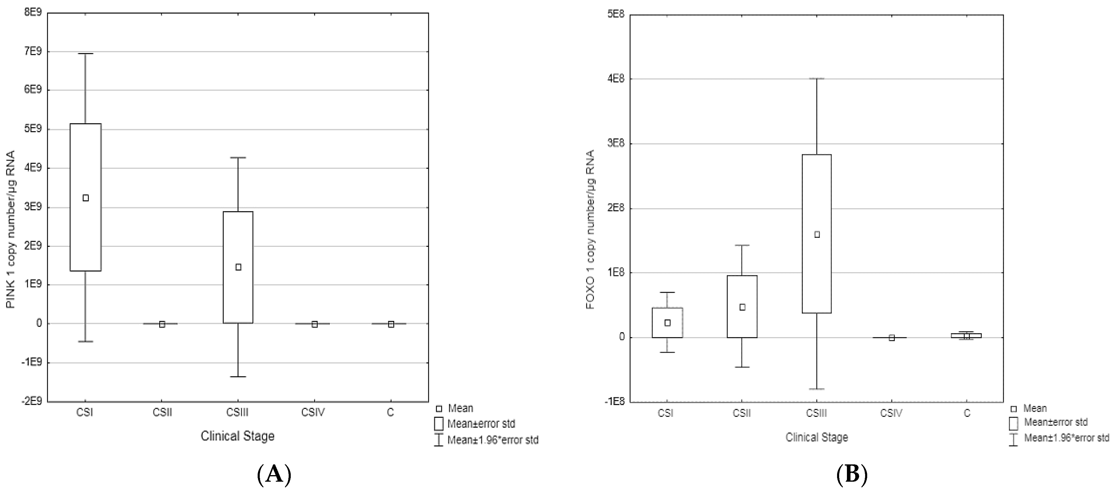
The results obtained using the qRT-PCR technique were developed based on the Statistica 12.5 program (StatSoft, Tulsa, OK, USA).
For each analyzed parameter, the most important elements of descriptive statistics were determined: mean, median, minimum and maximum value, standard deviation, and upper (75%) and lower (25%) quartiles. The normality of value distribution was checked using the Shapiro-Wilk test. Then, the results with a Gaussian distribution were analyzed with Student’s test. In case of Gaussian distribution, the results are expressed as mean and standard deviation (SD), while in the case of non-Gaussian distribution, the results are expressed as median and standard deviation.

3. Results

The transcriptional activity of genes involved in the autophagy process (LAMP-2, BECN1, PINK1, FOXO1) in colorectal cancer biopsies in four clinical stages of adenocarcinoma (CSI, CSII, CSIII, CSIV) was compared with that of controls (colon samples assessed as histopathologically normal). The results were normalized to the endogenous b-Actin control. Comparing the number of copies of BECN1 mRNA/µg RNA and LAMP-2 mRNA/µg RNA in the control and study samples, no statistically significant differences were found (one-way ANOVA), assuming that p < 0.05. The lack of statistically significant differences may be due to the small size of the study group in each stage of clinical advancement of adenocarcinoma.
The ANOVA test showed statistically significant differences only in the expression of PINK1 (p = 0.044194). It was also noted that PINK1 expression was statistically significantly higher in the CSI about the normal intestine (p = 0.004188) and to CSII (p = 0.024910)—Table 3. The post-hoc LSD test allowed us to observe a significant increase in the transcriptional activity of the FOXO1 gene in CSIII compared to the control (p = 0.020474).
To determine the potential role of BECN1 in the development of colorectal cancer, a RT-qPCR test was performed, which showed a decrease in BECN1 expression in tumor samples compared to controls (Figure 1). In CSI, CSII, and CSIII, the level of expression is at a similar level, while in CSIV it was significantly reduced (Table 3).
The results of the RT-qPCR reaction for LAMP-2 show the highest transcriptional activity for CSII.
In the results of the RT-qPCR reaction about PINK1 gene—the highest transcriptional activity is observed for CSI and CSIII, FOXO1 gene expression increases gradually from CSI, reaching a maximum in CSIII (Figure 2).
To sum up the results, no statistically significant differences were observed for BECN1 and LAMP-2 among the examined genes (LSD test).
The direction of changes in the expression of the examined genes determined by the qRT-PCR method in colorectal cancer and controls is presented in Table 4.

4. Discussion

The molecular mechanisms of colorectal cancer involved in the proliferation and apoptosis of colorectal cancer cells are poorly understood [34].
As previous studies have shown, autophagy has a double effect on carcinogenesis. It can inhibit or promote tumor growth. It plays a very important role in all stages of oncogenesis, including initiation, progression, promotion, metastasis, and treatment resistance. Therefore, it is believed that the modulation of autophagy may be a new therapeutic strategy in the treatment of cancer [35,36,37].
Among the genes associated with autophagy, the BECN1 gene represents the first link between autophagy and cancer. BECN1 is also considered a tumor suppressor gene. It takes part in the formation of the autophagosome already in the first phase of aerophagy [38,39,40]. In addition, it interacts with many positive and negative regulators of autophagy, which also affects the activity of the process and carcinogenesis [38]. It is involved in many signaling pathways related to the regulation of autophagy. Moreover, it is associated with poor prognosis and metastasis [41,42]. The results of our study are in line with studies by Hu et al., which also showed lower expression of BECN1 in CRC compared to healthy intestine samples. In addition, Hu’s team showed that the lower expression of BECN1 was associated with a poor prognosis in CRC, suggesting that BECN1 may act as a suppressor and represent a new prognostic marker for patients with colorectal cancer. Data presented by Hu et al. indicate that BECN1 deficiency in CRC samples has no effect on cancer cell growth but significantly increases their mobility and invasion. Interestingly, the role of BECN1 in CRC samples varied in different studies, which may be related to the use of multiple research methods and indicate that BECN1 may play a multidirectional role depending on the clinical stage of the disease [43]. There are indications that the Beclin1 gene plays an important role in tumor growth, although the mechanism is not fully understood. On the other hand, therapy targeted at the BECN1 molecule can control tumor growth [44,45,46]. In a study by Park et al., in turn, a higher expression of BECN1 in CRC samples was demonstrated; however, the patients had previously undergone chemotherapy (adjuvant chemotherapy based on 5-fluorouracil) [47]. There are conflicting reports regarding the relationship between BECN1 expression and prognosis in human cancers [48,49,50,51,52,53]. The Park team showed that overexpression of BECN1 in CRC was significantly associated with reduced survival in patients undergoing 5-FU adjuvant chemotherapy [47]. In contrast, other CRC studies have shown that high vs. low BECN1 expression is associated with a poorer prognosis and shorter survival in patients treated with surgery alone, and high vs. low BECN1 expression is associated with longer survival in patients after surgical removal of the tumor also undergoing chemotherapy based on 5-FU [52,53]. The Park study, as well as other reports, did not identify the association of BECN1 with clinicopathological variables [47,54,55]. Therefore, the prognostic effect of BECN1 may be due to its ability to confer chemoresistance to cancer cells. Inhibition of autophagy has been shown to sensitize colorectal cancer cells to 5-FU-induced apoptosis, which is consistent with the pro-survival role of autophagy [56,57]. However, further research is needed to determine the role of BECN1 as a predictive versus prognostic biomarker.
There have been many other studies suggesting that BECN1 may be closely related to survival. Beclin1 is a central regulator of autophagy that interacts with multiple proteins (UVRAG, Atg14L, Bif-1, Rubicon, Ambra1, Survivin). The Beclin1 signaling pathway regulates both autophagy and apoptosis, the balance of which determines the effectiveness of the anti-cancer treatment. The BH3 domain of Beclin1 is bound and inhibited by the anti-apoptotic proteins Bcl-2 or Bcl-XL, which may reduce its ability to induce autophagy [58,59].
Studies by Grasso et al. showed that combining various agents, such as selumetinib and cytarabine, with autophagy inhibitors (bafilomycin A1, chloroquine, or 3-methyladenine) enhanced the activity of selumetinib and cytarabine against colorectal cancer cells and leukemia cells, respectively [60,61]. Caspases, on the other hand, can split Beclin1 during apoptosis, thus preventing its autophagic activity [62,63]. Beclin1 plays an anti-apoptotic role in response to chemotherapy, but the mechanism has not yet been fully described [64]. All of this data suggests that Beklina1 exerts extensive regulatory control over the processes of cancer cell death, which contributes to influencing the outcome of CRC patients.
Research by Park et al. suggests that BECN1 expression predicts the efficacy of cytotoxic chemotherapy in CRC patients. In addition, the results of the study support the targeting of autophagy in vivo to enhance the efficacy of cytotoxic drugs used in the treatment of colorectal cancer. Currently, nearly 20 clinical trials have been registered with the National Cancer Institute investigating autophagy inhibition as a therapeutic strategy against a variety of human cancers. Hydroxychloroquine is most commonly used to inhibit autophagy; however, stronger and more specific autophagy inhibitors are needed [47,65].
Summarizing the results for BECN1, we showed reduced expression of BECN1 in all stages of CRC compared to controls, with the lowest expression being observed in CSIV. Therefore, it has been suggested that autophagy may contribute to the development or progression of colorectal cancer [48,66]. In the future, BECN1 may be a target for molecular therapies. Beclin1 silencing can be lethal in tumors that depend on an enhanced autophagic cell response for survival.
LAMP-2 (Lysosome-associated membrane protein 2) is a membrane protein present in lysosomes. It is the main regulator of chaperone-dependent autophagy (CMA) [67]. The main difference between CMA and other types of autophagy, such as macroautophagy and microautophagy, is the selective targeting and degradation of specific substrate proteins, without affecting organelles or neighboring proteins [68]. Previous research into the physiology or diseases associated with LAMP-2 primarily focused on aging [69], renal hypertrophy [70], and neurodegenerative diseases such as Parkinson’s disease [71,72] and Alzheimer’s disease [73,74]. Recently, there have been reports revealing the pro-neoplastic role of LAMP-2 [75,76]. According to the current state of knowledge, the increase in CMA activity, and thus the overexpression of the LAMP-2 protein, causes the progression of cancers, including colorectal cancers. This finding indicates that the inhibition of chaperone-dependent autophagy may, in the future, become an effective treatment for highly advanced stages of cancer [77].
Wang et al. showed that LAMP-2 is overexpressed in many types of cancer and is associated with a poor prognosis. In turn, inhibition of LAMP-2 reverses macrophage activation, increasing tumor cytotoxicity and inhibiting cancer progression [78]. In another study, analogous results were obtained; colorectal cancer cells were characterized by increased expression of LAMP-2 [79]. Numerous studies indicate that neoplastic transformation is associated with various structural changes in carbohydrates on the surface of cells, in particularly increased sialation and β1-6-linked branching of complex oligosaccharides related to asparagine (Asn) [80]. LAMPs are the main carriers of polylactosaminoglycans in various cells. The ability of LAMP to bind to the components of the extracellular matrix is inversely proportional to the degree of its glycosylation, and it is believed that increased branching of β1–6 molecules contributes to the increased metastasis potential by reducing adhesion [81]. Although increased glycosylation may play a role in reducing adhesion, LAMP-2 likely carries oligosaccharide ligands that are recognized by adhesion molecules [82]. It was found that highly metastatic tumor cells adhere more effectively to endothelial cells compared to tumor cells with a low metastatic potential [83].
Additionally, a study by Futura et al. showed that in non-cancerous areas of samples taken from cancer patients, there was no significant increase in LAMP-2 activity, which may suggest that there is no relationship between LAMP-2 expression and cell proliferation. It can be assumed that LAMP-2 is associated with cancer progression through a mechanism other than cell proliferation [79].
Lichter-Koneckie et al. provide potential clues to the function of LAMP-2 in tumorigenesis. Indeed, the researchers found that the expression pattern of LAMP-2 was specific to tissue type and cell type depending on the progress of differentiation. The researchers suggest that two distinct mechanisms at the transcriptional and post-transcriptional levels generate a variety of LAMP-2 proteins that perform different developmental functions. However, to confirm this hypothesis, it is necessary to conduct additional studies of LAMP-2 expression patterns at the level of mRNA and proteins in various tumor tissues, as well as in embryonic tissues at various stages of development [84].
Our study showed the highest level of LAMP-2 transcriptional activity in the clinical stage II of CRC, suggesting that the CMA process has a tumor-inhibitory effect in the later stages of the disease. The decrease in LAMP-2 transcriptional activity in subsequent stages of cancer advancement indicates the inhibition of chaperone-dependent autophagy, which suggests the accumulation of damaged proteins in the cell, which may lead to tumor progression. Therefore, further research at the molecular level is needed in this field, which will help determine the relationship of the LAMP-2 protein with cancer progression and identify signaling pathways contributing to the development of the disease.
PINK1 (PTEN-induced putative kinase 1), unlike other genes studied, participates in the third type of autophagy, which is mitophagy, responsible for the removal of damaged mitochondria. This is a type of selective autophagy. Damaged mitochondria can be a signal of cell death, inflammation, or aging. Increased levels of dysfunctional mitochondria contribute to the pathogenesis of many diseases, including cancer [85,86,87,88,89]. PINK1, as well as E3 ubiquitin ligase Parkin, are defense mechanisms by which cancer cells resist mitochondrial apoptosis [89,90]. In contrast, defective mitophagy affects many cellular pathways characteristic of neoplastic tumors, and adequate mitochondrial clearance probably contributes to tumor suppression at multiple molecular levels [91]. Once mitochondrial damage occurs, cells initiate mitophagy to remove damaged mitochondria and even induce mitochondrial apoptosis resulting in cell death [92]. Many studies have confirmed that mitophagy has a double effect on the development of cancer, it can both induce and inhibit cancer progression [93]. Anticancer drugs cause stress in the body by inducing autophagy, which reduces the effectiveness of these drugs [94]. There are two signaling pathways for mitophagy, dependent and independent of ubiquitin. Ubiquitin-dependent mitophagy plays an important role in the elimination of dysfunctional organelles through various mitochondrial physiological properties [95]. It has been shown that multidrug-resistant cancer cells become more sensitive to chemotherapeutic drugs when ubiquitin-dependent mitophagy is inhibited [92]. Therefore, it is believed that the induction of apoptosis combined with the inhibition of mitophagy may be a potential treatment strategy for, for example, breast cancer [96]. PINK1 in healthy cells is present at low levels; it accumulates during mitochondrial damage, increased mitochondrial ROS, depolarization, and accumulation of damaged proteins [97].
In summary, PINK1 is a tumor suppressor that modulates cellular metabolism and promotes colorectal cancer cell death. PINK1 inhibits CRC formation through metabolic reprogramming mediated by activating p53 and reducing acetyl-CoA production. These findings can potentially be used as therapeutic strategies in the treatment of CRC [98].
In our study, PINK1 showed reduced expression only in SCII, while in other stages it showed overexpression. Therefore, the results are consistent with other results published in the literature, where it has been shown that PINK1 may play a dual role in tumor progression. Loss of this protein reduces the expression of key genes involved in autophagy and inhibits the division of cancer cells, thus the development of the disease. In turn, overexpression induces proliferation, colony formation, migration, and invasive potential of tumor cells [93]. This is confirmed by the analysis of PINK1 expression in the Human Protein Atlas, which states that PINK1 may be either beneficial or detrimental depending on the type of cancer. For example, increased PINK1 expression in liver, kidney, pancreatic and endometrial cancers was associated with improved overall survival, while breast, cervical, ovarian, lung, glioblastoma, and melanoma cancers were associated with a poor prognosis. According to data from The Cancer Genome Atlas (TCGA), high expression of PINK1 was associated with a better prognosis for renal and uterine cancers. However, in the case of lung, esophageal, and ovarian cancers, low PINK1 expression was associated with a better overall survival rate [99].
PINK1 is a complex regulator in cancer etiology, regulated in part by the early and late stages of the disease pathogenesis. Summarizing the obtained results, PINK1 can be used as a potential target in the treatment of cancer. Reducing the expression of this gene limits proliferation and inhibits cell division, and thus may be a direct blocker of the cell cycle in cancer. In addition, the inability of cells to divide in PINK1 deficiency causes an increase in chromosomal aberrations, genetic instability, or aneuploidy, which can lead to the development of cancer.
Analyzing the literature data, PINK1 may also be a potential biomarker to predict responses to treatment, especially in the case of therapies that are based on mitochondrial processes and autophagy. Further studies on PINK1 and its function in tumor biology are urgently needed to better understand disease mechanisms and to determine the therapeutic effect of PINK1 inhibition in cancer. The results of our study should stimulate additional research into PINK1 as a potential master regulator of cellular metabolism, aging, and cancer.
The last gene tested was FOXO1 (Forkhead box O1). It is a transcription factor that regulates the transcription of genes that play a role in tumor suppression, energy metabolism, lifespan extension, apoptosis, and resistance to oxidative stress [100,101,102,103]. Recent studies have shown that by regulating pro-apoptotic genes such as BIM, FasL, and TRAIL, FOXOs act as tumor suppressors, resulting in growth inhibition of many types of cancer, including prostate, breast, glioblastoma, and colorectal cancer [104,105,106,107].
The post-translational modification of proteins is important for various cellular processes such as cell regulation and development. There is more and more talk about FOXO1 methylation. Chae et al. showed that insulin promotes FOXO1 methylation, causing FOXO1 degradation mediated by HMTase G9a activity [108]. In contrast, previous studies have shown that many types of cancer, such as colorectal, prostate, and breast cancer, were promoted by abnormal insulin production [109,110,111]. Chae et al. also found differences in FOXO1 and G9a expression between healthy and colorectal cancer samples. Tissue matrix analysis showed lower levels of FOXO1 expression in human colon cancer crypt cells compared to controls. The Human Protein Atlas database confirmed these results. It is hypothesized that the increase in crypt foci and colon cancer risk through insulin activity may be related to the induction of G9a, resulting in methylation-mediated degradation of FOXO1. A strong effect of FOXO1 on colon cancer cell proliferation was also observed, strongly suggesting the role of G9a-mediated FOXO1 methylation in CRC growth [109]. The Human Protein Atlas database has shown that the survival rate of patients with cancers of lower FOXO1 expression is shorter than those of patients with cancers of higher FOXO1 expression in colorectal, renal, and hepatic adenocarcinomas [112,113,114]. Chae et al. confirmed the results published earlier in their study. Overexpression of G9a and decreased expression of FOXO1 was observed in a large group of patients. FOXO1 expression was downregulated by G9a in most patients, especially the more advanced the disease. Together, this data suggests that G9a-mediated degradation of FOXO1 may play an important role in colon cancer progression. To sum up, the results showed that FOXO1 methylation is important for cell proliferation and colony formation in CRC cells. Studies suggest that the G9a inhibitor may have therapeutic potential in the treatment of CRC by inhibiting FOXO1 degradation [108].
Our results are only partially consistent with the literature data, since in CSI, II, and III, we observe increased FOXO1 expression, while in CSIV—decreased expression. The differences may be due to post-translational modifications of FOXO1, e.g., acetylation, which promotes autophagy, affecting the survival of cancer cells [115].
In turn, in other types of cancer, the increased transcriptional activity of FOXO1 has also been demonstrated, e.g., in squamous cell carcinoma of the esophagus [116] and breast cancer [117]. FOXO1 may be a potential target for epigenetic diagnosis or treatment of cancer in the future and may serve as a potential marker for assessing cancer prognosis.
The main challenge in developing a therapy targeted at specific genes is investigating the molecular mechanisms of activation, inhibition, and function of these genes. Currently, several studies have been conducted in which the inhibition of autophagy enhances the response to radiotherapy, e.g., in patients with melanoma, esophageal cancer, and ovarian cancer [118,119]. However, thus far, autophagy inhibition alone is not an effective therapeutic method [120]. The efficacy of autophagy in promoting cell death has been demonstrated in many other cancer models such as breast cancer, leukemia, prostate cancer, and myeloma. However, to date, clinical trials have not conclusively demonstrated that the inhibition of autophagy associated with anti-cancer therapy provides patients with effective therapeutic benefits [120,121,122]. Currently, in oncology, protocols aimed at autophagy induction instead of inhibition are undergoing intensive study [35,37,121]. Nevertheless, to date, none of the currently licensed drugs have been developed to modulate autophagy, although some drugs do activate autophagy to some extent [123,124].
Traditional cancer treatment includes chemotherapy, surgery, and radiation therapy. To date, none of these treatments guarantee full recovery. Therefore, further research into innovative therapeutic strategies is needed. With the development of molecular biology techniques, additional markers for predicting treatment response and developing potential targeted therapies are becoming increasingly important.
Our preliminary findings on the high correlation between genes involved in autophagy and colorectal cancer suggest that the genes under study may be used as target therapy in the treatment of CRC.
The limitations of our study are the following:
There were only a small number of colorectal cancer specimens in each stage of clinical cancer—which may have influenced the results of the findings from the research. This might be a limitation of the study design, although it is possible that, even if a larger panel of specimens were analyzed, the observations would be supported. We will continue the study this topic in future research.
Intestinal specimens were taken using the classic technique of surgery—which includes both the epithelial and the stromal parts of the tissue—and by doing so, it is possible that there is greater variability in the genes analyzed, compared to taking biological material only from the epithelial part or only from the stromal part, also considering that in advanced tumors the amount of stromal tissue increases [125].
Only one reference gene (beta-actin) was used for endogenous control in the Colorectal Cancer RT-qPCR experiment. β-actin is commonly used to normalize molecular expression studies due to its high conservation as an endogenous housekeeping gene, but we are planning to use novel reference genes identified via NGS and “classical” reference genes [126] in our next study.

5. Conclusions

Autophagy plays a dual role in both tumor progression and suppression. Many studies conducted so far confirm the important role of aerophagy in carcinogenesis, both as a suppressor in the early stage of cancer and as a promoter in the later stage of cancer advancement.
This publication highlights the role of autophagy in oncogenesis, as well as the potential of autophagy as a therapeutic target in the treatment of colorectal adenocarcinoma.
The regulation of autophagy can be used as an effective intervention strategy in cancer prevention and therapy by preventing cancer development, limiting tumor progression, and increasing the effectiveness of treatment.
The standard methods of cancer treatment used so far are capable of significantly prolonging life and stopping the progression of the disease. However, a serious problem presented here is the progression of the tumor and the recurrence of the tumor after treatment, mainly due to the emergence of resistance to treatment. Autophagy certainly facilitates the survival of cancer cells in unfavorable environmental conditions. Therefore, in the near future, standard cancer treatments combined with the regulation of autophagy activity through activators or inhibitors can be considered as a potential anti-cancer therapy. However, further research is needed in this direction to understand how autophagy contributes to cancer development and treatment, and how the aerophagy pathway can be targeted and regulated during cancer prevention and treatment.
Molecular studies on the regulatory pathways of autophagy will certainly contribute to the development of effective preventative and therapeutic methods for colorectal cancer.

Author Contributions

Conceptualization, M.B., M.M.-W., D.W.; methodology, M.B.; validation, M.B., D.W.; formal analysis, M.B., S.D.-G.; investigation, M.B., M.M.-W.; resources, D.W.; data curation, M.B., D.W.; writing—original draft preparation, M.B., M.M.-W.; writing—review and editing, M.B., S.D.-G., E.F.; visualization, E.F., D.W.; supervision, M.M.-W. All authors have read and agreed to the published version of the manuscript.

Funding

This research received a grant from the Medical University of Silesia in Katowice—decision No. KNW/0022/KB1/42/14.

Institutional Review Board Statement

The study was conducted according to the guidelines of the Declaration of Helsinki and approved by the Ethics Committee of the Medical University of Silesia in Katowice (Poland)—decision No. KNW/0022/KB1/42/14.

Informed Consent Statement

Written informed consent was obtained from all subjects involved in the study.

Data Availability Statement

The data presented in this study are available on request from the corresponding author.

Conflicts of Interest

The authors declare no conflict of interest.

References

  1. Xie, Z.; Klionsky, D.J. Autophagosome formation: Core machinery and adaptations. Nature 2007, 9, 1102–1109. [Google Scholar] [CrossRef]
  2. Glick, D.; Barth, S.; MacLeod, K.F. Autophagy: Cellular and molecular mechanisms. J. Pathol. 2010, 221, 3–12. [Google Scholar] [CrossRef] [Green Version]
  3. Levine, B.; Klionsky, D.J. Development by Self-Digestion: Molecular Mechanisms and Biological Functions of Autophagy. Dev. Cell 2004, 6, 463–477. [Google Scholar] [CrossRef]
  4. Yu, L.; Chen, Y.; Tooze, S.A. Autophagy pathway: Cellular and molecular mechanisms. Autophagy 2017, 14, 207–215. [Google Scholar] [CrossRef] [Green Version]
  5. Klionsky, D.J. Autophagy: From phenomenology toą molecular understanding in less than a decade. Nat. Rev. Mol. Cell Biol. 2007, 8, 931–937. [Google Scholar] [CrossRef]
  6. Lu, G.; Wang, Y.; Shi, Y.; Zhang, Z.; Huang, C.; He, W.; Wang, C.; Shen, H. Autophagy in health and disease: From molecular mechanisms to therapeutic target. Medcomm 2022, 3, e150. [Google Scholar] [CrossRef]
  7. Rabinowitz, J.D.; White, E. Autophagy and metabolism. Science 2010, 330, 1344–1348. [Google Scholar] [CrossRef] [Green Version]
  8. Mizushima, N.; Komatsu, M. Autophagy: Renovation of Cells and Tissues. Cell 2011, 147, 728–741. [Google Scholar] [CrossRef] [Green Version]
  9. Sargazi, S.; Sheervalilou, R.; Rokni, M.; Shirvaliloo, M.; Shahraki, O.; Rezaei, N. The role of autophagy in controlling SARS-CoV-2 infection: An overview on virophagy-mediated molecular drug targets. Cell Biol. Int. 2021, 45, 1599–1612. [Google Scholar] [CrossRef]
  10. Chen, Y.; Zhao, Y.; Mishra, P.K. Editorial: Autophagy-Mediated Cell Survival and Death in Disease Progression and Treatment. Front. Cell Dev. Biol. 2022, 10, 916347. [Google Scholar] [CrossRef]
  11. Yu, L.; Wan, F.; Dutta, S.; Welsh, S.; Liu, Z.; Freundt, E.; Baehrecke, E.H.; Lenardo, M. Autophagic programmed cell death by selective catalase degradation. Proc. Natl. Acad. Sci. 2006, 103, 4952–4957. [Google Scholar] [CrossRef] [Green Version]
  12. Mathew, R.; Karantza-Wadsworth, V.; White, E. Role of autophagy in cancer. Nat. Rev. Cancer 2007, 7, 961–967. [Google Scholar] [CrossRef]
  13. Karthika, C.; Sureshkumar, R.; Zehravi, M.; Akter, R.; Ali, F.; Ramproshad, S.; Mondal, B.; Kundu, M.K.; Dey, A.; Rahman, H.; et al. Multidrug Resistance in Cancer Cells: Focus on a Possible Strategy Plan to Address Colon Carcinoma Cells. Life 2022, 12, 811. [Google Scholar] [CrossRef]
  14. Badadani, M. Autophagy Mechanism, Regulation, Functions, and Disorders. Cell Biol. 2012, 2012, 927064. [Google Scholar] [CrossRef] [Green Version]
  15. Barnard, R.A.; Regan, D.P.; Hansen, R.J.; Maycotte, P.; Thorburn, A.; Gustafson, D.L. Autophagy Inhibition Delays Early but Not Late-Stage Metastatic Disease. Experiment 2016, 358, 282–293. [Google Scholar] [CrossRef] [Green Version]
  16. Guo, J.Y.; Xia, B.; White, E. Autophagy-Mediated Tumor Promotion. Cell 2013, 155, 1216–1219. [Google Scholar] [CrossRef] [Green Version]
  17. Fung, C.; Lock, R.; Gao, S.; Salas, E.; Debnath, J. Induction of Autophagy during Extracellular Matrix Detachment Promotes Cell Survival. Mol. Biol. Cell 2008, 19, 797–806. [Google Scholar] [CrossRef] [Green Version]
  18. Lu, Z.; Luo, R.Z.; Lu, Y.; Zhang, X.; Yu, Q.; Khare, S.; Kondo, S.; Kondo, Y.; Yu, Y.; Mills, G.B.; et al. The tumor suppressor gene ARHI regulates autophagy and tumor dormancy in human ovarian cancer cells. J. Clin. Investig. 2008, 118, 3917–3929. [Google Scholar] [CrossRef] [Green Version]
  19. Ahn, C.H.; Jeong, E.G.; Lee, J.W.; Kim, M.S.; Kim, S.H.; Kim, S.S.; Yoo, N.J.; Lee, S.H. Expression of beclin-1, an autophagy-related protein, in gastric and colorectal cancers. Apmis 2007, 115, 1344–1349. [Google Scholar] [CrossRef]
  20. Tang, H.; Da, L.; Mao, Y.; Li, Y.; Li, D.; Xu, Z.; Li, F.; Wang, Y.; Tiollais, P.; Li, T.; et al. Hepatitis B virus X protein sensitizes cells to starvation-induced autophagy via up-regulation of beclin 1 ex-pression. Hepatology 2009, 49, 60–71. [Google Scholar] [CrossRef]
  21. Karantza-Wadsworth, V.; White, E. Role of autophagy in breast cancer. Autophagy 2007, 3, 610–613. [Google Scholar] [CrossRef] [PubMed] [Green Version]
  22. Sun, Y.; Liu, J.H.; Jin, L.; Lin, S.M.; Yang, Y.; Sui, Y.X.; Shi, H. Over-expression of the Beclin1 gene upregulates chemosensitivity to anti-cancer drugs by enhancing therapy-induced apoptosis in cervix squamous carcinoma CaSki cells. Cancer Lett. 2010, 294, 204–210. [Google Scholar] [CrossRef] [PubMed]
  23. Tam, S.Y.; Wu, V.W.C.; Law, H.K.W. Influence of autophagy on the efficacy of radiotherapy. Radiat. Oncol. 2017, 12, 57. [Google Scholar] [CrossRef] [PubMed] [Green Version]
  24. Classen, F.; Kranz, P.; Riffkin, H.; Pompsch, M.; Wolf, A.; Göpelt, K.; Baumann, M.; Baumann, J.; Brockmeier, U.; Metzen, E. Autophagy induced by ionizing radiation promotes cell death over survival in human colorectal cancer cells. Exp. Cell Res. 2019, 374, 29–37. [Google Scholar] [CrossRef] [PubMed]
  25. Xiao, M.; Benoit, A.; Hasmim, M.; Duhem, C.; Vogin, G.; Berchem, G.; Noman, M.Z.; Janji, B. Targeting Cytoprotective Autophagy to Enhance Anticancer Therapies. Front. Oncol. 2021, 11, 626309. [Google Scholar] [CrossRef]
  26. Garbar, C.; Mascaux, C.; Giustiniani, J.; Merrouche, Y.; Bensussan, A. Chemotherapy treatment induces an increase of autophagy in the luminal breast cancer cell MCF7, but not in the triple-negative MDA-MB231. Sci. Rep. 2017, 7, 7201. [Google Scholar] [CrossRef] [Green Version]
  27. Ramirez, J.A.Z.; Romagnoli, G.G.; Kaneno, R. Inhibiting autophagy to prevent drug resistance and improve anti-tumor therapy. Life Sci. 2020, 265, 118745. [Google Scholar] [CrossRef]
  28. White, E. Deconvoluting the context-dependent role for autophagy in cancer. Nat. Rev. Cancer 2012, 12, 401–410. [Google Scholar] [CrossRef] [Green Version]
  29. Dereń-Wagemann, I.; Kiełbiński, M.; Kuliczkowski, K. Autofagia-proces o dwóch obliczach. Acta Haematol. Pol. 2013, 44, 383–391. [Google Scholar] [CrossRef]
  30. Sawicki, T.; Ruszkowska, M.; Danielewicz, A.; Niedźwiedzka, E.; Arłukowicz, T.; Przybyłowicz, K. A Review of Colorectal Cancer in Terms of Epidemiology, Risk Factors, Development, Symptoms and Diagnosis. Cancers 2021, 13, 2025. [Google Scholar] [CrossRef]
  31. Bednarczyk, M.; Muc-Wierzgoń, M.; Waniczek, D.; Fatyga, E.; Klakla, K.; Mazurek, U.; Wierzgoń, J. Autophagy-related gene expression in colorectal cancer patients. J. Biol. Regul. Homeost. Agents 2017, 31, 923–927. [Google Scholar]
  32. Available online: https://www.qiagen.com/us/products/discovery-and-translational-research/pcr-qpcr-dpcr/real-time-pcr-enzymes-and-kits/probe-based-one-step-qrt-pcr/quantitect-rt-pcr-kits (accessed on 28 December 2022).
  33. Ponchel, F.; Toomes, C.; Bransfield, K.; Leong, F.T.; Douglas, S.H.; Field, S.L.; Bell, S.M.; Combaret, V.; Puisieux, A.; Mighell, A.J.; et al. Real-time PCR based on SYBR-Green I fluorescence: An alternative to the TaqMan assay for a relative quantification of gene rearrangements, gene amplifications and micro gene deletions. BMC Biotechnol. 2003, 3, 18. [Google Scholar] [CrossRef] [Green Version]
  34. Al-Joufi, F.A.; Setia, A.; Salem-Bekhit, M.M.; Sahu, R.K.; Alqahtani, F.Y.; Widyowati, R.; Aleanizy, F.S. Molecular Pathogenesis of Colorectal Cancer with an Emphasis on Recent Advances in Biomarkers, as Well as Nanotechnology-Based Diagnostic and Therapeutic Approaches. Nanomaterials 2022, 12, 169. [Google Scholar] [CrossRef]
  35. Bednarczyk, M.; Zmarzły, N.; Grabarek, B.; Mazurek, U.; Muc-Wierzgoń, M. Gene involved in the regulation of different types of autophagy and their participation in cancer pathogenesis. Oncotarget 2018, 9, 34413–34428. [Google Scholar] [CrossRef] [Green Version]
  36. Yun, C.W.; Jeon, J.; Go, G.; Lee, J.H.; Lee, S.H. The Dual Role of Autophagy in Cancer Development and a Therapeutic Strategy for Cancer by Targeting Autophagy. Int. J. Mol. Sci. 2020, 22, 179. [Google Scholar] [CrossRef]
  37. Fulda, S. Autophagy in Cancer Therapy. Front. Oncol. 2017, 7, 128. [Google Scholar] [CrossRef] [Green Version]
  38. Liu, J.-L.; Chen, F.-F.; Chang, S.-F.; Chen, C.-N.; Lung, J.; Lo, C.-H.; Lee, F.-H.; Lu, Y.-C.; Hung, C.-H. Expression of Beclin Family Proteins Is Associated with Tumor Progression in Oral Cancer. PLoS ONE 2015, 10, e0141308. [Google Scholar] [CrossRef]
  39. Hill, S.M.; Wrobel, L.; Rubinsztein, D.C. Post-translational modifications of Beclin 1 provide multiple strategies for autophagy regulation. Cell Death Differ. 2018, 26, 617–629. [Google Scholar] [CrossRef]
  40. Menon, M.B.; Dhamija, S. Beclin 1 Phosphorylation–at the Center of Autophagy Regulation. Front. Cell Dev. Biol. 2018, 6, 137. [Google Scholar] [CrossRef] [Green Version]
  41. Wu, S.; Sun, C.; Tian, D.; Li, Y.; Gao, X.; He, S.; Li, T. Expression and clinical significances of Beclin1, LC3 and mTOR in colorectal cancer. Int. J. Clin. Exp. Pathol. 2015, 8, 3882–3891. [Google Scholar]
  42. Zhang, M.Y.; Gou, W.F.; Zhao, S.; Mao, X.Y.; Zheng, Z.H.; Takano, Y.; Zheng, H.C. Beclin 1 expression is closely linked to colorectal carcinogenesis and distant metastasis of colorectal carci-noma. Int. J. Mol. Sci. 2014, 15, 14372–14385. [Google Scholar] [CrossRef] [Green Version]
  43. Hu, F.; Li, G.; Huang, C.; Hou, Z.; Yang, X.; Luo, X.; Feng, Y.; Wang, G.; Hu, J.; Cao, Z. The autophagy-independent role of BECN1 in colorectal cancer metastasis through regulating STAT3 signaling pathway activation. Cell Death Dis. 2020, 11, 304. [Google Scholar] [CrossRef]
  44. Prerna, K.; Dubey, V.K. Beclin1-mediated interplay between autophagy and apoptosis: New understanding. Int. J. Biol. Macromol. 2022, 204, 258–273. [Google Scholar] [CrossRef]
  45. Zhang, M.; Wang, L.; Zhao, S.; Guo, X.; Xu, Y.; Zheng, Z.; Lu, H.; Zheng, H. Effects of Beclin 1 overexpression on aggressive phenotypes of colon cancer cells. Oncol. Lett. 2018, 17, 2441–2450. [Google Scholar] [CrossRef]
  46. Shen, H.; Yin, L.; Deng, G.; Guo, C.; Han, Y.; Li, Y.; Cai, C.; Fu, Y.; Liu, S.; Zeng, S. Knockdown of Beclin-1 impairs epithelial-mesenchymal transition of colon cancer cells. J. Cell. Biochem. 2018, 119, 7022–7031. [Google Scholar] [CrossRef]
  47. Park, J.K.; Huang, S.; Wu, T.T. Prognostic impact of Beclin 1, p62/sequestosome 1 and LC3 protein expression in colon carci-nomas from patients receiving 5-fluorouracil as adjuvant chemotherapy. Cancer Biol Ther. 2013, 14, 100–107. [Google Scholar] [CrossRef] [Green Version]
  48. Pan, Y.; Zhao, Z.; Li, J.; Li, J.; Luo, Y.; Li, W.; You, W.; Zhang, Y.; Li, Z.; Yang, J.; et al. Nuclear Beclin 1 Destabilizes Retinoblastoma Protein to Promote Cell Cycle Progression and Colorectal Cancer Growth. Cancers 2022, 14, 4735. [Google Scholar] [CrossRef]
  49. Chen, Y.; Lu, Y.; Lu, C.; Zhang, L. Beclin-1 Expression is a Predictor of Clinical Outcome in Patients with Esophageal Squamous Cell Carcinoma and Correlated to Hypoxia-Inducible Factor (HIF)-1α Expression. Pathol. Oncol. Res. 2009, 15, 487–493. [Google Scholar] [CrossRef] [Green Version]
  50. Ding, Z.-B.; Shi, Y.-H.; Zhou, J.; Qiu, S.-J.; Xu, Y.; Dai, Z.; Shi, G.-M.; Wang, X.-Y.; Ke, A.-W.; Wu, B.; et al. Association of Autophagy Defect with a Malignant Phenotype and Poor Prognosis of Hepatocellular Carcinoma. Cancer Res 2008, 68, 9167–9175. [Google Scholar] [CrossRef] [Green Version]
  51. Huang, J.-J.; Li, H.-R.; Huang, Y.; Jiang, W.-Q.; Xu, R.-H.; Huang, H.-Q.; Lv, Y.; Xia, Z.-J.; Zhu, X.-F.; Lin, T.-Y.; et al. Beclin 1 expression: A predictor of prognosis in patients with extranodal natural killer T-cell lymphoma, nasal type. Autophagy 2010, 6, 777–783. [Google Scholar] [CrossRef] [Green Version]
  52. Li, B.X.; Li, C.Y.; Peng, R.Q.; Wu, X.J.; Wang, H.Y.; Wan, D.S.; Zhu, X.F.; Zhang, X.S. The expression of beclin 1 is associated with favorable prognosis in stage IIIB colon cancers. Autophagy 2009, 5, 303–306. [Google Scholar] [CrossRef] [Green Version]
  53. Koukourakis, M.I.; Giatromanolaki, A.; Sivridis, E.; Pitiakoudis, M.; Gatter, K.C.; Harris, A.L. Beclin 1 over- and underex-pression in colorectal cancer: Distinct patterns relate to prognosis and tumour hypoxia. Br J Cancer. 2010, 103, 1209–1214. [Google Scholar] [CrossRef]
  54. Hu, D.; Huo, Y.; Xue, Y.; Feng, H.; Sun, W.; Wang, H.; Wu, J.; Wang, X. Clinical application of autophagy proteins as prognostic biomarkers in colorectal cancer: A meta-analysis. Futur. Oncol. 2022, 18, 3537–3549. [Google Scholar] [CrossRef]
  55. Li, J.; Hou, N.; Faried, A.; Tsutsumi, S.; Kuwano, H. Inhibition of autophagy augments 5-fluorouracil chemotherapy in human colon cancer in vitro and in vivo model. Eur. J. Cancer 2010, 46, 1900–1909. [Google Scholar] [CrossRef]
  56. Apel, A.; Herr, I.; Schwarz, H.; Rodemann, H.P.; Mayer, A. Blocked Autophagy Sensitizes Resistant Carcinoma Cells to Radiation Therapy. Cancer Res 2008, 68, 1485–1494. [Google Scholar] [CrossRef] [Green Version]
  57. Guo, X.-L.; Li, D.; Hu, F.; Song, J.-R.; Zhang, S.-S.; Deng, W.-J.; Sun, K.; Zhao, Q.-D.; Xie, X.-Q.; Song, Y.-J.; et al. Targeting autophagy potentiates chemotherapy-induced apoptosis and proliferation inhibition in hepatocarcinoma cells. Cancer Lett. 2012, 320, 171–179. [Google Scholar] [CrossRef]
  58. Pattingre, S.; Tassa, A.; Qu, X.; Garuti, R.; Liang, X.H.; Mizushima, N.; Packer, M.; Schneider, M.D.; Levine, B. Bcl-2 Antiapoptotic Proteins Inhibit Beclin 1-Dependent Autophagy. Cell 2005, 122, 927–939. [Google Scholar] [CrossRef] [Green Version]
  59. Maiuri, M.C.; Le Toumelin, G.; Criollo, A.; Rain, J.C.; Gautier, F.; Juin, P.; Tasdemir, E.; Pierron, G.; Troulinaki, K.; Tavernarakis, N.; et al. Functional and physical interaction between Bcl-X(L) and a BH3-like domain in Beclin-1. EMBO J. 2007, 26, 2527–2539. [Google Scholar] [CrossRef]
  60. Grasso, S.; Pereira, G.J.; Palmeira-Dos-Santos, C.; Calgarotto, A.K.; Martínez-Lacaci, I.; Ferragut, J.A.; Smaili, S.S.; Bincoletto, C. Autophagy regulates Selumetinib (AZD6244) induced-apoptosis in colorectal cancer cells. Eur. J. Med. Chem. 2016, 122, 611–618. [Google Scholar] [CrossRef]
  61. Palmeira-Dos-Santos, C.; Pereira, G.J.S.; Barbosa, C.M.V.; Jurkiewicz, A.; Smaili, S.S.; Bincoletto, C. Comparative study of autophagy inhibition by 3MA and CQ on Cytarabine-induced death of leukaemia cells. J. Cancer Res. Clin. Oncol. 2014, 140, 909–920. [Google Scholar] [CrossRef]
  62. Djavaheri-Mergny, M.; Maiuri, M.C.; Kroemer, G. Cross talk between apoptosis and autophagy by caspase-mediated cleavage of Beclin 1. Oncogene 2010, 29, 1717–1729. [Google Scholar] [CrossRef] [PubMed]
  63. Luo, S.; Rubinsztein, D.C. Apoptosis blocks Beclin 1-dependent autophagosome synthesis: An effect rescued by Bcl-xL. Cell Death Differ. 2010, 17, 268–277. [Google Scholar] [CrossRef]
  64. Kang, R.; Zeh, H.J.; Lotze, M.T.; Tang, D. The Beclin 1 network regulates autophagy and apoptosis. Cell Death Differ. 2011, 18, 571–580. [Google Scholar] [CrossRef] [PubMed]
  65. Yang, Z.J.; Chee, C.E.; Huang, S.; Sinicrope, F.A. The Role of Autophagy in Cancer: Therapeutic Implications. Mol. Cancer Ther. 2011, 10, 1533–1541. [Google Scholar] [CrossRef] [PubMed] [Green Version]
  66. Pirtoli, L.; Cevenini, G.; Tini, P.; Vannini, M.; Oliveri, G.; Marsili, S.; Mourmouras, V.; Rubino, G.; Miracco, C. The prognostic role of Beclin 1 protein expression in high-grade gliomas. Autophagy 2009, 5, 930–936. [Google Scholar] [CrossRef] [Green Version]
  67. Cuervo, A.M.; Dice, J.F. A Receptor for the Selective Uptake and Degradation of Proteins by Lysosomes. Science 1996, 273, 501–503. [Google Scholar] [CrossRef]
  68. Kiffin, R.; Christian, C.; Knecht, E.; Cuervo, A.M. Activation of Chaperone-mediated Autophagy during Oxidative Stress. Mol. Biol. Cell 2004, 15, 4829–4840. [Google Scholar] [CrossRef] [Green Version]
  69. Wanh, Y.; Lu, J. Chaperone-Mediated Autophagy in Neurodegenerative Diseases: Molecular Mechanisms and Pharmacological Opportunities. Cells 2022, 1, 2250–2255. [Google Scholar]
  70. Sooparb, S.; Price, S.R.; Shaoguang, J.; Franch, H.A. Suppression of chaperone-mediated autophagy in the renal cortex during acute diabetes mellitus. Kidney Int. 2004, 65, 2135–2144. [Google Scholar] [CrossRef] [Green Version]
  71. Vogiatzi, T.; Xilouri, M.; Vekrellis, K.; Stefanis, L. Wild Type α-Synuclein Is Degraded by Chaperone-mediated Autophagy and Macroautophagy in Neuronal Cells. J. Biol. Chem. 2008, 283, 23542–23556. [Google Scholar] [CrossRef] [Green Version]
  72. Auzmendi-Iriarte, J.; Matheu, A. Impact of Chaperone-Mediated Autophagy in Brain Aging: Neurodegenerative Diseases and Glioblastoma. Front. Aging Neurosci. 2021, 12, 630743. [Google Scholar] [CrossRef]
  73. Wang, Y.; Martinez-Vicente, M.; Krüger, U.; Kaushik, S.; Wong, E.; Mandelkow, E.-M.; Cuervo, A.M.; Mandelkow, E. Tau fragmentation, aggregation and clearance: The dual role of lysosomal processing. Hum. Mol. Genet. 2009, 18, 4153–4170. [Google Scholar] [CrossRef] [Green Version]
  74. Liu, H.; Wang, P.; Song, W.; Sun, X. Degradation of regulator of calcineurin 1 (RCAN1) is mediated by both chaper-one-mediated autophagy and ubiquitin proteasome pathways. FASEB J. 2009, 23, 3383–3392. [Google Scholar] [CrossRef]
  75. Kon, M.; Kiffin, R.; Koga, H.; Chapochnick, J.; Macian, F.; Varticovski, L.; Cuervo, A.M. Chaperone-Mediated Autophagy Is Required for Tumor Growth. Sci. Transl. Med. 2011, 3, 109ra117. [Google Scholar] [CrossRef] [Green Version]
  76. Saha, T. LAMP2A overexpression in breast tumors promotes cancer cell survival via chaperone-mediated autophagy. Autophagy 2012, 8, 1643–1656. [Google Scholar] [CrossRef] [Green Version]
  77. Eskelinen, E.-L.; Saftig, P. Autophagy: A lysosomal degradation pathway with a central role in health and disease. Biochim. Biophys. Acta (BBA) Mol. Cell Res. 2009, 1793, 664–673. [Google Scholar] [CrossRef] [Green Version]
  78. Wang, R.; Liu, Y.; Liu, L.; Chen, M.; Wang, X.; Yang, J.; Gong, Y.; Ding, B.-S.; Wei, Y.; Wei, X. Tumor cells induce LAMP2 a expression in tumor-associated macrophage for cancer progression. Ebiomedicine 2019, 40, 118–134. [Google Scholar] [CrossRef] [Green Version]
  79. Furuta, K.; Ikeda, M.; Nakayama, Y.; Nakamura, K.; Tanaka, M.; Hamasaki, N.; Himeno, M.; Hamilton, S.R.; August, J.T. Expression of Lysosome-Associated Membrane Proteins in Human Colorectal Neoplasms and Inflammatory Diseases. Am. J. Pathol. 2001, 159, 449–455. [Google Scholar] [CrossRef] [Green Version]
  80. Dennis, J.W.; Laferté, S.; Waghorne, C.; Breitman, M.L.; Kerbel, R.S. β1-6 Branching of Asn-Linked Oligosaccharides Is Directly Associated with Metastasis. Science 1987, 236, 582–585. [Google Scholar] [CrossRef]
  81. Laferté, S.; Dennis, J.W. Glycosylation-dependent collagen-binding activities of two membrane glycoproteins in MDAY-D2 tumor cells. Cancer Res 1988, 48, 4743–4748. [Google Scholar]
  82. Bierhuizen, M.; Maemura, K.; Fukuda, M. Expression of a differentiation antigen and poly-N-acetyllactosaminyl O-glycans directed by a cloned core 2 beta-1,6-N-acetylglucosaminyltransferase. J. Biol. Chem. 1994, 269, 4473–4479. [Google Scholar] [CrossRef] [PubMed]
  83. Tang, T.; Yang, Z.-Y.; Wang, D.; Yang, X.-Y.; Wang, J.; Li, L.; Wen, Q.; Gao, L.; Bian, X.-W.; Yu, S.-C. The role of lysosomes in cancer development and progression. Cell Biosci. 2020, 10, 131. [Google Scholar] [CrossRef] [PubMed]
  84. Lichter-Konecki, U.; Moter, S.E.; Krawisz, B.R.; Schlotter, M.; Hipke, C.; Konecki, D.S. Expression patterns of murine lyso-some-associated membrane protein 2 (LAMP-2) transcripts during morphogenesis. Differentiation 1999, 65, 43–58. [Google Scholar] [CrossRef] [PubMed]
  85. Silvian, L.F. PINK1/Parkin Pathway Activation for Mitochondrial Quality Control—Which Is the Best Molecular Target for Therapy? Front. Aging Neurosci. 2022, 14, 890823. [Google Scholar] [CrossRef] [PubMed]
  86. Chourasia, A.; Boland, M.; Macleod, K. Mitophagy and cancer. Cancer Metab. 2015, 3, 4. [Google Scholar] [CrossRef] [Green Version]
  87. Parganlija, D.; Klinkenberg, M.; Dominguez-Bautista, J.A.; Hetzel, M.; Gispert, S.; Chimi, M.A.; Dröse, S.; Mai, S.; Brandt, U.; Auburger, G.; et al. Loss of PINK1 Impairs Stress-Induced Autophagy and Cell Survival. PLoS ONE 2014, 9, e95288. [Google Scholar] [CrossRef] [Green Version]
  88. Shirihai, O.S.; Song, M.; Dorn, G.W. II How mitochondrial dynamism orchestrates mitophagy. Circ. Res. 2015, 116, 1835–1849. [Google Scholar] [CrossRef] [Green Version]
  89. Durcan, T.M.; Fon, E.A. The three ‘P’s of mitophagy: PARKIN, PINK1, and post-translational modifications. Genes Dev. 2015, 29, 989–999. [Google Scholar]
  90. Li, S.; Zhang, J.; Liu, C.; Wang, Q.; Yan, J.; Hui, L.; Jia, Q.; Shan, H.; Tao, L.; Zhang, M. The Role of Mitophagy in Regulating Cell Death. Oxidative Med. Cell. Longev. 2021, 2021, 6617256. [Google Scholar] [CrossRef]
  91. Vara-Perez, M.; Felipe-Abrio, B.; Agostinis, P. Mitophagy in Cancer: A Tale of Adaptation. Cells 2019, 8, 493. [Google Scholar] [CrossRef] [Green Version]
  92. Zhao, C.; He, R.; Shen, M. PINK1/Parkin-Mediated Mitophagy Regulation by Reactive Oxygen Species Alleviates Rocaglamide A-Induced Apoptosis in Pancreatic Cancer Cells. Front Pharmacol. 2019, 10, 968. [Google Scholar] [CrossRef] [Green Version]
  93. Zeng, C.; Zou, T.; Qu, J.; Chen, X.; Zhang, S.; Lin, Z. Cyclovirobuxine D induced-mitophagy through the p65/BNIP3/LC3 axis potentiates its apoptosis-inducing effects in lung cancer cells. Int. J. Mol. Sci. 2021, 22, 5820. [Google Scholar] [CrossRef]
  94. Delgado, M.E.; Dyck, L.; Laussmann, M.A.; Rehm, M. Modulation of apoptosis sensitivity through the interplay with au-tophagic and proteasomal degradation pathways. Cell Death Dis. 2014, 5, e1011. [Google Scholar] [CrossRef] [Green Version]
  95. Palikaras, K.; Lionaki, E.; Tavernarakis, N. Mechanisms of mitophagy in cellular homeostasis, physiology and pathology. Nat. Cell Biol. 2018, 20, 1013–1022. [Google Scholar] [CrossRef]
  96. Mao, L.; Liu, H.; Zhang, R. PINK1/Parkin-mediated mitophagy inhibits warangalone-induced mitochondrial apoptosis in breast cancer cells. Aging 2021, 13, 12955–12972. [Google Scholar] [CrossRef]
  97. Sekine, S. PINK1 import regulation at a crossroad of mitochondrial fate: The molecular mechanisms of PINK1 import. J. Biochem. 2020, 167, 217–224. [Google Scholar]
  98. Yin, K.; Lee, J.; Liu, Z. Mitophagy protein PINK1 suppresses colon tumor growth by metabolic reprogramming via p53 acti-vation and reducing acetyl-CoA production. Cell Death Differ. 2021, 28, 2421–2435. [Google Scholar] [CrossRef]
  99. Wang, M.; Luan, S.; Fan, X.; Wang, J.; Huang, J.; Gao, X.; Han, D. The emerging multifaceted role of PINK1 in cancer biology. Cancer Sci. 2022, 113, 4037–4047. [Google Scholar] [CrossRef]
  100. Salih, D.A.; Brunet, A. FoxO transcription factors in the maintenance of cellular homeostasis during aging. Curr. Opin. Cell Biol. 2008, 20, 126–136. [Google Scholar] [CrossRef] [Green Version]
  101. Cheng, Z. FoxO transcription factors in mitochondrial homeostasis. Biochem. J. 2022, 479, 525–536. [Google Scholar] [CrossRef]
  102. Essers, M.; Weijzen, S.; De Vries-Smits, A.M.M.; Saarloos, I.; De Ruiter, N.D.; Bos, J.L.; Burgering, B.M.T. FOXO transcription factor activation by oxidative stress mediated by the small GTPase Ral and JNK. EMBO J. 2004, 23, 4802–4812. [Google Scholar] [CrossRef] [PubMed] [Green Version]
  103. Brunet, A.; Bonni, A.; Zigmond, M.J.; Lin, M.Z.; Juo, P.; Hu, L.S.; Anderson, M.J.; Arden, K.C.; Blenis, J.; Greenberg, M.E. Akt Promotes Cell Survival by Phosphorylating and Inhibiting a Forkhead Transcription Factor. Cell 1999, 96, 857–868. [Google Scholar] [CrossRef] [PubMed] [Green Version]
  104. Greer, E.L.; Brunet, A. FOXO transcription factors in ageing and cancer. Acta Physiol. 2007, 192, 19–28. [Google Scholar] [CrossRef] [PubMed]
  105. Hu, M.C.T.; Lee, D.F.; Xia, W.; Golfman, L.S.; Ou-Yang, F.; Yang, J.Y.; Zou, Y.; Bao, S.; Hanada, N.; Saso, H.; et al. IkappaB kinase promotes tumorigenesis through inhibition of forkhead FOXO3a. Cell 2004, 117, 225–237. [Google Scholar] [CrossRef] [PubMed] [Green Version]
  106. Seoane, J.; Le, H.-V.; Shen, L.; Anderson, S.A.; Massagué, J. Integration of Smad and Forkhead Pathways in the Control of Neuroepithelial and Glioblastoma Cell Proliferation. Cell 2004, 117, 211–223. [Google Scholar] [CrossRef] [Green Version]
  107. Modur, V.; Nagarajan, R.; Evers, B.M.; Milbrandt, J. FOXO Proteins Regulate Tumor Necrosis Factor-related Apoptosis Inducing Ligand Expression. J. Biol. Chem. 2002, 277, 47928–47937. [Google Scholar] [CrossRef]
  108. Chae, Y.-C.; Kim, J.-Y.; Park, J.W.; Kim, K.-B.; Oh, H.; Lee, K.-H.; Seo, S.-B. FOXO1 degradation via G9a-mediated methylation promotes cell proliferation in colon cancer. Nucleic Acids Res. 2018, 47, 1692–1705. [Google Scholar] [CrossRef] [Green Version]
  109. Giovannucci, E. Metabolic syndrome, hyperinsulinemia, and colon cancer: A review. Am. J. Clin. Nutr. 2007, 86, 836–842. [Google Scholar] [CrossRef] [Green Version]
  110. Venkateswaran, V.; Haddad, A.Q.; Fleshner, N.E.; Fan, R.; Sugar, L.M.; Nam, R.; Klotz, L.H.; Pollak, M. Association of Diet-Induced Hyperinsulinemia With Accelerated Growth of Prostate Cancer (LNCaP) Xenografts. Gynecol. Oncol. 2007, 99, 1793–1800. [Google Scholar] [CrossRef] [Green Version]
  111. Wysocki, P.J.; Wierusz-Wysocka, B. Obesity, hyperinsulinemia and breast cancer: Novel targets and a novel role for metformin. Expert Rev. Mol. Diagn. 2010, 10, 509–519. [Google Scholar] [CrossRef]
  112. Ding, J.; Li, T.; Wang, X.; Zhao, E.; Choi, J.-H.; Yang, L.; Zha, Y.; Dong, Z.; Huang, S.; Asara, J.M.; et al. The Histone H3 Methyltransferase G9A Epigenetically Activates the Serine-Glycine Synthesis Pathway to Sustain Cancer Cell Survival and Proliferation. Cell Metab. 2013, 18, 896–907. [Google Scholar] [CrossRef] [Green Version]
  113. Khan, M.A.; Massey, S.; Ahmad, I.; Sadaf; Akhter, N.; Habib, M.; Mustafa, S.; Deo, S.V.S.; Husain, S.A. FOXO1 Gene Downregulation and Promoter Methylation Exhibits Significant Correlation With Clinical Parameters in Indian Breast Cancer Patients. Front. Genet. 2022, 13, 842943. [Google Scholar] [CrossRef]
  114. Zhang, J.; He, P.; Xi, Y.; Geng, M.; Chen, Y.; Ding, J. Down-regulation of G9a triggers DNA damage response and inhibits colorectal cancer cells proliferation. Oncotarget 2015, 6, 2917–2927. [Google Scholar] [CrossRef] [Green Version]
  115. Daitoku, H.; Sakamaki, J.; Fukamizu, A. Regulation of FoxO transcription factors by acetylation and protein–protein interac-tions. Biochim. Biophys. Acta 2011, 1813, 1954–1960. [Google Scholar] [CrossRef] [Green Version]
  116. Wang, Y.; Lyu, Z.; Qin, Y.; Wang, X. FOXO1 promotes tumor progression by increased M2 macrophage infiltration in esoph-ageal squamous cell carcinoma. Theranostics 2020, 10, 11535–11548. [Google Scholar] [CrossRef]
  117. Han, C.-Y.; Cho, K.-B.; Choi, H.-S.; Han, H.-K.; Kang, K.-W. Role of FoxO1 activation in MDR1 expression in adriamycin-resistant breast cancer cells. Carcinog. 2008, 29, 1837–1844. [Google Scholar] [CrossRef]
  118. Liang, B.; Kong, D.; Liu, Y.; Liang, N.; He, M.; Ma, S.; Liu, X. Autophagy inhibition plays the synergetic killing roles with radiation in the mul-ti-drug resistant SKVCR ovarian cancer cells. Radiat. Oncol. 2012, 7, 213. [Google Scholar] [CrossRef] [Green Version]
  119. Chen, Y.; Li, X.; Guo, L.; Wu, X.; He, C.; Zhang, S.; Xiao, Y.; Yang, Y.; Hao, D. Combining radiation with autophagy inhibition enhances suppression of tumor growth and angiogenesis in esophageal cancer. Mol. Med. Rep. 2015, 12, 1645–1652. [Google Scholar] [CrossRef] [Green Version]
  120. Gewirtz, D.A. The Challenge of Developing Autophagy Inhibition as a Therapeutic Strategy. Cancer Res. 2016, 76, 5610–5614. [Google Scholar] [CrossRef] [Green Version]
  121. Galluzzi, L.; Bravo-San Pedro, J.M.; Demaria, S.; Formenti, S.C.; Kroemer, G. Activating autophagy to potentiate immunogenic chemotherapy and radiation therapy. Nat. Rev. Clin. Oncol. 2017, 14, 247–258. [Google Scholar] [CrossRef]
  122. Chen, N.; Karantza, V. Autophagy as a therapeutic target in cancer. Cancer Biol. Ther. 2011, 11, 157–168. [Google Scholar] [CrossRef] [PubMed] [Green Version]
  123. Vakifahmetoglu-Norberg, H.; Xia, H.-G.; Yuan, J. Pharmacologic agents targeting autophagy. J. Clin. Investig. 2015, 125, 5–13. [Google Scholar] [CrossRef] [PubMed] [Green Version]
  124. Ha, J.; Kim, J. Novel pharmacological modulators of autophagy: An updated patent review (2012–2015). Expert Opin. Ther. Pat. 2016, 26, 1273–1289. [Google Scholar] [CrossRef] [PubMed]
  125. Sullivan, L.; Pacheco, R.R.; Kmeid, M.; Chen, A.; Lee, H. Tumor Stroma Ratio and Its Significance in Locally Advanced Colorectal Cancer. Curr. Oncol. 2022, 29, 3232–3241. [Google Scholar] [CrossRef]
  126. Xu, L.; Luo, H.; Wang, R.; Wu, W.W.; Phue, J.-N.; Shen, R.-F.; Juhl, H.; Wu, L.; Alterovitz, W.-L.; Simonyan, V.; et al. Novel reference genes in colorectal cancer identify a distinct subset of high stage tumors and their associated histologically normal colonic tissues. BMC Med. Genet. 2019, 20, 138. [Google Scholar] [CrossRef]
Figure 1. (A). Comparison of the number of BECN1 mRNA molecules in clinical stages (I–IV) of CRC vs. controls (C). (B). Comparison of the number of LAMP-2 mRNA molecules in clinical stages (I–IV) of CRC vs. controls (C).
Biomedicines 11 00418 g001
Figure 2. (A). Comparison of the number of PINK mRNA molecules in clinical stages (I–IV) of CRC vs. controls (C). (B). Comparison of the number of FOXO mRNA molecules in clinical stages (I–IV) of CRC vs. controls (C).
Biomedicines 11 00418 g002
Table 1. Clinicopathological characteristics of the study group.
FeatureValue
Tumor locationColon: 26 (66.67%); Rectum: 13 (33.33%)
TI: 1 (2.56%); II: 16 (41.03%); III: 20 (51.28%); IV: 2 (5.13%)
NN0: 24 (61.54%); N1: 4 (10.26%); N2: 11 (28.21%)
MM0: 36 (92.31%); M1: 3 (7.69%)
TNM stagingI: 14 (35.9%); II: 9 (23.08%); III: 13 (33.33%); IV: 3 (7.69%)
Table 2. Sequences of the primers used for the RT-qPCR reaction.
GenePrimer Sequences 5′ → 3′
Starter ForwardStarter Reverse
BECN 1CAGTATCAGAGAGAATACAGTGTGGAAGGTTGCATTAAAGAC
LAMP-2AACAAAGAGCAGACTGTTTCCAGCTGTAGAATACTTTCCTTG
PINK 1GGACGCTGTTCCTCGTTAATCTGCGATCACCAGCCA
FOXO 1GTCAAGACAACGACACATAGAAACTAAAAGGGAGTTGGTG
Table 3. Values of descriptive statistics, ANOVA test and post hoc test of mRNA coding for the studied genes in colorectal cancer specimens and in the control specimens.
Colon SamplesAvMeQ1Q3SDANOVA TestPost Hoc (LSD) Test
BECN1
CSI13,90110,245.0013,90110,245.0019,344p = 0.759580NS
CSII92925981.0092925981.0013,083
CSIII12,2586141.5012,2586141.5017,626
CSIV49342629.0049342629.005475
C8,530,21910,560.008,530,21910,560.0057,606,226
LAMP-2
CSI23,38914,735.008417.0023,340.0025,031p = 0.617352NS
CSII5,856,39622,940.0012,870.0035,040.0026,703,506
CSIII27,63619,830.009676.0047,850.0022,862
CSIV17,46717,775.0014,455.0020,895.004931
C3,786,23733,850.0014,310.0051,660.0020,487,421
PINK1
CSI2248 × 10914052248 × 1091.4058879 × 109p = 0.044194CSII vs. CSI
p = 0.024910
CSI vs. C
p = 0.004188
CSII1257 × 1041745.001257 × 1041745.003342 × 104
CSIII1452 × 109153.001452 × 109153.008245 × 109
CSIV3586 × 1047202.003586 × 1047202.007905 × 104
C2285 × 10411.7802285 × 10411.7801809 × 105
FOXO1
CSI23,485,542337.200134.85003658.5081,347,958p = 0.219372CSIII vs. C
p = 0.020474
CSII47,997,855563.25050.40005226.00179,571,390
CSIII1,606,633,3882143.50048.100034,430.00575,189,334
CSIV6128275.50074.334512,685.0010,116
C3,116,668262.8001.378014,070.0023,029,138
Av—mathematical average; Me—median; Q1—lower quartile; Q3—upper quartile; SD—standard deviation; C—control group; CSI, CSII, CSIII, CSIV—clinical stages of colorectal cancer; NS—statistically insignificant difference; LSD—last significant differences test.
Table 4. Change in the expression of the studied genes determined by RT-qPCR in colorectal cancer and control (C).
GeneClinical Stage
CSI vs. CCSII vs. CCSIII vs. CCSIV vs. C
BECN1
LAMP-2
PINK1
FOXO1
↑—increase in gene expression, ↓—decrease in gene expression.
Disclaimer/Publisher’s Note: The statements, opinions and data contained in all publications are solely those of the individual author(s) and contributor(s) and not of MDPI and/or the editor(s). MDPI and/or the editor(s) disclaim responsibility for any injury to people or property resulting from any ideas, methods, instructions or products referred to in the content.

