Article

Solid Phase Synthesis and TAR RNA-Binding Activity of Nucleopeptides Containing Nucleobases Linked to the Side Chains via 1,4-Linked-1,2,3-triazole

1
Laboratory of Biologically Active Compounds Chemistry, Department of Molecular Biochemistry, Faculty of Chemistry, University of Gdansk, Wita Stwosza 63, 80-308 Gdansk, Poland
2
Environmental Nucleic Acid Laboratory, Faculty of Chemistry, University of Gdansk, Wita Stwosza 63, 80-308 Gdansk, Poland
3
Laboratory of Photobiophysics, Department of Biomedical Chemistry, Faculty of Chemistry, University of Gdansk, Wita Stwosza 63, 80-308 Gdansk, Poland
*
Author to whom correspondence should be addressed.
These authors contributed equally to this work.
Biomedicines 2024, 12(3), 570; https://doi.org/10.3390/biomedicines12030570
Submission received: 20 December 2023 / Revised: 1 February 2024 / Accepted: 21 February 2024 / Published: 3 March 2024
(This article belongs to the Special Issue Vaccines and Antivirals against Emerging Viruses)

Abstract

:
Nucleopeptides (NPs) represent synthetic polymers created by attaching nucleobases to the side chains of amino acid residues within peptides. These compounds amalgamate the characteristics of peptides and nucleic acids, showcasing a unique ability to recognize RNA structures. In this study, we present the design and synthesis of Fmoc-protected nucleobase amino acids (1,4-TzlNBAs) and a new class of NPs, where canonical nucleobases are affixed to the side chain of L-homoalanine (Hal) through a 1,4-linked-1,2,3-triazole (HalTzl). Fmoc-protected 1,4-TzlNBAs suitable for HalTzl synthesis were obtained via Cu(I)-catalyzed azide–alkyne cycloaddition (CuAAC) conjugation of Fmoc-L-azidohomoalanine (Fmoc-Aha) and N1- or N9-propargylated nucleobases or their derivatives. Following this, two trinucleopeptides, HalTzlAAA and HalTzlAGA, and the hexanucleopeptide HalTzlTCCCAG, designed to complement bulge and outer loop structures of TAR (trans-activation response element) RNA HIV-1, were synthesized using the classical solid-phase peptide synthesis (SPPS) protocol. The binding between HalTzls and fluorescently labeled 5′-(FAM(6))-TAR UCU and UUU mutant was characterized using circular dichroism (CD) and fluorescence spectroscopy. CD results confirmed the binding of HalTzls to TAR RNA, which was evident by a decrease in ellipticity band intensity around 265 nm during complexation. CD thermal denaturation studies indicated a relatively modest effect of complexation on the stability of TAR RNA structure. The binding of HalTzls at an equimolar ratio only marginally increased the melting temperature (Tm) of the TAR RNA structure, with an increment of less than 2 °C in most cases. Fluorescence spectroscopy revealed that HalTzlAAA and HalTzlAGA, complementary to UUU or UCU bulges, respectively, exhibited disparate affinities for the TAR RNA structure (with Kd ≈ 30 and 256 µM, respectively). Hexamer HalTzlTCCCAG, binding to the outer loop of TARUCU, demonstrated a moderate affinity with Kd ≈ 38 µM. This study demonstrates that newly designed HalTzls effectively bind the TAR RNA structure, presenting a potential new class of RNA binders and may be a promising scaffold for the development of a new class of antiviral drugs.

1. Introduction

The conventional depiction of RNA portrays it as an intermediary facilitating the transfer of genetic information stored in DNA. However, its role extends far beyond mere mediation. RNA is not a passive intermediary but a primary regulator associated with the flow of genetic information in the cell [1,2]. Consequently, RNA has emerged as a potential therapeutic target or tool [3,4,5]. Due to limitations in the ribose phosphate backbone of RNA, such as low cellular stability, constrained transport across cell membranes, and moderate affinity for target nucleic acid sequences, numerous efforts have been made to modify RNA’s structure to obtain ligands with improved physicochemical, biochemical, or pharmacological parameters [6,7,8]. One avenue of this research involves replacing the natural RNA backbone with classic peptide or peptide-like backbones [9,10,11,12,13]. Such biomimetics often have better physicochemical and biochemical parameters than natural nucleic acids.
While many small- and large-molecule RNA inhibitors exist, and despite their advantages, such as high specificity and affinity for recognizing RNA structures, researchers continue to seek new mimetics with even better properties [14,15,16,17,18]. Nucleopeptides (NPs) are commonly used inhibitors of RNA activity [9,10,11,19,20,21]. NPs are artificial polymers created by attaching nucleobases to the side chains of amino acid residues in peptides. These hybrid chemical structures possess the unique ability to recognize RNA. Among the well-known examples are peptide nucleic acids (PNAs) [22,23,24], which contain nucleobases connected through a short acetyl linker to a nonclassical peptide chain consisting of N-(2-aminoethyl)glycine residues. PNA, due to its high specificity and nucleic acid recognition affinity, is currently the most efficient type of nucleopeptide designed to date. However, it is burdened by poor solubility and a tendency to self-aggregate, especially for longer sequences [25,26]. Other limitations to the use of PNAs as nucleic acid activity-blocking ligands include their inability to enter the cell by penetrating the cell membrane [25]. However, despite their advantages, researchers are still in pursuit of new nucleic acid mimetics with improved properties. NP analogs containing the classic peptide chain are also recognized. The plasticity and ease of modification of the peptide chain mean that NPs based on the classical peptide backbone offer hope of eliminating the disadvantages of PNA [27,28,29,30]. Recently, NPs have emerged as an alternative to PNA, demonstrating the ability to penetrate the cell nucleus and exhibit antisense properties without cytotoxic effects [19,20,21,22,23,24,31,32]. The phenomenon of chirality of NPs, unlike non-chiral PNAs, can also be used to modulate their activity in nucleic acid recognition. Data available in the literature show that L-NPs form stable complexes with RNA, unlike their D-enantiomers [31]. The potential for using NPs as antiviral drugs goes much further than the inhibition of nucleic acid activity associated with viral replication [19]. Results from some experiments show that NPs block the activity of viral enzymes such as reverse transcriptase (RT), which catalyzes one of the key steps in HIV replication [29]. This suggests that these compounds may exhibit a much broader spectrum of biological activity than was assumed when designing their structures.
RNA plays a pivotal role in controlling various pathological processes, including those involved in viral replication. As a result, several RNA molecules or their fragments have garnered interest for their potential use as therapeutic targets or as model systems to understand the mechanisms of RNA recognition [33,34,35,36]. One extensively studied RNA-based model is the trans-activation responsive region (TAR) RNA located at the 5′-end of nascent transcripts of HIV-1, responsible for elongating proviral DNA during integration into the host chromosome. TAR RNA features a 59 bp stem–bulge–loop structure [37,38,39]. The three-nucleotide bulge (U23CU25 or U23UU25) interacts with an arginine-rich region (ARR) of the 86-amino acid Tat HIV-1 protein. Short peptides containing an isolated ARR sequence can also recognize the TAR RNA bulge [18,39,40,41]. Additionally, the six-nucleotide apical loop of the upper hairpin binds cellular proteins like transactivating factors crucial for the hyperphosphorylation of RNA polymerase II (RNAPII) and the elongation of viral transcripts [40,41,42,43,44].
TAR is one of the key regulatory structures of HIV [45]. When it is deleted or mutations are introduced into the TAR structure, especially in the looping and outer loop region, the process of transcription of proviral DNA by RNAP II is inhibited, preventing the formation of new viral particles [45]. For this reason, the TAR structure is a promising yet pharmacologically untapped target for designing inhibitors that block HIV replication at its TAR-dependent transactivation stage [45].
Designing ligands to inhibit interactions by binding to the bulge and apical loop structures of TAR RNA presents a potential opportunity to enhance the arsenal of antiretroviral compounds capable of impeding HIV replication. Numerous TAR RNA inhibitors, including NPs, have been documented in the literature; however, none are currently employed in clinical therapy [46,47,48,49,50]. Unfortunately, the high genetic variability of the virus necessitates an ongoing search for new chemical compounds adept at recognizing mutating sequences within the viral genetic material. The limited understanding of the molecular rules governing the RNA recognition process prompted the design of a new class of ligands that could potentially serve as inhibitors of HIV replication. Capitalizing on the popularity and success of the concept of click chemistry, along with the biological compatibility of the triazole moiety and the nucleobases’ ability to recognize complementary sequences, we designed and synthesized Fmoc-protected 1,4-TzlNBAs and HalTzls. These structures contain canonical nucleobases attached to the side chains via a 1,4-linked-1,2,3-triazole linker. The data on the design and synthesis of 1,4-TzlNBAs and HalTzls, along with their ability to bind to the structure of TAR RNA, are presented.

2. Materials and Methods

All reagents were utilized as purchased from commercial suppliers without additional purification. The progression of the reactions was monitored using TLC with Merck precoated aluminum plates 60 F254 (Merck Life Science, Poznan, Poland) featuring a 0.2 mm layer of silica gel containing a fluorescence indicator. Analytical RP HPLC analyses were conducted on a Beckman instrument (Beckman Instruments, Fullerton, CA, USA) using a Phenomenex Kinetex C18 column (4.6 mm × 150 mm, 5 µm particle size). Preparative HPLC purifications were performed on a Spot Prep II 250 instrument (Armen Instrument, Saint-Ave, France) utilizing a C18 reversed-phase column. The 1H and 13C measurements were carried out in DMSO-d6 using a Bruker AVANCE III 500 MHz spectrometer (Bruker Daltonics, Billerica, MA, USA) and standard Bruker software (Analyst TF 1.7.1.); δ in parts per million; J in hertz. Mass spectra were recorded on a triple quadrupole time-of-flight (QTOF) mass spectrometer AB SCIEX 5600+ (AB Sciex LLC, Framingham, MA, USA) equipped with a duo-electrospray interface and operated in a positive ionization mode.

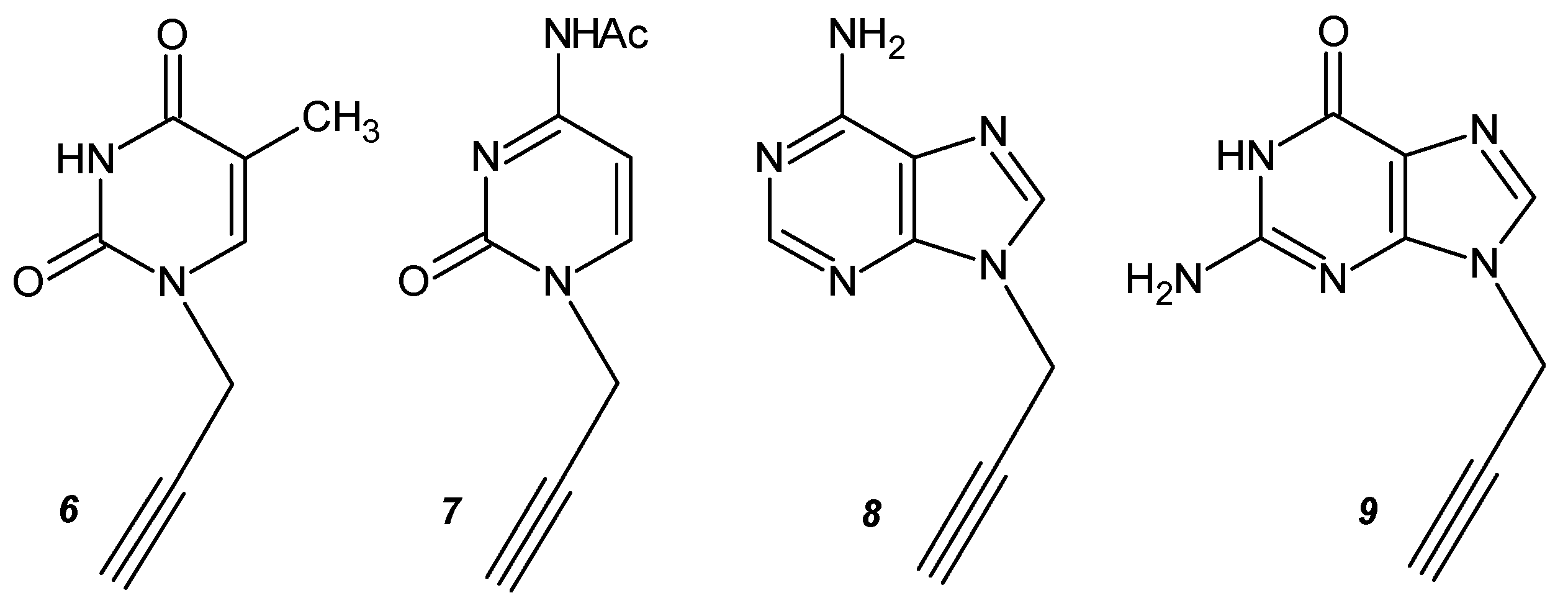
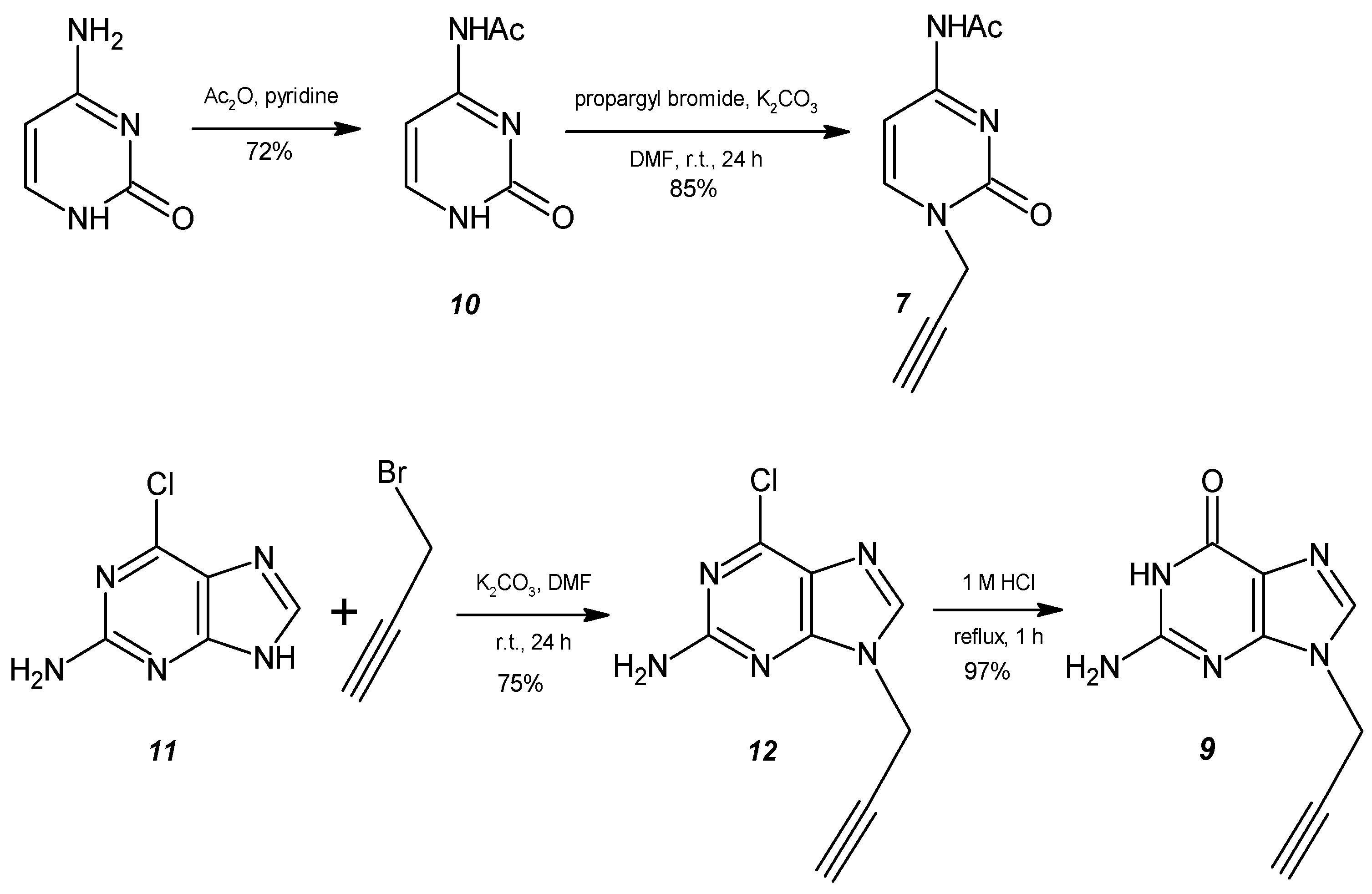
2.1. General Procedure for the Synthesis of the Monopropargyl Nucleobases (5)

N1-propargylthymine, N4-acetyl-N1-propargylcytosine, N9-propargyladenine, and N9-propargylguanine were obtained by reacting thymine, N4-acetylcytosine, adenine, and 2-amino-6-chloropurine with propargyl bromide in the presence of potassium carbonate in DMF at room temperature, as described in the literature [51]. The mixture of 2 mmol of the heterocyclic base (thymine, N4-acetylcytosine, adenine, 2-amino-6-chloropurine), 1.9 mmol of K2CO3, and 2 mmol of propargyl bromide in 20 mL of anhydrous DMF was stirred at room temperature for 24 h. After removing the solvent under reduced pressure, the residue obtained was purified on a silica gel column (DCM:MeOH, 95:5 v/v).
N1-propargylthymine (6) was obtained as a pale yellow solid (91.93 mg, 56%). ESI-MS: m/z calcd and found for [M+H]+ 165.06.
N4-acetylcytosine (10) was obtained as a white solid (220 mg, 72%). ESI-MS: m/z calcd for [M+H]+ 154.01; found: 154.03.
N4-acetyl-N1-propargylcytosine (7) was obtained as a white solid (120 mg, 85%). ESI-MS: m/z calcd for [M+H]+ 192.08; found: 192.07.
N9-propargyladenine (8) was obtained as a white solid (98.71 mg, 57%). ESI-MS: m/z calcd and found for [M+H]+ 174.07.
2-amino-6-chloro-N9-propargylpurine (12) was obtained as a pale yellow solid (311 mg, 75%). ESI-MS: m/z calcd and found for [M+H]+ 208.03.
N9-propargylguanine (9): A suspension of 12 (190.00 mg, 0.915 mmol) was dissolved in 1 M HCl (4 mL). The solution was heated at reflux for 1 h. After cooling, the solution was neutralized to pH 7 with 10% NaOH. The solvent was removed under reduced pressure, and the residue was purified by preparative C18 RP-HPLC to yield 5 as a white solid (167.9 mg, 97%). ESI-MS: m/z calcd and found for [M+H]+ 190.07.

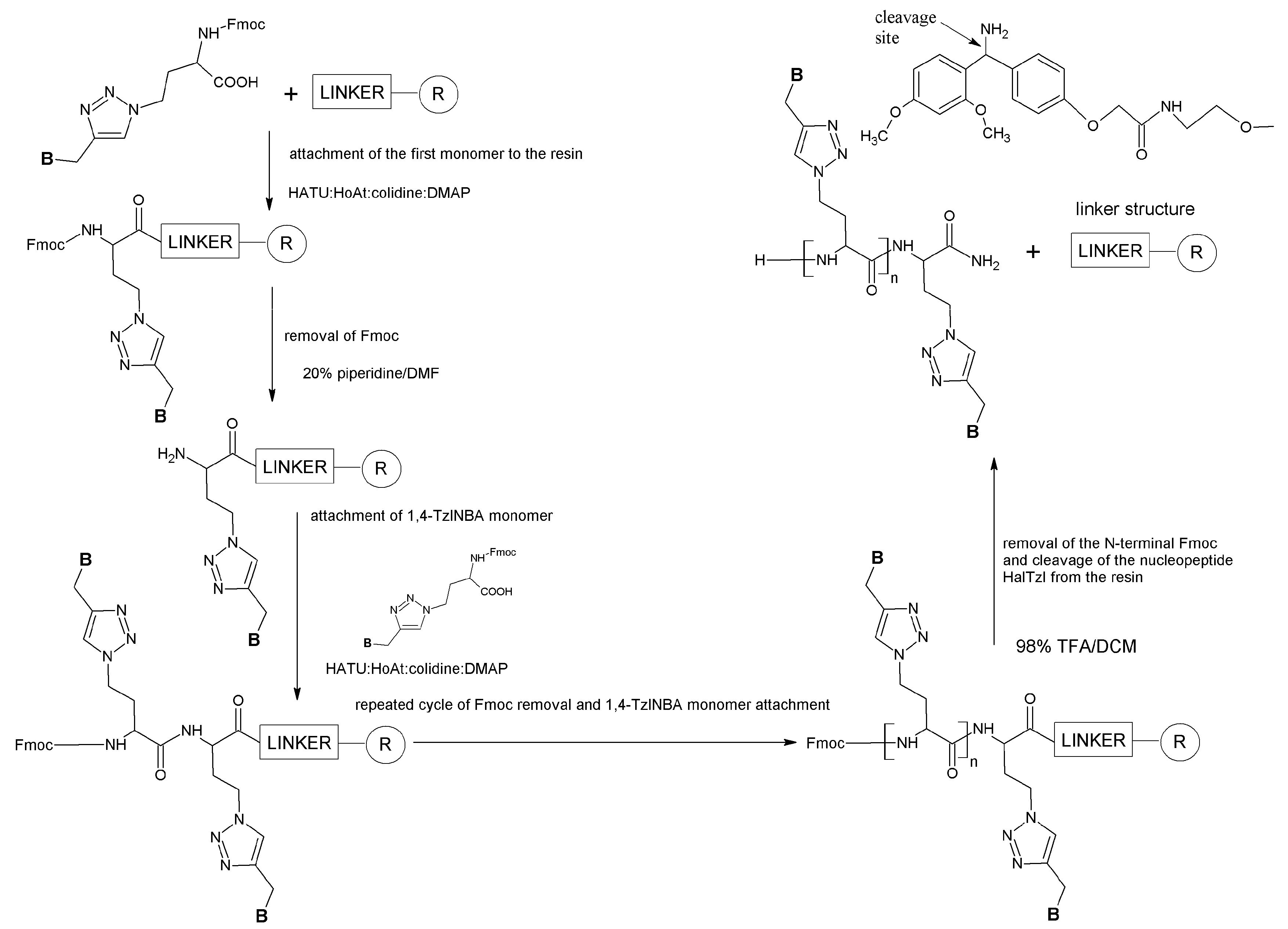
2.2. General Procedure for the Synthesis of 1,4-TzlNBAs

To a mixture of the corresponding N-propargyl nucleobase (1 equiv) and Fmoc-Aha (1.2 equiv) in H2O/t-BuOH (1:1), a 100 mM solution of CuSO4 × 5H2O (1.2 equiv) and a freshly prepared 500 mM solution of sodium ascorbate (4 equiv) were added. The reaction mixture was stirred at room temperature for 1 day and monitored by RP-HPLC. After completion, the resulting slurry was filtered, and the solvent was removed under reduced pressure. The crude product was pre-purified by precipitation from DMSO with cold water. A preparative C18 RP HPLC was then applied.
2-(Fmoc-amino)-4-[4-(N1-methylthymine)-1H-1,2,3-triazol-1-yl)butanoic acid (1)
According to the general procedure, from alkyne 6 (100 mg, 0.609 mmol) and Fmoc-Aha (267.8 mg, 0.731 mmol), monomer 1 (239.09 mg, 74%) was obtained after a preparative C18 RP-HPLC purification as a white solid. ESI-MS: m/z calcd for [M+H]+ 531.19; found: 531.15.
1H NMR (DMSO-d6, δ): 12.73 (1H, COOH, s); 11.23 (1H, NH(T), s); 7.98 (1H, H5(Tzl), s); 7.83 (2H, Fmoc, d, J = 7.55 Hz); 7.73 (1H, NH(Hal), d, J = 8.25 Hz); 7.66 (2H, Fmoc, d, J = 7.4 Hz); 7.53 (1H, H6, s); 7.35 (2H, Fmoc, ddd, J = 2.9 Hz); 7.27 (2H, Fmoc, m); 4.82 (2H, Tzl-CH2-T, s); 4.32 (2H, Tzl-CH2-CH2, m); 4.26 (2H, OCH2, d, J = 7.05 Hz); 4.18 (1H, CH(Fmoc), t, J = 6.8 Hz); 3.91 (1H, CH(Hal), m); 2.23 i 2.03 (2H, Tzl-CH2-CH2, m); 1.68 (3H, CH3, s).
13C NMR (DMSO-d6, δ): 173.50; 164.73; 156.61; 151.19; 144.27; 143.01; 141.58; 141.21; 128.12; 127.57; 125.69; 124.07; 120.59; 109.32; 66.10; 51.66; 47.13; 46.97; 42.65; 31.77; 12.41.
2-(Fmoc-amino)-4-[4-(N1-methyl-N4-acetylcytosine)-1H-1,2,3-triazol-1-yl)butanoic acid (2)
According to the general procedure, from alkyne 7 (100 mg, 0.523 mmol) and Fmoc-Aha (230 mg, 0.628 mmol), monomer 2 (201.2 mg, 69%) was obtained after precipitation from DMSO with cold water and purification of the resulting precipitate using preparative C18 RP HPLC as a white solid as a white solid. ESI-MS: m/z calcd for [M+H]+ 558.21; found: 558.25.
1H NMR (DMSO-d6, δ): 12.81 (1H, COOH, s); 10.84 (1H, NH(C), s); 8.19 (1H, H6, d, J = 7.25 Hz); 8.07 (1H, H5(Tzl), s); 7.90 (2H, Fmoc, d, J = 7.55 Hz); 7.80 (1H, NH(Hal), d, J = 8.05 Hz); 7.73 (2H, Fmoc, d, J = 7.45 Hz); 7.42 (2H, Fmoc, ddd, J = 3.35 Hz); 7.34 (2H, Fmoc, m); 7.17 (1H, H5, d, J = 7.25 Hz); 5.06 (2H, Tzl-CH2-C, s); 4.39 (2H, Tzl-CH2-CH2, m); 4.33 (2H, OCH2, m); 4.25 (1H, CH(Fmoc), t, J = 6.9 Hz); 3.92 (1H, CH(Hal), m); 2.30 (2H, Tzl-CH2-CH2, m); 2.09 (3H, CH3(Ac), s).
13C NMR (DMSO-d6, δ): 173.50; 171.37; 162.98; 156.61; 155.42; 150.60; 144.26; 141.21; 128.12; 127.57; 125.69; 124.46; 120.61; 95.78; 66.10; 51.68; 47.05; 44.87; 31.72; 24.78.
2-(Fmoc-amino)-4-[4-(N9-methyladenine)-1H-1,2,3-triazol-1-yl)butanoic acid (3)
According to the general procedure, from alkyne 8 (173 mg, 1 mmol) and Fmoc-Aha (439.7 mg, 1.2 mmol), monomer 3 (415.45 mg, 77%) was obtained after precipitation from DMSO with cold water and purification of the resulting precipitate using preparative C18 RP HPLC as a white solid. ESI-MS: m/z calcd for [M+H]+ 540.21; found: 540.16.
1H NMR (DMSO-d6, δ): 12.81 (1H, COOH, s); 8.26 i 8.21 (2H, H2 i H8, 2s); 8.09 (1H, H5(Tzl), s); 7.90 (2H, Fmoc, d, J = 7.4 Hz); 7.79 (1H, NH(Hal), d, J = 8.15 Hz); 7.73 (2H, Fmoc, d, J = 5.5 Hz); 7.61 (2H, NH2, s); 7.42 (2H, Fmoc, ddd, J = 4.15 Hz); 7.33 (2H, Fmoc, dd, J = 6.4 Hz); 5.46 (2H, Tzl-CH2-A, s); 4.38 (2H, Tzl-CH2-CH2, m); 4.33 (2H, OCH2, d, J = Hz); 4.25 (1H, CH(Fmoc), t, J = 6.9 Hz); 3.91 (1H, CH(Hal), m); 2.30 i 2.11 (2H, Tzl-CH2-CH2, m).
13C NMR (DMSO-d6, δ): 173.49; 156.60; 151.60; 149.60; 144.26; 142.84; 141.21; 128.12; 127.56; 125.70; 124.17; 120.61; 66.09; 51.63; 47.06; 38.64; 31.67.
2-(Fmoc-amino)-4-[4-(N9-methylguanine)-1H-1,2,3-triazol-1-yl)butanoic acid (4)
According to the general procedure, from alkyne 9 (100 mg, 0.529 mmol) and Fmoc-Aha (232.6 mg, 0.635 mmol), monomer 4 (211.59 mg, 72%) was obtained after precipitation from DMSO with cold water and purification of the resulting precipitate using preparative C18 RP HPLC as a white solid. ESI-MS: m/z calcd for [M+H]+ 556.20; found: 556.09.
1H NMR (DMSO-d6, δ): 12.83 (1H, COOH, s); 10.91 (1H, NH(G), s); 8.19 (1H, H8, s); 8.06 (1H, H5(Tzl), s); 7.90 (2H, Fmoc, d, J = 7.55 Hz); 7.80 (1H, NH(Hal), d, J = 8.15 Hz); 7.73 (2H, Fmoc, d, J = 6.05 Hz); 7.42 (2H, Fmoc, ddd, J = 3.85 Hz); 7.34 (2H, Fmoc, q, J = 6.95 Hz); 6.69 (2H, NH2(G), s); 5.30 (2H, Tzl-CH2-G, s); 4.40 (2H, Tzl-CH2-CH2, m); 4.34 (2H, OCH2, d, J = 7 Hz); 4.25 (1H, CH(Fmoc), t, J = 6.9 Hz); 3.91 (1H, CH(Hal), m); 2.30 i 2.11 (2H, Tzl-CH2-CH2, m).
13C NMR (DMSO-d6, δ): 173.50; 156.63; 154.80; 144.25; 141.21; 137.40; 128.12; 127.56; 125.69; 124.19; 120.60; 92.90; 66.11; 51.62; 47.12; 42.70; 38.60; 31.50.

2.3. Solid-Phase Synthesis of HalTzls

Nucleopeptides HalTzls were synthesized as C-terminal amides using a semi-automatic peptide synthesizer, Peptide Synthesizer SP650 Labortec AG (Bubendorf, Switzerland). The classical SPPS method with the Fmoc strategy was used [50]. Fmoc-protected 1,4-TzlNBA monomers were assembled on TentaGel S RAM resin (capacity 0.22 mmol Fmoc-NH/1g resin; 50 mg resin was used) as active derivatives in a 3-fold molar excess of coupling reagents (Fmoc-1,4-TzlNBA: HATU: HOAt: colidine: DMAP, 1:1:1:2:0.001 molar ratio) in the N,N-dimethylformamide (DMF) solution for 60 min. Each coupling process was repeated using a 1.5-fold excess of reagents for 60 min. The removal of the Fmoc groups was carried out with a 20% piperidine/DMF in two cycles lasting 5 and 15 min, respectively. The cleavage from the resin and deprotection of the HalTzls were performed with a 98% TFA/DCM solution for 120 min. After evaporation of TFA and DCM, the crude nucleopeptide was dissolved in water and, after freezing, lyophilized. The crude HalTzls were purified using a semi-preparative RP HPLC Knauer chromatograph (Knauer, WG GmbH, Berlin, Germany) system using a Kromasil C8 column (20 × 250 mm, 10 µm particle size). A gradient of 0–60% ACN with 0.08% TFA, at a flow rate of 3.5 mL/min, was used for purification. The column was maintained at ambient temperature. The eluates were monitored with a UV detector at λ = 254 nm. Fractions with the highest purity (>95%) were analyzed by analytical RP-HPLC using a Beckman System Gold chromatograph (Beckman Coulter, Brea, USA) using a Kinetex C18 column (Phenomenex, 150 × 4.6 mm, 5 µm particle size) with a 0–100% ACN gradient with the addition of 0.08% TFA (solvent B) and 0.08% TFA (solvent A) in 30 min. The column was maintained at ambient temperature. The flow rate was 1 mL/min, and the eluates were monitored using a UV detector at λ = 254 nm. The identity of HalTzls was confirmed by mass spectrometry using a QTOF SCIEX 5600+ ESI spectrometer operating in positive mode. A summary of the physical and chemical parameters of HalTzls can be found in Table 1.

2.4. TAR RNA Synthesis

5′-FAM(6)-TAR RNA UCU/UUU and its unlabeled forms were obtained from FutureSynthesis (Poznan, Poland). The lyophilized samples were dissolved in TB or Tris buffer and denatured at 95 °C for 5 min before CD and fluorescence anisotropy experiments.

2.5. Circular Dichroism (CD) Spectropolarimetry

CD spectra were recorded using a Jasco J-815 spectropolarimeter equipped with a Peltier-controlled cell holder in a quartz cuvette with a 0.1 cm path length spanning from 200 to 310 nm at 25 °C. CD temperature-dependent experiments were conducted over the range of 25–95 °C. TAR RNA was maintained at a concentration of 5 μM for all CD experiments. To prevent changes in ellipticity values linked to potential sample dilution, the solution containing TAR RNA was introduced to the dry lyophilized HalTzl sample, resulting in a final TAR RNA concentration of 5 μM and a 10-fold excess (50 μM) of nucleopeptide concentration. Before initiating CD experiments, each sample was equilibrated for at least 30 min at the initial temperature to ensure the required temperature and achieve equilibrium in TAR—HalTzl interactions. The spectra were averaged over three scans, each recorded at a speed of 100 nm/min with a 1 nm data pitch. As HalTzls nucleopeptides exhibit a negative ellipticity effect in the 200–240 nm range, ellipticity values for TAR—HalTzls complexes were corrected for the contribution associated with HalTzls. The observed ellipticity for both free and bound (corrected ellipticity) TAR was expressed in mdeg. The baseline was corrected for every CD spectrum. Origin 6.5 software (OriginLab, Northampton, MA, USA) was utilized for the analysis of obtained data in CD melting experiments. The Boltzmann function implemented in this program was employed for fitting melting curves and calculating Tm values.

2.6. Fluorescence Anisotropy Spectroscopy

The binding affinities between HalTzls and TAR UUU/UCU structures were determined through fluorescence anisotropy measurements. This was conducted using a Fluoromax-4 spectrofluorophotometer (Horiba Jobin Yvon IBH Ltd., Kyoto, Japan) with a cylindrical quartz cuvette featuring an optical path of 3 mm. Fluorescence anisotropy was computed from the intensities detected at 518 nm with excitation at 480 nm. Each data point was measured three times with a 500 ms integration time and then averaged. A 5′-FAM(6)-TAR RNA solution (final concentration 100 nM) underwent titration with increasing amounts of HalTzl. The reactions occurred in TB buffer at 25 °C. HalTzl (ligand) solutions were prepared as serial dilutions in TB buffer. Before initiating the experiment, the FAM-labeled TAR RNA sample was refolded by heating to 90 °C for 5 min and then being allowed to cool freely to 25 °C. Subsequently, the titration was performed. After adding the ligand to TAR RNA, the sample was gently mixed by hand and incubated to achieve complexation equilibrium at 25 °C for 20 min. Binding data were analyzed using a single binding site curve-fitting procedure implemented in GraphPad Prism 3.0 software (GraphPad Software Inc., San Diego, CA, USA).

3. Results and Discussion

3.1. Synthesis of N-Propargyl Nucleobases and 1,4-TzlNBA Monomers

In the ongoing pursuit of designing and synthesizing novel NBAs, we present the synthesis of nucleobase 1,2,3-triazole amino acid derivatives (1,4-TzlNBAs) (1–4) featuring the complete set of canonical nucleobases connected to the side chain of the commercially available Fmoc-Aha (13) through a 1,4-disubstituted 1,2,3-triazole linkage (Figure 1).
The monomers (1–4) were conceived as building blocks for nucleobase peptides, aligning with the Fmoc strategy of SPPS. In the initial step, we synthesized N-propargylated nucleobase derivatives (6–9) as the dipolarophile components of the CuAAC reaction (Scheme 1, Figure 2).
These compounds had been previously prepared using free or protected nucleobases and propargyl bromide [51]. The nucleobases were subjected to treatment with propargyl bromide in DMF in the presence of K2CO3 as a basic catalyst (Scheme 1 and Scheme 2). The reactions were conducted for 24 h at room temperature. Thymine and adenine were utilized as unprotected substrates, while cytosine and guanine, due to their reactive amino groups, were employed as N4-acetyl (10) and 2-amino-6-chloropurine (11) derivatives, respectively (Scheme 2).
We chose to use N4-acetyl (10) instead of the commonly used N4-benzoylcytosine because propargylation of the latter resulted in N1-propargyl-N4-benzoylcytosine, which was hardly soluble in organic solvents. The acetylation of cytosine was conducted with acetic anhydride in pyridine. N9-propargyl guanine was obtained through 2-amino-6-chloropurine propargylation (11), followed by its hydrolysis (12) in 1 M HCl for 1 h (Scheme 2). The short duration of hydrolysis was crucial, as prolonged hydrolysis led to an increased number and amount of side products. In all cases, propargylation resulted in N1- or N9-monopropargyl nucleobase as the main product of the reaction. This shows that the bromoalkylation reaction of nitrogenous bases with 1,2-dibromoethane is regioselective, albeit for some nucleobases, due to the reactivity of their functional groups, semipermanent or complete protection is necessary. In the final step, CuAAC was applied to “click” N-propargyl nucleobase and the azido moiety of Fmoc-L-Aha (13) (Scheme 3).
The four target Fmoc-1,4-TzlNBAs (1–4) were successfully prepared. The reaction was carried out overnight in an H2O/t-BuOH solution using CuSO4 and a freshly prepared sodium ascorbate solution. The final products were purified by column chromatography, and their structures were characterized by 1H and 13C NMR. Since none of the reactions used in the synthesis of the NBA monomers and HalTzls oligomers resulted in a change in the configuration of the asymmetry center (α-carbon atom) of the starting compound (Fmoc-L-Aha), all the synthesized compounds (Figure 1, Scheme 4) retained the L-configuration of the asymmetry center (or centers). The resulting Fmoc derivatives 1-4 are soluble in DMF, making it possible to use the standard solid-phase peptide synthesis (SPPS) protocol commonly used for the synthesis of peptides for the synthesis of HalTzls [52].

3.2. HalTzls Synthesis

After the synthesis of NBA monomers, we proceeded to synthesize HalTzls nucleopeptides using the SPPS method with the Fmoc strategy (Scheme 4).
To mitigate the unfavorable impact of electrostatic repulsion between the negatively charged phosphate groups in the TAR RNA structure and the C-terminal carboxylate group of HalTzls nucleopeptides, the latter were synthesized in the form of C-terminal amides. Various combinations of NBA coupling conditions were tested to optimize synthesis efficiency. The best results were achieved using a dual coupling mixture of Fmoc-1,4-TzlNBA:HATU:HOAt:collidine:DMAP (1:1:1:2:0.001 molar ratio) in DMF for two 60 min cycles. After separation from the resin and purification by RP HPLC, HalTzls were obtained with moderate to good yields, ranging from about 17% to 37% (Table 1). Only in the case of HalTzlTCCCAG did the cleavage procedure from the resin prove insufficient, resulting in partial removal of the protective acetyl groups located at the C4 position of the cytosine residues (Supplement data, Figure S1). Therefore, in this case, an additional procedure involved dissolving and leaving the resin-cleaved crude HalTzlTCCCAG in a 1 M K2CO3/MeOH solution for 1 h. After this time, all acetyl protective groups of cytosine were removed (Supplement data, Figure S1).
Following the purification and physicochemical characterization of HalTzls (Table 1, Figure S1), their ability to bind complementary RNA sequences using 5′-FAM(6)-TAR UCU/UUU as a model system of pharmacological relevance was assessed (Figure 3). For CD experiments, an unlabeled version of TAR UCU/UUU was used.

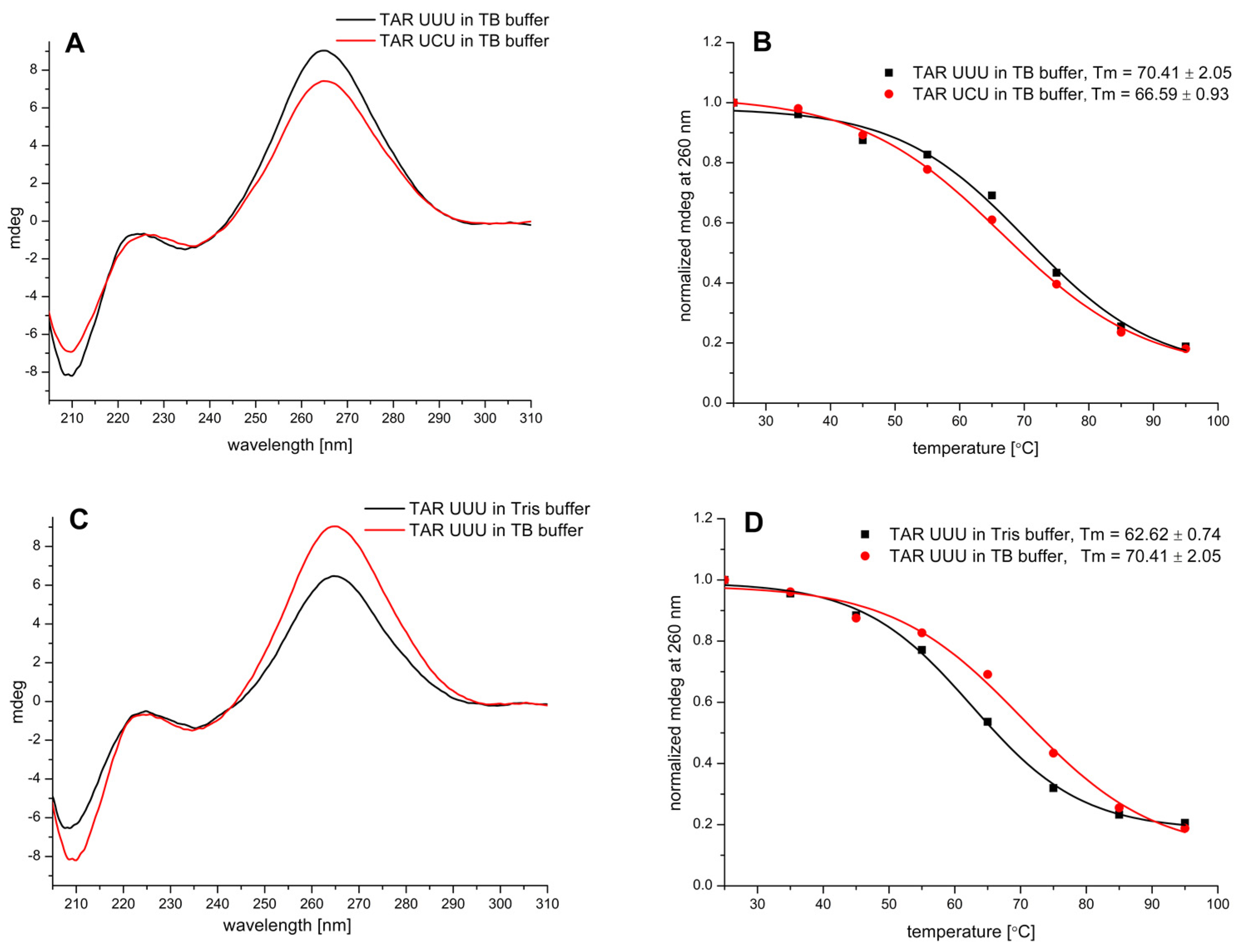
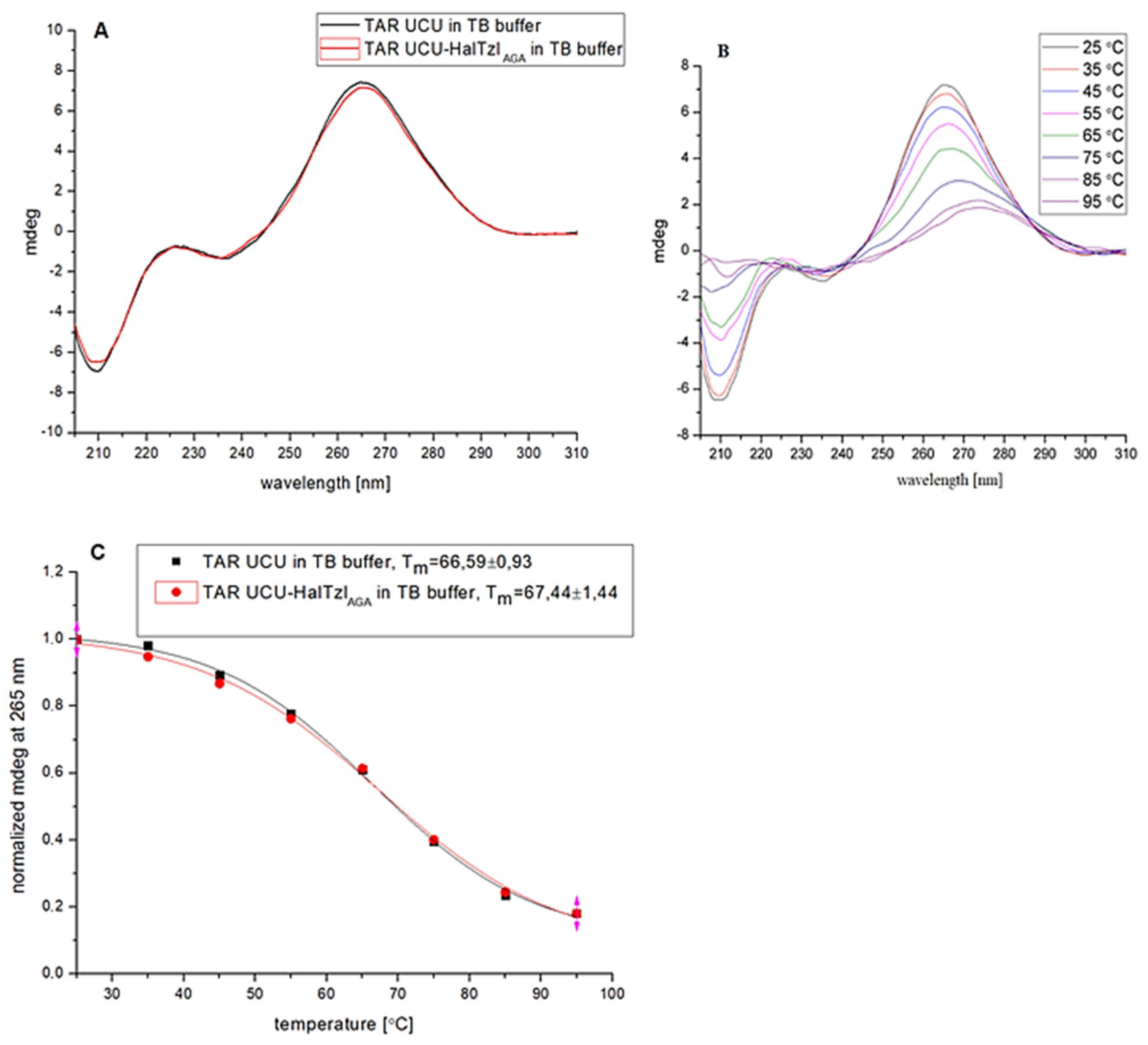
3.3. CD Study of TAR RNA—HalTzls Interactions

The CD method is a widely employed technique for studying conformational changes in both free and bound RNA [53,54]. In this study, we utilized this method to characterize the behavior of TAR UCU and UUU structures during the binding of HalTzls ligands. Temperature-dependent CD studies were employed to characterize both uncomplexed and bound TARs. CD spectra of RNA (form A) typically exhibit a negative minimum around 210 and 235 nm and a positive maximum at 265 nm [54,55]. The latter serves as a diagnostic band for ligand binding, as its intensity and position are sensitive to changes in the nucleobase stacking profile. These changes correlate with conformational alterations in RNA induced by physicochemical factors (e.g., temperature) or ligand binding.
The CD spectrum of free TAR UCU and TAR UUU recorded in the TB buffer is similar for both and is characteristic of the A form of RNA (Figure 4A).
The spectra exhibit three extremes: a minimum at about 208 nm and 235 nm and a maximum at 265 nm. In the case of TAR UCU, the intensity of the extrema was slightly lower (except for 235 nm) compared to the TAR UUU variant (Figure 4A). The CD experiment conducted for TAR UUU revealed that the intensity of the ellipticity signal is sensitive to environmental parameters, such as buffer type. The CD curves of TAR UUU recorded in Tris and TB buffers differ slightly in intensity at the extreme points of these curves, likely reflecting the small conformational changes occurring in the single-stranded bulge and apical loop fragments (Figure 4C).
To assess the stability of TAR’s hairpin-bulge structure, temperature-dependent CD measurements were conducted. CD curves recorded in the range of 25–95 °C showed that the structure of both TAR variants was temperature-dependent (Figure 4B). A gradual decrease in the intensity of both extremes correlated with increasing temperature. In the case of the maximum, a shift of the positive extremum toward longer wavelengths was observed. These changes reflect conformational changes in single-stranded fragments, the bulge, and the apical loop, as well as the unraveling of double-stranded helix fragments located both above and below the bulge. Temperature-dependent changes in ellipticity at a maximum of around 265 nm were used to characterize the stability of unbound TAR structures by determining their melting temperatures (Tm). TAR’s CD melting profiles are shown in Figure 4B,D. The curves were characterized by the distinctive sigmoid-like shape typical of unraveling double-stranded TAR fragments. The determined Tm values indicated that the structure of TAR UUU is much more stable in TB than in Tris buffer (Tm 70.41 ± 2.05 vs. 62.62 ± 0.74 °C) (Table 2). The difference is almost 4 °C. This highlights the crucial role of environmental conditions (e.g., buffer) in RNA activity studies. The greater stability of a TAR structure containing a U-rich UUU bulge relative to one containing a UCU bulge has been observed previously [56]. Our temperature-dependent CD studies have shown that the observed changes in ellipticity signal intensity, especially around the extremum at 265 nm, correlate with conformational changes affecting the stability of TAR structures.
Subsequently, CD was employed to characterize the binding of HalTzls ligands to the TAR UCU/UUU structure. As the quantitative characterization of the target interactions was determined by dissociation constants (Kd) using fluorescence anisotropy spectroscopy, CD experiments focused on the analysis of complexes formed under conditions of a 10-fold excess of HalTzls ligands relative to TAR. In general, the CD curves illustrating the interaction of TAR with HalTzls ligands are characterized by a reduction in the intensity of the band ellipticity at 265 nm and a less intense effect for the band at 210 nm (Figure 5, Figure 6 and Figure 7). Since the HalTzls ligands themselves exhibit a negative ellipticity value in the 200–240 nm range, the CD curves of the complexes were corrected for this effect. CD curves recorded for the TAR UUU—HalTzlAAA complexes showed similar binding effects in both Tris and TB buffers (Figure 5).
In both cases, the decrease in ellipticity intensity for the band at approximately 265 nm was approximately 15%. However, some differences related to the stability of the complex associated with the Tm parameter were observed (Figure 5, Table 2). With increasing temperature, CD melting curves were characterized by a gradual decrease in the intensity of the CD signal at a maximum of 265 nm and a shift of this maximum toward longer wavelengths. At the highest temperature investigated (95 °C), the CD maximum was moved to about 275 nm (Figure 5A,B). CD melting curves show that the stability of the complex TAR UUU—HalTzlAAA characterized by the Tm value is higher in the TB than in the Tris buffer. These differences are derived from the primary effect of differences in the stability of unbound TAR UUU in these buffers (Figure 4B). A small difference in the Tm parameter between the unbound and bound form of TAR UUU was recorded in the Tris buffer (Table 2). In the TB buffer, these values are almost the same. These may indicate a relatively low affinity of HalTzlAAA for the TAR UUU bulge. The CD spectrum revealed that the interaction between TAR UCU and the HalTzlAGA ligand, complementary to the bulge structure, caused only a 4% decrease in ellipticity intensity at 265 nm (Figure 6).
This effect was significantly smaller, almost four times, compared to the observed effect for TAR UUU interacting with HalTzlAAA. Analysis of the CD melting curves indicated that this change correlated with a slight increase in stability (approximately 1 °C) of the bound TAR UCU structure (Table 1). For the TAR UCU interaction with HalTzlTCCCAG, complementary to the apical loop of TAR UCU, almost a 27% decrease in ellipticity intensity at 265 nm was observed (Figure 7). This effect was correlated with a 2 °C increase in the stability of the bound vs. free TAR UCU structure.

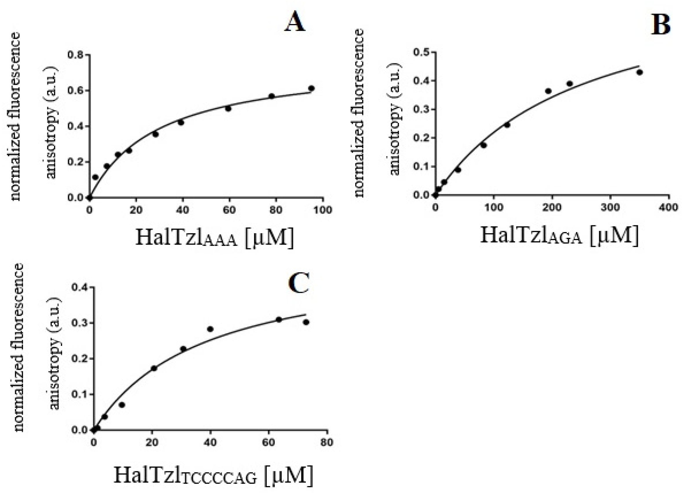
3.4. Fluorescence Anisotropy Study of TAR RNA—HalTzls Interactions

Fluorescence anisotropy was employed to quantify TAR—HalTzl interactions. 5′-FAM(6)-labeled TAR UUU/UCU RNA fragments were utilized as target molecules to characterize the binding properties of HalTzls ligands. The binding experiments involved measuring the decreasing fluorescence anisotropy of FAM-TAR caused by the addition of increasing amounts of HalTzls (Figure 8).
The obtained data were used to determine binding curves. The dissociation constants (Kd) of the FAM-TAR—HalTzls complexes were then calculated (Table 3). To derive the binding curves and Kd values, it was assumed that the interaction corresponds to the one-binding-site model, where the HalTzl ligand binds to the complementary sequences of the bulge or the apical loop regions of TAR.
The analysis of TAR UUU interactions with HalTzlAAA revealed a medium-strength binding characterized by a dissociation constant (Kd) of approximately 30 µM (Figure 8A, Table 3). In contrast, when the HalTzAGA ligand bound to the TAR UCU structure, the binding was nearly 9-fold weaker compared to TAR UUU—HalTzlAAA (Figure 8B, Table 3), with a Kd of ~256 µM. This behavior of the HalTzlAGA ligand, despite the presence of a guanine (G) residue capable of forming three hydrogen bonds (compared to the two formed by adenine (A)), can only be explained by the steric constraints present during the interaction. This is intriguing because, in both TAR UUU and UCU variants, the bulges have a high degree of conformational freedom and should easily adapt to the structure of the ligands. For TAR UCU interaction with HalTzlTCCCAG, a much stronger binding was observed with a Kd of ~38 µM compared to the interaction with the HalTzlAGA ligand (Kd ~256 µM) (Figure 8C, Table 3). In these cases, an almost 7-fold difference in the strength of the ligand binding by the TAR structure was observed. This indicates that the HalTzlTCCCAG hexamer binds much more tightly to the apical loop of TAR UCU than the HalTzlAGA trimer to the UCU bulge. Although the ligand and the apical loop and bulge sequences are complementary to each other, the ligand sequences differ in length (HalTzlTCCCAG sequence is 2-fold longer than that of HalTzlAGA). Surprisingly, the interaction strength for the TAR UCU—HalTzlTCCCAG system is similar to that observed for the TAR UUU—HalTzlAAA. This may indicate that, in addition to the complementarity of nucleosides connected with the ability to form hydrogen bonds, other factors like interactions of nucleobases ring systems with triazole moieties of side chains of the ligands play an important role in the TAR—HalTzl interactions. The pronounced differences observed in the binding of ligands of the same size trimers HalTzlAAA and HalTzlAGA to the UCU bulge suggest that steric factors may also play an important role in the recognition of TAR structure by HalTzl ligands. The tested ligands demonstrated moderate (approximately 30–40 µM) or low (approximately 260 µM) affinity for TAR structures. This implies that interactions, such as hydrogen bonding or hydrophobic interactions involving complementary nucleobases and triazole rings, are insufficient to yield ligands with high affinity for TAR structures. Introducing positively charged amino acid residues, such as lysine (Lys) or arginine (Arg), into newly designed structures based on HalTzl sequences would be a reasonable strategy. This approach, commonly used in ligand design, including nucleopeptides binding to RNA/DNA, involves additional electrostatic interactions between the positively charged side chains of Lys or Arg and the negatively charged phosphate groups of the sugar–phosphate chain of RNA. Such modifications are expected to significantly increase the affinity of the HalTzl ligands for the TAR structure.
The popularity of click chemistry, especially the CuAAC variant, and the ease of formation of the triazole ring have made the design and synthesis of new systems containing this system an important branch of synthetic organic chemistry [57]. In many cases, the newly obtained triazole derivatives of known chemical structures showed greater biological activity than their unmodified counterparts [57,58,59]. In most cases, the triazole ring was introduced into small-molecule compounds, although examples of the introduction of this system into large macromolecules such as peptides or nucleic acids are known [60,61,62]. The molecular basis of such modifications underlying the changes in physicochemical properties and biological activity of such modified biomolecules is poorly understood. Previous results have shown that the presence of the triazole system strongly influences the structure and stability of nucleic acid duplexes [63]. The effect of triazole-modified 2′-deoxyuridines on the stability of DNA/DNA and DNA/RNA duplexes has been reported [64,65]. Authors observed that although a single triazole ring incorporation decreased DNA/DNA duplex stability, the stacking of a few (2–4 triazole residues) consecutive modifications led to increased stability due to additional base stacking interactions of triazole moieties. This demonstrates the complex nature of the interactions of the triazole system with the structure of the nucleic acid chain, both nucleobases and the backbone, which are likely to involve electrostatic and hydrophobic interactions, including base stacking, the possibility of forming hydrogen bonds, as well as steric hindrance, as the triazole ring is a flat aromatic system similar in size to the pyrimidine nucleobase. To our knowledge, the triazole system has not yet been introduced as a structural element in NPs. Our strategy of introducing the triazole system into each side chain and linking the nucleobase to the rest of the amino acid molecule through the triazole ring is the first step towards understanding the influence of the triazole ring on the process of nucleic acid recognition by such modified NPs.

4. Conclusions

In summary, a complete set of Fmoc-protected nucleobase 1,2,3-triazole-linked amino acid derivatives (1,4-TzlNBAs) has been synthesized using click chemistry (CuAAC). These monomers were suitable for the solid-phase synthesis of a new type of nucleopeptides, HalTzls. Interaction studies using circular dichroism and fluorescence anisotropy have demonstrated that the designed and synthesized HalTzl ligands bind to complementary sequences of TAR RNA HIV-1, albeit with moderate affinity. This suggests that introducing additional positively charged amino acid residues, such as Lys and Arg, into the structure of HalTzls ligands could, through extra electrostatic interactions, enhance the affinity of HalTzls for TAR RNA. This enhancement may contribute to the development of new inhibitors blocking the TAR RNA-dependent transactivation during HIV replication.

Supplementary Materials

The following supporting information can be downloaded at: https://www.mdpi.com/article/10.3390/biomedicines12030570/s1.

Author Contributions

Data curation, P.M.; Formal Analysis, P.M., M.P., J.R. and P.R.; Funding Acquisition, P.M. and P.R.; Investigation, P.M., M.P., I.B., W.W. and K.P.; Methodology, P.M., M.P., I.B. and W.W.; Project Administration, P.M.; Resources, P.M.; Supervision, P.M.; Validation, P.M.; Visualization, P.M., M.P. and J.R.; Writing—Original Draft, P.M. and M.P.; Writing—Review and Editing, P.M., I.B., J.R. and P.R. All authors have read and agreed to the published version of the manuscript.

Funding

This research was funded by the National Science Centre, Poland, grant number N N204 355540 to P.M.

Institutional Review Board Statement

Not applicable.

Informed Consent Statement

Not applicable.

Data Availability Statement

Data are contained within the article and Supplementary Materials.

Conflicts of Interest

The authors declare no conflicts of interest.

Abbreviations

1,4-TzlNBAnucleobase amino acid bearing of nucleobase attached to the side chain via 1,4-linked 1,2,3-triazole moiety
Ac acetyl
ACN acetonitrile
ARR arginine-rich region
B, BH nucleobase
C18 octadodecane chain
CuAAC Cu(I)-catalyzed azide-alkyne cycloaddition
DCM dichloromethane
DMAP 4-Dimethylaminopyridine
DMF N,N-dimethylformamide
FAM fluorescein
Fmoc fluorenylmethyloxycarbonyl
Fmoc-AhaFmoc-L-azidohomoalanine
Hal L-homoalanine
HalTzl nucleopeptide designed on the basis of L-azidohomoalanine (Hal), containing nucleobases attached through a 1,4-linked-triazole linker to the Hal’s side chain
HATU [dimethylamino(triazolo[4,5-b]pyridin-3-yloxy)methylidene]-dimethylazanium;3-hydroxytriazolo[4,5-b]pyridine hexafluorophosphate
HIV-1 human immunodeficiency virus type 1
HOAt 1-Hydroxy-7-azabenzotriazole
MeOH methanol
NBA nucleobase amino acid
NP nucleopeptide
PNA peptide nucleic acid
RNAP II RNA polymerase II
RT reverse transcriptase
SPPS solid-phase peptide synthesis
TAR trans-activation response element
TB tris/borate buffer
t-BuOH tert-butanol
TFA trifluoroacetic acid
Tzl 1,2,3-triazol

References

  1. Harries, L.W. RNA Biology Provides New Therapeutic Targets for Human Disease. Front. Genet. 2019, 10, 205. [Google Scholar] [CrossRef] [PubMed]
  2. Jeong, S. RNA in Biology and Therapeutics. Mol. Cells 2023, 46, 1–2. [Google Scholar] [CrossRef] [PubMed]
  3. Zhu, Y.; Zhu, L.; Wang, X.; Jin, H. RNA-based therapeutics: An overview and prospectus. Cell Death Dis. 2022, 13, 644. [Google Scholar] [CrossRef] [PubMed]
  4. Damase, T.R.; Sukhovershin, R.; Boada, C.; Taraballi, F.; Pettigrew, R.I.; Cooke, J.P. The Limitless Future of RNA Therapeutics. Front. Bioeng. Biotechnol. 2021, 9, 628137. [Google Scholar] [CrossRef] [PubMed]
  5. Mucha, P.; Szyk, A.; Rekowski, P.; Weiss, P.A.; Agris, P.F. Anticodon domain methylated nucleosides of yeast tRNA(Phe) are significant recognition determinants in the binding of a phage display selected peptide. Biochemistry 2001, 40, 14191–14199. [Google Scholar] [CrossRef] [PubMed]
  6. Singh, G.; Monga, V. Peptide Nucleic Acids: Recent Developments in the Synthesis and Backbone Modifications. Bioorg. Chem. 2023, 141, 106860. [Google Scholar] [CrossRef]
  7. Wang, F.; Li, P.; Chu, H.C.; Lo, P.K. Nucleic Acids and Their Analogues for Biomedical Applications. Biosensors 2022, 12, 93. [Google Scholar] [CrossRef]
  8. Coppock, M.B.; Williams, M.E. Nucleic Acid Mimetics Supramolecular Aspects of Chemical Biology. In Supramolecular Chemistry: From Molecules to Nanomaterials; John Wiley & Sons, Ltd.: Hoboken, NJ, USA, 2012. [Google Scholar]
  9. Du, X.; Zhou, J.; Li, X.; Xu, B. Self-assembly of nucleopeptides to interact with DNAs. Interface Focus. 2017, 7, 20160116. [Google Scholar] [CrossRef]
  10. Robles, J.; Pedroso, E.; Grandas, A. Solid-phase synthesis of a nucleopeptide from the linking site of adenovirus-2 nucleoprotein, -Ser(p5′CATCAT)-Gly-Asp-. Convergent versus stepwise strategy. Nucleic Acids Res. 1995, 23, 4151–4161. [Google Scholar] [CrossRef]
  11. Tomassi, S.; Ieranò, C.; Del Bene, A.; D’Aniello, A.; Napolitano, M.; Rea, G.; Auletta, F.; Portella, L.; Capiluongo, A.; Mazzarella, V.; et al. Tailoring the Structure of Cell Penetrating DNA and RNA Binding Nucleopeptides. Int. J. Mol. Sci. 2022, 23, 8504. [Google Scholar] [CrossRef]
  12. Wojciechowska, M.; Ruczyński, J.; Rekowski, P.; Alenowicz, M.; Mucha, P.; Pieszko, M.; Miszka, A.; Dobkowski, M.; Bluijssen, H. Synthesis and hybridization studies of a new CPP-PNA conjugate as a potential therapeutic agent in atherosclerosis treatment. Protein Pept. Lett. 2014, 21, 672–678. [Google Scholar] [CrossRef]
  13. Nandhini, K.P.; Shaer, D.A.; Albericio, F.; de la Torre, B.G. The challenge of peptide nucleic acid synthesis. Chem. Soc. Rev. 2023, 52, 2764–2789. [Google Scholar] [CrossRef]
  14. Childs-Disney, J.L.; Yang, X.; Gibaut, Q.M.R.; Tong, Y.; Batey, R.T.; Disney, M.D. Targeting RNA structures with small molecules. Nat. Rev. Drug Discov. 2022, 21, 736–762. [Google Scholar] [CrossRef]
  15. Warner, K.; Hajdin, C.; Weeks, K. Principles for targeting RNA with drug-like small molecules. Nat. Rev. Drug Discov. 2018, 17, 547–558. [Google Scholar] [CrossRef]
  16. Falese, J.P.; Donlic, A.; Hargrove, A.E. Targeting RNA with small molecules: From fundamental principles towards the clinic. Chem. Soc. Rev. 2021, 50, 2224–2243. [Google Scholar] [CrossRef]
  17. Pant, S.; Jena, N.R. Inhibition of the RNA-dependent RNA Polymerase of the SARS-CoV-2 by Short Peptide Inhibitors. Eur. J. Pharm. Sci. 2021, 167, 106012. [Google Scholar] [CrossRef]
  18. Mucha, P.; Szyk, A.; Rekowski, P.; Barciszewski, J. Structural requirements for conserved Arg52 residue for interaction of the human immunodeficiency virus type 1 trans-activation responsive element with trans-activator of transcription protein (49-57). Capillary electrophoresis mobility shift assay. J. Chrom. A. 2002, 968, 211–220. [Google Scholar] [CrossRef]
  19. Musumeci, D.; Roviello, V.; Roviello, G.N. DNA- and RNA-binding ability of oligoDapT, a nucleobase-decorated peptide, for biomedical applications. Int. J. Nanomed. 2018, 13, 2613–2629. [Google Scholar] [CrossRef]
  20. Mercurio, M.E.; Tomassi, S.; Gaglione, M.; Russo, R.; Chambery, A.; Lama, S.; Stiuso, P.; Cosconati, S.; Novellino, E.; Di Maro, S.; et al. Switchable Protecting Strategy for Solid Phase Synthesis of DNA and RNA Interacting Nucleopeptides. J. Org. Chem. 2016, 81, 11612–11625. [Google Scholar] [CrossRef]
  21. van der Heden van Noort, G.J.; van Delft, P.; Meeuwenoord, N.J.; Overkleeft, H.S.; van der Marel, G.A.; Filippov, D.V. Fully automated sequential solid phase approach towards viral RNA-nucleopeptides. Chem. Commun. 2012, 48, 8093–8095. [Google Scholar] [CrossRef]
  22. Verona, M.; Verdolino, V.; Palazzesi, F.; Corradini, R. Focus on PNA Flexibility and RNA Binding using Molecular Dynamics and Metadynamics. Sci. Rep. 2017, 7, 42799. [Google Scholar] [CrossRef]
  23. Pellestor, F.; Paulasova, P. The peptide nucleic acids (PNAs), powerful tools for molecular genetics and cytogenetics. Eur. J. Hum. Genet. 2004, 12, 694–700. [Google Scholar] [CrossRef]
  24. Kiliszek, A.; Banaszak, K.; Dauter, Z.; Rypniewski, W. The first crystal structures of RNA-PNA duplexes and a PNA-PNA duplex containing mismatches--toward anti-sense therapy against TREDs. Nucleic Acids Res. 2016, 44, 1937–1943. [Google Scholar] [CrossRef]
  25. Quijano, E.; Bahal, R.; Ricciardi, A.; Saltzman, W.M.; Glazer, P.M. Therapeutic Peptide Nucleic Acids: Principles, Limitations, and Opportunities. Yale J. Biol. Med. 2017, 90, 583–598. [Google Scholar]
  26. MacLelland, V.; Kravitz, M.; Gupta, A. Therapeutic and diagnostic applications of antisense peptide nucleic acids. Mol. Ther. Nucleic Acids 2023, 35, 102086. [Google Scholar] [CrossRef]
  27. Musumeci, D.; Mokhir, A.; Roviello, G.N. Synthesis and nucleic acid binding evaluation of a thyminyl l-diaminobutanoic acid-based nucleopeptide. Bioorg. Chem. 2020, 100, 103862. [Google Scholar] [CrossRef]
  28. Roviello, G.N.; Musumeci, D. Synthetic approaches to nucleopeptides containing all four nucleobases, and nucleic acid-binding studies on a mixed-sequence nucleo-oligolysine. RSC Adv. 2016, 6, 63578–63585. [Google Scholar] [CrossRef]
  29. Roviello, G.N.; Vicidomini, C.; Di Gaetano, S.; Capasso, D.; Musumeci, D.; Roviello, V. Solid phase synthesis and RNA-binding activity of an arginine-containing nucleopeptide. RSC Adv. 2016, 6, 14140–14148. [Google Scholar] [CrossRef]
  30. Roviello, G.N.; Musumeci, D.; D’Alessandro, C.; Pedone, C. Binding ability of a thymine-functionalized oligolysine towards nucleic acids. Bioorg. Med. Chem. 2014, 22, 997–1002. [Google Scholar] [CrossRef]
  31. Tomassi, S.; Montalban, F.F.; Russo, R.; Novellino, E.; Messere, A.; Di Maro, S. Investigation of the Stereochemical-Dependent DNA and RNA Binding of Arginine-Based Nucleopeptides. Symmetry 2019, 11, 567. [Google Scholar] [CrossRef]
  32. Kriek, N.M.; Meeuwenoord, N.J.; van den Elst, H.; Heus, H.A.; van der Marel, G.A.; Filippov, D.V. Chemical synthesis of picornaviral protein primers of RNA replication. Org. Biomol. Chem. 2006, 4, 3576–3586. [Google Scholar] [CrossRef]
  33. Gotora, P.T.; van der Sluis, R.; Williams, M.E. HIV-1 Tat amino acid residues that influence Tat-TAR binding affinity: A scoping review. BMC Infect. Dis. 2023, 23, 164. [Google Scholar] [CrossRef]
  34. Singh, R.R.; Mondal, I.; Janjua, T.; Popat, A.; Kulshreshtha, R. Engineered smart materials for RNA based molecular therapy to treat Glioblastoma. Bioact. Mater. 2023, 33, 396–423. [Google Scholar] [CrossRef]
  35. Yu, A.M.; Choi, Y.H.; Tu, M.J. RNA Drugs and RNA Targets for Small Molecules: Principles, Progress, and Challenges. Pharmacol. Rev. 2020, 72, 862–898. [Google Scholar] [CrossRef]
  36. Garner, A.L. Contemporary Progress and Opportunities in RNA-Targeted Drug Discovery. ACS Med. Chem. Lett. 2023, 14, 251–259. [Google Scholar] [CrossRef]
  37. Abulwerdi, F.A.; Le Grice, S.F.J. Recent Advances in Targeting the HIV-1 Tat/TAR Complex. Curr. Pharm. Des. 2017, 23, 4112–4121. [Google Scholar] [CrossRef]
  38. Ne, E.; Palstra, R.J.; Mahmoudi, T. Transcription: Insights From the HIV-1 Promoter. Int. Rev. Cell Mol. Biol. 2018, 335, 191–243. [Google Scholar]
  39. Rice, A.P. The HIV-1 Tat Protein: Mechanism of Action and Target for HIV-1 Cure Strategies. Curr. Pharm. Des. 2017, 23, 4098–4102. [Google Scholar] [CrossRef]
  40. Schulze-Gahmen, U.; Hurley, J.H. Structural mechanism for HIV-1 TAR loop recognition by Tat and the super elongation complex. Proc. Natl. Acad. Sci. USA 2018, 115, 12973–12978. [Google Scholar] [CrossRef]
  41. Ronsard, L.; Rai, T.; Rai, D.; Ramachandran, V.G.; Banerjea, A.C. In silico Analyses of Subtype Specific HIV-1 Tat-TAR RNA Interaction Reveals the Structural Determinants for Viral Activity. Front. Microbiol. 2017, 8, 1467. [Google Scholar] [CrossRef]
  42. Obayashi, C.M.; Shinohara, Y.; Masuda, T.; Kawai, G. Influence of the 5′-terminal sequences on the 5′-UTR structure of HIV-1 genomic RNA. Sci. Rep. 2021, 11, 10920. [Google Scholar] [CrossRef]
  43. Frankel, A.D.; Young, J.A. HIV-1: Fifteen proteins and an RNA. Annu. Rev. Biochem. 1998, 67, 1–25. [Google Scholar] [CrossRef]
  44. Dutilleul, A.; Rodari, A.; Van Lint, C. Depicting HIV-1 Transcriptional Mechanisms: A Summary of What We Know. Viruses 2020, 12, 1385. [Google Scholar] [CrossRef]
  45. Chavali, S.S.; Bonn-Breach, R.; Wedekind, J.E. Face-time with TAR: Portraits of an HIV-1 RNA with diverse modes of effector recognition relevant for drug discovery. J. Biol. Chem. 2019, 294, 9326–9341. [Google Scholar] [CrossRef]
  46. Chavali, S.S.; Mali, S.M.; Jenkins, J.L.; Fasan, R.; Wedekind, J.E. Co-crystal structures of HIV TAR RNA bound to lab-evolved proteins show key roles for arginine relevant to the design of cyclic peptide TAR inhibitors. J. Biol. Chem. 2020, 295, 16470–16486. [Google Scholar] [CrossRef]
  47. Chavali, S.S.; Mali, S.M.; Bonn, R.; Saseendran Anitha, A.; Bennett, R.P.; Smith, H.C.; Fasan, R.; Wedekind, J.E. Cyclic peptides with a distinct arginine-fork motif recognize the HIV trans-activation response RNA in vitro and in cells. J. Biol. Chem. 2021, 297, 101390. [Google Scholar] [CrossRef]
  48. Kaushik, N.; Basu, A.; Palumbo, P.; Myers, R.L.; Pandey, V.N. Anti-TAR polyamide nucleotide analog conjugated with a membrane-permeating peptide inhibits human immunodeficiency virus type 1 production. J. Virol. 2002, 76, 3881–3891. [Google Scholar] [CrossRef]
  49. Chaubey, B.; Tripathi, S.; Ganguly, S.; Harris, D.; Casale, R.A.; Pandey, V.N. A PNA-transportan conjugate targeted to the TAR region of the HIV-1 genome exhibits both antiviral and virucidal properties. Virology 2005, 331, 418–428. [Google Scholar] [CrossRef]
  50. Tan, X.; Bruchez, M.P.; Armitage, B.A. Closing the Loop: Constraining TAT Peptide by γPNA Hairpin for Enhanced Cellular Delivery of Biomolecules. Bioconjug Chem. 2018, 29, 2892–2898. [Google Scholar] [CrossRef]
  51. Lazrek, H.B.; Taourirte, M.; Oulih, T.; Barascut, J.L.; Imbach, J.L.; Pannecouque, C.; Witrouw, M.; De Clercq, E. Synthesis and anti-HIV activity of new modified 1,2,3-triazole acyclonucleosides. Nucleosides Nucleotides Nucleic Acids 2001, 20, 1949–1960. [Google Scholar] [CrossRef]
  52. Dobkowski, M.; Szychowska, A.; Pieszko, M.; Barciszewski, J.; Mucha, P. ‘Click’ chemistry synthesis and capillary electrophoresis study of 1,4-linked 1,2,3-triazole AZT-systemin conjugate. J. Pep. Sci. 2014, 20, 696–703. [Google Scholar] [CrossRef]
  53. Qi, L.; Zhang, J.; Liu, Q.; Gao, X. Ligand “switching on” fluorescence of HIV-1 RNA-templated copper nanoclusters for ligand-RNA interaction assays. Int. J. Biol. Macromol. 2023, 256, 127779. [Google Scholar] [CrossRef]
  54. Kumar, S.; Maiti, S. The effect of N-acetylation and N-methylation of lysine residue of Tat peptide on its interaction with HIV-1 TAR RNA. PLoS ONE 2013, 8, e77595. [Google Scholar] [CrossRef]
  55. Garg, A.; Heinemann, U. A novel form of RNA double helix based on G·U and C·A+ wobble base pairing. RNA 2018, 24, 209–218. [Google Scholar] [CrossRef]
  56. Carter-O’Connell, I.; Booth, D.; Eason, B.; Grover, N. Thermodynamic examination of trinucleotide bulged RNA in the context of HIV-1 TAR RNA. RNA 2008, 14, 2550–2556. [Google Scholar] [CrossRef]
  57. Zhao, R.; Zhu, J.; Jiang, X.; Bai, R. Click chemistry-aided drug discovery: A retrospective and prospective outlook. Eur. J. Med. Chem. 2024, 264, 116037. [Google Scholar] [CrossRef]
  58. Manoharan, A.; Jayan, J.; Rangarajan, T.M.; Bose, K.; Benny, F.; Ipe, R.S.; Kumar, S.; Kukreti, N.; Abdelgawad, M.A.; Ghoneim, M.M.; et al. “Click Chemistry”: An Emerging Tool for Developing a New Class of Structural Motifs against Various Neurodegenerative Disorders. ACS Omega 2023, 8, 44437–44457. [Google Scholar] [CrossRef]
  59. Kaur, J.; Saxena, M.; Rishi, N. An Overview of Recent Advances in Biomedical Applications of Click Chemistry. Bioconjugate Chem. 2021, 32, 1455–1471. [Google Scholar] [CrossRef]
  60. Guarrochena, X.; Kaudela, B.; Mindt, T.L. Automated solid-phase synthesis of metabolically stabilized triazolo-peptidomimetics. J. Pept. Sci. 2023, 29, e3488. [Google Scholar] [CrossRef]
  61. Carter, E.P.; Ang, C.G.; Chaiken, I.M. Peptide Triazole Inhibitors of HIV-1: Hijackers of Env Metastability. Curr. Protein Pept. Sci. 2023, 24, 59–77. [Google Scholar] [CrossRef]
  62. Baraniak, D.; Boryski, J. Triazole-Modified Nucleic Acids for the Application in Bioorganic and Medicinal Chemistry. Biomedicines 2021, 9, 628. [Google Scholar] [CrossRef] [PubMed]
  63. Efthymiou, T.; Gong, W.; Desaulniers, J.P. Chemical architecture and applications of nucleic acid derivatives containing 1,2,3-triazole functionalities synthesized via click chemistry. Molecules 2012, 17, 12665–12703. [Google Scholar] [CrossRef] [PubMed]
  64. Hornum, M.; Kumar, P.; Podsiadly, P.; Nielsen, P. Increasing the Stability of DNA/RNA Duplexes by Introducing Stacking Phenyl-Substituted Pyrazole, Furan, and Triazole Moieties in the Major Groove. J. Org. Chem. 2015, 80, 9592–9602. [Google Scholar] [CrossRef] [PubMed]
  65. Kumar, P.; Hornum, M.; Nielsen, L.J.; Enderlin, G.; Andersen, N.K.; Len, C.; Herve, G.; Sartori, G.; Nielsen, P. High-affinity RNA targeting by oligonucleotides displaying aromatic stacking and amino groups in the major groove. Comparison of triazoles and phenyl substituents. J. Org. Chem. 2014, 79, 2854–2863. [Google Scholar] [CrossRef]
Figure 1. Structures of Fmoc-protected 1,4-TzlNBAs.
Biomedicines 12 00570 g001
Scheme 1. Scheme of synthesis of N-propargyl nucleobases.
Biomedicines 12 00570 sch001
Figure 2. Structures of N-propargyl nucleobases used to synthesize 1,4-TzlNBAs (B-nucleobase).
Biomedicines 12 00570 g002
Scheme 2. Synthesis of N4-acetyl-N1-propargylcytosine (7) and N9-propargylguanine (9).
Biomedicines 12 00570 sch002
Scheme 3. Scheme of the synthesis of 1,4-TzlNBAs.
Biomedicines 12 00570 sch003
Scheme 4. Scheme of HalTzls SPPS synthesis. Labels: R—resin; B—nucleobase.
Biomedicines 12 00570 sch004
Figure 3. Secondary structure of 5′-FAM(6)-TAR UCU/UUU RNA.
Biomedicines 12 00570 g003
Figure 4. CD spectra of free TAR UCU/UUU structure (A,C) and temperature-dependent CD melting curves (B,D).
Biomedicines 12 00570 g004
Figure 5. Temperature-dependent CD spectra for the TAR UUU—HalTzlAAA complex in Tris (A) and TB (B) buffer. CD spectra of TAR UUU—HalTzlAAA in Tris buffer (C) and TB (E). CD melting curves for TAR UUU—HalTzlAAA complex in Tris buffer (D) and TB (F).
Biomedicines 12 00570 g005
Figure 6. CD spectra of free TAR UCU and TAR UCU—HalTzlAGA complex in TB buffer (A), temperature-dependent CD spectra of TAR UCU—HalTzlAGA complex (B) and CD melting curves for free and complexed TAR UCU (C).
Biomedicines 12 00570 g006
Figure 7. CD spectra of free TAR UCU and TAR UCU—HalTzlTCCCAG complex in TB buffer (A), temperature-dependent CD spectra of TAR UCU—HalTzlTCCCAG complex (B), and CD melting curves for free and complexed TAR UCU (C).
Biomedicines 12 00570 g007
Figure 8. Fluorescence anisotropy binding curves obtained by titration of TAR UUU with HalTzlAAA (A) and TAR UCU with HalTzlAGA (B) and HalTzlTCCCAG (C).
Biomedicines 12 00570 g008
Table 1. Summary of physicochemical parameters of HalTzls.
Nucleopeptide
Label
[M+H]+ RP-HPLCQuantity [mg]
(Yield %)
CalculatedDeterminedAnalytical
RP HPLC Gradient
tR [min]
HalTzlAGA931.40931.400–100%B
in 30 min
7.323.90 (33.5)
HalTzlAAA915.42915.420–100%B
in 30 min
7.064.20 (36.7)
HalTzlTCCCAG1747.74874.43 * 0–100%B
in 30 min
8.593.20 (16.6)
* [M+2H]2+.
Table 2. Summary of melting temperatures (Tm) determined from CD melting curves.
Free/Complexed TARBufferTm [°C]ΔTm [°C]
TAR UUUTris62.62 ± 0.74-
TAR UUU—HalTzlAAA63.31 ± 0.850.69
TAR UUUTB70.41 ± 2.05-
TAR UUU—HalTzlAAA70.27 ± 2.07−0.14
TAR UCUTB66.59 ± 0.93-
TAR UCU—HalTzlAGA67.44 ± 1.440.85
TARUCU—HalTzlTCCCAG68.34 ± 0.991.75
Table 3. Dissociation constants (Kd) of FAM-TAR—HalTzls interaction.
ComplexKd [µM]
TAR UUU—HalTzlAAA30.07 ± 4.76
TAR UCU—HalTzlAGA255.9 ± 53.19
TAR UCU—HalTzlTCCCAG38.27 ± 9.87
Disclaimer/Publisher’s Note: The statements, opinions and data contained in all publications are solely those of the individual author(s) and contributor(s) and not of MDPI and/or the editor(s). MDPI and/or the editor(s) disclaim responsibility for any injury to people or property resulting from any ideas, methods, instructions or products referred to in the content.

