

Exploring the Link between Inflammatory Biomarkers and Head and Neck Cancer: Understanding the Impact of Smoking as a Cancer-Predisposing Factor

 , , , ,
Abstract
:1. Introduction
2. Materials and Methods
2.1. Study Subjects
2.2. Study Design
2.3. Biochemical Analysis
2.4. Statistical Analysis
3. Results
4. Discussion
5. Conclusions
Supplementary Materials
Author Contributions
Funding
Institutional Review Board Statement
Informed Consent Statement
Data Availability Statement
Acknowledgments
Conflicts of Interest
References
- Hui, D.; dos Santos, R.; Chisholm, G.; Bansal, S.; Silva, T.B.; Kilgore, K.; Crovador, C.S.; Yu, X.; Swartz, M.D.; Perez-Cruz, P.E.; et al. Clinical Signs of Impending Death in Cancer Patients. Oncologist 2014, 19, 681–687. [Google Scholar] [CrossRef] [PubMed]
- Torre, L.A.; Siegel, R.L.; Ward, E.M.; Jemal, A. Global Cancer Incidence and Mortality Rates and Trends—An Update. Cancer Epidemiol. Biomark. Prev. 2016, 25, 16–27. [Google Scholar] [CrossRef] [PubMed]
- Marur, S.; Forastiere, A.A. Head and Neck Squamous Cell Carcinoma: Update on Epidemiology, Diagnosis, and Treatment. Mayo Clin. Proc. 2016, 91, 386–396. [Google Scholar] [CrossRef] [PubMed]
- Resteghini, C.; Trama, A.; Borgonovi, E.; Hosni, H.; Corrao, G.; Orlandi, E.; Calareso, G.; De Cecco, L.; Piazza, C.; Mainardi, L.; et al. Big Data in Head and Neck Cancer. Curr. Treat. Options Oncol. 2018, 19, 62. [Google Scholar] [CrossRef] [PubMed]
- Rettig, E.M.; D’Souza, G. Epidemiology of Head and Neck Cancer. Surg. Oncol. Clin. N. Am. 2015, 24, 379–396. [Google Scholar] [CrossRef] [PubMed]
- Mathew, A.; Tirkey, A.J.; Li, H.; Steffen, A.; Lockwood, M.B.; Patil, C.L.; Doorenbos, A.Z. Symptom Clusters in Head and Neck Cancer: A Systematic Review and Conceptual Model. Semin. Oncol. Nurs. 2021, 37, 151215. [Google Scholar] [CrossRef]
- Alterio, D.; Marvaso, G.; Ferrari, A.; Volpe, S.; Orecchia, R.; Jereczek-Fossa, B.A. Modern Radiotherapy for Head and Neck Cancer. Semin. Oncol. 2019, 46, 233–245. [Google Scholar] [CrossRef]
- Vučičević Boras, V.; Fučić, A.; Baranović, S.; Blivajs, I.; Milenović, M.; Bišof, V.; Rakušić, Z.; Ceppi, M.; Bruzzone, M. Environmental and Behavioural Head and Neck Cancer Risk Factors. Cent. Eur. J. Public Health 2019, 27, 106–109. [Google Scholar] [CrossRef] [PubMed]
- Gormley, M.; Creaney, G.; Schache, A.; Ingarfield, K.; Conway, D.I. Reviewing the Epidemiology of Head and Neck Cancer: Definitions, Trends and Risk Factors. Br. Dent. J. 2022, 233, 780–786. [Google Scholar] [CrossRef]
- Cohen, N.; Fedewa, S.; Chen, A.Y. Epidemiology and Demographics of the Head and Neck Cancer Population. Oral Maxillofac. Surg. Clin. N. Am. 2018, 30, 381–395. [Google Scholar] [CrossRef]
- World Health Organization. WHO Global Report: Mortality Attributable to Tobacco. Available online: https://apps.who.int/iris/rest/bitstreams/53361/retrieve (accessed on 27 February 2023).
- Britton, J.; Bogdanovica, I. Tobacco Control Efforts in Europe. Lancet 2013, 381, 1588–1595. [Google Scholar] [CrossRef] [PubMed]
- Agnihotri, R.; Gaur, S. Implications of Tobacco Smoking on the Oral Health of Older Adults. Geriatr. Gerontol. Int. 2014, 14, 526–540. [Google Scholar] [CrossRef] [PubMed]
- Ruback, M.J.C.; Galbiatti, A.L.; Arantes, L.M.R.B.; Marucci, G.H.; Russo, A.; Ruiz-Cintra, M.T.; Raposo, L.S.; Maniglia, J.V.; Pavarino, É.C.; Goloni-Bertollo, E.M. Clinical and Epidemiological Characteristics of Patients in the Head and Neck Surgery Department of a University Hospital. Sao Paulo Med. J. 2012, 130, 307–313. [Google Scholar] [CrossRef] [PubMed]
- Talhout, R.; Schulz, T.; Florek, E.; Van Benthem, J.; Wester, P.; Opperhuizen, A. Hazardous Compounds in Tobacco Smoke. Int. J. Environ. Res. Public Health 2011, 8, 613–628. [Google Scholar] [CrossRef] [PubMed]
- Wu, H.; Lo, Y.-C.; Yin, Q. Structural Studies of NEMO and TRAF6: Implications in NF-ΚB Activation. In Advances in Experimental Medicine and Biology; Springer: Berlin/Heidelberg, Germany, 2011; Volume 691, pp. 89–91. ISBN 9781441966117. [Google Scholar]
- Xiao, C.; Eldridge, R.C.; Beitler, J.J.; Higgins, K.A.; Chico, C.E.; Felger, J.C.; Wommack, E.C.; Knobf, T.; Saba, N.F.; Shin, D.M.; et al. Association Among Glucocorticoid Receptor Sensitivity, Fatigue, and Inflammation in Patients With Head and Neck Cancer. Psychosom. Med. 2020, 82, 508–516. [Google Scholar] [CrossRef] [PubMed]
- World Health Organization. International Classification of Diseases for Mortality and Morbidity Statistics (11th Revision). Available online: https://icd.who.int/browse11/l-m/en (accessed on 16 January 2023).
- Lippitz, B.E. Cytokine Patterns in Patients with Cancer: A Systematic Review. Lancet Oncol. 2013, 14, e218–e228. [Google Scholar] [CrossRef] [PubMed]
- Ferrari, E.; Pezzi, M.E.; Cassi, D.; Pertinhez, T.A.; Spisni, A.; Meleti, M. Salivary Cytokines as Biomarkers for Oral Squamous Cell Carcinoma: A Systematic Review. Int. J. Mol. Sci. 2021, 22, 6795. [Google Scholar] [CrossRef] [PubMed]
- Singh, N.; Baby, D.; Rajguru, J.; Patil, P.; Thakkannavar, S.; Pujari, V. Inflammation and Cancer. Ann. Afr. Med. 2019, 18, 121. [Google Scholar] [CrossRef] [PubMed]
- Mantovani, A.; Allavena, P.; Sica, A.; Balkwill, F. Cancer-Related Inflammation. Nature 2008, 454, 436–444. [Google Scholar] [CrossRef]
- Diakos, C.I.; Charles, K.A.; McMillan, D.C.; Clarke, S.J. Cancer-Related Inflammation and Treatment Effectiveness. Lancet Oncol. 2014, 15, e493–e503. [Google Scholar] [CrossRef]
- Khandia, R.; Munjal, A. Interplay between Inflammation and Cancer. In Advances in Protein Chemistry and Structural Biology; Elsevier: Amsterdam, The Netherlands, 2020; Volume 119, pp. 199–245. [Google Scholar]
- Strzelak, A.; Ratajczak, A.; Adamiec, A.; Feleszko, W. Tobacco Smoke Induces and Alters Immune Responses in the Lung Triggering Inflammation, Allergy, Asthma and Other Lung Diseases: A Mechanistic Review. Int. J. Environ. Res. Public Health 2018, 15, 1033. [Google Scholar] [CrossRef] [PubMed]
- Rom, O.; Avezov, K.; Aizenbud, D.; Reznick, A.Z. Cigarette Smoking and Inflammation Revisited. Respir. Physiol. Neurobiol. 2013, 187, 5–10. [Google Scholar] [CrossRef]
- Caliri, A.W.; Tommasi, S.; Besaratinia, A. Relationships among Smoking, Oxidative Stress, Inflammation, Macromolecular Damage, and Cancer. Mutat. Res. Mutat. Res. 2021, 787, 108365. [Google Scholar] [CrossRef] [PubMed]
- McCarter, K.; Baker, A.L.; Wolfenden, L.; Wratten, C.; Bauer, J.; Beck, A.K.; Forbes, E.; Carter, G.; Leigh, L.; Oldmeadow, C.; et al. Smoking and Other Health Factors in Patients with Head and Neck Cancer. Cancer Epidemiol. 2022, 79, 102202. [Google Scholar] [CrossRef] [PubMed]
- Koyanagi, Y.N.; Matsuo, K.; Ito, H.; Wakai, K.; Nagata, C.; Nakayama, T.; Sadakane, A.; Tanaka, K.; Tamakoshi, A.; Sugawara, Y.; et al. Cigarette Smoking and the Risk of Head and Neck Cancer in the Japanese Population: A Systematic Review and Meta-Analysis. Jpn. J. Clin. Oncol. 2016, 46, 580–595. [Google Scholar] [CrossRef] [PubMed]
- Wyss, A.; Hashibe, M.; Chuang, S.-C.; Lee, Y.-C.A.; Zhang, Z.-F.; Yu, G.-P.; Winn, D.M.; Wei, Q.; Talamini, R.; Szeszenia-Dabrowska, N.; et al. Cigarette, Cigar, and Pipe Smoking and the Risk of Head and Neck Cancers: Pooled Analysis in the International Head and Neck Cancer Epidemiology Consortium. Am. J. Epidemiol. 2013, 178, 679–690. [Google Scholar] [CrossRef]
- Du, E.; Mazul, A.L.; Farquhar, D.; Brennan, P.; Anantharaman, D.; Abedi-Ardekani, B.; Weissler, M.C.; Hayes, D.N.; Olshan, A.F.; Zevallos, J.P. Long-term Survival in Head and Neck Cancer: Impact of Site, Stage, Smoking, and Human Papillomavirus Status. Laryngoscope 2019, 129, 2506–2513. [Google Scholar] [CrossRef]
- Yu, V.X.; Long, S.; Tassler, A. Smoking and Head and Neck Cancer. JAMA Otolaryngol. Neck Surg. 2023, 149, 470. [Google Scholar] [CrossRef] [PubMed]
- Merlano, M.C.; Denaro, N.; Galizia, D.; Abbona, A.; Paccagnella, M.; Minei, S.; Garrone, O.; Bossi, P. Why Oncologists Should Feel Directly Involved in Persuading Patients with Head and Neck Cancer to Quit Smoking. Oncology 2023, 101, 252–256. [Google Scholar] [CrossRef]
- Bonomi, M.; Patsias, A.; Posner, M.; Sikora, A. The Role of Inflammation in Head and Neck Cancer. In Advances in Experimental Medicine and Biology; Springer: Berlin/Heidelberg, Germany, 2014; Volume 816, pp. 107–127. [Google Scholar]
- Wang, F.; Arun, P.; Friedman, J.; Chen, Z.; Van Waes, C. Current and Potential Inflammation Targeted Therapies in Head and Neck Cancer. Curr. Opin. Pharmacol. 2009, 9, 389–395. [Google Scholar] [CrossRef]
- Saroul, N.; Puechmaille, M.; Lambert, C.; Hassan, A.S.; Biau, J.; Lapeyre, M.; Mom, T.; Bernadach, M.; Gilain, L. Prognosis in Head and Neck Cancer: Importance of Nutritional and Biological Inflammatory Status. Otolaryngol. Neck Surg. 2022, 166, 118–127. [Google Scholar] [CrossRef] [PubMed]
- Allen, C.; Duffy, S.; Teknos, T.; Islam, M.; Chen, Z.; Albert, P.S.; Wolf, G.; Van Waes, C. Nuclear Factor-ΚB–Related Serum Factors as Longitudinal Biomarkers of Response and Survival in Advanced Oropharyngeal Carcinoma. Clin. Cancer Res. 2007, 13, 3182–3190. [Google Scholar] [CrossRef] [PubMed]
- Sehgal, A.; Irvine, K.M.; Hume, D.A. Functions of Macrophage Colony-Stimulating Factor (CSF1) in Development, Homeostasis, and Tissue Repair. Semin. Immunol. 2021, 54, 101509. [Google Scholar] [CrossRef] [PubMed]
- Fleetwood, A.J.; Achuthan, A.; Hamilton, J.A. Colony Stimulating Factors (CSFs). In Encyclopedia of Immunobiology; Elsevier: Amsterdam, The Netherlands, 2016; Volume 2, pp. 586–596. ISBN 9780080921525. [Google Scholar]
- Rao, Q.; Zheng, G.-G.; Li, G.; Lin, Y.-M.; Wu, K.-F. Membrane-Bound Macrophage Colony-Stimulating Factor Mediated Auto-Juxtacrine Downregulates Matrix Metalloproteinase-9 Release on J6-1 Leukemic Cell. Exp. Biol. Med. 2004, 229, 946–953. [Google Scholar] [CrossRef] [PubMed]
- Chockalingam, S.; Ghosh, S.S. Macrophage Colony-Stimulating Factor and Cancer: A Review. Tumor Biol. 2014, 35, 10635–10644. [Google Scholar] [CrossRef] [PubMed]
- Sampaio, N.G.; Yu, W.; Cox, D.; Wyckoff, J.; Condeelis, J.; Stanley, E.R.; Pixley, F.J. Phosphorylation of CSF-1R Y721 Mediates Its Association with PI3K to Regulate Macrophage Motility and Enhancement of Tumor Cell Invasion. J. Cell Sci. 2011, 124, 2021–2031. [Google Scholar] [CrossRef]
- Murga-Zamalloa, C.; Rolland, D.C.M.; Polk, A.; Wolfe, A.; Dewar, H.; Chowdhury, P.; Onder, O.; Dewar, R.; Brown, N.A.; Bailey, N.G.; et al. Colony-Stimulating Factor 1 Receptor (CSF1R) Activates AKT/MTOR Signaling and Promotes T-Cell Lymphoma Viability. Clin. Cancer Res. 2020, 26, 690–703. [Google Scholar] [CrossRef]
- Lee, A.W. The Role of Atypical Protein Kinase C in CSF-1-Dependent Erk Activation and Proliferation in Myeloid Progenitors and Macrophages. PLoS ONE 2011, 6, e25580. [Google Scholar] [CrossRef]
- Xu, S.; Zhang, Z.; Xia, L.-X.; Huang, J. Role of Macrophage Colony-Stimulating Factor (M-CSF) in Human Granulosa Cells. Gynecol. Endocrinol. 2016, 32, 1005–1008. [Google Scholar] [CrossRef]
- McDermott, R.S.; Deneux, L.; Mosseri, V.; Védrenne, J.; Clough, K.; Fourquet, A.; Rodriguez, J.; Cosset, J.-M.; Sastre, X.; Beuzeboc, P.; et al. Circulating Macrophage Colony Stimulating Factor as a Marker of Tumour Progression. Eur. Cytokine Netw. 2002, 13, 121–127. [Google Scholar]
- Pan, Y.; Yu, Y.; Wang, X.; Zhang, T. Tumor-Associated Macrophages in Tumor Immunity. Front. Immunol. 2020, 11, 583084. [Google Scholar] [CrossRef] [PubMed]
- Köttstorfer, J.; Kaiser, G.; Thomas, A.; Gregori, M.; Kecht, M.; Domaszewski, F.; Sarahrudi, K. The Influence of Non-Osteogenic Factors on the Expression of M-CSF and VEGF during Fracture Healing. Injury 2013, 44, 930–934. [Google Scholar] [CrossRef] [PubMed]
- Islam, H.; Neudorf, H.; Mui, A.L.; Little, J.P. Interpreting ‘Anti-inflammatory’ Cytokine Responses to Exercise: Focus on Interleukin-10. J. Physiol. 2021, 599, 5163–5177. [Google Scholar] [CrossRef] [PubMed]
- Oft, M. IL-10: Master Switch from Tumor-Promoting Inflammation to Antitumor Immunity. Cancer Immunol. Res. 2014, 2, 194–199. [Google Scholar] [CrossRef] [PubMed]
- Costa Brandão Berti, F.; Brajão de Oliveira, K. IL-10 in Cancer: Just a Classical Immunosuppressive Factor or Also an Immunostimulating One? AIMS Allergy Immunol. 2018, 2, 88–97. [Google Scholar] [CrossRef]
- Dennis, K.L.; Blatner, N.R.; Gounari, F.; Khazaie, K. Current Status of Interleukin-10 and Regulatory T-Cells in Cancer. Curr. Opin. Oncol. 2013, 25, 637–645. [Google Scholar] [CrossRef]
- Bornstein, S.; Schmidt, M.; Choonoo, G.; Levin, T.; Gray, J.; Thomas, C.R.; Wong, M.; McWeeney, S. IL-10 and Integrin Signaling Pathways Are Associated with Head and Neck Cancer Progression. BMC Genomics 2016, 17, 38. [Google Scholar] [CrossRef]
- Huang, W.; Song, J.; Jia, X.-W.; Chen, Y.-X.; Shi, J.; Jiang, X. Interleukin-10 Rs1800896 Polymorphism Is Associated with Increased Head and Neck Cancer Risk but Not Associated with Its Clinical Stages. Oncotarget 2017, 8, 37217–37224. [Google Scholar] [CrossRef] [PubMed]
- Ma, S.; Caligiuri, M.A.; Yu, J. Harnessing IL-15 Signaling to Potentiate NK Cell-Mediated Cancer Immunotherapy. Trends Immunol. 2022, 43, 833–847. [Google Scholar] [CrossRef]
- Waldmann, T.A.; Dubois, S.; Miljkovic, M.D.; Conlon, K.C. IL-15 in the Combination Immunotherapy of Cancer. Front. Immunol. 2020, 11, 868. [Google Scholar] [CrossRef]
- Rautela, J.; Huntington, N.D. IL-15 Signaling in NK Cell Cancer Immunotherapy. Curr. Opin. Immunol. 2017, 44, 1–6. [Google Scholar] [CrossRef]
- Diab, A.; Cohen, A.D.; Alpdogan, O.; Perales, M.-A. IL-15: Targeting CD8 + T Cells for Immunotherapy. Cytotherapy 2005, 7, 23–35. [Google Scholar] [CrossRef]
- Cai, M.; Huang, X.; Huang, X.; Ju, D.; Zhu, Y.Z.; Ye, L. Research Progress of Interleukin-15 in Cancer Immunotherapy. Front. Pharmacol. 2023, 14, 1184703. [Google Scholar] [CrossRef] [PubMed]
- Chen, Y.; Jiang, L.; Lyu, K.; Lu, J.; Long, L.; Wang, X.; Liu, T.; Li, S. A Promising Candidate in Tendon Healing Events—PDGF-BB. Biomolecules 2022, 12, 1518. [Google Scholar] [CrossRef]
- Zaidi, M.; Lizneva, D.; Yuen, T. The Role of PDGF-BB in the Bone-Vascular Relationship during Aging. J. Clin. Investig. 2021, 131, e153644. [Google Scholar] [CrossRef]
- Li, T.; Guo, T.; Liu, H.; Jiang, H.; Wang, Y. Platelet-derived Growth Factor-BB Mediates Pancreatic Cancer Malignancy via Regulation of the Hippo/Yes-associated Protein Signaling Pathway. Oncol. Rep. 2020, 45, 83–94. [Google Scholar] [CrossRef] [PubMed]
- Zhou, J.; Shao, L.; Yu, J.; Huang, J.; Feng, Q. PDGF-BB Promotes Vascular Smooth Muscle Cell Migration by Enhancing Pim-1 Expression via Inhibiting MiR-214. Ann. Transl. Med. 2021, 9, 1728. [Google Scholar] [CrossRef] [PubMed]
- Inanc, M.; Sirakaya, H.A.; Karaman, H.; Bozkurt, O. The Prognostic Importance of VEGF-A, PDGF-BB and c-MET in Patients with Metastatic Colorectal Cancer. J. Oncol. Pharm. Pract. 2020, 26, 1878–1885. [Google Scholar] [CrossRef]
- Chen, H.; Teng, Y.; Chen, X.; Liu, Z.; Geng, F.; Liu, Y.; Jiang, H.; Wang, Z.; Yang, L. Platelet-derived Growth Factor (PDGF)-BB Protects Dopaminergic Neurons via Activation of Akt/ERK/CREB Pathways to Upregulate Tyrosine Hydroxylase. CNS Neurosci. Ther. 2021, 27, 1300–1312. [Google Scholar] [CrossRef]
- Er, P.; Qian, D.; Zhang, W.; Zhang, B.; Wei, H.; Zhang, T.; Chen, X.; Wang, Y.; Zhao, J.; Wang, Q.; et al. The Expression of PDGF-BB Predicts Curative Effect in Locally Advanced Esophageal Squamous Cell Carcinoma Treated by Radiotherapy. Aging 2020, 12, 6586–6599. [Google Scholar] [CrossRef]


| Parameter | HNC | Control | p-Value | Power of a Test | ||
|---|---|---|---|---|---|---|
| Smoking | Non-Smoking | |||||
| n (Female/Male) | 25 (11/14) | 25 (10/15) | 30 (13/17) | - | - | |
| Age [years] | Mean | 61.600 | 61.920 | 62.467 | 0.8247 | 0.1497 |
| SEM | 1.377 | 1.008 | 0.655 | |||
| Median | 60.000 | 62.000 | 63.000 | |||
| IQR | 6.000 | 5.000 | 5.000 | |||
| Body Mass [kg] | Mean | 70.600 | 70.924 | 67.500 | 0.2325 | 1.0000 |
| SEM | 2.805 | 2.288 | 1.251 | |||
| Median | 70.000 | 73.000 | 67.000 | |||
| IQR | 17.000 | 18.000 | 11.000 | |||
| Height [m] | Mean | 1.69 | 1.71 | 1.69 | 0.5397 | 0.3995 |
| SEM | 0.0181 | 0.0124 | 0.0101 | |||
| Median | 1.69 | 1.73 | 1.70 | |||
| IQR | 0.12 | 0.08 | 0.10 | |||
| BMI [kg/m2] | Mean | 24.369 | 24.037 | 23.369 | 0.0931 | 0.8827 |
| SEM | 0.584 | 0.606 | 0.279 | |||
| Median | 25.059 | 24.676 | 23.939 | |||
| IQR | 4.444 | 5.897 | 2.227 | |||
| Average number of cigarettes smoked per day [n] | Mean | 20.600 | 0 | 0 | - | - |
| SEM | 1.261 | 0 | 0 | |||
| Median | 21.000 | 0 | 0 | |||
| IQR | 9.000 | 0 | 0 | |||
| Min | 10 | 0 | 0 | |||
| Max | 32 | 0 | 0 | |||
| Frequency of strong alcohol consumption [n] | A few times a week | 1 | 1 | 0 | - | - |
| Once a week | 10 | 7 | 4 | |||
| Once every two weeks | 6 | 7 | 5 | |||
| Less often | 5 | 6 | 10 | |||
| No alcohol consumption | 3 | 4 | 11 | |||
| Frequency of physical activity [n] | Every day | 0 | 1 | 3 | - | - |
| A few times a week | 1 | 3 | 9 | |||
| Once a week | 3 | 4 | 10 | |||
| Less often | 4 | 4 | 6 | |||
| No physical activity | 17 | 13 | 2 | |||
| Parameter | HNC | Control n = 30 | p-Value | Power of a Test | ||
|---|---|---|---|---|---|---|
| Smoking n = 25 | Non-Smoking n = 25 | |||||
| CTACK [pg/mL] | Mean | 1458.197 | 1752.868 | 902.949 | <0.0001 | 1.0000 |
| SEM | 113.280 | 149.048 | 57.056 | |||
| Median | 1507.790 | 1687.180 | 880.405 | |||
| IQR | 853.170 | 856.340 | 414.760 | |||
| Eotaxin [pg/mL] | Mean | 139.020 | 127.476 | 102.908 | 0.2146 | 1.0000 |
| SEM | 17.180 | 12.629 | 10.215 | |||
| Median | 125.250 | 114.190 | 90.440 | |||
| IQR | 126.000 | 46.100 | 54.570 | |||
| Basic FGF [pg/mL] | Mean | 58.025 | 67.913 | 28.800 | <0.0001 | 1.0000 |
| SEM | 4.713 | 3.878 | 2.886 | |||
| Median | 66.490 | 66.490 | 26.730 | |||
| IQR | 23.460 | 13.550 | 9.320 | |||
| G-CSF [pg/mL] | Mean | 120.454 | 150.841 | 36.364 | <0.0001 | 1.0000 |
| SEM | 12.121 | 13.436 | 6.013 | |||
| Median | 109.420 | 157.250 | 32.885 | |||
| IQR | 97.370 | 64.310 | 33.610 | |||
| GM-CSF [pg/mL] | Mean | 1.244 | 1.151 | 0.480 | 0.0344 | 0.7426 |
| SEM | 0.343 | 0.287 | 0.000 | |||
| Median | 0.480 | 0.480 | 0.480 | |||
| IQR | 0.000 | 0.000 | 0.000 | |||
| GRO-α [pg/mL] | Mean | 785.867 | 682.210 | 649.428 | 0.4965 | 1.0000 |
| SEM | 54.894 | 45.123 | 62.209 | |||
| Median | 720.350 | 705.670 | 662.705 | |||
| IQR | 295.730 | 216.280 | 529.260 | |||
| HGF [pg/mL] | Mean | 865.029 | 1264.327 | 288.109 | <0.0001 | 1.0000 |
| SEM | 124.092 | 166.989 | 17.531 | |||
| Median | 765.640 | 1081.750 | 276.405 | |||
| IQR | 660.440 | 1006.650 | 82.460 | |||
| IFN-α2 [pg/mL] | Mean | 20.631 | 19.700 | 0.950 | <0.0001 | 1.0000 |
| SEM | 0.944 | 1.377 | 0.000 | |||
| Median | 19.960 | 19.960 | 0.950 | |||
| IQR | 4.370 | 7.900 | 0.000 | |||
| IFN-γ [pg/mL] | Mean | 16.040 | 20.529 | 7.042 | <0.0001 | 1.0000 |
| SEM | 2.705 | 2.609 | 0.463 | |||
| Median | 11.720 | 15.650 | 6.595 | |||
| IQR | 12.960 | 15.120 | 2.510 | |||
| IL-1α [pg/mL] | Mean | 17.118 | 17.347 | 4.671 | <0.0001 | 1.0000 |
| SEM | 4.142 | 3.371 | 0.654 | |||
| Median | 11.260 | 13.250 | 3.730 | |||
| IQR | 17.130 | 23.930 | 0.000 | |||
| IL-1β | Mean | 3.396 | 3.704 | 0.498 | <0.0001 | 1.0000 |
| SEM | 0.499 | 0.482 | 0.078 | |||
| Median | 3.050 | 3.090 | 0.290 | |||
| IQR | 2.020 | 2.200 | 0.000 | |||
| IL-1ra [pg/mL] | Mean | 505.377 | 561.774 | 169.045 | <0.0001 | 1.0000 |
| SEM | 71.457 | 80.716 | 13.833 | |||
| Median | 398.820 | 528.540 | 156.015 | |||
| IQR | 267.100 | 506.610 | 67.130 | |||
| IL-2 [pg/mL] | Mean | 3.129 | 4.931 | 1.290 | 0.0002 | 1.0000 |
| SEM | 0.687 | 1.190 | 0.000 | |||
| Median | 1.290 | 1.290 | 1.290 | |||
| IQR | 2.010 | 5.030 | 0.000 | |||
| IL-2Rα [pg/mL] | Mean | 84.220 | 110.756 | 45.417 | <0.0001 | 1.0000 |
| SEM | 6.701 | 9.901 | 3.019 | |||
| Median | 86.120 | 102.280 | 41.550 | |||
| IQR | 46.190 | 59.580 | 17.010 | |||
| IL-3 [pg/mL] | Mean | 1.362 | 1.699 | 0.130 | <0.0001 | 0.9976 |
| SEM | 0.335 | 0.187 | 0.000 | |||
| Median | 0.620 | 1.690 | 0.130 | |||
| IQR | 1.860 | 0.980 | 0.000 | |||
| IL-4 [pg/mL] | Mean | 2.248 | 2.383 | 1.988 | 0.5138 | 0.2324 |
| SEM | 0.100 | 0.231 | 0.192 | |||
| Median | 2.330 | 2.250 | 2.110 | |||
| IQR | 0.600 | 1.280 | 1.360 | |||
| IL-5 [pg/mL] | Mean | 3.647 | 3.647 | 3.647 | 0.9766 | 0.0500 |
| SEM | 0.014 | 0.014 | 0.012 | |||
| Median | 3.630 | 3.630 | 3.630 | |||
| IQR | 0.000 | 0.000 | 0.000 | |||
| IL-6 [pg/mL] | Mean | 4.487 | 7.092 | 0.380 | <0.0001 | 1.0000 |
| SEM | 1.037 | 1.293 | 0.000 | |||
| Median | 2.410 | 5.540 | 0.380 | |||
| IQR | 6.040 | 12.830 | 0.000 | |||
| IL-7 [pg/mL] | Mean | 16.690 | 15.493 | 1.920 | <0.0001 | 1.0000 |
| SEM | 2.198 | 1.763 | 0.000 | |||
| Median | 17.090 | 17.090 | 1.920 | |||
| IQR | 10.590 | 9.980 | 0.000 | |||
| IL-8 [pg/mL] | Mean | 16.126 | 16.941 | 7.337 | 0.0008 | 1.0000 |
| SEM | 3.140 | 2.701 | 0.635 | |||
| Median | 11.020 | 14.290 | 6.415 | |||
| IQR | 7.380 | 18.750 | 2.900 | |||
| IL-9 [pg/mL] | Mean | 123.837 | 88.550 | 502.017 | <0.0001 | 1.0000 |
| SEM | 15.556 | 9.262 | 3.948 | |||
| Median | 90.110 | 81.090 | 506.195 | |||
| IQR | 33.570 | 36.240 | 36.050 | |||
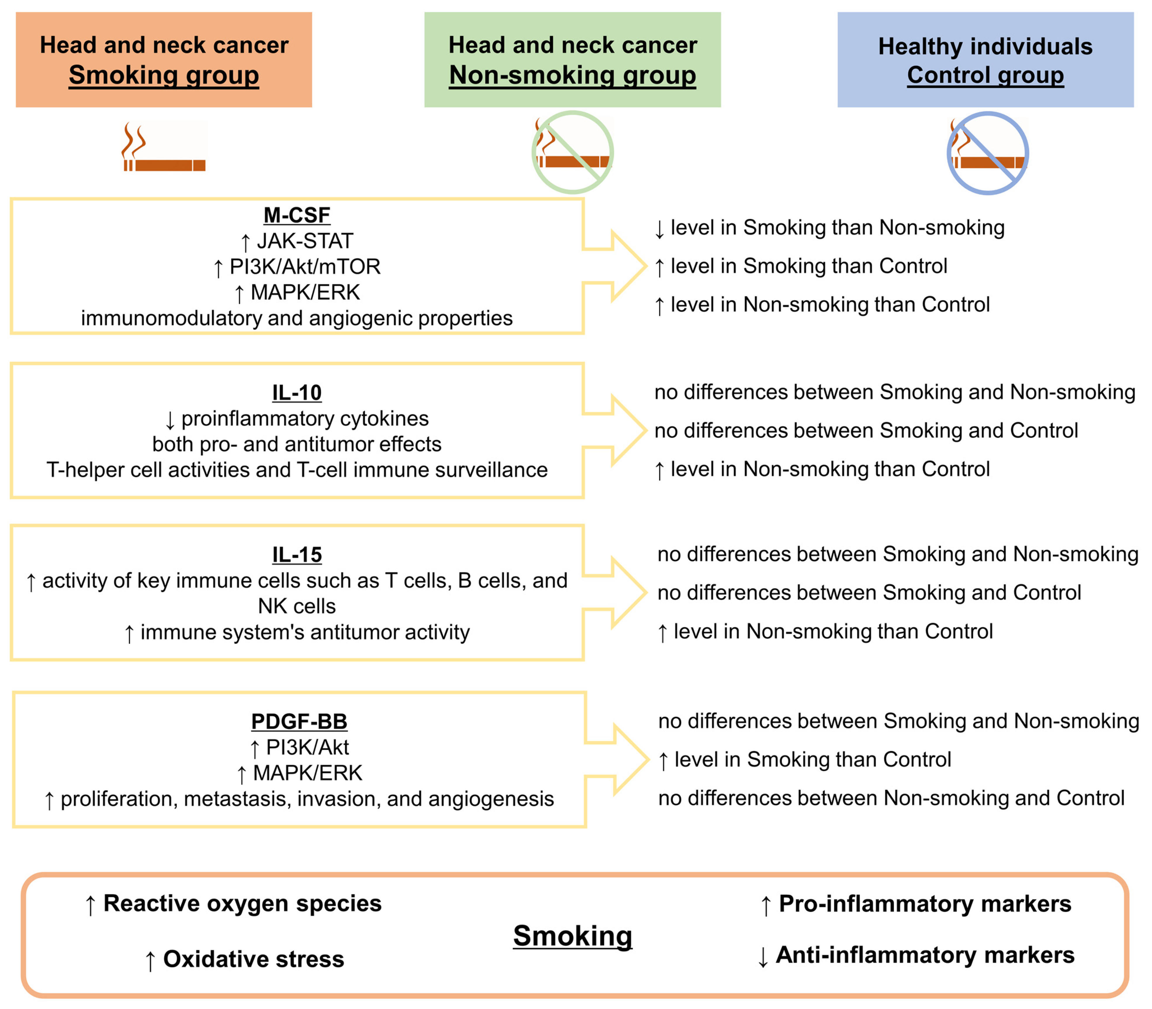
| IL-10 [pg/mL] | Mean | 1.456 | 1.796 | 1.060 | 0.0053 | 0.6258 |
| SEM | 0.280 | 0.404 | 0.000 | |||
| Median | 1.060 | 1.060 | 1.060 | |||
| IQR | 0.000 | 0.010 | 0.000 | |||
| IL-12 (p70) [pg/mL] | Mean | 3.824 | 3.341 | 1.430 | 0.0004 | 1.0000 |
| SEM | 0.869 | 1.048 | 0.000 | |||
| Median | 1.430 | 1.430 | 1.430 | |||
| IQR | 3.120 | 0.870 | 0.000 | |||
| IL-12 (p40) [pg/mL] | Mean | 167.134 | 205.251 | 14.680 | <0.0001 | 1.0000 |
| SEM | 25.555 | 28.714 | 0.000 | |||
| Median | 161.010 | 178.960 | 14.680 | |||
| IQR | 110.890 | 147.680 | 0.000 | |||
| IL-13 [pg/mL] | Mean | 3.340 | 4.489 | 0.928 | <0.0001 | 1.0000 |
| SEM | 0.683 | 1.000 | 0.306 | |||
| Median | 2.220 | 2.880 | 0.310 | |||
| IQR | 5.020 | 4.240 | 0.000 | |||
| IL-15 [pg/mL] | Mean | 52.302 | 60.704 | 12.420 | 0.0458 | 1.0000 |
| SEM | 19.055 | 21.310 | 0.000 | |||
| Median | 12.420 | 12.420 | 12.420 | |||
| IQR | 0.000 | 0.000 | 0.000 | |||
| IL-16 [pg/mL] | Mean | 67.369 | 82.065 | 48.114 | 0.6491 | 1.0000 |
| SEM | 13.513 | 17.297 | 3.614 | |||
| Median | 51.610 | 54.770 | 44.055 | |||
| IQR | 108.310 | 121.390 | 21.650 | |||
| IL-17A [pg/mL] | Mean | 10.338 | 12.556 | 2.440 | <0.0001 | 1.0000 |
| SEM | 1.171 | 1.775 | 0.000 | |||
| Median | 11.580 | 12.420 | 2.440 | |||
| IQR | 12.260 | 14.360 | 0.000 | |||
| IL-18 [pg/mL] | Mean | 72.940 | 97.220 | 35.260 | <0.0001 | 1.0000 |
| SEM | 9.330 | 10.921 | 3.650 | |||
| Median | 57.420 | 84.630 | 31.030 | |||
| IQR | 40.020 | 30.850 | 26.200 | |||
| IP-10 [pg/mL] | Mean | 1250.649 | 1459.909 | 495.298 | <0.0001 | 1.0000 |
| SEM | 215.017 | 159.842 | 69.284 | |||
| Median | 985.360 | 1400.980 | 406.375 | |||
| IQR | 1323.870 | 734.530 | 161.390 | |||
| LIF [pg/mL] | Mean | 45.032 | 54.508 | 15.098 | 0.0004 | 1.0000 |
| SEM | 7.005 | 9.041 | 2.691 | |||
| Median | 45.330 | 57.260 | 10.345 | |||
| IQR | 61.310 | 73.010 | 18.400 | |||
| MCP-1 [pg/mL] | Mean | 84.806 | 83.477 | 37.471 | <0.0001 | 1.0000 |
| SEM | 10.490 | 8.800 | 3.939 | |||
| Median | 83.460 | 78.570 | 31.470 | |||
| IQR | 87.060 | 34.420 | 27.130 | |||
| MCP-3 [pg/mL] | Mean | 2.112 | 2.598 | 0.480 | <0.0001 | 0.9999 |
| SEM | 0.441 | 0.449 | 0.000 | |||
| Median | 0.620 | 2.640 | 0.480 | |||
| IQR | 3.710 | 3.570 | 0.000 | |||
| M-CSF [pg/mL] | Mean | 24.916 | 37.500 | 5.783 | <0.0001 | 1.0000 |
| SEM | 2.802 | 3.498 | 0.632 | |||
| Median | 26.540 | 35.250 | 5.295 | |||
| IQR | 15.820 | 19.300 | 3.360 | |||
| MIF [pg/mL] | Mean | 1771.194 | 2417.451 | 591.863 | <0.0001 | 1.0000 |
| SEM | 379.186 | 426.807 | 102.379 | |||
| Median | 1058.540 | 1718.620 | 456.030 | |||
| IQR | 1394.900 | 2148.950 | 422.190 | |||
| MIG [pg/mL] | Mean | 771.724 | 1041.315 | 145.324 | <0.0001 | 1.0000 |
| SEM | 129.188 | 145.282 | 25.763 | |||
| Median | 705.950 | 853.010 | 103.865 | |||
| IQR | 763.680 | 864.400 | 70.850 | |||
| MIP-1α [pg/mL] | Mean | 3.761 | 4.362 | 1.758 | <0.0001 | 1.0000 |
| SEM | 0.395 | 0.354 | 0.199 | |||
| Median | 3.470 | 4.360 | 1.490 | |||
| IQR | 2.110 | 1.890 | 0.620 | |||
| MIP-1β [pg/mL] | Mean | 104.682 | 68.332 | 411.826 | <0.0001 | 1.0000 |
| SEM | 14.570 | 5.921 | 4.481 | |||
| Median | 72.370 | 61.810 | 416.440 | |||
| IQR | 75.160 | 29.150 | 31.030 | |||
| β-NGF [pg/mL] | Mean | 2.164 | 1.480 | 2.137 | 0.6782 | 0.6390 |
| SEM | 0.780 | 0.625 | 0.762 | |||
| Median | 0.470 | 0.470 | 0.470 | |||
| IQR | 1.720 | 0.670 | 0.000 | |||
| PDGF-BB [pg/mL] | Mean | 6882.342 | 6390.921 | 3455.909 | 0.0225 | 1.0000 |
| SEM | 982.017 | 996.719 | 279.008 | |||
| Median | 5566.670 | 4419.040 | 3288.955 | |||
| IQR | 4841.070 | 5696.230 | 2451.180 | |||
| RANTES [ng/mL] | Mean | 33.573 | 30.619 | 12.535 | <0.0001 | 1.0000 |
| SEM | 4.136 | 4.304 | 0.994 | |||
| Median | 28.127 | 24.290 | 11.121 | |||
| IQR | 28.106 | 26.923 | 3.176 | |||
| SCF [pg/mL] | Mean | 79.349 | 95.429 | 62.904 | 0.0009 | 1.0000 |
| SEM | 5.175 | 8.688 | 3.130 | |||
| Median | 81.580 | 88.430 | 62.630 | |||
| IQR | 40.170 | 35.810 | 23.970 | |||
| SCGF-β [ng/mL] | Mean | 191.594 | 216.834 | 36.824 | <0.0001 | 1.0000 |
| SEM | 23.854 | 26.456 | 1.345 | |||
| Median | 197.690 | 189.664 | 37.639 | |||
| IQR | 176.292 | 75.318 | 8.147 | |||
| SDF-1α+β [pg/mL] | Mean | 1008.943 | 868.606 | 1596.160 | <0.0001 | 1.0000 |
| SEM | 84.795 | 59.952 | 56.487 | |||
| Median | 883.690 | 831.120 | 1556.050 | |||
| IQR | 305.530 | 281.040 | 509.760 | |||
| TNF-α [pg/mL] | Mean | 59.754 | 67.218 | 48.602 | 0.0026 | 1.0000 |
| SEM | 3.705 | 6.845 | 2.589 | |||
| Median | 61.030 | 64.010 | 45.940 | |||
| IQR | 25.030 | 29.810 | 6.200 | |||
| TNF-β [pg/mL] | Mean | 116.192 | 61.449 | 1207.996 | <0.0001 | 1.0000 |
| SEM | 20.697 | 5.545 | 10.790 | |||
| Median | 63.030 | 59.440 | 1209.405 | |||
| IQR | 44.050 | 28.350 | 65.120 | |||
| TRAIL [pg/mL] | Mean | 38.398 | 44.284 | 19.951 | <0.0001 | 1.0000 |
| SEM | 3.791 | 4.316 | 1.215 | |||
| Median | 34.180 | 41.890 | 18.380 | |||
| IQR | 16.800 | 12.710 | 5.710 | |||
| VEGF [pg/mL] | Mean | 258.338 | 258.141 | 18.010 | <0.0001 | 1.0000 |
| SEM | 44.980 | 37.547 | 0.000 | |||
| Median | 231.320 | 249.770 | 18.010 | |||
| IQR | 353.430 | 199.070 | 0.000 | |||
| Parameter | p-Value | ||
|---|---|---|---|
| Smoking HNC vs. Non-Smoking HNC | Smoking HNC vs. Control | Non-Smoking HNC vs. Control | |
| CTACK | 0.3244 | 0.0003 | <0.0001 |
| IL-7 | 0.9650 | <0.0001 | <0.0001 |
| Basic FGF | 1.0000 | 0.0006 | <0.0001 |
| G-CSF | 0.3339 | <0.0001 | <0.0001 |
| GM-CSF | 1.0000 | 0.0347 | 0.0347 |
| HGF | 0.1570 | <0.0001 | <0.0001 |
| IFN-α2 | 1.0000 | <0.0001 | <0.0001 |
| IFN-γ | 0.1565 | 0.0038 | <0.0001 |
| IL-1α | 1.0000 | <0.0001 | <0.0001 |
| IL-1β | 1.0000 | <0.0001 | <0.0001 |
| IL-1ra | 1.0000 | <0.0001 | <0.0001 |
| IL-2 | 0.7074 | 0.0029 | 0.0001 |
| IL-2Rα | 0.1998 | <0.0001 | <0.0001 |
| IL-3 | 0.1059 | <0.0001 | <0.0001 |
| IL-6 | 0.5234 | <0.0001 | <0.0001 |
| IL-8 | 1.0000 | 0.0010 | 0.0149 |
| IL-9 | 0.3684 | <0.0001 | <0.0001 |
| IL-10 | 0.2873 | 0.3726 | 0.0069 |
| IL-12 (p70) | 0.9328 | 0.0002 | 0.0029 |
| IL-12 (p40) | 1.0000 | <0.0001 | <0.0001 |
| IL-13 | 1.0000 | 0.0004 | <0.0001 |
| IL-15 | 1.0000 | 0.0765 | 0.0347 |
| IL-17A | 1.0000 | <0.0001 | <0.0001 |
| IL-18 | 0.1308 | 0.0001 | <0.0001 |
| IP-10 | 0.3760 | 0.0021 | <0.0001 |
| LIF | 1.0000 | 0.0025 | 0.0021 |
| MCP-1 | 1.0000 | 0.0009 | <0.0001 |
| MCP-3 | 0.6000 | <0.0001 | <0.0001 |
| M-CSF | 0.0484 | <0.0001 | <0.0001 |
| MIF | 0.6647 | 0.0011 | <0.0001 |
| MIG | 0.5514 | <0.0001 | <0.0001 |
| MIP-1α | 0.3406 | <0.0001 | <0.0001 |
| MIP-1β | 0.4054 | <0.0001 | <0.0001 |
| PDGF-BB | 1.0000 | 0.0362 | 0.1105 |
| RANTES | 1.0000 | <0.0001 | <0.0001 |
| SCF | 0.6643 | 0.0480 | 0.0009 |
| SCGF-β | 1.0000 | <0.0001 | <0.0001 |
| SDF-1α+β | 1.0000 | <0.0001 | <0.0001 |
| TNF-α | 1.0000 | 0.0224 | 0.0053 |
| TNF-β | 0.2971 | <0.0001 | <0.0001 |
| TRAIL | 0.4868 | <0.0001 | <0.0001 |
| VEGF | 1.0000 | <0.0001 | <0.0001 |
Disclaimer/Publisher’s Note: The statements, opinions and data contained in all publications are solely those of the individual author(s) and contributor(s) and not of MDPI and/or the editor(s). MDPI and/or the editor(s) disclaim responsibility for any injury to people or property resulting from any ideas, methods, instructions or products referred to in the content. |
© 2024 by the authors. Licensee MDPI, Basel, Switzerland. This article is an open access article distributed under the terms and conditions of the Creative Commons Attribution (CC BY) license (https://creativecommons.org/licenses/by/4.0/).
Share and Cite
Nuszkiewicz, J.; Wróblewska, J.; Budek, M.; Czuczejko, J.; Woźniak, A.; Maruszak-Parda, M.; Szewczyk-Golec, K. Exploring the Link between Inflammatory Biomarkers and Head and Neck Cancer: Understanding the Impact of Smoking as a Cancer-Predisposing Factor. Biomedicines 2024, 12, 748. https://doi.org/10.3390/biomedicines12040748
Nuszkiewicz J, Wróblewska J, Budek M, Czuczejko J, Woźniak A, Maruszak-Parda M, Szewczyk-Golec K. Exploring the Link between Inflammatory Biomarkers and Head and Neck Cancer: Understanding the Impact of Smoking as a Cancer-Predisposing Factor. Biomedicines. 2024; 12(4):748. https://doi.org/10.3390/biomedicines12040748
Chicago/Turabian StyleNuszkiewicz, Jarosław, Joanna Wróblewska, Marlena Budek, Jolanta Czuczejko, Alina Woźniak, Marta Maruszak-Parda, and Karolina Szewczyk-Golec. 2024. "Exploring the Link between Inflammatory Biomarkers and Head and Neck Cancer: Understanding the Impact of Smoking as a Cancer-Predisposing Factor" Biomedicines 12, no. 4: 748. https://doi.org/10.3390/biomedicines12040748