
Post-Operative Adhesions: A Comprehensive Review of Mechanisms

 , ,
, ,
Abstract
:
1. Introduction
2. Clinical Significance of Post-Surgical Adhesions
2.1. Pelvic and Abdominal Adhesions
2.2. Thoracic Adhesions
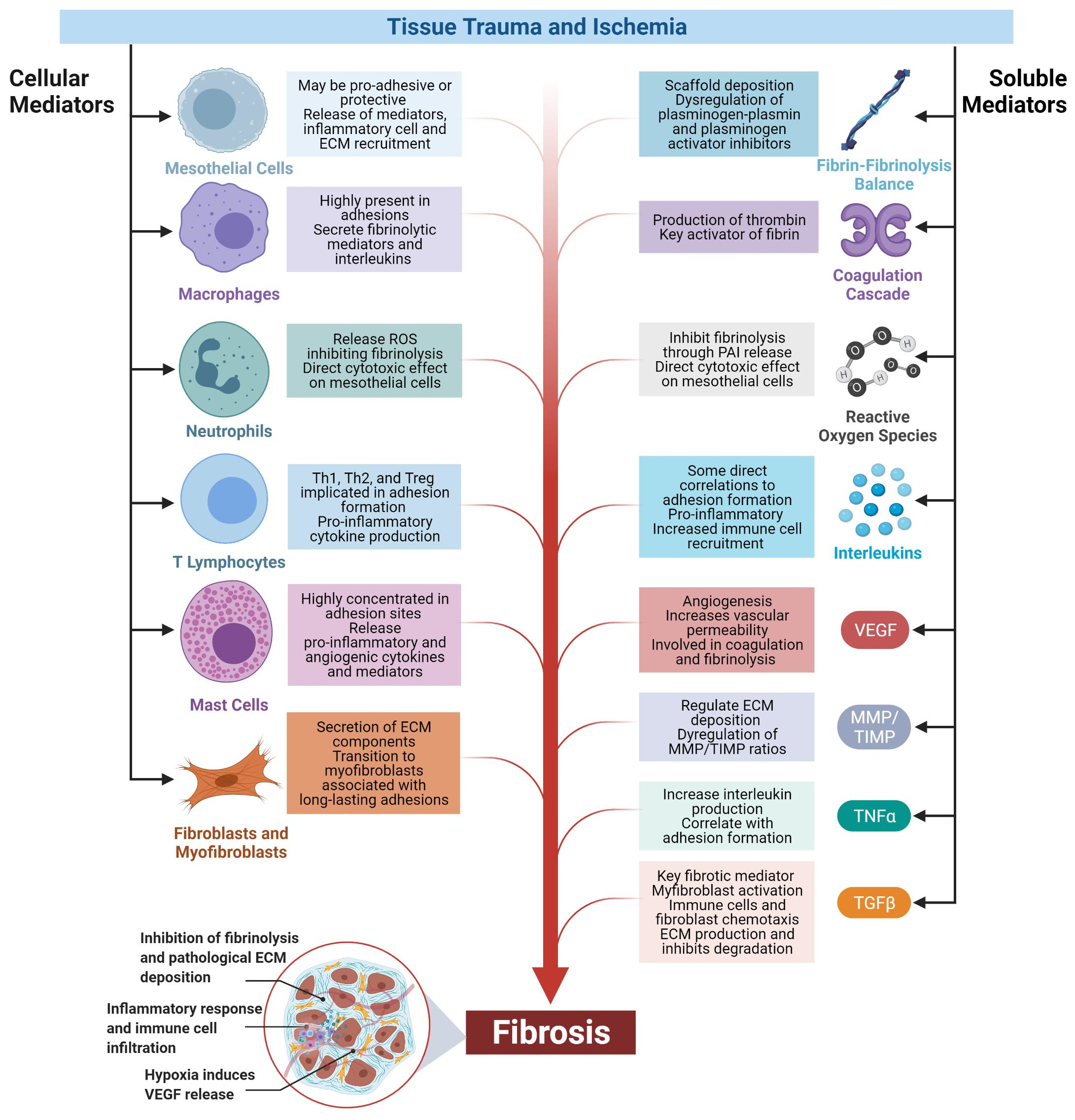
3. Mechanisms Driving Post-Surgical Adhesion Formation
3.1. Fibrin
3.2. Cellular and Immune Mechanisms
3.2.1. The Mesothelium
3.2.2. Fibroblasts and Myofibroblasts
3.2.3. Macrophages
3.2.4. Neutrophils
3.2.5. T-Cells
3.2.6. Mast Cells
3.3. Inflammation and Angiogenesis
3.3.1. Interleukins and Tumor Necrosis Factor-Alpha
3.3.2. Transforming Growth Factor-Beta
3.3.3. Vascular Endothelial Growth Factor A
3.3.4. Matrix Metalloproteinases (MMPs)
3.3.5. Substance P and Serotonin
3.4. Hypoxia and Genetic Biomarkers
4. Perspective: Clinical Translation and Future Work
5. Conclusions
Author Contributions
Funding
Data Availability Statement
Conflicts of Interest
References
- Diamond, M.P.; Freeman, M.L. Clinical implications of postsurgical adhesions. Hum. Reprod. Update 2001, 7, 567–576. [Google Scholar] [CrossRef] [PubMed] [Green Version]
- Liakakos, T.; Thomakos, N.; Fine, P.M.; Dervenis, C.; Young, R.L. Peritoneal adhesions: Etiology, pathophysiology, and clinical significance. Recent advances in prevention and management. Dig. Surg. 2001, 18, 260–273. [Google Scholar] [CrossRef]
- Goel, G.; King, T.; Daveson, A.J.; Andrews, J.M.; Krishnarajah, J.; Krause, R.; Brown, G.J.E.; Fogel, R.; Barish, C.F.; Epstein, R. Epitope-specific immunotherapy targeting CD4-positive T cells in coeliac disease: Two randomised, double-blind, placebo-controlled phase 1 studies. Lancet Gastroenterol. Hepatol. 2017, 2, 479–493. [Google Scholar] [CrossRef]
- Herrick, S.E.; Mutsaers, S.E.; Ozua, P.; Sulaiman, H.; Omer, A.; Boulos, P.; Foster, M.L.; Laurent, G.J. Human peritoneal adhesions are highly cellular, innervated, and vascularized. J. Pathol. 2000, 192, 67–72. [Google Scholar] [CrossRef]
- Lauder, C.I.; Garcea, G.; Strickland, A.; Maddern, G.J. Abdominal adhesion prevention: Still a sticky subject? Digestive Surg. 2010, 27, 347–358. [Google Scholar] [CrossRef]
- Parker, M.C.; Wilson, M.S.; Menzies, D.; Sunderland, G.; Thompson, J.N.; Clark, D.N.; Knigh, A.D.; Crowe, A.D. Colorectal surgery: The risk and burden of adhesion-related complications. Off. J. Assoc. Coloproctol. Great Br. Irel. 2004, 6, 506–511. [Google Scholar] [CrossRef]
- Ellis, H.; Moran, B.J.; Thompson, J.N.; Parker, M.C.; Wilson, M.S.; Menzies, D.; McGuire, A.; Lower, A.M.; Hawthorn, R.J.S.; O’Brien, F. Adhesion-related hospital readmissions after abdominal and pelvic surgery: A retrospective cohort study. Lancet 1999, 353, 1476–1480. [Google Scholar] [CrossRef]
- Schnuriger, B.; Barmparas, G.; Branco, B.C.; Lustenberger, T.; Inaba, K.; Demetriades, D. Prevention of postoperative peritoneal adhesions: A review of the literature. Am. J. Surg. 2011, 201, 111–121. [Google Scholar] [CrossRef]
- Parker, M.C.; Wilson, M.S.; Menzies, D.; Sunderland, G.; Clark, D.N.; Knight, A.D.; Crowe, A.M. The SCAR-3 study: 5-year adhesion-related readmission risk following lower abdominal surgical procedures. Off. J. Assoc. Coloproctol. Great Br. Irel. 2005, 7, 551–558. [Google Scholar] [CrossRef] [PubMed]
- Parker, M.C.; Ellis, H.; Moran, B.J.; Thompson, J.N.; Wilson, M.S.; Menzies, D.; McGuire, A.B.A.; Lower, A.M.; Hawthorn, R.J.S.; O’Brien, F. Postoperative adhesions: Ten-year follow-up of 12,584 patients undergoing lower abdominal surgery. Dis. Colon Rectum. 2001, 44, 822–829. [Google Scholar] [CrossRef] [PubMed]
- Ray, N.F.; Denton, W.G.; Thamer, M.; Henderson, S.C.; Perry, S. Abdominal adhesiolysis: Inpatient care and expenditures in the United States in 1994. J. Am. Coll. Surg. 1998, 186, 1–9. [Google Scholar] [CrossRef]
- Sikirica, V.; Bapat, B.; Candrilli, S.D.; Davis, K.L.; Wilson, M.; Johns, A. The inpatient burden of abdominal and gynecological adhesiolysis in the US. BMC Surg. 2011, 11, 13. [Google Scholar] [CrossRef] [Green Version]
- Weibel, M.A.; Majno, G. Peritoneal adhesions and their relation to abdominal surgery: A postmortem study. Am. J. Surg. 1973, 126, 345–353. [Google Scholar] [CrossRef]
- Menzies, D.; Ellis, H. Intestinal obstruction from adhesions--how big is the problem? Ann. R. Coll. Surg. Engl. 1990, 72, 60–63. [Google Scholar]
- Ellis, H. The clinical significance of adhesions: Focus on intestinal obstruction. Eur. J. Surg. Suppl. 1997, 5–9. [Google Scholar]
- Coleman, M.G.; McLain, A.D.; Moran, B.J. Impact of previous surgery on time taken for incision and division of adhesions during laparotomy. Dis. Colon Rectum. 2000, 43, 1297–1299. [Google Scholar] [CrossRef] [PubMed]
- Ten Broek, R.P.; Issa, Y.; van Santbrink, E.J.; Bouvy, N.D.; Kruitwagen, R.F.; Jeekel, J.; Bakkum, E.A.; Rovers, M.M. Burden of adhesions in abdominal and pelvic surgery: Systematic review and met-analysis. BMJ 2013, 347, f5588. [Google Scholar] [CrossRef] [Green Version]
- Lower, A.M.; Hawthorn, R.J.S.; Emeritus, H.E.; O’Brien, F.; Buchan, S.; Crowe, A.M. The impact of adhesions on hospital readmissions over ten years after 8849 open gynaecological operations: An assessment from the Surgical and Clinical Adhesions Research Study. BJOG Int. J. Obstet. Gynaecol. 2000, 107, 855–862. [Google Scholar] [CrossRef]
- Shahian, D.M.; O’Brien, S.M.; Filardo, G.; Ferraris, V.A.; Haan, C.K.; Rich, J.B.; Normand, S.-L.T.; DeLong, E.R.; Shewan, C.M.; Dokholyan, R.S. The Society of Thoracic Surgeons 2008 cardiac surgery risk models: Part 1—coronary artery bypass grafting surgery. Ann. Thorac Surg. 2009, 88 (Suppl. 1), S2–S22. [Google Scholar] [CrossRef]
- O’Brien, S.M.; Shahian, D.M.; Filardo, G.; Ferraris, V.A.; Haan, C.K.; Rich, J.B.; Normand, S.-L.T.; DeLong, E.R.; Shewan, C.M.; Dokholyan, R.S. The Society of Thoracic Surgeons 2008 cardiac surgery risk models: Part 2—isolated valve surgery. Ann. Thorac. Surg. 2009, 88 (Suppl. 1), S23–S42. [Google Scholar] [CrossRef] [PubMed]
- Jacobs, J.P.; Mavroudis, C.; Quintessenza, J.A.; Chai, P.J.; Pasquali, S.K.; Hill, K.D.; Vricella, L.A.; Jacobs, M.L.; Dearani, J.A.; Cameron, D. Reoperations for pediatric and congenital heart disease: An analysis of the Society of Thoracic Surgeons (STS) congenital heart surgery database. Semin Thorac. Cardiovasc Surg. Pediatr. Card. Surg. Annu. 2014, 17, 2–8. [Google Scholar] [CrossRef] [Green Version]
- Cannata, A.; Petrella, D.; Russo, C.F.; Bruschi, G.; Fratto, P.; Gambacorta, M.; Martinelli, L. Postsurgical intrapericardial adhesions: Mechanisms of formation and prevention. Ann. Thorac. Surg. 2013, 95, 1818–1826. [Google Scholar] [CrossRef]
- Nkere, U.U. Postoperative adhesion formation and the use of adhesion preventing techniques in cardiac and general surgery. Am. Soc. Artif. Intern. Organs 2000, 46, 654–656. [Google Scholar] [CrossRef]
- Nkere, U.U.; Whawell, S.A.; Sarraf, C.E.; Schofield, J.B.; Thompson, J.N.; Taylor, K.M. Pericardial trauma and adhesions in relation to reoperative cardiac surgery. Thorac. Cardiovasc. Surg. 1995, 43, 338–346. [Google Scholar] [CrossRef]
- Coccolini, F.; Ansaloni, L.; Manfredi, R.; Campanati, L.; Poiasina, E.; Bertoli, P.; Capponi, M.G.; Sartelli, M.; Di Saverio, S.; Cucchi, M. Peritoneal adhesion index (PAI): Proposal of a score for the "ignored iceberg" of medicine and surgery. World J. Emerg Surg. 2013, 8, 6. [Google Scholar] [CrossRef] [Green Version]
- Milligan, D.W.; Raftery, A.T. Observations on the pathogenesis of peritoneal adhesions: A light and electron microscopical study. Br. J. Surg. 1974, 61, 274–280. [Google Scholar] [CrossRef] [PubMed]
- Binda, M.M.; Molinas, C.R.; Koninckx, P.R. Reactive oxygen species and adhesion formation: Clinical implications in adhesion prevention. Hum. Reproduction. 2003, 18, 2503–2507. [Google Scholar] [CrossRef] [Green Version]
- Holmdahl, L.; Eriksson, E.; al-Jabreen, M.; Risberg, B. Fibrinolysis in human peritoneum during operation. Surgery 1996, 119, 701–705. [Google Scholar] [CrossRef]
- Holmdahl, L.; Ivarsson, M.L. The role of cytokines, coagulation, and fibrinolysis in peritoneal tissue repair. Eur. J. Surg. 1999, 165, 1012–1019. [Google Scholar] [PubMed]
- Sun, Z.; Sessler, D.I.; Dalton, J.E.; Devereaux, P.J.; Shahinyan, A.; Naylor, A.J.; Hutcherson, M.T.; Finnegan, P.S.; Tandon, V.; Darvish-Kazem, S. Postoperative Hypoxemia Is Common and Persistent: A Prospective Blinded Observational Study. Anesth. Analg. 2015, 121, 709–715. [Google Scholar] [CrossRef] [Green Version]
- Risberg, B.; Stenberg, B. Modulation of tissue fibrinolysis from hypoxia and hyperoxia. Thromb Res. 1985, 38, 129–136. [Google Scholar] [CrossRef]
- Yan, S.-F.; Mackman, N.; Kisiel, W.; Stern, D.M.; Pinsky, D.J. Hypoxia/Hypoxemia-Induced Activation of the Procoagulant Pathways and the Pathogenesis of Ischemia-Associated Thrombosis. Arterioscler. Thromb. Vasc. Biol. 1999, 19, 2029–2035. [Google Scholar] [CrossRef] [PubMed] [Green Version]
- Gertler, J.P.; Perry, L.; L’Italien, G.; Chung-Welch, N.; Cambria, R.P.; Orkin, R.; Abbot, W.M. Ambient oxygen tension modulates endothelial fibrinolysis. J. Vasc. Surg. 1993, 18, 939–945. [Google Scholar] [CrossRef] [Green Version]
- Palta, S.; Saroa, R.; Palta, A. Overview of the coagulation system. Indian J. Anaesth. 2014, 58, 515–523. [Google Scholar] [CrossRef] [PubMed]
- DiZerega, G.S. Biochemical events in peritoneal tissue repair. Eur. J. Surg. Supplement. Acta Chir. Suppl. 1997, 10–16. [Google Scholar]
- Di Filippo, C.; Falsetto, A.; de Pascale, V.; Tufariello, E.; de Lucia, D.; Rossi, F.; D’amico, M.; Cenamo, A. Plasma levels of t-PA and PAI-1 correlate with the formation of experimental post-surgical peritoneal adhesions. Mediat. Inflamm. 2006, 2006, 13901. [Google Scholar] [CrossRef] [PubMed]
- Chegini, N. Peritoneal molecular environment, adhesion formation and clinical implication. Front. Biosci. 2002, 7, e91–e115. [Google Scholar]
- Chegini, N. TGF-beta system: The principal profibrotic mediator of peritoneal adhesion formation. Semin. Reprod. Med. 2008, 26, 298–312. [Google Scholar] [CrossRef]
- Fortin, C.N.; Saed, G.M.; Diamond, M.P. Predisposing factors to post-operative adhesion development. Hum. Reprod. Update. 2015, 21, 536–551. [Google Scholar] [CrossRef] [Green Version]
- Mutsaers, S.E.; Birnie, K.; Lansley, S.; Herrick, S.E.; Lim, C.-B.; Prêle, C.M. Mesothelial cells in tissue repair and fibrosis. Front. Pharmacol. 2015, 6, 113. [Google Scholar] [CrossRef] [Green Version]
- Tsai, J.M.; Shoham, M.; Fernhoff, N.B.; George, B.M.; Marjon, K.D.; McCracken, M.N.; Kao, C.S.; Sinha, R.; Volmer, A.K.; Miyanishi, M. Neutrophil and monocyte kinetics play critical roles in mouse peritoneal adhesion formation. Blood Adv. 2019, 3, 2713–2721. [Google Scholar]
- Sandoval, P.; Jimenez-Heffernan, J.A.; Guerra-Azcona, G.; Perez-Lozano, M.L.; Rynne-Vidal, A.; Albar-Vizcaino, P.; Gil-Vera, F.; Martin, P.; Martin, P.; Coronado, M.J.; et al. Mesothelial-to-mesenchymal transition in the pathogenesis of post-surgical peritoneal adhesions. J. Pathol. 2016, 239, 48–59. [Google Scholar] [CrossRef] [PubMed]
- Jin, X.; Ren, S.; Macarak, E.; Rosenbloom, J. Pathobiological mechanisms of peritoneal adhesions: The mesenchymal transition of rat peritoneal mesothelial cells induced by TGF-β1 and IL-6 requires activation of Erk1/2 and Smad2 linker region phosphorylation. Matrix Biol. 2016, 51, 55–64. [Google Scholar] [CrossRef]
- Yáñez-Mó, M.; Lara-Pezzi, E.; Selgas, R.; Ramírez-Huesca, M.; Domínguez-Jiménez, C.; Jiménez-Heffernan, J.A.; Aguilera, A.; Sánchez-Tomero, J.A.; Bajo, M.A.; Álvarez, V.; et al. Peritoneal Dialysis and Epithelial-to-Mesenchymal Transition of Mesothelial Cells. N. Engl. J. Med. 2003, 348, 403–413. [Google Scholar] [CrossRef] [PubMed]
- Raftery, A.T. Regeneration of parietal and visceral peritoneum in the immature animal: A light and electron microscopical study. Br. J. Surg. 1973, 60, 969–975. [Google Scholar] [CrossRef] [PubMed]
- Menzies, D. Peritoneal adhesions. Incidence, cause, and prevention. Surg. Annu. 1992, 24 Pt 1, 27–45. [Google Scholar] [PubMed]
- Cheong, Y.C.; Laird, S.M.; Li, T.C.; Shelton, J.B.; Ledger, W.L.; Cooke, I.D. Peritoneal healing and adhesion formation/reformation. Hum. Reprod. Update 2001, 7, 556–566. [Google Scholar] [CrossRef] [PubMed]
- Jaworska-Wilczynska, M.; Trzaskoma, P.; Szczepankiewicz, A.A.; Hryniewiecki, T. Pericardium: Structure and function in health and disease. Folia Histochem. Cytobiol. 2016, 54, 121–125. [Google Scholar] [CrossRef] [Green Version]
- Schade, D.S.; Williamson, J.R. The pathogenesis of peritoneal adhesions: An ultrastructural study. Ann. Surg. 1968, 167, 500–510. [Google Scholar] [CrossRef] [PubMed]
- DiZerega, G.S.; Campeau, J.D. Peritoneal repair and post-surgical adhesion formation. Hum. Reprod. Update. 2001, 7, 547–555. [Google Scholar] [CrossRef] [PubMed]
- Suzuki, T.; Kono, T.; Bochimoto, H.; Hira, Y.; Watanabe, T.; Furukawa, H. An injured tissue affects the opposite intact peritoneum during postoperative adhesion formation. Sci. Rep. 2015, 5, 7668. [Google Scholar] [CrossRef] [PubMed] [Green Version]
- Inagaki, N.F.; Inagaki, F.F.; Kokudo, N.; Miyajima, A. Use of mouse liver mesothelial cells to prevent postoperative adhesion and promote liver regeneration after hepatectomy. J. Hepatol. 2015, 62, 1141–1147. [Google Scholar] [CrossRef] [Green Version]
- Betjes, M.G.; Tuk, C.W.; Struijk, D.G.; Krediet, R.T.; Arisz, L.; Beelen, R.H. Adherence of Staphylococci to plastic, mesothelial cells and mesothelial extracellular matrix. Adv. Perit. Dial. 1992, 8, 215–218. [Google Scholar] [PubMed]
- Tsai, J.M.; Sinha, R.; Seita, J.; Fernhoff, N.; Christ, S.; Koopmans, T.; Krampitz, G.W.; McKenna, K.M.; Xing, L.; Sandholzer, M.; et al. Surgical adhesions in mice are derived from mesothelial cells and can be targeted by antibodies against mesothelial markers. Sci. Transl. Med. 2018, 10. [Google Scholar] [CrossRef] [Green Version]
- Fischer, A.; Koopmans, T.; Ramesh, P.; Christ, S.; Strunz, M.; Wannemacher, J.; Aichler, M.; Feuchtinger, A.; Walch, A.; Ansari, M.; et al. Post-surgical adhesions are triggered by calcium-dependent membrane bridges between mesothelial surfaces. Nat. Commun. 2020, 11, 3068. [Google Scholar] [CrossRef]
- Boland, G.M.; Weigel, R.J. Formation and Prevention of Postoperative Abdominal Adhesions. J. Surg. Res. 2006, 132, 3–12. [Google Scholar] [CrossRef]
- Ellis, H.; Harrison, W.; Hugh, T.B. The healing of the peritneum under normal and pathological conditions. Br. J. Surg. 1965, 524, 71–76. [Google Scholar]
- Raftery, A.T. Regeneration of parietal and visceral peritoneum: An electron microscopical study. J. Anat. 1973, 115 Pt 3, 375–392. [Google Scholar]
- Hellebrekers, B.W.; Kooistra, T. Pathogenesis of postoperative adhesion formation. Br. J. Surg. 2011, 98, 1503–1516. [Google Scholar] [CrossRef]
- Lucas, P.A.; Warejcka, D.J.; Young, H.E.; Lee, B.Y. Formation of Abdominal Adhesions Is Inhibited by Antibodies to Transforming Growth Factor-β1. J. Surg. Res. 1996, 65, 135–138. [Google Scholar] [CrossRef] [PubMed]
- Foster, D.S.; Marshall, C.D.; Gulati, G.S.; Chinta, M.S.; Nguyen, A.; Salhotra, A.; Jines, R.E.; Burcham, A.; Lerbs, T.; Cui, L. Elucidating the fundamental fibrotic processes driving abdominal adhesion formation. Nat. Commun. 2020, 11, 4061. [Google Scholar] [CrossRef]
- Buckman, R.F.; Woods, M.; Sargent, L.; Gervin, A.S. A unifying pathogenetic mechanism in the etiology of intraperitoneal adhesions. J. Surg. Res. 1976, 20, 1–5. [Google Scholar] [CrossRef]
- Holmadhl, L.; al-Jabreen, M.; Xia, G.; Risberg, B. The impact of starch-powdered gloves on the formation of adhesions in rats. Acta Chirurgica. 1994, 160, 257–261. [Google Scholar]
- Belluco, C.; Meggiolaro, F.; Pressato, D.; Pavesio, A.; Bigon, E.; Donà, M.; Forlin, M.; Nitti, D.; Lise, M. Prevention of postsurgical adhesions with an autocrosslinked hyaluronan derivative gel. J. Surg. Res. 2001, 100, 217–221. [Google Scholar] [CrossRef] [PubMed]
- Xu, X.; Rivkind, A.; Pappo, O.; Pikarsky, A.; Levi-Schaffer, F. Role of mast cells and myofibroblasts in human peritoneal adhesion formation. Ann. Surg. 2002, 236, 593–601. [Google Scholar] [CrossRef]
- Hinz, B.; Phan, S.H.; Thannickal, V.J.; Galli, A.; Bochaton-Piallat, M.-L.; Gabbiani, G. The Myofibroblast: One Function, Multiple Origins. Am. J. Pathol. 2007, 170, 1807–1816. [Google Scholar] [CrossRef]
- Wei, G.; Chen, X.; Wang, G.; Jia, P.; Xu, Q.; Ping, G.; Wang, K.; Li, X. Inhibition of cyclooxygenase-2 prevents intra-abdominal adhesions by decreasing activity of peritoneal fibroblasts. Drug Des. Devel. Ther. 2015, 9, 3083–3098. [Google Scholar]
- Rout, U.K.; Saed, G.M.; Diamond, M.P. Expression pattern and regulation of genes differ between fibroblasts of adhesion and normal human peritoneum. Reprod. Biol. Endocrinol. 2005, 3, 1. [Google Scholar] [CrossRef] [Green Version]
- Saed, G.M.; Diamond, M.P. Hypoxia-induced irreversible up-regulation of type I collagen and transforming growth factor-β1 in human peritoneal fibroblasts. Fertil. Steril. 2002, 78, 144–147. [Google Scholar] [CrossRef]
- Saed, G.M.; Munkarah, A.R.; Diamond, M.P. Cyclooxygenase-2 is expressed in human fibroblasts isolated from intraperitoneal adhesions but not from normal peritoneal tissues. Fertil Steril. 2003, 79, 1404–1408. [Google Scholar] [CrossRef]
- Saed, G.M.; Zhang, W.; Chegini, N.; Holmdahl, L.; Diamond, M.P. Transforming growth factor beta isoforms production by human peritoneal mesothelial cells after exposure to hypoxia. Am. J. Reprod. Immunol. 2000, 43, 285–291. [Google Scholar] [CrossRef] [PubMed]
- Saed, G.M.; Munkarah, A.R.; Abu-Soud, H.M.; Diamond, M.P. Hypoxia upregulates cyclooxygenase-2 and prostaglandin E2 levels in human peritoneal fibroblasts. Fertil. Steril. 2005, 83, 1216–1219. [Google Scholar] [CrossRef]
- Jiang, Z.L.; Zhu, X.; Diamond, M.P.; Abu-Soud, H.M.; Saed, G.M. Nitric oxide synthase isoforms expression in fibroblasts isolated from human normal peritoneum and adhesion tissues. Fertil. Steril. 2008, 90, 769–774. [Google Scholar] [CrossRef] [PubMed] [Green Version]
- Saed, G.M.; Diamond, M.P. Differential expression of alpha smooth muscle cell actin in human fibroblasts isolated from intraperitoneal adhesions and normal peritoneal tissues. Fertil. Steril. 2004, 82 (Suppl. 3), 1188–1192. [Google Scholar] [CrossRef]
- Chen, Y.T.; Chang, Y.T.; Pan, S.Y.; Chou, Y.H.; Chang, F.C.; Yeh, P.Y.; Liu, Y.H.; Chiang, W.C.; Chen, Y.M.; Wu, K.D.; et al. Lineage tracing reveals distinctive fates for mesothelial cells and submesothelial fibroblasts during peritoneal injury. J. Am. Soc. Nephrol. 2014, 25, 2847–2858. [Google Scholar] [CrossRef] [Green Version]
- Park, D.S.J.; Regmi, S.C.; Svystonyuk, D.A.; Teng, G.; Belke, D.; Turnbull, J.; Guzzardi, D.G.; Kang, S.; Cowman, M.K.; Schmidt, T.A.; et al. Human pericardial proteoglycan 4 (lubricin): Implications for postcardiotomy intrathoracic adhesion formation. J. Thorac. Cardiovasc. Surg. 2018, 156, 1598–1608.e1. [Google Scholar] [CrossRef] [PubMed]
- Williams, R.S.; Rossi, A.M.; Chegini, N.; Schultz, G. Effect of transforming growth factor β on postoperative adhesion formation and intact peritoneum. J. Surg. Res. 1992, 52, 65–70. [Google Scholar] [CrossRef]
- Haney, A.F. Identification of macrophages at the site of peritoneal injury: Evidence supporting a direct role for peritoneal macrophages in healing injured peritoneum. Fertil. Steril. 2000, 73, 988–995. [Google Scholar] [CrossRef]
- Binnebösel, M.; Rosch, R.; Junge, K.; Lynen-Jansen, P.; Schumpelick, V.; Klinge, U. Macrophage and T-lymphocyte Infiltrates in Human Peritoneal Adhesions Indicate a Chronic Inflammatory Disease. World J. Surg. 2008, 32, 296–304. [Google Scholar] [CrossRef]
- Turza, K.C.; Butler, C.E. Adhesions and meshes: Synthetic versus bioprosthetic. Plast. Reconstr. Surg. 2012, 130 (Suppl. 2), 206s–213s. [Google Scholar] [CrossRef]
- Ergul, E.; Korukluoglu, B. Peritoneal adhesions: Facing the enemy. Int. J. Surg. 2008, 6, 253–260. [Google Scholar] [CrossRef] [Green Version]
- Canturk, N.Z.; Vural, B.; Esen, N.; Canturk, Z.; Oktay, G.; Kirkali, G.; Solakoglu, S. Effects of granulocyte-macrophage colony-stimulating factor on incisional wound healing in an experimental diabetic rat model. Endocr. Res. 1999, 25, 105–116. [Google Scholar] [CrossRef]
- Honjo, K.; Munakata, S.; Tashiro, Y.; Salama, Y.; Shimazu, H.; Eiamboonsert, S.; Dhahri, D.; Ichimura, A.; Dan, T.; Miyata, T.; et al. Plasminogen activator inhibitor-1 regulates macrophage-dependent postoperative adhesion by enhancing EGF-HER1 signaling in mice. FASEB J. 2017, 31, 2625–2637. [Google Scholar] [CrossRef] [PubMed] [Green Version]
- Ar’Rajab, A.; Dawidson, I.; Sentementes, J.; Sikes, P.; Harris, R.; Mileski, W. Enhancement of Peritoneal Macrophages Reduces Postoperative Peritoneal Adhesion Formation. J. Surg. Res. 1995, 58, 307–312. [Google Scholar] [CrossRef] [PubMed]
- Hoshino, A.; Kawamura, Y.I.; Yasuhara, M.; Toyama-Sorimachi, N.; Yamamoto, K.; Matsukawa, A.; Lira, S.A.; Dohi, T. Inhibition of CCL1-CCR8 Interaction Prevents Aggregation of Macrophages and Development of Peritoneal Adhesions. J. Immunol. 2007, 178, 5296–5304. [Google Scholar] [CrossRef]
- Sindrilaru, A.; Scharffetter-Kochanek, K. Disclosure of the Culprits: Macrophages-Versatile Regulators of Wound Healing. Adv. Wound Care 2013, 2, 357–368. [Google Scholar] [CrossRef] [Green Version]
- Orecchioni, M.; Ghosheh, Y.; Pramod, A.B.; Ley, K. Macrophage Polarization: Different Gene Signatures in M1(LPS+) vs. Classically and M2(LPS–) vs. Alternatively Activated Macrophages. Front. Immunol. 2019, 10, 1084. [Google Scholar] [CrossRef] [PubMed]
- Martinez, F.O.; Gordon, S. The M1 and M2 paradigm of macrophage activation: Time for reassessment. F1000Prime Rep. 2014, 6, 13. [Google Scholar] [CrossRef] [PubMed] [Green Version]
- Hong, G.S.; Schwandt, T.; Stein, K.; Schneiker, B.; Kummer, M.P.; Heneka, M.T.; Kitamura, K.; Kallf, J.K.; Wehner, S. Effects of macrophage-dependent peroxisome proliferator-activated receptor γ signalling on adhesion formation after abdominal surgery in an experimental model. Br. J. Surg. 2015, 102, 1506–1516. [Google Scholar] [CrossRef]
- Rodgers, K.E.; diZerega, G.S. Modulation of peritoneal re-epithelialization by postsurgical macrophages. J. Surg. Research. 1992, 53, 542–548. [Google Scholar] [CrossRef]
- Uyama, N.; Tsutsui, H.; Wu, S.; Yasuda, K.; Hatano, E.; Qin, X.-Y.; Kojima, S.; Fujimoto, J. Anti-interleukin-6 receptor antibody treatment ameliorates postoperative adhesion formation. Sci. Reports. 2019, 9, 17558. [Google Scholar] [CrossRef] [Green Version]
- Ribeiro, R.A.; Flores, C.A.; Cunha, F.Q.; Ferreira, S.H. IL-8 causes in vivo neutrophil migration by a cell-dependent mechanism. Immunology 1991, 73, 472–477. [Google Scholar]
- Vural, B.; Cantürk, N.Z.; Esen, N.; Solakoglu, S.; Cantürk, Z.; Kirkali, G.; Sökmensüer, C. The role of neutrophils in the formation of peritoneal adhesions. Hum. Reprod. 1999, 14, 49–54. [Google Scholar] [CrossRef] [PubMed] [Green Version]
- Ten Raa, S.; van den Tol, M.P.; Sluiter, W.; Hofland, L.J.; van Eijck, C.H.; Jeekel, H. The role of neutrophils and oxygen free radicals in post-operative adhesions. J. Surg. Res. 2006, 136, 45–52. [Google Scholar] [CrossRef] [PubMed]
- Arfors, K.E.; Lundberg, C.; Lindbom, L.; Lundberg, K.; Beatty, P.G.; Harlan, J.M. A monoclonal antibody to the membrane glycoprotein complex CD18 inhibits polymorphonuclear leukocyte accumulation and plasma leakage in vivo. Blood 1987, 69, 338–340. [Google Scholar] [CrossRef] [PubMed]
- Ar’Rajab, A.; Mileski, W.; Sentementes, J.T.; Sikes, P.; Harris, R.B.; Dawidson, I.J. The role of neutrophils in peritoneal adhesion formation. J. Surg. Res. 1996, 61, 143–146. [Google Scholar] [CrossRef]
- Awonuga, A.O.; Belotte, J.; Abuanzeh, S.; Fletcher, N.M.; Diamond, M.P.; Saed, G.M. Advances in the Pathogenesis of Adhesion Development: The Role of Oxidative Stress. Reprod. Sci. 2014, 21, 823–836. [Google Scholar] [CrossRef] [PubMed] [Green Version]
- Roy, S.; Clark, C.J.; Mohebali, K.; Bhatt, U.; Wallace, W.A.; Nahman, N.S.; Ellison, E.C.; Melvin, W.S.; Sen, C.K. Reactive oxygen species and EGR-1 gene expression in surgical postoperative peritoneal adhesions. World J. Surg. 2004, 28, 316–320. [Google Scholar]
- Binnebösel, M.; Klink, C.D.; Serno, J.; Jansen, P.L.; von Trotha, K.T.; Neumann, U.P.; Junge, K. Chronological evaluation of inflammatory mediators during peritoneal adhesion formation using a rat model. Langenbeck’s Arch. Surg. 2011, 396, 371–378. [Google Scholar] [CrossRef]
- Chung, D.R.; Chitnis, T.; Panzo, R.J.; Kasper, D.L.; Sayegh, M.H.; Tzianabos, A.O. CD4+ T cells regulate surgical and postinfectious adhesion formation. J. Exp. Medicine. 2002, 195, 1471–1478. [Google Scholar] [CrossRef] [Green Version]
- Tzianabos, A.O.; Holsti, M.A.; Zheng, X.X.; Stucchi, A.F.; Kuchroo, V.K.; Strom, T.B.; Glimcher, L.H.; Cruikshank, W.W. Functional Th1 cells are required for surgical adhesion formation in a murine model. J. Immunol. 2008, 180, 6970–6976. [Google Scholar] [CrossRef] [Green Version]
- Ozbilgin, K.; Üner, M.A.; Ozkut, M.; Hasdemir, P.S. The effects of pirfenidone on T helper cells in prevention of intraperitoneal adhesions. Kaohsiung J. Med. Sciences. 2017, 33, 271–276. [Google Scholar] [CrossRef] [Green Version]
- McLoughlin, R.M.; Jenkins, B.J.; Grail, D.; Williams, A.S.; Fielding, C.A.; Parker, C.R.; Ernst, M.; Topley, N.; Jones, S.A. IL-6 trans-signaling via STAT3 directs T cell infiltration in acute inflammation. Proc. Natl. Acad. Sci. USA 2005, 102, 9589–9594. [Google Scholar] [CrossRef] [Green Version]
- Maciver, A.H.; McCall, M.; James Shapiro, A.M. Intra-abdominal adhesions: Cellular mechanisms and strategies for prevention. Int. J. Surgery. 2011, 9, 589–594. [Google Scholar] [CrossRef] [Green Version]
- Canturk, N.Z.; Vural, B.; Cubukcu, A.; Duzcen, E.; Utkan, Z.; Dulger, M. Experimental study on the role of mast cells in peritoneal adhesion formation. East. Afr. Med. J. 1999, 76, 233–236. [Google Scholar]
- Yao, Y.-L.; Ishihara, T.; Takai, S.; Miyazaki, M.; Mita, S. Association between the Expression of Mast Cell Chymase and Intraperitoneal Adhesion Formation in Mice. J. Surg. Res. 2000, 92, 40–44. [Google Scholar] [CrossRef] [PubMed]
- Soga, Y.; Takai, S.; Koyama, T.; Okamoto, Y.; Ikeda, T.; Nishimura, K.; Miyazaki, M.; Komeda, M. Attenuation of adhesion formation after cardiac surgery with a chymase inhibitor in a hamster model. J. Thorac. Cardiovasc. Surg. 2004, 127, 72–78. [Google Scholar] [CrossRef] [Green Version]
- Cahill, R.A.; Wang, J.H.; Soohkai, S.; Redmond, H.P. Mast cells facilitate local VEGF release as an early event in the pathogenesis of postoperative peritoneal adhesions. Surgery 2006, 140, 108–112. [Google Scholar] [CrossRef] [PubMed]
- Arjmand, M.H. The association between visceral adiposity with systemic inflammation, oxidative stress, and risk of post-surgical adhesion. Arch. Physiol. Biochem. 2020, 1–6. [Google Scholar] [CrossRef]
- Pismensky, S.V.; Kalzhanov, Z.R.; Eliseeva, M.Y.; Kosmas, I.P.; Mynbaev, O.A. Severe inflammatory reaction induced by peritoneal trauma is the key driving mechanism of postoperative adhesion formation. BMC Surg. 2011, 11, 30. [Google Scholar] [CrossRef] [Green Version]
- Reber, P.U.; Andrén, S.; Schmied, B.; Büchler, M.W. Cytokines in surgical trauma: Cholecystectomy as an example. Dig. Surg. 1998, 15, 92–101. [Google Scholar] [CrossRef] [PubMed]
- Buyalos, R.P.; Funari, V.A.; Azziz, R.; Watson, J.M.; Martinez-Maza, O. Elevated interleukin-6 levels in peritoneal fluid of patients with pelvic pathology. Fertil. Steril. 1992, 58, 302–306. [Google Scholar] [CrossRef]
- Kaidi, A.A.; Gurchumelidze, T.; Nazzal, M.; Figert, P.; Vanterpool, C.; Silva, Y. Tumor necrosis factor-alpha: A marker for peritoneal adhesion formation. J. Surg. Res. 1995, 58, 516–518. [Google Scholar] [CrossRef] [PubMed]
- Cheong, Y.C.; Laird, S.M.; Shelton, J.B.; Ledger, W.L.; Li, T.C.; Cooke, I.D. The correlation of adhesions and peritoneal fluid cytokine concentrations: A pilot study. Hum. Reprod. 2002, 17, 1039–1045. [Google Scholar] [CrossRef] [Green Version]
- Saba, A.A.; Godziachvili, V.; Mavani, A.K.; Silva, Y.J. Serum levels of interleukin 1 and tumor necrosis factor alpha correlate with peritoneal adhesion grades in humans after major abdominal surgery. Am. Surg. 1998, 64, 734–736. [Google Scholar] [PubMed]
- Tsukada, K.; Katoh, H.; Suzuki, T.; Takenoshita, S.; Nagamachi, Y. Correlations of peritoneal interleukin-6, serum beta-2 microglobulin and urinary beta-2 microglobulin after elective abdominal surgery. Acta Pathol. Microbiol. Et Immunol. Scand. 1993, 101, 409–412. [Google Scholar] [CrossRef] [PubMed]
- Yoshikawa, H.; Kawamura, I.; Fujita, M.; Tsukada, H.; Arakawa, M.; Mitsuyama, M. Membrane damage and interleukin-1 production in murine macrophages exposed to listeriolysin O. Infect. Immun. 1993, 61, 1334–1339. [Google Scholar] [CrossRef] [Green Version]
- Whawell, S.A.; Thompson, J.N. Cytokine-induced release of plasminogen activator inhibitor-1 by human mesothelial cells. Eur. J. Surg. 1995, 161, 315–318. [Google Scholar]
- Kaidi, A.A.; Nazzal, M.; Gurchumelidze, T.; Ali, M.A.; Dawe, E.J.; Silva, Y.J. Preoperative administration of antibodies against tumor necrosis factor-alpha (TNF-alpha) and interleukin-1 (IL-1) and their impact on peritoneal adhesion formation. Am. Surg. 1995, 61, 569–572. [Google Scholar]
- Whawell, S.A.; Wang, Y.; Fleming, K.A.; Thompson, E.M.; Thompson, J.N. Localization of plasminogen activator inhibitor-1 production in inflamed appendix by in situ mRNA hybridization. J. Pathol. 1993, 169, 67–71. [Google Scholar] [CrossRef]
- Shenkin, A.; Fraser, W.D.; Series, J.; Winstanley, F.P.; McCartney, A.C.; Burns, H.J.; Van Damme, J. The serum interleukin 6 response to elective surgery. Lymphokine Res. 1989, 8, 123–127. [Google Scholar]
- Topley, N.; Jörres, A.; Luttmann, W.; Petersen, M.M.; Lang, M.J.; Thierauch, K.H.; Müller, M.; Coles, G.A.; Davies, M.; Williams, J.D. Human peritoneal mesothelial cells synthesize interleukin-6: Induction by IL-1 beta and TNF alpha. Kidney Int. 1993, 43, 226–233. [Google Scholar] [CrossRef] [Green Version]
- Cheong, Y.C.; Shelton, J.B.; Laird, S.M.; Richmond, M.; Kudesia, G.; Li, T.C.; Letger, W.L. IL-1, IL-6 and TNF-α concentrations in the peritoneal fluid of women with pelvic adhesions. Hum. Reproduction. 2002, 17, 69–75. [Google Scholar] [CrossRef]
- Witowski, J.; Jörres, A.; Coles, G.A.; Williams, J.D.; Topley, N. Superinduction of IL-6 synthesis in human peritoneal mesothelial cells is related to the induction and stabilization of IL-6 mRNA. Kidney Int. 1996, 50, 1212–1223. [Google Scholar] [CrossRef] [Green Version]
- Jonjić, N.; Peri, G.; Bernasconi, S.; Sciacca, F.L.; Colotta, F.; Pelicci, P.; Lanfrancone, L.; Mantovani, A. Expression of adhesion molecules and chemotactic cytokines in cultured human mesothelial cells. J. Exp. Med. 1992, 176, 1165–1174. [Google Scholar] [CrossRef] [PubMed] [Green Version]
- De Oliveira, S.; Reyes-Aldasoro, C.C.; Candel, S.; Renshaw, S.A.; Mulero, V.; Calado, A. Cxcl8 (IL-8) mediates neutrophil recruitment and behavior in the zebrafish inflammatory response. J. Immunol. 2013, 190, 4349–4359. [Google Scholar] [CrossRef] [PubMed]
- Ouyang, W.; Rutz, S.; Crellin, N.K.; Valdez, P.A.; Hymowitz, S.G. Regulation and functions of the IL-10 family of cytokines in inflammation and disease. Annu. Rev. Immunol. 2011, 29, 71–109. [Google Scholar] [CrossRef] [PubMed]
- Wang, S.; Gao, X.; Shen, G.; Wang, W.; Li, J.; Zhao, J.; Wei, Y.Q.; Edwards, C.K. Interleukin-10 deficiency impairs regulatory T cell-derived neuropilin-1 functions and promotes Th1 and Th17 immunity. Sci. Rep. 2016, 6, 24249. [Google Scholar] [CrossRef] [Green Version]
- Saed, G.M.; Zhang, W.; Diamond, M.P. Molecular characterization of fibroblasts isolated from human peritoneum and adhesions. Fertil. Steril. 2001, 75, 763–768. [Google Scholar] [CrossRef]
- Montz, F.J.; Holschneider, C.H.; Bozuk, M.; Gotlieb, W.H.; Martinez-Maza, O. Interleukin 10: Ability to minimize postoperative intraperitoneal adhesion formation in a murine model. Fertil. Steril. 1994, 61, 1136–1140. [Google Scholar] [CrossRef]
- Holschneider, C.H.; Cristoforoni, P.M.; Ghosh, K.; Punyasavatsut, M.; Abed, E.; Montz, F.J. Endogenous versus exogenous IL-10 in postoperative intraperitoneal adhesion formation in a murine model. J. Surg. Res. 1997, 70, 138–143. [Google Scholar] [CrossRef]
- Wang, G.; Wu, K.; Li, W.; Zhao, E.; Shi, L.; Wang, J.; Shuai, X.; Cai, K.; Lu, X.; Tao, K. Role of IL-17 and TGF-beta in peritoneal adhesion formation after surgical trauma. Off. Publ. Wound Heal. Soc. Eur. Tissue Repair Soc. 2014, 22, 631–639. [Google Scholar]
- Rong, H.; Tang, X.M.; Zhao, Y.; Juneja, S.C.; Fay, M.F.; Williams, R.S. Postsurgical intraperitoneal exposure to glove powders modulates inflammatory and immune-related cytokine production. Off. Publ. Wound Heal. Soc. Eur. Tissue Repair Soc. 1997, 5, 89–96. [Google Scholar] [CrossRef] [PubMed]
- Wang, Q.; Huang, Y.; Zhou, R.; Wu, K.; Li, W.; Shi, L.; Xia, Z.; Tao, K.; Wang, G.; Wang, G. Regulation and function of IL-22 in peritoneal adhesion formation after abdominal surgery. Wound Repair Regen. 2020, 28, 105–117. [Google Scholar] [CrossRef] [PubMed]
- Betjes, M.G.; Tuk, C.W.; Struijk, D.G.; Krediet, R.T.; Arisz, L.; Hart, M.; Beelen, R.H.J. Interleukin-8 production by human peritoneal mesothelial cells in response to tumor necrosis factor-alpha, interleukin-1, and medium conditioned by macrophages cocultured with Staphylococcus epidermidis. J. Infect. Dis. 1993, 168, 1202–1210. [Google Scholar] [CrossRef]
- Chegini, N. The role of growth factors in peritoneal healing: Transforming growth factor beta (TGF-beta). Eur. J. Surg. Suppl. 1997, 577, 17–23. [Google Scholar]
- Holmdahl, L.; Kotseos, K.; Bergström, M.; Falk, P.; Ivarsson, M.L.; Chegini, N. Overproduction of transforming growth factor-beta1 (TGF-beta1) is associated with adhesion formation and peritoneal fibrinolytic impairment. Surgery 2001, 129, 626–632. [Google Scholar] [CrossRef]
- Hobson, K.G.; DeWing, M.; Ho, H.S.; Wolfe, B.M.; Cho, K.; Greenhalgh, D.G. Expression of Transforming Growth Factor β1 in Patients with and Without Previous Abdominal Surgery. Arch. Surg. 2003, 138, 1249–1252. [Google Scholar] [CrossRef] [PubMed] [Green Version]
- Chegini, N.; Kotseos, K.; Zhao, Y.; Bennett, B.; McLean, F.W.; Diamond, M.P.; Holmdahl, L.; Burns, J. Differential expression of TGF-β1 and TGF-β3 in serosal tissues of human intraperitoneal organs and peritoneal adhesions. Hum. Reprod. 2001, 16, 1291–1300. [Google Scholar] [CrossRef] [Green Version]
- Freeman, M.L.; Saed, G.M.; Elhammady, E.F.; Diamond, M.P. Expression of transforming growth factor beta isoform mRNA in injured peritoneum that healed with adhesions and without adhesions and in uninjured peritoneum. Fertil. Steril. 2003, 80 (Suppl. 2), 708–713. [Google Scholar] [CrossRef]
- Tyrone, J.; Krause, D.K.; Cathrine, J.; Wheeler, S.E.R.D.M. Increased Levels of Surgical Adhesions in TGFbeta1 Heterozygous Mice. J. Investig. Surg. 1999, 12, 31–38. [Google Scholar] [CrossRef] [PubMed]
- Tsauo, J.; Song, H.-Y.; Choi, E.Y.; Kim, D.-K.; Kim, K.Y.; Park, J.-H.; Kim, M.T.; Yoo, S.H.; Lim, Y.J. EW-7197, an oral transforming growth factor β type I receptor kinase inhibitor, for preventing peritoneal adhesion formation in a rat model. Surgery 2018, 164, 1100–1108. [Google Scholar] [CrossRef]
- Soleimani, A.; Asgharzadeh, F.; Rahmani, F.; Avan, A.; Mehraban, S.; Fakhraei, M.; Arjmand, M.H.; Binabaj, M.M.; Parizadeh, M.R.; Ferns, G.A. Novel oral transforming growth factor-β signaling inhibitor potently inhibits postsurgical adhesion band formation. J. Cell. Physiol. 2020, 235, 1349–1357. [Google Scholar] [CrossRef]
- Cahill, R.A.; Redmond, H.P. Cytokine orchestration in post-operative peritoneal adhesion formation. World J. Gastroenterol. 2008, 14, 4861–4866. [Google Scholar] [CrossRef]
- Imudia, A.N.; Kumar, S.; Saed, G.M.; Diamond, M.P. Pathogenesis of Intra-abdominal and pelvic adhesion development. Semin. Reprod. Med. 2008, 26, 289–297. [Google Scholar] [CrossRef]
- Molinas, C.R.; Binda, M.M.; Koninckx, P.R. Angiogenic factors in peritoneal adhesion formation. Gynecol. Surg. 2006, 3, 157–167. [Google Scholar] [CrossRef] [Green Version]
- Futami, R.; Miyashita, M.; Nomura, T.; Makino, H.; Matsutani, T.; Sasajima, K.; Tajiri, T. Increased serum vascular endothelial growth factor following major surgical injury. J. Nippon Med. Sch. 2007, 74, 223–229. [Google Scholar] [CrossRef] [PubMed] [Green Version]
- Wiczyk, H.P.; Grow, D.R.; Adams, L.A.; O’Shea, D.L.; Reece, M.T. Pelvic adhesions contain sex steroid receptors and produce angiogenesis growth factors. Fertil. Steril. 1998, 69, 511–516. [Google Scholar] [CrossRef]
- Saltzman, A.K.; Olson, T.A.; Mohanraj, D.; Carson, L.F.; Ramakrishnan, S. Prevention of postoperative adhesions by an antibody to vascular permeability factor/vascular endothelial growth factor in a murine model. Am. J. Obs. Gynecol. 1996, 174, 1502–1506. [Google Scholar] [CrossRef]
- Molinas, C.R.; Binda, M.M.; Carmeliet, P.; Koninckx, P.R. Role of vascular endothelial growth factor receptor 1 in basal adhesion formation and in carbon dioxide pneumoperitoneum-enhanced adhesion formation after laparoscopic surgery in mice. Fertil. Steril. 2004, 82 (Suppl 3), 1149–1153. [Google Scholar] [CrossRef] [Green Version]
- Jelveh Moghaddam, H.; Aghajani, M.; Raeis-Abdollahi, E.; Faghihi, M.; Dabbagh, A.; Imani, A. Decrease in VEGF-Induced Pericardial Adhesion Formation Using Bevacizumab After Surgery. Surg. Innov. 2019, 26, 21–26. [Google Scholar] [CrossRef]
- Cheong, Y.C.; Shelton, J.B.; Laird, S.M.; Li, T.C.; Ledger, W.L.; Cooke, I.D. Peritoneal fluid concentrations of matrix metalloproteinase-9, tissue inhibitor of metalloproteinase-1, and transforming growth factor-beta in women with pelvic adhesions. Fertil. Steril. 2003, 79, 1168–1175. [Google Scholar] [CrossRef]
- Chegini, N.; Kotseos, K.; Bennett, B.; Diamond, M.P.; Holmdahl, L.; Burns, J. Matrix metalloproteinase (MMP-1) and tissue inhibitor of MMP in peritoneal fluids and sera and correlation with peritoneal adhesions. Fertil. Steril. 2001, 76, 1207–1211. [Google Scholar] [CrossRef]
- Chegini, N.; Kotseos, K.; Zhao, Y.; Ma, C.; McLean, F.; Diamond, M.P.; Holmdahl, L.; Burns, J. Expression of matrix metalloproteinase (MMP-1) and tissue inhibitor of MMP in serosal tissue of intraperitoneal organs and adhesions. Fertil. Steril. 2001, 76, 1212–1219. [Google Scholar] [CrossRef]
- Christodoulidis, G.; Tsilioni, I.; Spyridakis, M.-E.; Kiropoulos, T.; Oikonomidi, S.; Koukoulis, G.; Tepetes, K. Matrix Metaloproteinase-2 and -9 Serum Levels as Potential Markers of Intraperitoneal Adhesions. J. Investig. Surg. 2013, 26, 134–140. [Google Scholar] [CrossRef]
- Atta, H.; El-Rehany, M.; Roeb, E.; Abdel-Ghany, H.; Ramzy, M.; Gaber, S. Mutant matrix metalloproteinase-9 reduces postoperative peritoneal adhesions in rats. Int. J. Surgery. 2016, 26, 58–63. [Google Scholar] [CrossRef]
- Liu, X.; Wei, Y.; Bai, X.; Li, M.; Li, H.; Wang, L.; Zhang, S.; Li, X.; Zhao, T.; Liu, Y. Berberine prevents primary peritoneal adhesion and adhesion reformation by directly inhibiting TIMP-1. Acta Pharm. Sin. B 2020, 10, 812–824. [Google Scholar] [CrossRef] [PubMed]
- Chegini, N.; Zhao, Y.; Kotseos, K.; Ma, C.; Bennett, B.; Diamond, M.P.; Holmdahl, L.; Skinner, K. Differential expression of matrix metalloproteinase and tissue inhibitor of MMP in serosal tissue of intraperitoneal organs and adhesions. BJOG 2002, 109, 1041–1049. [Google Scholar] [CrossRef] [PubMed]
- Reed, K.L.; Fruin, A.B.; Gower, A.C.; Stucchi, A.F.; Leeman, S.E.; Becker, J.M. A neurokinin 1 receptor antagonist decreases postoperative peritoneal adhesion formation and increases peritoneal fibrinolytic activity. Proc. Natl. Acad. Sci. USA 2004, 101, 9115–9120. [Google Scholar] [CrossRef] [Green Version]
- Reed, K.L.; Fruin, A.B.; Bishop-Bartolomei, K.K.; Gower, A.C.; Nicolaou, M.; Stucchi, A.F.; Leeman, S.E.; Becker, J.M. Neurokinin-1 receptor and substance P messenger RNA levels increase during intraabdominal adhesion formation. J. Surg. Res. 2002, 108, 165–172. [Google Scholar] [CrossRef] [PubMed]
- Esposito, A.J.; Heydrick, S.J.; Cassidy, M.R.; Gallant, J.; Stucchi, A.F.; Becker, J.M. Substance P is an early mediator of peritoneal fibrinolytic pathway genes and promotes intra-abdominal adhesion formation. J. Surg. Res. 2013, 181, 25–31. [Google Scholar] [CrossRef]
- Cassidy, M.R.; Sheldon, H.K.; Gainsbury, M.L.; Gillespie, E.; Kosaka, H.; Heydrick, S.; Stucchi, A.F. The neurokinin 1 receptor regulates peritoneal fibrinolytic activity and postoperative adhesion formation. J. Surg. Res. 2014, 191, 12–18. [Google Scholar] [CrossRef]
- Bi, J.; Zhang, S.; Du, Z.; Zhang, J.; Deng, Y.; Liu, C.; Zhang, J. Peripheral serotonin regulates postoperative intra-abdominal adhesion formation in mice. Sci. Rep. 2017, 7, 10001. [Google Scholar] [CrossRef]
- Shavell, V.I.; Saed, G.M.; Diamond, M.P. Review: Cellular metabolism: Contribution to postoperative adhesion development. Reprod. Sci. 2009, 16, 627–634. [Google Scholar] [CrossRef] [PubMed]
- Fletcher, N.M.; Jiang, Z.L.; Diamond, M.P.; Abu-Soud, H.M.; Saed, G.M. Hypoxia-generated superoxide induces the development of the adhesion phenotype. Free Radic Biol Med. 2008, 45, 530–536. [Google Scholar] [CrossRef] [Green Version]
- Braun, K.M.; Diamond, M.P. The biology of adhesion formation in the peritoneal cavity. Semin. Pediatr. Surg. 2014, 23, 336–343. [Google Scholar] [CrossRef] [PubMed] [Green Version]
- Rinkevich, Y.; Mori, T.; Sahoo, D.; Xu, P.X.; Bermingham, J.R., Jr.; Weissman, I.L. Identification and prospective isolation of a mesothelial precursor lineage giving rise to smooth muscle cells and fibroblasts for mammalian internal organs, and their vasculature. Nat. Cell Biol. 2012, 14, 1251–1260. [Google Scholar] [CrossRef] [PubMed] [Green Version]
- Rinkevich, Y.; Walmsley, G.G.; Hu, M.S.; Maan, Z.N.; Newman, A.M.; Drukker, M.; Januszyk, M.; Krampitz, G.W.; Gurtner, G.C.; Lorenz, H.P.; et al. Skin fibrosis. Identification and isolation of a dermal lineage with intrinsic fibrogenic potential. Science 2015, 348, aaa2151. [Google Scholar] [CrossRef] [PubMed] [Green Version]
- Thakur, M.; Rambhatla, A.; Qadri, F.; Chatzicharalampous, C.; Awonuga, M.; Saed, G.; Diamond, M.P.; Awonuga, A.O. Is There a Genetic Predisposition to Postoperative Adhesion Development? Reprod. Sci. 2020, 1–11. [Google Scholar] [CrossRef]
- Gemmati, D.; Occhionorelli, S.; Tisato, V.; Vigliano, M.; Longo, G.; Gonelli, A.; Sibilla, M.G.; Serino, M.L.; Zamboni, P. Inherited genetic predispositions in F13A1 and F13B genes predict abdominal adhesion formation: Identification of gender prognostic indicators. Sci. Rep. 2018, 8, 16916. [Google Scholar] [CrossRef] [PubMed]
- Bian, Y.-Y.; Yang, L.-L.; Yan, Y.; Zhao, M.; Chen, Y.-Q.; Zhou, Y.-Q.; Wang, Z.X.; Li, W.L.; Zeng, L. Identification of candidate biomarkers correlated with pathogenesis of postoperative peritoneal adhesion by using microarray analysis. World J. Gastrointest. Oncol. 2020, 12, 54–65. [Google Scholar] [CrossRef] [PubMed]

| Cellular Target | Role in Adhesions |
|---|---|
| Mesothelial Cells | Loss of protective mesothelium * Activated mesothelial cells * Profibrotic phenotype upon traumatic insult MMT may be a source of adhesiogenic myofibroblasts Secrete inflammatory mediators, chemokines, growth factors, and ECM components Directly recruit neutrophils and monocytes under mechanical stress Inflammatory mediators promote detachment of mesothelial cells ↑ surface marker mesothelin Cytoskeletal and calcium-dependent membrane bridges may drive early adhesion formation |
| Fibroblasts | ↑ in adhesions correlated with development of vessel structures and long-lasting connective tissue elements Acquisition of myofibroblast phenotype Secretion of ECM components |
| Myofibroblasts | Stimulated by profibrotic TGF-β ↑ α-SMA expression indicating increased cellular activity and profibrotic response ↑ expression of type I and II collagen, MMP-1, TIMP-1, fibronectin, COX-2, and TGF-β Hyperplasia Myofibroblast activation may facilitate transition to dense fibrous adhesions |
| Macrophages | Predominant cell population after surgical trauma Unique autocrine activation in response to tissue damage Identified in long-lasting adhesions Recruitment of inflammatory cells Secretion of PA, PAI, IL-1, IL-6, and TNFα Recruitment of mesothelial cells to injury M2 macrophage response inversely correlated with peritoneal adhesion formation |
| Neutrophils | Predominant cell population in early period of adhesion formation Respond to mesothelial and inflammatory cell Release IL-8 and IL-1β Role in TGF-β secretion Generation of ROS NET formation contributes to pathogenesis Reduction or depletion has beneficial effects for reduction of adhesion formation * Inhibition increases adhesion formation * |
| T-Cells | Predominant cell population on third day after insult Persist in quality and quantity in mature adhesions Fibrotic disorders share T-cell abnormalities Adhesion formation requires of CD4+ αβ T-cells Production of IL-17, IL-6 IFN-γ and T-bet drives adhesion formation * Reduction of Th2 and Treg drives adhesion formation * Th1 CD4+ phenotype drives adhesion formation * |
| Mast Cells | High concentrations in adhesion sites Release histamine, serotonin, cytokines, serine proteases, chymase, and VEGF Deficient mice demonstrate adhesions |
| Signaling Molecule | Brief Role in Adhesions |
|---|---|
| Fibrin | Disruption of fibrin production and fibrinolysis homeostasis Formation of polymeric matrix for cellular adhesion and ECM production Scaffold for fibroblasts and capillary formation ↑ plasminogen activation |
| Tissue plasminogen activator (tPA) | Regulation of fibrinolytic system ↓ plasma levels in rat peritoneal adhesion model |
| Plasminogen activator inhibitor (PAI) | Inhibit tissue plasminogen activator, ↓ fibrinolysis ↑ plasma levels in rat peritoneal adhesion model |
| Reactive Oxygen Species (ROS) | Enhanced in peritoneal adhesion Direct cytotoxic effect on mesothelial cells Inhibit fibrinolysis through PAI-1 release in mesothelial cells Treatment by ROS scavengers reduces adhesion formation |
| Chymase | Activates profibrotic TGF-β |
| IL-6 | Marker for early tissue damage Promotes fibrosis via MMP and TIMP expression ↑ in intra-abdominal adhesion models ↑ in human adhesions 12 h associated with reformation ↑ directly correlated with extent of adhesion formation Inhibition in mouse model ↓ adhesion formation Stimulate mesothelial and inflammatory cells to ↑ PAI-1/2 |
| IL-8 | Neutrophil chemo-attractant |
| IL-10 | Modulates macrophage and lymphocyte responses Inhibit secretion of pro-inflammatory mediators ↑ in adhesion fibroblasts |
| IL-1 | Early elevations are biological marker ↑ IL-1 in human adhesions at 48 h ↓ local fibrinolytic capacity IL-1β increases release of PAI-1 ↓ fibrinolysis |
| IL-17 | Peak levels 6–12 h after surgery ↑ IL-17 ↑ PAI-1 and ↓ tPA |
| IFN-γ | Necessary for adhesion development ↑ PAI-1 and ↓ tPA |
| TNF-α | ↑ directly correlated with extent of adhesion formation ↑ IL production |
| TGF-β | Principal fibrotic mediator ↑ in adhesion tissue, peritoneal fluid, and animal models ↓ reduces incidence, quality, and tenacity of adhesions |
| VEGF | Increases vascular permeability Promotes deposition of fibrin matrix ↑ in endothelial cells in pelvic adhesions ↓ reduces adhesions |
| MMP and TIMP | MMP-2/9 proposed as biomarkers MMP/TIMP ratio alterations decrease enzymatic activity Inactivating MMP results in reduction of severity Blocking TIMP-1 prevents primary adhesion formation |
| Substance P | ↓ fibrinolytic activity |
| Serotonin | ↑ inflammation, oxidative stress, angiopoiesis ↑ TGF-β expression Regulation of fibrinolytic system |
Publisher’s Note: MDPI stays neutral with regard to jurisdictional claims in published maps and institutional affiliations. |
© 2021 by the authors. Licensee MDPI, Basel, Switzerland. This article is an open access article distributed under the terms and conditions of the Creative Commons Attribution (CC BY) license (https://creativecommons.org/licenses/by/4.0/).
Share and Cite
Fatehi Hassanabad, A.; Zarzycki, A.N.; Jeon, K.; Deniset, J.F.; Fedak, P.W.M. Post-Operative Adhesions: A Comprehensive Review of Mechanisms. Biomedicines 2021, 9, 867. https://doi.org/10.3390/biomedicines9080867
Fatehi Hassanabad A, Zarzycki AN, Jeon K, Deniset JF, Fedak PWM. Post-Operative Adhesions: A Comprehensive Review of Mechanisms. Biomedicines. 2021; 9(8):867. https://doi.org/10.3390/biomedicines9080867
Chicago/Turabian StyleFatehi Hassanabad, Ali, Anna N. Zarzycki, Kristina Jeon, Justin F. Deniset, and Paul W. M. Fedak. 2021. "Post-Operative Adhesions: A Comprehensive Review of Mechanisms" Biomedicines 9, no. 8: 867. https://doi.org/10.3390/biomedicines9080867
APA StyleFatehi Hassanabad, A., Zarzycki, A. N., Jeon, K., Deniset, J. F., & Fedak, P. W. M. (2021). Post-Operative Adhesions: A Comprehensive Review of Mechanisms. Biomedicines, 9(8), 867. https://doi.org/10.3390/biomedicines9080867