
Synthesis, In Silico, and In Vitro Biological Evaluation of New Furan Hybrid Molecules


1
Department of Organic Chemistry, Faculty of Chemistry, University of Plovdiv, 24 Tzar Assen Str., 4000 Plovdiv, Bulgaria
2
Department of Pharmacognosy, Faculty of Pharmacy, Medical University of Sofia, 2 Dunav Str., 1000 Sofia, Bulgaria
*
Author to whom correspondence should be addressed.
Processes 2022, 10(10), 1997; https://doi.org/10.3390/pr10101997
Submission received: 24 August 2022
/
Revised: 22 September 2022
/
Accepted: 29 September 2022
/
Published: 2 October 2022
(This article belongs to the Special Issue Synthesis, Application and Biological Evaluation of Chemical Organic Compounds)
Abstract
:Herein, we report the synthesis of new hybrid molecules between furan and N-containing heterocyclic compounds such as pyrrolidine, 1,2,3,4-tetrahydroquinoline, 1,2,3,4-tetrahydroisoquinoline, and piperidine. The obtained compounds were fully characterized using 1H- and 13C-NMR, UV-Vis, and HRMS spectra. All compounds were assessed for their anti-inflammatory, anti-arthritic, antioxidant, reducing power ability, and chelating activity. The less lipophilic molecules H2 (60.1 ± 8.16) and H4 (62.23 ± 0.83) had almost 12 times higher ATA compared with the used ketoprofen (720.57 ± 19.78) standard. The inhibition of albumin denaturation results makes the newly obtained hybrids potential anti-inflammatory drugs, as the expressed values are higher than the ketoprofen standard (126.58 ± 5.00), except H3 (150.99 ± 1.16). All four compounds show significant activity regarding the in vitro biological activities, which makes them great candidates for potential future drugs.

1. Introduction
Furan is a five-membered heterocyclic organic compound containing one oxygen and four carbon atoms. Its derivatives are a wide group of heterocyclic molecules owing to a vast array of biological activities. Organic compounds in which the furan structure is a building block have been described in the literature as possessing antibacterial, antiviral, pesticidal, cytotoxic, antitumorigenic, psychotropic, and anti-inflammatory properties. They also have an effect on the cardiovascular system, as well as hypoglycemic, antifertility, and ulcer-healing properties [1].
There are many different kinds of bacteria that are susceptible to Nitrofurantoin 1. It is used for treating urinary tract infections that are caused by Escherichia coli, enterococci, Staphylococcus aureus, and certain strains of Klebsiella and Enterobacter species that are susceptible to Nitrofurantoin 1 [2] (Figure 1).
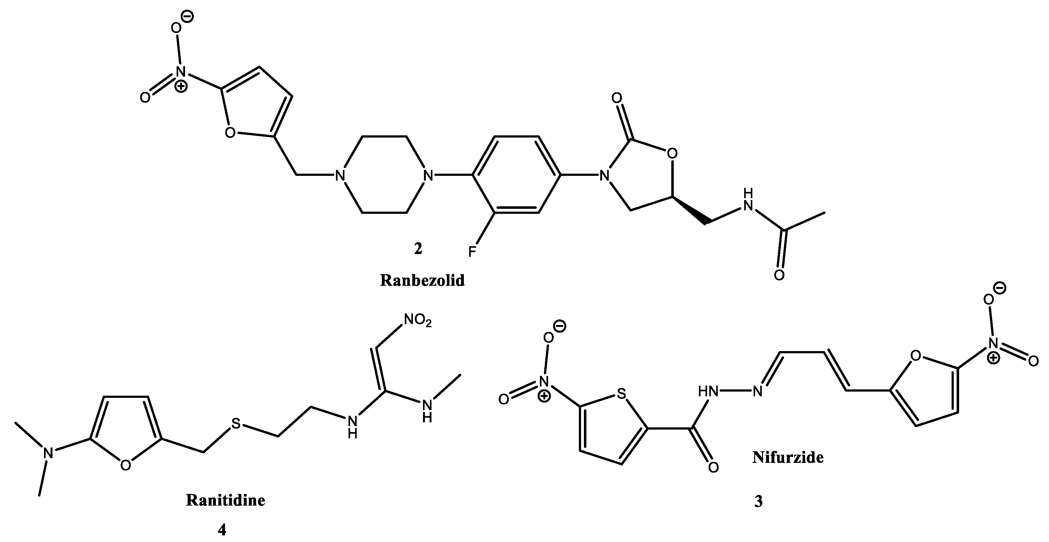
Some well-known furan derivatives are ranbezolid 2 [3], nifurzide 3 [4], and ranitidine 4 [5] widely used as anti-bacterial, anti-infective, and H2 receptor antagonists, respectively (Figure 2).
On other hand, the N-containing heterocycles are of great importance, not only because of their affluence, but above all because of their chemical, biological, and technical significance. Together, they create a large group of compounds that play a powerful role in biological investigations such as antibacterial, anticancer, anti-inflammatory, antiviral, anti-tumor, and anti-diabetic applications [6].
In the search for new biofunctional compounds, we were interested in obtaining new hybrid molecules containing both a furan fragment and a nitrogen-containing core in their structure.
In a recent study, Li and co-workers synthesized new tetrahydroquinoline and tetrahydroisoquinolines containing 2-phenyl-5-furan moiety as PDE4 inhibitors [7].
Using Pd-catalyzed coupling reactions of 2-(alkyltelluro)furan with terminal alkynes, Zeni et al. managed to synthesize a series of acetylenic furan hybrids [8]. The obtained compounds were tested for anti-inflammatory activity and showed very promising results.
2. Materials and Methods
2.1. Synthesis
The reagents were purchased from commercial suppliers (Sigma-Aldrich S.A. and Riedel-de Haën, Sofia, Bulgaria) and used as received. A Bruker Advance II+600 spectrometer was used for the recording of the NMR spectral data (BAS-IOCCP—Sofia, Bruker, Billerica, MA, USA). All compounds were analyzed in CDCl3 at 600 MHz and 150.9 MHz for 1H-NMR and 13C-NMR, respectively. Chemical shifts were determined to tetramethylsilane (TMS) (δ = 0.00 ppm) as an internal standard; the coupling constants are given in Hz. Recorded NMR spectra were taken at room temperature (approx. 295 K). The MS analysis was carried out on a Q Exactive Plus high-resolution mass spectrometer (HRMS) with a heated electrospray ionization source (HESI-II) (Thermo Fisher Scientific, Inc., Bremen, Germany) equipped with a Dionex Ultimate 3000RSLC ultrahigh-performance liquid chromatography (UHPLC) system (Thermo Fisher Scientific, Inc.). For the TLC analysis, precoated 0.2 mm Fluka silica gel 60 plates (Merck KGaA, Darmstadt, Germany) were used.
Synthesis of hybrid compounds 7.
Furan-2-carbonyl chloride 6 (1 mmol) was added to a solution of the corresponding amine 5a–d (1 mmol) in dichloromethane (15 mL). Triethylamine (1.2 mmol) was added after 10 min. After 30 min, the reaction mixture was washed with diluted hydrochloric acid (H2O:HCl = 4:1 v/v), a saturated solution of Na2CO3, and water. The organic layer was dried using anhydrous Na2SO4, concentrated, and filtered on a short column with neutral Al2O3.
Furan-2-yl(pyrrolidin-1-yl)methanone 7a: light yellow oil, yield 94% (0.155 g), 1H NMR (600 MHz, CDCl3) δ 7.51 (dd, J = 1.7, 0.8 Hz, 1H), 7.06 (dd, J = 3.5, 0.8 Hz, 1H), 6.49 (dd, J = 3.5, 1.8 Hz, 1H), 3.84 (t, J = 6.8 Hz, 2H), 3.66 (t, J = 6.9 Hz, 2H), 2.00 (p, J = 6.7 Hz, 2H), 1.91 (p, J = 6.7 Hz, 2H). 13C NMR (151 MHz, CDCl3) δ 158.1 (C=O), 148.8 (O=C-C-O), 144.0 (O-CH=CH), 115.7 (C=CH-CH), 111.3 (C-CH=CH), 47.8 (CH2-CH2-N-), 47.0 (CH2-CH2-N-), 26.6 (CH2), 23.8 (CH2). UV λmax, MeOH: 272 (ε = 12,200) nm. HRMS Electrospray ionization (ESI) m/z calcd for [M+H]+ C9H12NO2+ = 166.0863, found 166.0860 (mass error Δm = −1.82 ppm), calcd for [M+Na]+ C9H11NO2Na+ = 188.0682, found 188.0679 (mass error Δm = −1.60 ppm).
Furan-2-yl(piperidin-1-yl)methanone 7b: light yellow oil, yield 92% (0.165 g), 1H NMR (600 MHz, CDCl3) δ 7.40 (dd, J = 1.7, 0.8 Hz, 1H), 6.85 (dd, J = 3.4, 0.8 Hz, 1H), 6.39 (dd, J = 3.4, 1.8 Hz, 1H), 3.63 (s, 4H), 1.65–1.60 (m, 2H), 1.59–1.54 (m, 4H). 13C NMR (151 MHz, CDCl3) δ 159.3 (C=O), 148.2 (O=C-C-O), 143.4 (O-CH=CH), 115.5 (C=CH-CH=CH-O), 111.1 (C=CH-CH=CH-O), 47.7 (N-CH2-CH2), 26.2 (N-CH2-CH2), 24.7 (CH2-CH2-CH2). UV λmax, MeOH: 272 (ε = 14,800) nm. HRMS Electrospray ionization (ESI) m/z calcd for [M+H]+ C10H14NO2+ = 180.1019, found 180.1017 (mass error Δm = −1.06 ppm), calcd for [M+Na]+ C10H13NO2Na+ = 202.0838, found 202.0836 (mass error Δm = −0.99 ppm).
(3,4-dihydroquinolin-1(2H)-yl)(furan-2-yl)methanone 7c: light yellow oil, yield 95% (0.216 g), 1H NMR (600 MHz, CDCl3) δ 7.27 (dd, J = 1.7, 0.8 Hz, 1H), 7.11 (dd, J = 7.5, 0.8 Hz, 1H), 7.01 (td, J = 7.4, 1.2 Hz, 1H), 6.95 (ddd, J = 8.0, 4.5, 1.1 Hz, 1H), 6.84 (d, J = 8.0 Hz, 1H), 6.60 (d, J = 3.5 Hz, 1H), 6.30 (dd, J = 3.5, 1.7 Hz, 1H), 3.84 (t, J = 6.6 Hz, 2H), 2.73 (t, J = 6.6 Hz, 2H), 1.96 (p, J = 6.6 Hz, 2H). 13C NMR (151 MHz, CDCl3) δ 159.8, 147.9, 143.9, 138.9, 132.0, 128.4, 125.9, 124.9, 124.3, 116.4, 111.2, 44.2, 26.8, 24.0. UV λmax, MeOH: 291 (ε = 10,800) nm. HRMS Electrospray ionization (ESI) m/z calcd for [M+H]+ C14H14NO2+ = 228.1019, found 228.1015 (mass error Δm = −1.75 ppm), calcd for [M+Na]+ C14H13NO2Na+ = 250.0838, found 250.0834 (mass error Δm = 2.40 ppm).
(3,4-dihydroisoquinolin-2(1H)-yl)(furan-2-yl)methanone 7d: light yellow oil, yield 90% (0.205 g), 1H NMR (600 MHz, CDCl3) δ 7.55 (s, 1H), 7.24–7.17 (m, 4H), 7.07 (dd, J = 3.5, 0.7 Hz, 1H), 6.53 (dd, J = 3.5, 1.8 Hz, 1H), 4.88 (s, 2H), 4.01 (s, 2H), 3.00 (s, 2H). 13C NMR (151 MHz, CDCl3) δ 159.6 (C=O), 148.1 (O=C-C-O), 143.9 (O-CH=CH), 133.1 (C, Ar), 129.9 (C, Ar), 127.4 (C, Ar), 126.8(C, Ar), 126.5 (C, Ar), 125.3 (C, Ar), 116.3 (C=CH-CH), 111.3 (CH-CH=CH), 48.3 (Ar-CH2-N), 44.5 (Ar-CH2-CH2-N), 28.5 (Ar-CH2-CH2-N). UV λmax, MeOH: 232 (ε = 13500) nm, 272 (ε = 12,000) nm. HRMS Electrospray ionization (ESI) m/z calcd for [M+H]+ C14H14NO2+ = 228.1019, found 228.1015 (mass error Δm = −1.75 ppm), calcd for [M+Na]+ C14H13NO2Na+ = 250.0838, found 250.0835 (mass error Δm = 2.80 ppm).
2.2. Biological Evaluation
Chemicals and Reagents
For HPLC analysis, chromatographic-grade methanol was used (VWR, Vienna, Austria). A Millipore purifier (Millipore, USA) was used to obtain water for HPLC. Potassium dihydrogen phosphate, potassium chloride, ascorbic acid, ibuprofen, ketoprofen, dipotassium hydrogen phosphate, sodium chloride, hydrogen peroxide, trypsin, Tris-HCl buffer, and perchloric acid were purchased from Sigma-Aldrich, Taufkirchen, Germany. Human albumin 20%—BB, 200 g/L was ordered from BB-NCIPD Ltd., Sofia, Bulgaria. Chromatographic plates, Kieselgel 60 F254, were purchased from Merck (Darmstadt, Germany).
2.3. Biological Experiments
2.3.1. Hydrogen Peroxide Scavenging Activity (HPSA)
The Manolov et al. approach was used to evaluate the furan hybrid molecules’ capacity to scavenge hydrogen peroxide [12].
A 43 mM solution of H2O2 was prepared in potassium phosphate buffer solution (0.2 M, pH 7.4). The analysis of the samples was carried out as follows: in test tubes, 0.6 mL H2O2 (43 mM), 1 mL sample/standard with different concentrations (20–1000 μg/mL), and 2.4 mL potassium phosphate buffer solution were mixed. The mixture was stirred and incubated in the dark for 10 min at 37 °C. Absorbance was measured at 230 nm with a spectrophotometer (Camspec M508, Leeds, UK) against a blank solution containing phosphate buffer and H2O2 without the sample. Ascorbic acid and quercetin were used as standards. The percentage HPSA of the samples was evaluated by comparing with a blank sample and calculated using the following formula:
where Ablank is the absorbance of the blank sample, ACS is the absorbance of the control sample, and ATS is the absorbance of the test sample. The mean IC50 value was estimated based on three replicates by means of interpolating the graphical dependence of scavenging hydrogen peroxide on concentration.
2.3.2. Metal-Chelating Activity (MChA) on Ferrous Ions
The iron-chelating ability of the furan derivatives was studied by the method in [13].
The ability of the different fractions to chelate Fe2+ was evaluated. The reaction mixture contained 1 mL of sample/standard, 3.7 mL of methanol, and 0.1 mL of 2 mmol/L ferric dichloride. With the addition of 0.2 mL of 5 mmol/L ferrozine, the reaction started. The reaction mixture was vortexed and left for 10 min at room temperature. After reaching equilibrium in the mixture, the absorbance of the sample was measured at 562 nm on a UV-Vis spectrophotometer (Camspec M508, Leeds, UK). The blank was prepared the same way as the test sample, but methanol was added instead of the sample/standard. The results were reported as percent inhibition of ferrozine Fe2+ complex formation with the sample. The mean IC50 value was estimated based on three replicates. Ethylenediaminetetraacetic acid (EDTA)-sodium (Na) (0.5 mg/mL) served as a positive control.
2.3.3. Inhibition of Albumin Denaturation (IAD)
The anti-inflammatory efficacy was evaluated using an inhibition of albumin denaturation (IAD) in vitro study. Sakat method [14] with a modest modification [15] was used to finish the analysis.
Human albumin was used in the experiment. In distilled water, an albumin (1%) solution was prepared (pH 7.4). The tested compounds/standard were first dissolved in 1.2 mL DMF and PBS up to 25 mL, resulting in a stock solution with a final concentration of 1000 μg/mL. Then, in PBS, a series of working solutions with varying concentrations (20–500 μg/mL) were prepared. The reaction mixture contained 2 mL of different concentrations of test sample/standard and 1 mL of albumin (1%). The mixture was incubated at 37 °C for 15 min before being heated in a water bath at 70 °C for 15 min. After cooling, the turbidity was measured with a spectrophotometer at 660 nm (Camspec M508, Leeds, UK). The experiment was carried out three times. The inhibition of albumin denaturation (IAD) was calculated as a percentage of the control. The control sample was albumin dissolved in distilled water at the same concentration.
2.3.4. Antitryptic Activity (ATA)
This method is also known to have anti-arthritic activity in vitro. The analysis was carried out using the method described by Oyedapo and Femurewa [16], with minor modifications as described by Manolov et al. [15].
Trypsin, 2 mL 0.06 mg/mL, 1 mL Tris-HCl buffer (20 mM, pH 7.4), and 1 mL test sample/standard of various concentrations (20–1000 μg/mL) were added to the reaction mixture. For 5 min, the mixture was incubated at 37 °C. Following that, 1 mL of human albumin (4% v/v) was added. The mixture was then incubated for an additional 20 min. To stop the reaction, 2 mL of 70% perchloric acid was added to the mixture. The cloudy suspension was cooled and centrifuged for 20 min at 5000 rpm. A spectrophotometer (Camspec M508, Leeds, UK) was used to measure the absorbance of the supernatant at 280 nm in comparison to the control solution. The control solution consisted of various concentrations of sample/standard in methanol. As a baseline, ibuprofen was used. The analysis was carried out three times. By comparing the samples to a blank sample, the percentage of antitryptic activity (ATA) was determined. The blank sample was prepared in the same way as the test sample, with the exception that perchloric acid was added before the albumin.
where Ablank is the absorbance of the blank sample, ACS is the absorbance of the control solution (test sample in different concentrations), and ATS is the absorbance of the test samples. The mean IC50 values were estimated by means of interpolating the graphical dependence of ATA on concentration.
2.4. Physicochemical Characterization
2.4.1. Determination of Lipophilicity as RM Values
The lipophilicity of the furan derivatives was estimated using the method described by Hadjipavlou-Litina and Pontiki [17].
RPTLC (reverse-phase TLC) was performed on silica gel plates impregnated with 55% (v/v) liquid paraffin in light petroleum ether. The mobile phase was a methanol/water mixture (77:23, v/v) containing 0.1 acetic acid. The plates were developed in closed chromatography tanks saturated with the mobile phase at 24 °C. Spots were detected under UV light or by iodine vapors. RM values were determined from the corresponding Rf values (from 10 individual measurements) using the equation RM = log [(1/Rf) − 1].
2.4.2. Prediction of Anti-Inflammatory and Anti-Arthritic Activity
2.5. Statistical Analysis
All analyses were performed in triplicate. The data were presented as mean ± SD. The significance level was set at p < 0.05.
3. Results
3.1. Synthesis
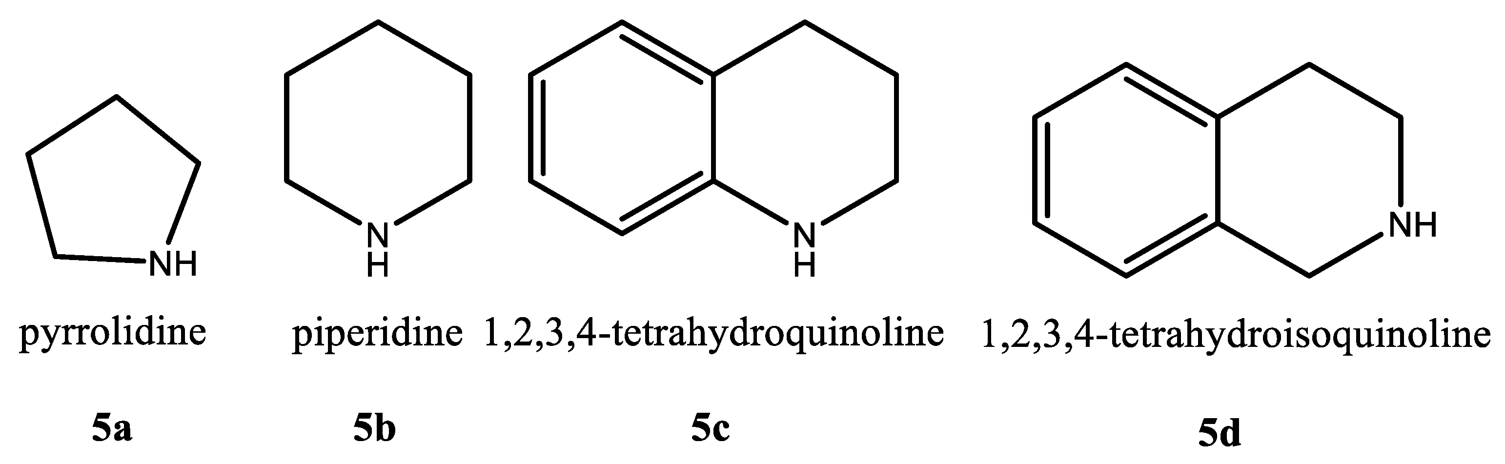
In the present article, we report the successful synthesis of new furan hybrid molecules with different N-containing compounds, such as pyrrolidine 5a, piperidine 5b, 1,2,3,4-tetrahydroquinoline 5c, and 1,2,3,4-tetrahydroisoquinoline 5d (Figure 3), as shown in Scheme 1. In Scheme 1, we report the successful synthesis of new furan hybrid molecules with various N-containing compounds, such as pyrrolidine 5a, piperidine 5b, 1,2,3,4-tetrahydroquinoline 5c, and 1,2,3,4-tetrahydroisoquinoline 5d.
For this purpose, furan-2-carbonyl chloride 6 is added to a solution of the corresponding amine 5 (1 mmol) in dichloromethane. The reaction mixture is stirred for a further 10 min and then an excess of triethylamine (1.5 mmol) is carefully added dropwise. The secondary amines 5a–d were fully acylated for the next 30 min (TLC).
The reaction generally works in high yields. All yields are above 90%, as can be seen in the experimental section. All compounds are fully characterized by UV, 1H-, and 13C- NMR and HRMS spectra.
All obtained compounds 7a, 7c, and 7d are new compounds. Compound 7b, the hybrid molecule of furan 6 and piperidine 5b, has been previously reported [20,21,22].
Comparing the 1H-NMR data for compound 7b obtained by us with the same reported by Ramkumar and Chandrasekaran can be seen the match in the spectra [23]. 1H NMR (CDCl3, 400 MHz): δ = 7.34 (d, J = 0.8 Hz, 1 H, ArH), 6.79 (dd, J1 = 3.4 Hz, J2 = 0.8 Hz, 1 H, ArH), 6.31–6.33 (m, 1 H, ArH), 3.55 (s, 4 H, CH2), 1.48–1.55 (m, 6H, CH2).
A broadened singlet is observed for the methylene groups on both sides of the nitrogen atom at 3.55 ppm by them and at 3.66 ppm by us. The protons from the furan core also match, being slightly shifted (from 7.34 ppm to 7.40 ppm, from 6.79 ppm to 6.85 ppm, and from 6.31–6.33 ppm to 6.39 ppm). The rest of the signals from the hydrogenated nitrogen-containing core appear as one multiplet for six protons in the interval from 1.48 to 1.55 (m, 6H, CH2) in the Ramkumar 1H-NMR spectra. In the 1H-NMR spectrum obtained by us, two multiplets stand out from 1.65–1.60 for two protons and from 1.59–1.54 for four protons, respectively. Based on the coincidence of all of these data, it can be asserted without doubt that we have succeed in obtaining the same compound.
The four compounds have been given the names hybrid 1 (H1)- 7a, hybrid 2 (H2)- 7b, hybrid 3 (H3)- 7c, and hybrid 4 (H4)- 7d for biological evaluation research.
The newly synthesized compounds (H1–4) differ mainly in the N-containing heterocycles. In order to investigate the structure of the new hybrid molecules, we used mass spectrometry. We used ESI in positive ionization mode. The fragmentation of the furan derivatives H1–4 proceeds according to the mechanism presented in Scheme 2.
The structural fragment that connects the furan (ring A) and heterocyclic (ring B) rings in furan derivatives is C-C(O)-N. Several fragmentation pathways originate from here. The main fragmentation pathways of compounds H1–4 involve the cleavage of C-N (path 1), C-C (path 2) bonds, and retro cyclization of heteroring B with a cleavage of bonds at position a/d for H1, b/f for H2, c/f, c/e for H3, and a/f for H4 (Scheme 2). The cleavage of the C-N bond (pathway 1) provides important information about the structure of the heterocyclic ring (m/z 70, 84, 132) and the furan ion at m/z 95. The structure of compounds H1 and H2 does not contain fused rings. Ring B, in their structure, is respectively pyrrolidine and piperidine. The retro cyclization of ring B is associated with the cleavage of the a/d bonds for H1, and b/f for H4, resulting in the same ion with m/z 124 (Figures S14 and S16).
The structure of compounds H3 and H4 contain fused rings: benzo[b]piperidine (1,2,3,4 tetrahydroquinoline nuclei) and benzo[c]piperidine (1,2,3,4 tetrahydroisoquinoline nuclei), respectively.
In the presence of an additional benzene nucleus during fragmentation, the formation of additional characteristic ions is observed, by which the isomers H3 and H4 are clearly distinguished. For both isomers, the ions m/z 132 (path 1) and m/z 160 (path 2) were obtained. Additional fragmentation pathways depend on the position of the nitrogen atom in the structure of compounds H3 and H4 (Scheme 2, Figures S18 and S20). For compound H3, which contains benzo[b]piperidine in its structure, the fragmentation proceeds in other ways, different from its isomer. Under MS conditions, a neutral CO molecule (28 au) is lost from the structure of compound H3, where a retro cyclization follows and leads to the m/z 172 ion. A molecule of H2O (18 au) is lost from the same ion and an ion of m/z 154 is obtained (Scheme 2, Figure S18). These two ions are not produced in the fragmentation of compound H4. Additionally, the fragmentation of the m/z 160 ions produced an m/z 118 ion. In fact, ions m/z 118, m/z 154 and m/z 172 are characteristic of compound H3 (Figure S18), while compound H4 is ion m/z 117, which is the result of the retro cyclization of benzo[c]piperidine in position a/f (Scheme 2, Figure S20).
3.2. Biological Evaluation
All synthesized furan hybrids were analyzed for their in vitro hydrogen peroxide-scavenging activity (HPSA), inhibition albumin denaturation (IAD), metal-chelating activity (MChA), and antitryptic activity (ATA). The results observed in vitro were contrasted with those predicted in silico. The results are presented in Table 1.
3.2.1. Hydrogen Peroxide Scavenging Activity
Copper and iron are vitally essential trace elements for humans, which participate as cofactors in numerous enzymes and various physiological processes. However, in their free form, they are toxic because Cu(II) catalyzes the oxidation of ascorbic acid, producing reactive oxygen species (ROS) such as superoxide radicals (O2•−) and H2O2. In this catalytic process, Cu(II) and Fe(II) react with H2O2, and hydroxyl radicals (•OH) are generated via the Fenton reaction [24]. It has been established that the harm they produce plays a role in the onset of a number of diseases, including Alzheimer’s disease, atherosclerosis, cancer, and cardiovascular diseases [25]. Therefore, in the human body, the sulfur-containing molecules glutathione, cysteine, and ergothioneine play an important role as endogenous antioxidants [24]. For this reason, the study of compounds with high oxygen-free radical scavenging activity is a current and significant area of research.
In the current study, the goal of the research was to stop hydrogen peroxide’s damaging effects. Hydrogen peroxide is an oxidant that is continuously produced in living tissues as a result of several metabolic processes. However, in order to avoid entering hazardous reactions like the Fenton reaction, its detoxification is crucial [26].
The inflammatory process also causes and accelerates the formation of ROS. Most significantly, the formation of other ROS species, such as H2O2, is linked to the production of superoxide anion radicals at the site of inflammation. Assuming that at least some of the oxygen produced in these processes is in the singlet state, it is also engaged in the reductive breakdown reactions of organic hydroperoxides ROOH and hydrogen peroxide (the so-called Haber–Weiss reaction) [27,28]. Therefore, it is crucial to inhibit H2O2 in order to stop the generation of •OH.
We compared the results obtained for the antioxidant activity of the synthetic analogs of furan with the standards of ascorbic acid and quercetin. They are natural compounds with demonstrable antioxidant activity [15].
The discovered furan hybrid molecules exhibit reduced in vitro antioxidant activity when compared to ascorbic acid (24.84 µg/mL) and quercetin (69.25 µg/mL). Compounds H3 (77.75 µg/mL) and H4 (71.72 µg/mL) demonstrate higher antioxidant activity compared to the rest of the synthesized compounds (H1, H2) (Table 1, Figure 4A).
The inclusion of benzene nuclei in the structure of the discovered compounds greatly boosts the antioxidant action. As a result, compounds H3 and H4 show strong activity.
3.2.2. Metal-Chelating Activity (MChA) on Ferrous Ions
Fe(II) is a transition metal well known for its increased propensity for the Fenton reaction, but it also makes it one of the most important pro-oxidants involved in lipid peroxidation, i.e., the oxidation of lipids leading to cell membrane damage [13,26]. This, in turn, accelerates the aging process. Chelation is one of the antioxidant methods for reducing the catalytic action of the transition metals Fe(II) and Cu(II). Chelating agents create sigma bonds with the metal and are regarded as strong secondary antioxidants [13,29] because they can lower the redox potential and stabilize the oxidized form of the iron ion.
The metal-chelating properties of the newly synthesized furan derivatives (H1–4) on ferrous iron are shown in Table 1 and Figure 4B. The results for the metal-chelating activity are presented as IC50. Chelating activity on ferrous iron is an important step in preventing lipid peroxidation. Therefore, we investigated the ability of compounds H1–4 to form chelate complexes with Fe(II) ions. The concentration gradient of H1–4 affects the chelating activity. As the concentration of compounds H1–4 increases, their ability to chelate Fe(II) increases. At low concentrations, H1–4 show low chelating activity. From the analysis, we found that compounds H3 (1.02 mg/mL) and H4 (1.46 mg/mL) showed significant metal-chelating activity four times higher than compounds H1 and H2.
Here, we must clarify that there is a connection between the two methods (HPSA and MChA). In living tissues, competing reactions are most likely taking place—both hydrogen peroxide deactivation and Fe(II) chelation. The research shows that there is a good correlation dependence of 0.9672 between the two methods. This relationship is beneficial because the chelation of Fe(II) will prevent the Fenton reaction from occurring. This approach provides more information about the antioxidant activity of the compounds, proving them to be reliable exogenous antioxidants.
3.2.3. Inhibition of Albumin Denaturation (IAD)
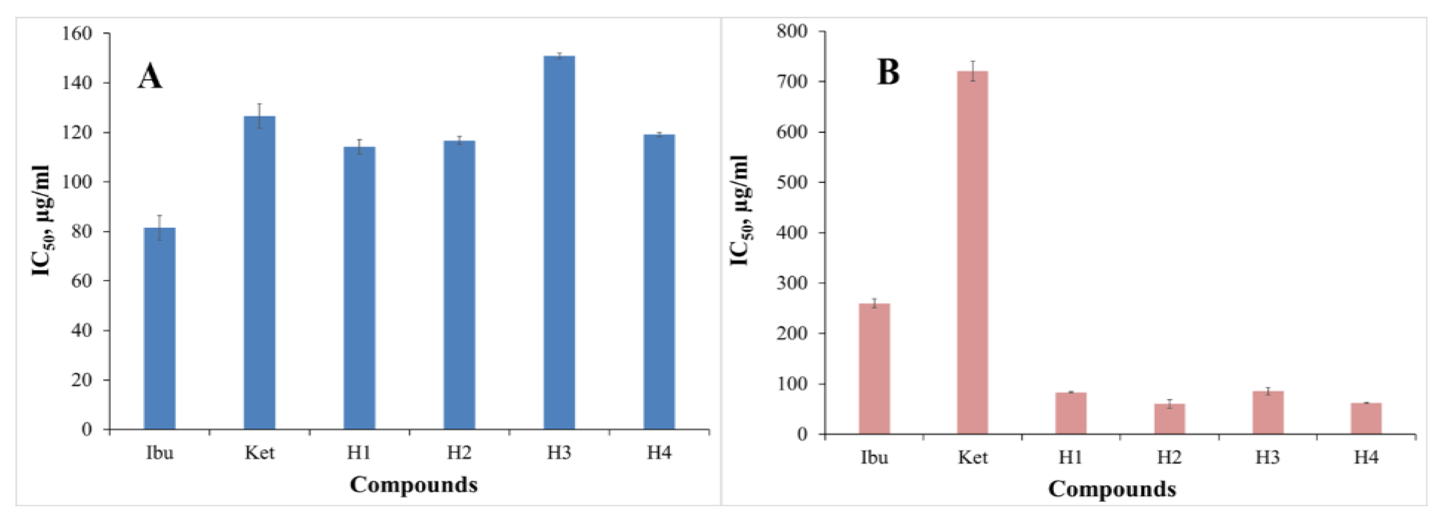
Living tissues’ response to harm is inflammation. Cell migration, tissue breakdown, mediator release, enzyme activation, fluid extravasation, and tissue healing are only a few of the numerous complicated processes involved [30]. Inflammation in rheumatoid arthritis is well known to be brought on by the denaturation of proteins. The ability of certain anti-inflammatory medications to prevent thermally induced protein denaturation has been demonstrated to depend on dose [31]. The obtained furan hybrids were examined for their ability to inhibit albumin denaturation. This method determines the extent to which albumin is protected from denaturation when heated. We used human albumin for this purpose. Figure 5A shows the percentages of inhibition of synthetic furan derivatives. The obtained results of the analysis are presented as IC50. Because ibuprofen and ketoprofen have well-established properties, we decided to utilize them as a standard to compare the activities of the newly synthesized furan derivatives. The IC50 values of ibuprofen and ketoprofen estimated as IAD are 81.50 μg/mL and 126.58 μg/mL, respectively (Table 1, Figure 5A). All of the obtained results show that the IC50 values of furan hybrid molecules are in the range of 114.31 to 150.99 μg/mL (Table 1, Figure 5A).
In general, the obtained compounds (H1–4) exhibited high IAD activity as profens. The in silico anti-inflammatory activity (cAnti-A) results show that the standards (ibuprofen and ketoprofen) have higher activity than the synthesized derivatives (H1–4), indicating that there is a directly proportional dependence between in vitro and in silico for ibuprofen and a reverse dependence for ketoprofen (Table 1).
In addition, IAD analysis reveals that lipophilicity is a significant physicochemical parameter. The lipophilicity (RM) of the synthetic furan derivatives studied ranges from 0.87 to 1.04, which influences albumin protection to some extent (Table 1).
For the stability of albumin, the hydrophobic pocket of subdomain IIA and IIIA plays an important key role, popularly known as Sudlow’s sites I and II, respectively. Due to the hydrophobic nature of the interior of Sudlow’s sites I pocket, the drug primarily formed hydrophobic interactions with Phe211, Trp214, Leu219, Phe223, Leu234, Leu238, Leu260, Ile264, and Ile290 [32].
As the interior of Sudlow’s sites I pocket is hydrophobic in nature, the drug predominantly formed hydrophobic interactions with Phe211, Trp214, Leu219, Phe223, Leu234, Leu238, Leu260, Ile264, and Ile290 [32]. The stabilization of the albumin molecule in this study is due to hydrophobic interactions between Sudlow’s sites I and the furan derivatives.
3.2.4. Antitryptic Activity (ATA)
Proteinases have been linked to the development of arthritic symptoms. Neutrophils are known to be a good source of proteinase because their lysosomal granules contain many serine proteinases. It has previously been reported that leukocyte proteinase is important in the development of tissue damage during inflammatory reactions and that proteinase inhibitors provide significant protection [16,31]. In vitro anti-arthritic activity was assessed as antitryptic activity [16]. The IC50 results for the ATA range from 60.21 to 85.33 μg/mL. The results reveal that the furan derivatives H1–4 show better antitryptic activity compared to ibuprofen and ketoprofen (Table 1, Figure 5B).
3.2.5. Lipophilicity
The most commonly used parameter in SAR drug discovery is lipophilicity. It can be determined experimentally or mathematically.
Increased permeability, solubility, target potency, and toxicity have all been linked to lipophilicity. The lipophilicity was determined as RM values using reverse-phase thin layer chromatography (RPTLC). This is regarded as a dependable, quick, and convenient method of expressing lipophilicity [33]. Apart from the importance of lipophilicity in biologically active compound kinetics, both hydrophilic and lipophilic antioxidants are required to act as radical scavengers in the aqueous phase or as chain-breaking antioxidants in biological membranes [17].
In the present work, we have examined the antioxidant, metal-chelating, and in vitro biological activity of the newly synthesized furan derivatives. Lipophilicity is confirmed to be an important factor in their activity. The results show that compounds H1–4 are lipophilic with good antioxidant and metal-chelating activity. This is what makes them reliable lipophilic exogenous antioxidants that are necessary to neutralize harmful radicals in the cell membrane.
In general, the in vitro studies results show that the compounds H1–4 exhibit IAD and ATA. We learned a lot about the properties of the potential new drugs from both experiments, and both experiments are related to keeping the albumin molecule intact. Albumin is protected from denaturation (IAD) in the first case. Compounds H1–4 allosterically bind to albumin. In the second case, it is protected from the action of the enzyme trypsin (ATA), which is inhibited by furan derivatives.
4. Conclusions
In conclusion, we have successfully obtained three new hybrid molecules containing a furan skeleton and N-heterocycle. The new hybrid molecules were fully characterized using UV, 1H- and 13C-NMR, and HRMS spectrometry. All four of the obtained compounds were in vitro biologically evaluated for their antioxidant, anti-inflammatory, anti-arthritic, and metal-chelating activity. Detailed HRMS analysis showed that the isomers H3 and H4 can be easily distinguished from each other. All compounds showed activity in all in vitro biological tests performed. As expected, hybrid molecules H3 and H4 show greater metal-chelating activity as well as antioxidant activity than hybrids H1 and H2. The anti-inflammatory activity confirmed by the in silico analysis is also proven by the in vitro biological evaluation, and the hybrids are ranked in the following descending order: H1 > H2 > H4 > H3.
Supplementary Materials
The following supporting information can be downloaded at: https://www.mdpi.com/article/10.3390/pr10101997/s1, Figures S1–S4: 1H-NMR spectrum of hybrids 1–4; Figures S5–S8: 13C-NMR spectrum of hybrids 1–4; Figures S9–S12: UV spectrum of hybrids 1–4; Figures S13–S20: HRMS chromatograms of hybrids 1–4.
Author Contributions
Conceptualization, I.I. and S.M.; methodology, S.M. and D.B.; software, S.M.; validation, D.B., S.M. and I.I.; formal analysis, S.M., D.B. and P.N.; investigation, S.M.; resources, I.I.; data curation, I.I.; writing—original draft preparation, S.M. and D.B.; writing—review and editing, S.M., D.B. and I.I.; visualization, D.B.; supervision, I.I.; project administration, S.M.; funding acquisition, I.I. All authors have read and agreed to the published version of the manuscript.
Funding
This research was funded by the National Science Fund of the Bulgarian Ministry of Education and Science, grant number KΠ 06 M29/1.
Institutional Review Board Statement
Not applicable.
Informed Consent Statement
Not applicable.
Data Availability Statement
The data presented in this study are available in this article and in the supporting Supplementary Material.
Conflicts of Interest
The authors declare no conflict of interest.
References
- Demicheva, L. Biological activity of furan derivatives (review). Chem. Heterocycl. Compd. 1993, 29, 243–267. [Google Scholar] [CrossRef]
- Duffy, L.; Smith, A.D. Nitrofurantoin macrocrystals prevent bacteriuria in intermittment self-catheterization. Urology 1982, 20, 47–49. [Google Scholar] [CrossRef]
- Naruganahalli, K.S.; Shirumalla, R.K.; Bansal, V.; Gupta, J.B.; Das, B.; Ray, A. Ranbezolid, a novel oxazolidinone antibacterial: In vivo characterisation of monoamine oxidase inhibitory potential in conscious rats. Eur. J. Pharmacol. 2006, 545, 167–172. [Google Scholar] [CrossRef]
- Delsarte, A.; Faway, M.; Frère, J.M.; Coyette, J.; Calberg-Bacq, C.M.; Heinen, E. Nifurzide, a nitrofuran antiinfectious agent: Interaction with Escherichia coli cells. Antimicro. Agents Chemother. 1981, 19, 477–486. [Google Scholar] [CrossRef] [Green Version]
- White, C.M.; Hernandez, A.V. Ranitidine and Risk of N-Nitrosodimethylamine (NDMA) Formation. JAMA 2021, 326, 225–227. [Google Scholar] [CrossRef]
- Mermer, A.; Keles, T.; Sirin, Y. Recent studies of nitrogen containing heterocyclic compounds as novel antiviral agents: A review. Bioorg. Chem. 2021, 114, 105076. [Google Scholar] [CrossRef]
- Li, Y.-S.; Liu, X.-Y.; Zhao, D.-S.; Liao, Y.-X.; Zhang, L.-H.; Zhang, F.-Z.; Song, G.-P.; Cui, Z.-N. Tetrahydroquinoline and tetrahydroisoquinoline derivatives as potential selective PDE4B inhibitors. Bioorg. Med. Chem. Lett. 2018, 28, 3271–3275. [Google Scholar] [CrossRef]
- Zeni, G.; Lüdtke, D.; Nogueira, C.; Panatieri, R.; Braga, A.; Silveira, C.; Stefani, H.; Rocha, J. New acetylenic furan derivatives: Synthesis and anti-inflammatory activity. Tetrahedron Lett. 2001, 42, 8927–8930. [Google Scholar] [CrossRef]
- Janczewski, Ł.; Zieliński, D.; Kolesińska, B. Synthesis of amides and esters containing furan rings under microwave-assisted conditions. Open Chem. 2021, 19, 265–280. [Google Scholar] [CrossRef]
- Qian, Z.; Li, Q.; Wang, L.; Fu, F.; Liu, X. The chemical effect of furfural amide on the enhanced performance of the diphenolic acid derived bio-polybenzoxazine resin. J. Poly. Sci. 2021, 59, 2057–2068. [Google Scholar] [CrossRef]
- Malladi, S.; Venkata Nadh, R.; Suresh Babu, K.; Suri Babu, P. Synthesis and antibacterial activity studies of 2,4-di substituted furan derivatives. Beni-Suef Univ. J. Basic App. Sci. 2017, 6, 345–353. [Google Scholar] [CrossRef]
- Manolov, S.; Ivanov, I.; Bojilov, D. Synthesis of New 1,2,3,4-Tetrahydroquinoline Hybrid of Ibuprofen and Its Biological Evaluation. Molbank 2022, 1, M1350. [Google Scholar] [CrossRef]
- Sirin, S.; Duyar, H.; Aslım, B.; Seferoğlu, Z. Synthesis and biological activity of pyrrolidine/piperidine substituted 3-amido-9-ethylcarbazole derivatives. J. Mol. Struct. 2021, 1242, 130687. [Google Scholar] [CrossRef]
- Sakat, S.S.; Juvekar, A.R.; Gambhire, M.N. In-vitro antioxidant and anti-inflammatory activity of methanol extract of Oxalis corniculata linn. Int. J. Pharm. Pharm. Sci. 2010, 2, 146–155. [Google Scholar]
- Manolov, S.; Ivanov, I.; Bojilov, D. Microwave-assisted synthesis of 1,2,3,4-tetrahydroisoquinoline sulfonamide derivatives and their biological evaluation. J. Serb. Chem. Soc. 2021, 86, 139–151. [Google Scholar] [CrossRef]
- Oyedapo, O.O.; Famurewa, A.J. Antiprotease and membrane stabilizing activities of extracts of fagara zanthoxyloides, olax subscorpioides and tetrapleura tetraptera. Int. J. Pharmacogn. 1995, 33, 65–69. [Google Scholar] [CrossRef]
- Pontiki, E.; Hadjipavlou-Litina, D. Synthesis and pharmacochemical evaluation of novel aryl-acetic acid inhibitors of lipoxygenase, antioxidants, and anti-inflammatory agents. Bioorg. Med. Chem. 2007, 15, 5819–5827. [Google Scholar] [CrossRef]
- Sadym, A.; Lagunin, A.; Filimonov, D.; Poroikov, V. Prediction of biological activity spectra via the Internet. SAR QSAR Environ. Res. 2003, 14, 339–347. [Google Scholar] [CrossRef] [PubMed]
- Filimonov, D.; Lagunin, A.; Gloriozova, T.; Rudik, A.; Druzhilovskii, D.; Pogodin, P.; Poroikov, V. Prediction of the Biological Activity Spectra of Organic Compounds Using the Pass Online Web Resource. Chem. Heterocycl. Compd. 2014, 50, 444–457. [Google Scholar] [CrossRef]
- Das, S.; Addis, D.; Zhou, S.; Junge, K.; Beller, M. Zinc-catalyzed reduction of amides: Unprecedented selectivity and functional group tolerance. J. Am. Chem. Soc. 2010, 132, 1770–1771. [Google Scholar] [CrossRef] [PubMed]
- Pathak, U.; Bhattacharyya, S.; Pandey, L.; Mathur, S.; Jain, R. An easy access to tertiary amides from aldehydes and N, N-dialkylchlorothiophosphoramidates. RSC Adv. 2014, 4, 3900. [Google Scholar] [CrossRef]
- Khosravi, K.; Naserifar, S. 1,1,2,2-Tetrahydroperoxy-1,2-diphenylethane: An efficient and high oxygen content oxidant in various oxidative reactions. Tetrahedron 2018, 74, 6584–6592. [Google Scholar] [CrossRef]
- Ramkumar, R.; Chandrasekaran, S. Catalyst-free, metal-free, and chemoselective transformation of activated secondary amides. Synthesis 2019, 51, 921–932. [Google Scholar] [CrossRef] [Green Version]
- Chalana, A.; Kumar, R.; Karri, R.; Kumar, K.; Kumar, B.; Roy, G. Interplay of the intermolecular and intramolecular interactions in stabilizing the thione-based copper(I) complexes and their significance in protecting the biomolecules against metal-mediated oxidative damage. Polyhedron 2022, 215, 115647. [Google Scholar] [CrossRef]
- Galano, A.; Macías-Ruvalcaba, A.; Campos, M.; Pedraza-Chaverri, J. Mechanism of the OH radical scavenging activity of nordihydroguaiaretic acid: A combined theoretical and experimental study. J. Phys. Chem. B 2010, 114, 6625–6635. [Google Scholar] [CrossRef] [PubMed]
- Halliwell, B.; Gutterdge, J.M.C. Free Radicals in Biology and Medicine, 5th ed.; Oxford Academic: Oxford, UK, 2015; Available online: https://doi.org/10.1093/acprof:oso/9780198717478.001.0001 (accessed on 5 August 2022).
- Khan, A.U. Singlet molecular oxygen from superoxide anion and sensitized fluorescence of organic molecules. Science 1970, 168, 467–477. [Google Scholar] [CrossRef]
- Kellog, E.W.; Fridovich, I. Superoxide, hydrogen peroxide, and singlet oxygen in lipid peroxidation by a xanthine oxidase system. J. Biol. Chem. 1975, 250, 8812–8817. Available online: https://www.jbc.org/article/S0021-9258(19)40745-X/pdf (accessed on 15 August 2022). [CrossRef]
- Bandgar, B.P.; Adsul, L.K.; Lonikar, S.V.; Chavan, H.V.; Shringare, S.N.; Patil, S.A.; Jalde, S.S.; Koti, B.A.; Dhole, N.A.; Gacche, R.N.; et al. Synthesis of novel carbazole chalcones as radical scavenger, antimicrobial and cancer chemopreventive agents. J. Enzyme Inhib. Med. Chem. 2013, 28, 593–600. [Google Scholar] [CrossRef] [Green Version]
- Vane, J.R.; Botting, R.M. New insights into the mode of action of anti-inflammatory drug. Inflamm. Res. 1995, 44, 1–10. [Google Scholar] [CrossRef]
- Jayashree, V.; Bagyalakshmi, S.; Manjula Devi, K.; Richard Daniel, D. In-vitro anti-inflamatory activity of 4-benzylpiperidine. Asian J. Pharm. Clin. Res. 2016, 9, 108–110. [Google Scholar] [CrossRef]
- Mondal, M.; Lakshmi, P.; Krishna, R.; Sakthivel, N. Molecular interaction between human serum albumin (HSA) and phloroglucinol derivative that shows selective anti-proliferative potential. J. Lumin. 2017, 192, 990–998. [Google Scholar] [CrossRef]
- Hansch, C.; Leo, D.; Hoekman, D.H. Exploring QSAR: Hydrophobic, Electronic, and Steric Constants; American Chemical Society: Washington, DC, USA, 1995; Available online: https://www.amazon.com/Exploring-QSAR-Hydrophobic-Electronic-Profession-al/dp/0841229910 (accessed on 2 August 2022).
Figure 1.
Structural formula of nitrofurantoin 1.

Figure 2.
Structural formulas of ranbezolid, nifurzide, and ranitidine.
Scheme 1.
Synthesis of hybrid molecules 7a–d.

Figure 3.
Secondary amines took place in the acylation reaction.
Scheme 2.
Possible fragmentation pathways of compounds H1–4 under ESI-MS/MS conditions. 1-H1—fragmentation of pyrrolidine-furan H1. 1-H2—fragmentation of H2 compounds. 1-H3—fragmentation of compounds H3. 1-H4—fragmentation of compounds H4. H3 and H4 can be considered derivatives of piperidine, since its structure contains a piperidine ring (ring B)—benzo[b]piperidine (1,2,3,4 tetrahydroquinoline skeleton) for H3 and benzo[c]piperidine (1,2,3,4 tetrahydroisoquinoline core) for H4.
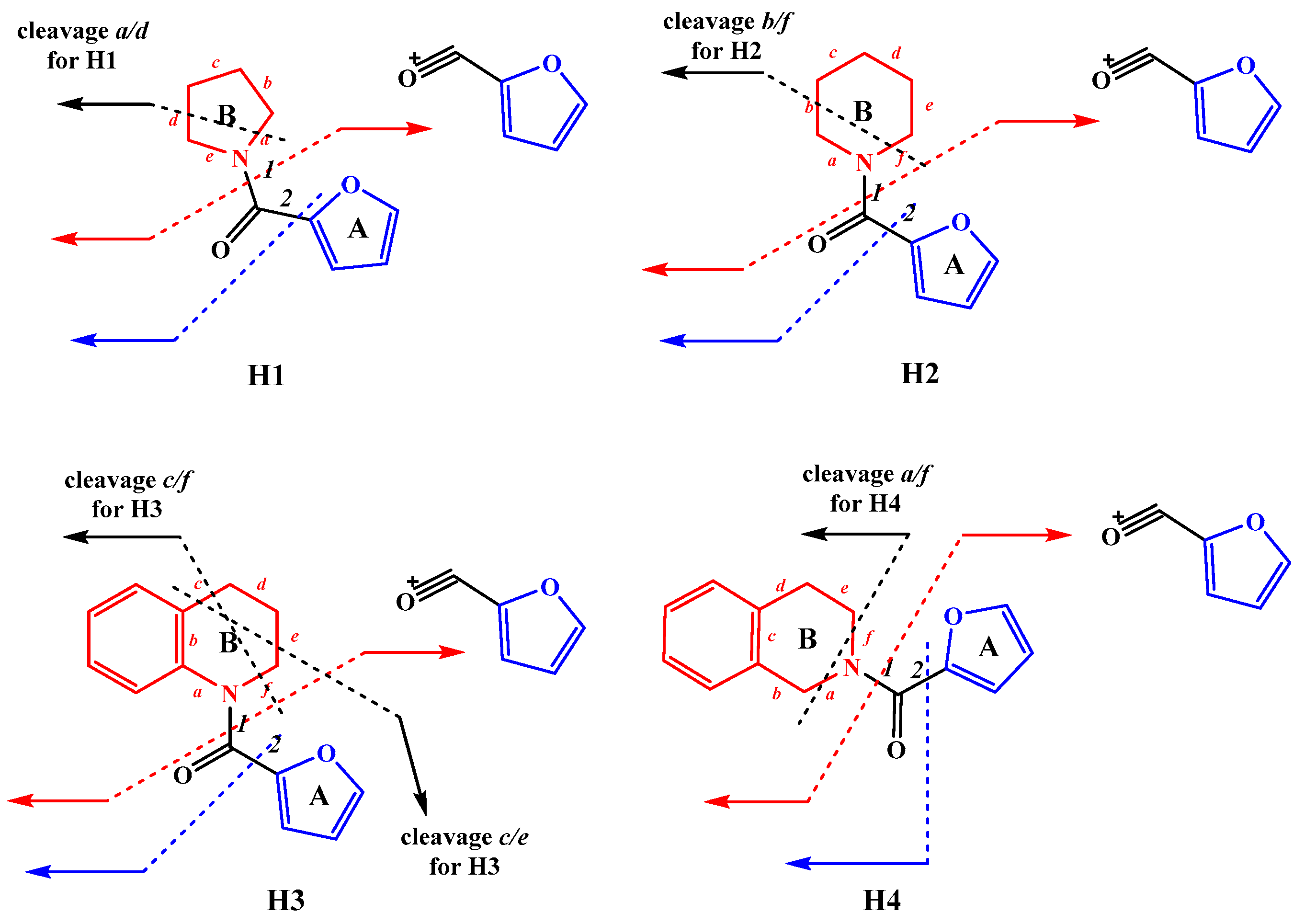
Figure 4.
Ability of furan derivatives to scavenge hydrogen peroxide (HPSA) (A) and form chelate complexes with Fe(II) ions (MChA) (B). Standards employed in this study were ascorbic acid (AA) and quercetin (Qrc). IC50 values were used to assess the HPSA and MChA outcomes.
Figure 4.
Ability of furan derivatives to scavenge hydrogen peroxide (HPSA) (A) and form chelate complexes with Fe(II) ions (MChA) (B). Standards employed in this study were ascorbic acid (AA) and quercetin (Qrc). IC50 values were used to assess the HPSA and MChA outcomes.
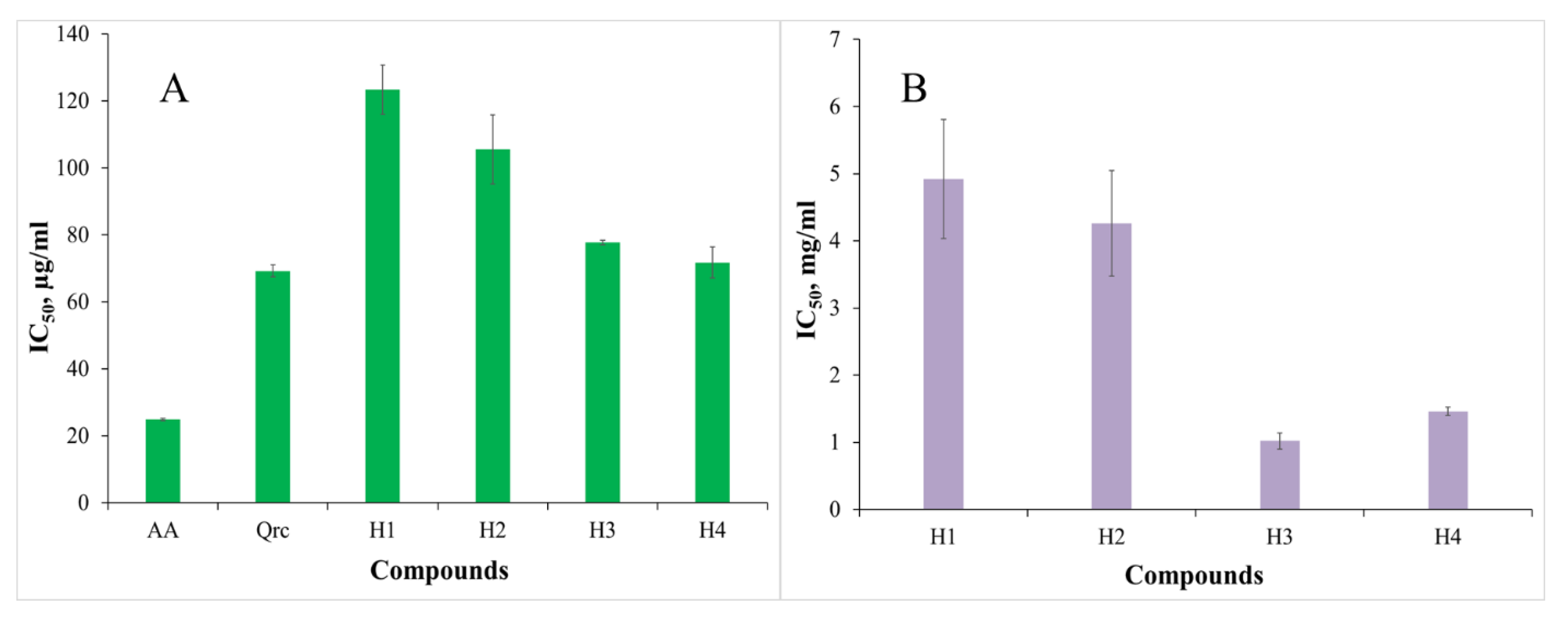
Figure 5.
In vitro biological activity was assessed as inhibition of albumin denaturation (IAD) (A) and antitryptic activity (ATA) (B). As benchmarks, anti-inflammatory medications such as ibuprofen and ketoprofen were utilized. The results of both methods are presented as IC50.
Figure 5.
In vitro biological activity was assessed as inhibition of albumin denaturation (IAD) (A) and antitryptic activity (ATA) (B). As benchmarks, anti-inflammatory medications such as ibuprofen and ketoprofen were utilized. The results of both methods are presented as IC50.

Table 1.
Biological evaluation findings from in vitro and in silico. The IC50 values are expressed for the following activities—hydrogen peroxide-scavenging activity (HPSA), inhibition of albumin denaturation (IAD), metal-chelating activity (MChA), and anti-tryptic activity (ATA). As reference points, ascorbic acid (AA), quercetin (Qrc), ibuprofen (Ibu), and ketoprofen (Ket) were used. Lipophilicity (RM) is a non-dimensional quantity because it is acquired via thin-layer chromatography and is a function of Rf. The terms “calculated anti-inflammatory activity (cAnti-I)” and “calculated anti-arthritic activity (cAnti-A)” are expressed as Pa (probability “to be active”), and assess the likelihood that the investigated molecule is a member of the sub-class of active compounds (resembles the most typical molecular structures in a subset of “actives” in the PASS training set). The molecule with the highest level of activity has a value of 1. H1–4—furan derivatives.
| Compounds | IC50± SD, μg/mL | IC50± SD, mg/mL | RM ± SD | Pa | |||
|---|---|---|---|---|---|---|---|
| HPSA | IAD | ATA | MChA | cAnti-I | cAnti-A | ||
| AA | 24.84 ± 0.35 | - | - | - | - | - | - |
| Qrc | 69.25 ± 1.82 | - | - | - | - | - | - |
| Ibu | - | 81.50 ± 4.95 | 259.82 ± 9.14 | - | 1.11 ± 0.010 | 0.903 | 0.573 |
| Ket | - | 126.58 ± 5.00 | 720.57 ± 19.78 | - | 1.54 ± 0.015 | 0.925 | 0.469 |
| H1 | 123.33 ± 7.41 | 114.31 ± 2.88 | 83.25 ± 1.69 | 4.92 ± 0.89 | 1.04 ± 0.023 | 0.224 | - |
| H2 | 105.52 ± 10.33 | 116.76 ± 1.61 | 60.21 ± 8.16 | 4.26 ± 0.79 | 0.94 ± 0.016 | 0.226 | - |
| H3 | 77.75 ± 0.67 | 150.99 ± 1.16 | 85.33 ± 7.26 | 1.02 ± 0.12 | 1.01 ± 0.030 | - | - |
| H4 | 71.72 ± 4.63 | 119.08 ± 0.92 | 62.23 ± 0.83 | 1.46 ± 0.06 | 0.87 ± 0.015 | 0.201 | - |
Publisher’s Note: MDPI stays neutral with regard to jurisdictional claims in published maps and institutional affiliations. |
© 2022 by the authors. Licensee MDPI, Basel, Switzerland. This article is an open access article distributed under the terms and conditions of the Creative Commons Attribution (CC BY) license (https://creativecommons.org/licenses/by/4.0/).
Share and Cite
MDPI and ACS Style
Manolov, S.; Ivanov, I.; Bojilov, D.; Nedialkov, P. Synthesis, In Silico, and In Vitro Biological Evaluation of New Furan Hybrid Molecules. Processes 2022, 10, 1997. https://doi.org/10.3390/pr10101997
AMA Style
Manolov S, Ivanov I, Bojilov D, Nedialkov P. Synthesis, In Silico, and In Vitro Biological Evaluation of New Furan Hybrid Molecules. Processes. 2022; 10(10):1997. https://doi.org/10.3390/pr10101997
Chicago/Turabian StyleManolov, Stanimir, Iliyan Ivanov, Dimitar Bojilov, and Paraskev Nedialkov. 2022. "Synthesis, In Silico, and In Vitro Biological Evaluation of New Furan Hybrid Molecules" Processes 10, no. 10: 1997. https://doi.org/10.3390/pr10101997
Note that from the first issue of 2016, this journal uses article numbers instead of page numbers. See further details here.