Article

Evaluation of Antiaging Effect of Sheep Placenta Extract Using SAMP8 Mice

1
International Aging Industry Research and Development Center (AIC), Providence University, Taichung 43301, Taiwan
2
Genius Bull International Ltd.No.220, Sec. 2, Taiwan Blvd., West Dist., Taichung 403, Taiwan
3
Department of Food and Nutrition, Providence University, Taichung 43301, Taiwan
4
Dong Wu Zhu Mu Qin Qi Yue Yi Biological and Technology Co., Ltd. Industry Area, Wu Li Ya Si Tai Town, Xiligol League, Inner Mongolia, China
5
Department of Hospitality Management, College of Agriculture, Tunghai University, Taichung 407224, Taiwan
*
Authors to whom correspondence should be addressed.
Processes 2022, 10(11), 2242; https://doi.org/10.3390/pr10112242
Submission received: 18 August 2022 / Revised: 21 October 2022 / Accepted: 25 October 2022 / Published: 1 November 2022

Abstract

:
Widely used in traditional medicine, sheep placenta extract (SPE) is known for its physiological effects such as wound healing, antioxidant, and anti-inflammatory properties. However, the effect of SPE on antiaging is still unclear. In this study, we investigated the effect of SPE on aging through the senescence-accelerated mouse prone 8 (SAMP8) strain. We designed an experiment using both male and female mice randomly divided into 4 groups (n = 10) as follows: Group A—control group; Group B—low-dose SPE (61.5 mg/kg BW/day); Group C—medium-dose SPE (123 mg/kg BW/day); and Group D—high-dose SPE (184.5 mg/kg BW/day). As a result of measuring the aging index parameters such as skin glossiness, spine lordosis, and kyphosis, it was found that the treatment of SPE lowered the aging index. In addition, we found that biochemical parameters such as lactic acid, glucose, ketone bodies, free fatty acids, tumor necrosis factor-alpha (TNF-α), and interleukin 6 (IL-6) were not changed in the experimental group treated with SPE for 13 weeks. Finally, we found that lipid peroxidation (LPO) was decreased, while the activities of catalase and superoxide dismutase (SOD) were significantly increased in the brain tissues of SPE-treated male and female mice. Supplementation of SPE lowered the oxidative stress caused by the aging process in mice without toxicity and decreased the aging index, suggesting the value of SPE as an effective antiaging treatment.

1. Introduction

The placenta joins the fetus to the mother and functions as a means of transport to transfer nutrients such as glucose, amino acids, fatty acids, vitamins, and other minerals essential for fetus growth and development [1]. The nutrients supplied by the placenta may be retained after the birth of the fetus [2,3]. Collagen, elastin, laminin, vitamins, trace minerals, nucleic acids, amino acids, peptides, cytokines, and growth factors have all been identified in human placental extracts [4,5,6,7].
With its high nutritional content and biologically active components, the placenta has historically been valued as a traditional folk medicine in China and other areas of the world. Placentophagy has a long history and has been practiced in many regions of the world. However, few scientific studies of the medicinal value of placental extract have been conducted [8]. Dried human placenta, also known as “Zi He Che”, was used in sixteenth-century China as a cure for impotence and for infertility, liver, and kidney issues, according to Li Shizen’s “Compendium of Materia Medica” [9]. It was also used to boost energy and stamina. The Araucanian Indians of Argentina use dried umbilical cord ground into powder to heal sick children. The Kol tribe in Central India consumes the placenta to enhance reproductive function [10].
In mainland China, since the Tang dynasty, practitioners of traditional Chinese medicine (TCM) have valued the placenta as a transitory organ that sustains the growth and development of a fetus in utero [11]. TCM practitioners have used the placenta to reduce mental tension and anxiety, enrich the blood, tonify Qi, increase essences, reduce fatigue and spasm, and detoxify the body [12]. In recent times, increased interest in the pharmacological effects and therapeutic value of the human placenta has led to more scientific studies, as well as increased usage in clinical settings [13].
Placenta treatment is now used for a variety of conditions, including adrenocortical hypofunction, lumbago, compromised immunity, infertility, depression, lack of lactation, and hair loss, as well as for antiaging purposes [9]. Within conventional medicine, practitioners are increasingly acknowledging the effectiveness of human placenta treatment, although restricted availability has prevented its widespread use to date. The benefits of treatment with sheep placenta (SP) extract include blood and skin nourishment, sedation effects, and increased longevity [14]. SP extract is one of the best medications for boosting essential vitality. Practitioners favor the use of sheep placenta because it shares the same nutritional composition and pharmacological properties as the human placenta [15]. However, fatigue in mice was also relieved by preparations made from goat placenta [16]. SP extract (SPE) is rich in tiny molecular peptides, nucleic acids, enzymes, amino acids, growth factors, collagen, and other active ingredients that may boost immunity, and reduce the effects of hypoxia [17].
The senescence-accelerated mouse (SAM) is a naturally occurring experimental mouse breed that exhibits early aging characteristics such as senile amyloidosis, senile osteoporosis, cataracts, a suppressed immune system, and deficiencies in learning and memory [18,19]. Breeders have developed a number of senescence-prone (SAMP) and senescence-resistant (SAMR) lines. Due to the early onset of brain shrinkage, which is accompanied by learning and/or memory impairment and affective disturbances, SAMP8 is a commonly utilized strain, particularly in dementia- and aging-related studies [20]. The longevity of SAMP8 is roughly half that of SAMR1 [21].
Integrative biological medicine is important for managing and restoring health and fitness. Placental extracts that still retain active components may promote rejuvenation, revitalization, and the restoration of youth and vitality by delaying aging processes. The placental extract is a powerful therapeutic agent with effective regeneration abilities that counter the aging process. In the present study, we evaluated the antiaging effect of SPE in SAMP8 mice.

2. Materials and Methods

2.1. Animals

We kept animals in transparent plastic cages of 30(W) × 20(D) × 10(H) cm3 in a dust-free automatic control room. We maintained room temperature at 22 ± 2 °C, with a relative humidity of 65 ± 5%. We controlled the light cycle using an automatic timer, with daily light and dark periods from 07:00 to 19:00 and 19:00 to 07:00 h, respectively. We provided animals with food and water ad libitum.

2.2. Sheep Placenta Extract Preparation and Dosage

For our experimental samples, we used dried sheep placenta powder provided by Zhenyuebo International Co., Ltd., produced from the placentas of Ujimqin sheep from Inner Mongolia, manufactured by East Ujimqin Banner Yueyi Biotechnology Co., Ltd., Inner Mongolia, China.
We determined dose conversion using the experimental evaluation method of the Ministry of Health and Welfare. First, we determined the recommended daily human intake per kilogram of body weight. We then expanded the recommended human dose by a factor of 12.3 to obtain the dose for mice in the medium-dose group. We then multiplied this dose by a factor of 0.5 for the low-dose group, and by 1.5 for the high-dose group.
Low-dose group (0.5 times)
For this group, we first calculated the recommended daily intake of human adults as 600 mg/60 kg BW/day × 1/2 = 5 mg/kg BW/day. We then calculated daily intake for mice based on the proportion of their body weight, as follows: 5 mg/kg BW/day × 12.3 = 61.5 mg/kg BW/day.
Medium-dose group (1 time)
For this group, we first calculated the recommended daily intake of human adults as 600 mg/60 kg BW/day × 1 = 10 mg/kg BW/day. We then calculated daily intake for mice based on the proportion of their body weight, as follows: 10 mg/kg BW/day × 12.3 = 123 mg/kg BW/day.
High-dose group (1.5 times)
For this group, we first calculated the recommended daily intake of human adults as 600 mg/60 kg BW/day × 1.5 = 15 mg/kg BW/day. We then calculated daily intake for mice based on the proportion of their body weight, as follows: 15 mg/kg BW/day × 12.3 = 184.5 mg/kg BW/day.

2.3. Experimental Animal Grouping

We used 3-month-old male and female SAMP8 mice as our experimental animals, and we randomly divided them into one control group and three experimental groups (low, medium, and high doses) with 10 mice in each group (n = 10), and 80 mice in total. For both male and female animals, Group A was the control group, Group B was the low-dose SPE group (61.5 mg/kg BW/day), Group C was the medium-dose SPE group (123 mg/kg BW/day), and Group D was the high-dose SPE group (184.5 mg/kg BW/day). We obtained approval for our animal experiment procedures from the Institutional Animal Care and Use Committee (IACUC) of Providence University (Approval No: 20201218 A008).
At the end of the treatment, we euthanized all mice using 95% CO2. We then collected blood and separated serum using centrifugation. This was then stored at −80 °C. We collected and weighed organs such as the liver, lung, heart, kidney, adipose tissue, and skeletal muscles, and stored them at −80 °C. We stored small portions of vital organ tissue sections in formalin for histological analysis. We used brain tissue to estimate LPO, catalase activities, and SOD activities.

2.4. Aging Index

We included behavioral aspects in the evaluation items of the aging index. For 30 s, we observed the reactivity of mice (reactivity) and also their escape response (passivity) following pinching of the skin on the nape of the neck. We also noted appearance aspects such as the glossiness of the skin (glossiness) and its roughness (coarseness), as well as any loss of hair (hair loss). We also sought to identify any skin ulceration (ulcer). With regard to animal eyes (eyes), we looked for mucositis around the eyes or edema of the eyelid (periophthalmic lesion). We also inspected animals using touch to assess any changes in spine lordokyphosis (lordokyphosis). We assessed each evaluation item by means of 5 grades, namely, 0, 1, 2, 3, and 4. The higher the score, the more serious the aging phenomenon observed.

2.5. Serum Biochemical Parameters Analysis

After 13 weeks of SPE treatment, we anesthetized all the mice and collected their blood. We then separated serum to analyze various biochemical parameters using an autoanalyzer (Hitachi 7060, Hitachi, Tokyo, Japan). These biochemical parameters were as follows: glucose, total protein, albumin, triglyceride, total cholesterol, HDL, LDL, AST, ALT, BUN, creatinine, sodium, potassium, uric acid, creatine kinase, ketones, free fatty acids, IL-6, and TNF-α.

2.6. Histological Analysis

We collected the liver, heart, lung, kidney, and brain from all the treatment groups. We carried out hematoxylin and eosin (H&E) staining on the collected organs, following previously reported methods [22].

2.7. Statistical Analysis

We analyzed the data obtained using the SPSS statistical software package. We subjected the experimental data to a one-way analysis of variance (one-way ANOVA) to identify differences between multiple groups. To compare the differences between groups, we used Duncan’s multiple-range test. A significant difference was indicated when p < 0.05.

3. Results

3.1. Effects of SPE on Food Intake, Water Intake, Body Weight, and Organ Weight of SAMP8 Mice

Table 1 and Table 2 show the effects of feeding 3-month-old SAMP8 mice with SPE in terms of changes in body weight, food intake, and water intake for male and female animals, respectively. For groups of both sexes, we found no significant changes in body weight, food intake, and water intake after the administration of SPE. Table 3 and Table 4 show weight changes in the brain, heart, liver, spleen, lung, and kidney of male and female mice, respectively. We found no significant changes in organ weights in the animal groups that received SPE treatment.

3.2. Effect of SPE on Aging Index of Senescence-Accelerated Mice

Table 5 and Table 6 show the effects of SPE administration on aging index assessment scores after 10 weeks for male and female mice, respectively. The skin glossiness, spinal curvature, and total aging index assessment scores were significantly lower for mice in the treatment groups (p < 0.05) compared with animals in the control group. SPE treatment produced the same effects in both male and female mice.

3.3. Effects of SPE on Serum Biochemical Parameters

Table 7 and Table 8 show the effects of SPE on the serum biochemical parameters glucose, total protein, albumin, triglyceride, total cholesterol, HDL, LDL, AST, ALT, BUN, creatinine, sodium, potassium, uric acid, creatine kinase, ketones, free fatty acids, IL-6, and TNF-α in male and female SAMP8 mice, respectively. For mice of both sexes, we found no significant changes between the treatment groups after SPE administration for 13 weeks.

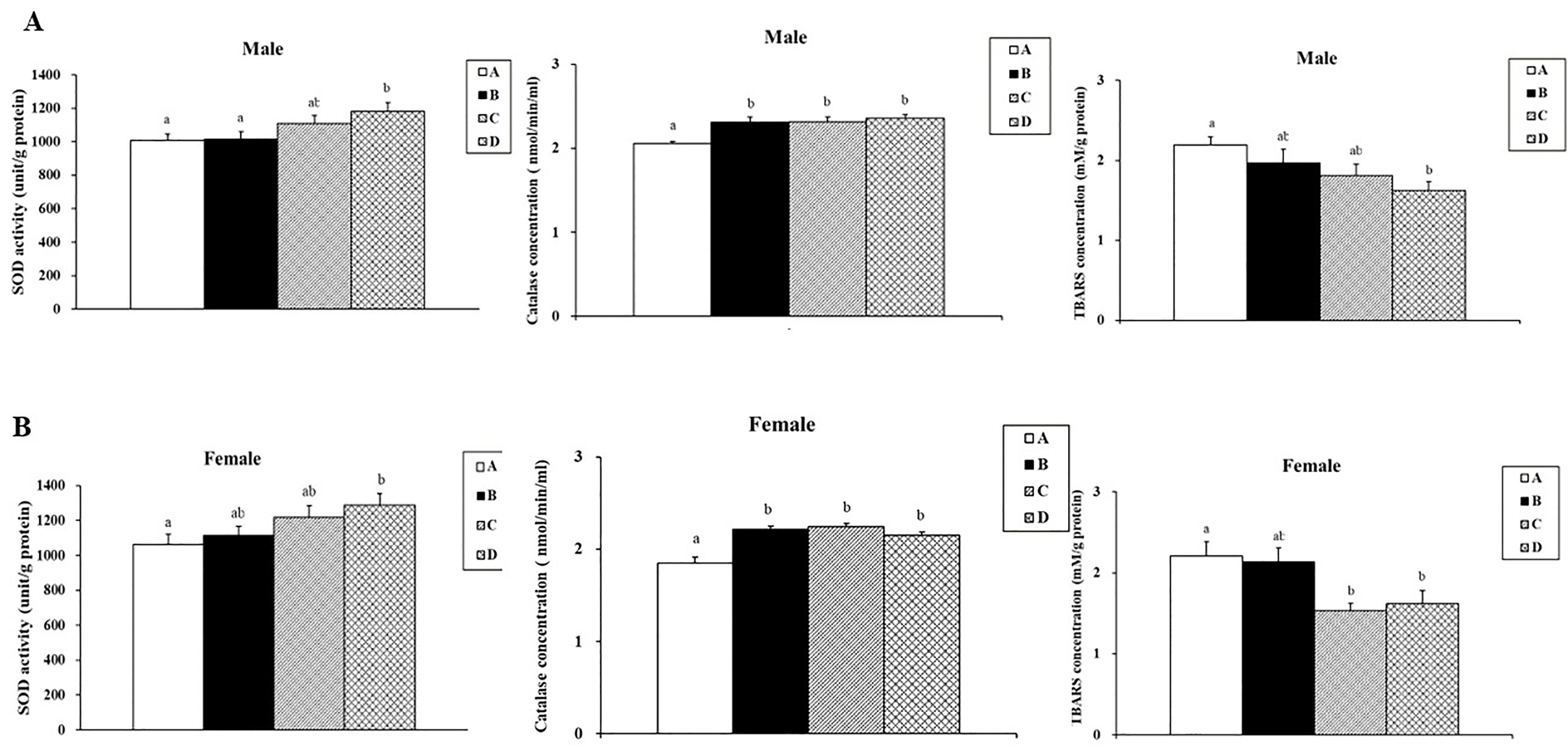
3.4. Effects of SPE on SOD Activities, Catalase Activities, and Lipid Peroxidation in Aging Mice

We quantified lipid peroxidation, SOD activities, and catalase activities in the brain tissues of male and female SAMP8 mice, as shown in Figure 1. For aging mice of both sexes, SPE treatment significantly reduced TBARS content (p < 0.05). At the same time, the activities of cellular antioxidant enzymes SOD and catalase significantly (p < 0.05) increased after SPE treatment in both male and female aging mice.

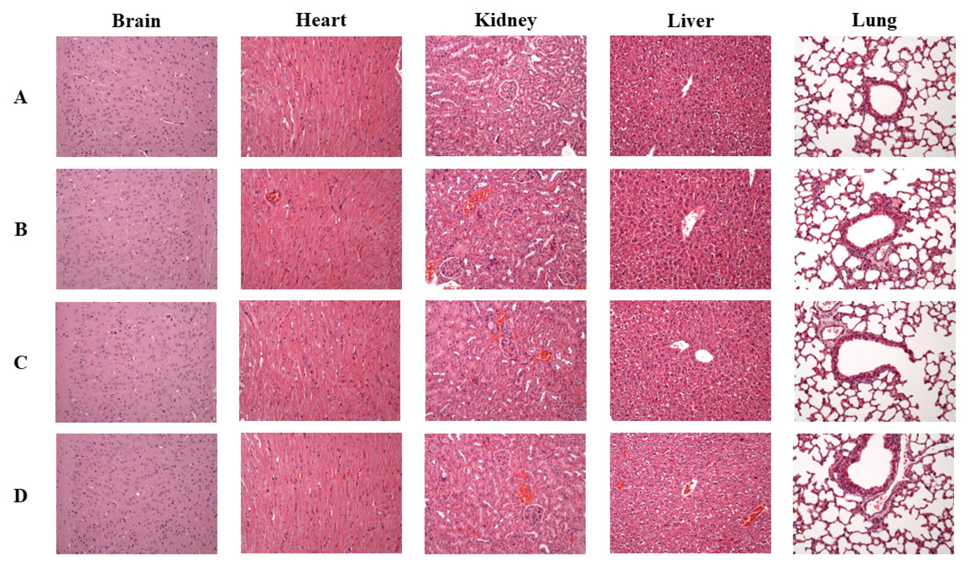
3.5. Effects of SPE on Histology of Vital Organs in Aging Mice

Figure 2 and Figure 3 show the effects of SPE treatment on changes in the histology of vital organs such as the brain, heart, kidney, liver, and lung in male and female mice, respectively. We found that a higher dose of SPE (184.5 mg/kg BW) administration to aging mice of either sex did not result in any significant structural changes in these vital organs. All organs displayed normal morphological architecture in the SPE-treated groups.

4. Discussion

Practitioners of traditional and folk medicine have used the placenta to treat a wide range of ailments. Along with its medicinal uses, it has also been used as an anesthetic in various clinical settings, including internal medicine, general surgery, ENT, ophthalmology, orthopedic surgery, plastic surgery, dermatology, obstetrics, and gynecology [9]. The main amino acids in SPE are glutamic acid, aspartic acid, lysine, and leucine. Eight essential amino acids constitute 31.20 g/100 g of SPE or 39.48 percent of total amino acids. Additionally, several cytokines and growth factors present in placenta extract contribute to their pharmacological activities [23].
Reactive oxygen species (ROS), which cause oxidative damage and lipid peroxidative injury, are produced throughout the aging process [24]. Free radicals and their metabolic byproducts must be removed by enzymes of antioxidant systems. The key enzymes in antioxidant systems are SOD and catalase [25]. Superoxide anion free radicals and hydrogen ions are dismutated into H2O2 and O2 by the antioxidant enzyme SOD, which is crucial for cleaning up free radicals [26]. Catalase eliminates oxidative risks by accelerating the synthesis of H2O and O2 from H2O2. One of the byproducts of lipid peroxidation is TBARS. During the aging process, TBARS levels increase [27]. In this study, we used these three defining indices to assess the antioxidant mechanism of SPE. We found that in SAMP8 mice, SOD and catalase activities decreased and the level of TBARS increased; however, SPE treatment for 13 weeks increased SOD and catalase antioxidant enzyme activities and lowered TABRAS levels in brain tissue. Antiaging processes may include regulating the activity of these antioxidant enzymes. There is strong evidence to support the original free radical hypothesis of aging, which states that free radical damage caused by numerous endogenous ROS is related to aging but is most likely the cause of it. Uracil, tyrosine, phenylalanine, and tryptophan are the main antioxidant substances found in placenta extract. These elements are responsible for 59% of PE’s anti-oxidative actions. Freeze-dried placenta powder delays D-galactose-induced aging in female KM mice by improving immunity and reducing LPO in the brain [28], which is in line with our present findings. Preparations from deer fetuses and placenta produce an antiaging effect by reducing monoamine oxidase activities in the brain and liver and lipofuscin activities in the heart and brain of aging rats [29].
High doses (1000 mg/kg) of swine placenta extract powder administered for 28 days do not produce any adverse effects in rats. Male and female rats treated with swine placenta powder showed no changes in hematological parameters and no changes to vital organs and tissues including the kidneys, heart, lung, liver, spleen, brain, adrenals, epididymides, prostate, seminal vesicles, thymus, testes, or ovaries. Uterus histology and organ weights were also unaffected [30]. In our study, we checked both male and female SAMP8 mice every day for symptoms of toxicity and death. In addition, we periodically checked the body weight, food and water consumption, and physical and visual health of the animals. We also carried out regular urinalysis and performed clinical biochemistry assessments of blood and plasma samples. We found that a higher dose of SPE (184.5 mg/kg BW) did not induce any changes in serum biochemical parameters or any histological changes in vital organs. This is in line with the previous findings and suggests that SPE is safe for human consumption.
Manufacturers employ a variety of techniques to produce placental extracts using raw materials from human, bovine, and porcine placentas. As a result, the components of placental extracts can be quite complex and may widely vary between products. Placental extracts include N-acetylneuraminic acid, glucosamine, omega-3 fatty acids, various fatty acids, and many other amino acids and nucleotides that have antioxidative or anti-inflammatory and wound-healing activities [31,32,33]. We postulated that most of these compounds/molecules would also be present in our sheep placenta extract. However, we were unable to validate the crucial elements and signaling pathways important for the prevention of aging. More research is needed to understand the molecular mechanism underpinning the antiaging activity of SPE, and this may be a challenging task.
As well as the aging index, serum biochemical parameters might be used as an indicator of aging. The liver function markers, ALT and AST, and the kidney function markers, creatinine and uric acid, are key indicators of tissue damage during the aging process [34]. However, in our study, these biochemical parameters did not significantly change after SPE administration. Inflammation plays a key role in the process of senescence [35]. Levels of cytokine IL-6 increase in elderly individuals, in comparison with young people, but levels of IL-1β and TNF-α do not increase [36]. In our study, we found no significant changes in levels of IL-6 or TNF-α.
In this study, we investigated the effect of different doses of SPE for 13 weeks on male and female SAMP8 in terms of the aging index, serum biochemical levels, and antioxidant status. We found that medium and high doses of sheep placenta reduced the appearance changes in aging, lowered levels of brain lipid peroxides, and increased SOD and catalase activities, thereby enhancing the antioxidant capacity of the body and helping to reduce the damage of oxidative stress, with the overall effect of delaying the aging process. SPE is rich in peptides and amino acids, whose strong antioxidant activities might contribute to antiaging effects. However, further characterization and molecular studies of SPE are needed to better understand its antiaging effects. In conclusion, SPE supplementation reduces the aging index and lowers the oxidative stress caused by the aging process in male and female mice, without toxicity, suggesting the value of SPE as an effective antiaging treatment.

Author Contributions

Conceptualization, W.-T.L.; methodology, C.-P.O.Y. and M.-Y.C.; validation, W.-C.L. and Y.-M.Y.; formal analysis, W.-C.L.; investigation, M.-Y.C. and Y.-J.H.; resources, M.-F.W.; data curation, M.-F.W.; writing—original draft preparation, M.-F.W. and W.-T.L.; writing—review and editing, M.-F.W. and W.-T.L.; supervision, W.-T.L.; project administration, M.-F.W.; funding acquisition, W.-T.L. All authors have read and agreed to the published version of the manuscript.

Funding

This research was funded by Providence University, fund number: 109: PU109-11150-A044, and Tunghai University Fund number: THU 111611.

Institutional Review Board Statement

All the animal treatment procedures were performed in accordance with the Guide for Care and Use of Laboratory Animals (National Institutes of Health Publication No. 85-23, raised 1996). Animal experiment procedures in the present study were approved by the Institutional Animal Care and Use Committee (IACUC) of Providence University (Approval No.: 20201218 A008).

Informed Consent Statement

Not applicable.

Data Availability Statement

Data are contained within the article.

Conflicts of Interest

The authors declare no conflict of interest.

References

  1. Sun, C.; Groom, K.M.; Oyston, C.; Chamley, L.W.; Clark, A.R.; James, J.L. The placenta in fetal growth restriction: What is going wrong? Placenta 2020, 96, 10–18. [Google Scholar] [CrossRef] [PubMed]
  2. Brett, K.E.; Ferraro, Z.M.; Yockell-Lelievre, J.; Gruslin, A.; Adamo, K.B. Maternal–fetal nutrient transport in pregnancy pathologies: The role of the placenta. Int. J. Mol. Sci. 2014, 15, 16153–16185. [Google Scholar] [CrossRef] [PubMed] [Green Version]
  3. Rasool, A.; Alvarado-Flores, F.; O’Tierney-Ginn, P. Placental impact of dietary supplements: More than micronutrients. Clin. Ther. 2021, 43, 226–245. [Google Scholar] [CrossRef] [PubMed]
  4. Ghoneum, M.; El-Gerbed, M.S. Human placental extract ameliorates methotrexate-induced hepatotoxicity in rats via regulating antioxidative and anti-inflammatory responses. Cancer Chemother. Pharmacol. 2021, 88, 961–971. [Google Scholar] [CrossRef] [PubMed]
  5. Goswami, S.; Sarkar, R.; Saha, P.; Maity, A.; Sarkar, T.; Das, D.; Chakraborty, P.D.; Bandyopadhyay, S.; Ghosh, C.K.; Karmakar, S. Effect of human placental extract in the management of biofilm mediated drug resistance–A focus on wound management. Microb. Pathog. 2017, 111, 307–315. [Google Scholar] [CrossRef]
  6. Kaneko, Y.; Sano, M.; Seno, K.; Oogaki, Y.; Takahashi, H.; Ohkuchi, A.; Yokozawa, M.; Yamauchi, K.; Iwata, H.; Kuwayama, T. Olive leaf extract (OleaVita) suppresses inflammatory cytokine production and NLRP3 inflammasomes in human placenta. Nutrients 2019, 11, 970. [Google Scholar] [CrossRef] [Green Version]
  7. Mumtaz, S.M.; Goyal, R.K.; Ameen, A.; Alexandrovich, B.I.; Gupta, M. Animal Placental Therapy: An Emerging Tool for Health Care. Curr. Tradit. Med. 2022, 8, 20–30. [Google Scholar] [CrossRef]
  8. Pogozhykh, O.; Prokopyuk, V.; Figueiredo, C.; Pogozhykh, D. Placenta and placental derivatives in regenerative therapies: Experimental studies, history, and prospects. Stem Cells Int. 2018, 2018, 4837930. [Google Scholar] [CrossRef]
  9. Pan, S.Y.; Chan, M.K.; Wong, M.B.; Klokol, D.; Chernykh, V. Placental therapy: An insight to their biological and therapeutic properties. Blood 2017, 4, 12. [Google Scholar]
  10. Ober, W.B. Notes on placentophagy. Bull. N. Y. Acad. Med. 1979, 55, 591. [Google Scholar]
  11. Hayes, E.H. Consumption of the placenta in the postpartum period. J. Obstet. Gynecol. Neonatal. Nurs. 2016, 45, 78–89. [Google Scholar] [CrossRef]
  12. Koike, K.; Yamamoto, Y.; Suzuki, N.; Yamazaki, R.; Yoshikawa, C.; Takano, F.; Takuma, K.; Sugiura, K.; Inoue, M. Efficacy of porcine placental extract on climacteric symptoms in peri-and postmenopausal women. Climacteric 2012, 16, 28–35. [Google Scholar] [CrossRef] [PubMed]
  13. Silini, A.R.; Cargnoni, A.; Magatti, M.; Pianta, S.; Parolini, O. The long path of human placenta, and its derivatives, in regenerative medicine. Front. Bioeng. Biotechnol. 2015, 3, 162. [Google Scholar] [CrossRef] [PubMed] [Green Version]
  14. Liu, W.; Hou, Y.; Cheng, Y. Polypeptide preparation of sheep placenta and its free radicals scavenging activity. China Brew. 2014, 33, 89–93. [Google Scholar]
  15. Wu, Y. Study on the new method of extraction placental peptide and analysis of nutritive composition in sheep placenta. Food Sci. 2005, 26, 295–297. [Google Scholar]
  16. Zhang, S.; Fang, F.; Gu, Y.; Zhang, Y.; Tao, Y.; Zhang, X. Effects of goat placental preparations on relieving fatigue and anti-oxidation in mice. J. Tradit. Chin. Vet. Med. 2008, 6, 8–10. [Google Scholar]
  17. Hou, Y.; Zhou, J.; Liu, W.; Cheng, Y.; Wu, L.; Yang, G. Preparation and characterization of antioxidant peptides from fermented goat placenta. Korean J. Food Sci. Anim. Resour. 2014, 34, 769. [Google Scholar] [CrossRef] [PubMed] [Green Version]
  18. Lai, P.-F.; Baskaran, R.; Kuo, C.-H.; Day, C.H.; Chen, R.-J.; Ho, T.-J.; Yeh, Y.-L.; Padma, V.V.; Lai, C.-H.; Huang, C.-Y. Bioactive dipeptide from potato protein hydrolysate combined with swimming exercise prevents high fat diet induced hepatocyte apoptosis by activating PI3K/Akt in SAMP8 mouse. Mol. Biol. Rep. 2021, 48, 2629–2637. [Google Scholar] [CrossRef] [PubMed]
  19. Takeda, T.; Matsushita, T.; Kurozumi, M.; Takemura, K.; Higuchi, K.; Hosokawa, M. Pathobiology of the senescence-accelerated mouse (SAM). Exp. Gerontol. 1997, 32, 117–127. [Google Scholar] [CrossRef]
  20. Akiguchi, I.; Pallàs, M.; Budka, H.; Akiyama, H.; Ueno, M.; Han, J.; Yagi, H.; Nishikawa, T.; Chiba, Y.; Sugiyama, H. SAMP8 mice as a neuropathological model of accelerated brain aging and dementia: Toshio Takeda’s legacy and future directions. Neuropathology 2017, 37, 293–305. [Google Scholar] [CrossRef] [Green Version]
  21. Lagartos-Donate, M.; Gonzáles-Fuentes, J.; Marcos-Rabal, P.; Insausti, R.; Arroyo-Jiménez, M. Pathological and non-pathological aging, SAMP8 and SAMR1. What do hippocampal neuronal populations tell us? bioRxiv 2019, bioRxiv:598599. [Google Scholar]
  22. Chen, Y.-J.; Baskaran, R.; Shibu, M.A.; Lin, W.-T. Anti-Fatigue and Exercise Performance Improvement Effect of Glossogyne tenuifolia Extract in Mice. Nutrients 2022, 14, 1011. [Google Scholar] [CrossRef]
  23. Liu, J.; Luo, S.; Yang, J.; Ren, F.; Zhao, Y.; Luo, H.; Ge, K.; Zhang, H. The protective effect of sheep placental extract on concanavalin A-induced liver injury in mice. Molecules 2018, 24, 28. [Google Scholar] [CrossRef] [PubMed] [Green Version]
  24. Bolduc, J.A.; Collins, J.A.; Loeser, R.F. Reactive oxygen species, aging and articular cartilage homeostasis. Free Radic. Biol. Med. 2019, 132, 73–82. [Google Scholar] [CrossRef]
  25. Padma, V.V.; Sowmya, P.; Felix, T.A.; Baskaran, R.; Poornima, P. Protective effect of gallic acid against lindane induced toxicity in experimental rats. Food Chem. Toxicol. 2011, 49, 991–998. [Google Scholar] [CrossRef] [PubMed]
  26. Baskaran, R.; Poornima, P.; Huang, C.Y.; Padma, V.V. Neferine prevents NF-κB translocation and protects muscle cells from oxidative stress and apoptosis induced by hypoxia. Biofactors 2016, 42, 407–417. [Google Scholar] [CrossRef] [PubMed]
  27. Ataie, Z.; Mehrani, H.; Ghasemi, A.; Farrokhfall, K. Cinnamaldehyde has beneficial effects against oxidative stress and nitric oxide metabolites in the brain of aged rats fed with long-term, high-fat diet. J. Funct. Foods 2019, 52, 545–551. [Google Scholar] [CrossRef]
  28. Song, H.; Zang, C.; Liu, X.; Hou, H.; LI, D. Anti-aging function of placenta freeze-dried powder on mice. Chin. J. Biochem. Pharm. 2014, 65–67. [Google Scholar]
  29. Xinghuai, Z.; Guiqin, Y.; Guangjin, H. Effect of preparations from deer fetus and placenta on anti-aging of old male rats. J. Shenyang Agric. Univ. 2005, 36, 233–235. [Google Scholar]
  30. Mitsui, Y.; Bagchi, M.; Marone, P.A.; Moriyama, H.; Bagchi, D. Safety and toxicological evaluation of a novel, fermented, peptide-enriched, hydrolyzed swine placenta extract powder. Toxicol. Mech. Methods 2015, 25, 13–20. [Google Scholar] [CrossRef]
  31. Park, S.; Phark, S.; Lee, M.; Lim, J.; Sul, D. Anti-oxidative and anti-inflammatory activities of placental extracts in benzo [a] pyrene-exposed rats. Placenta 2010, 31, 873–879. [Google Scholar] [CrossRef] [PubMed]
  32. Sur, T.K.; Biswas, T.K.; Ali, L.; Mukherjee, B. Anti-inflammatory and anti-platelet aggregation activity of human placental extract. Acta Pharmacol. Sin. 2003, 24, 187–192. [Google Scholar] [PubMed]
  33. Togashi, S.-I.; Takahashi, N.; Kubo, Y.; Shigihara, A.; Higashiyama, K.; Watanabe, S.; Fukui, T. Purification and identification of antioxidant substances in human-placenta extracts. J. Health Sci. 2000, 46, 117–125. [Google Scholar] [CrossRef] [Green Version]
  34. Lephart, E.D. Skin aging and oxidative stress: Equol’s anti-aging effects via biochemical and molecular mechanisms. Ageing Res. Rev. 2016, 31, 36–54. [Google Scholar] [CrossRef] [PubMed]
  35. Zhang, R.; Chen, J.; Mao, X.; Qi, P.; Zhang, X. Anti-inflammatory and anti-aging evaluation of pigment–protein complex extracted from Chlorella pyrenoidosa. Mar. Drugs 2019, 17, 586. [Google Scholar] [CrossRef] [PubMed] [Green Version]
  36. Roubenoff, R.; Harris, T.B.; Abad, L.W.; Wilson, P.W.; Dallal, G.E.; Dinarello, C.A. Monocyte cytokine production in an elderly population: Effect of age and inflammation. J. Gerontol. Ser. A Biol. Sci. Med. Sci. 1998, 53, M20–M26. [Google Scholar] [CrossRef]
Figure 1. Effects of SPE treatment on SOD activities, catalase activities, and LPO in brain tissues of SAMP8 mice. (A) Male SAMP8 mice. (B) Female SAMP8 mice. Data are expressed as the mean ± SEM and analyzed by one-way ANOVA. Groups with different letters indicate significant differences among each group (p < 0.05).
Processes 10 02242 g001
Figure 2. Effects of SPE treatment on histology of the brain, heart, liver, kidney, and lung of male SAMP8 mice (100×). Group (A)—control group, Group (B)—low-dose SPE (61.5 mg/kg BW/day), Group (C)—medium-dose SPE (123 mg/kg BW/day), and Group (D)—high-dose SPE (184.5 mg/kg BW/day).
Processes 10 02242 g002
Figure 3. Effects of SPE treatment on histology of the brain, heart, liver, kidney, and lung of female SAMP8 mice (100×). Group (A)—control group, Group (B)—low-dose SPE (61.5 mg/kg BW/day), Group (C)—medium-dose SPE (123 mg/kg BW/day), and Group (D)—high-dose SPE (184.5 mg/kg BW/day).
Processes 10 02242 g003
Table 1. Effects of SPE treatment on body weight, food intake, and water intake in male SAMP8 mice.
GroupBody Weight (g)Food Intake
(g/Day)
Water Intake
(mL/Day)
InitialFinalGain
A29.81 ± 0.5831.49 ± 2.021.69 ± 0.434.61 ± 0.086.54 ± 0.09
B30.24 ± 0.6732.01 ± 0.651.70 ± 0.564.73 ± 0.106.93 ± 0.14
C29.52 ± 0.6732.76 ± 0.632.80 ± 0.524.74 ± 0.086.65 ± 0.14
D29.68 ± 0.8530.76 ± 0.801.08 ± 0.314.87 ± 0.036.95 ± 0.14
Group A—control group, Group B—low-dose SPE (61.5 mg/kg BW/day), Group C—medium-dose SPE (123 mg/kg BW/day), and Group D—high-dose SPE (184.5 mg/kg BW/day). Data are expressed as the mean ± SEM and analyzed by one-way ANOVA.
Table 2. Effects of SPE treatment on body weight, food intake, and water intake in female SAMP8 mice.
GroupBody Weight (g)Food Intake
(g/Day)
Water Intake
(mL/Day)
InitialFinalGain
A25.58 ± 0.5326.73 ± 0.541.16 ± 0.244.28 ± 0.095.01 ± 0.10
B25.85 ± 0.5727.39 ± 0.431.59 ± 0.624.08 ± 0.095.00 ± 0.06
C25.80 ± 0.5527.17 ± 0.561.23 ± 0.274.16 ± 0.085.04 ± 0.04
D25.55 ± 0.4227.36 ± 0.501.82 ± 0.494.34 ± 0.095.18 ± 0.06
Group A—control group, Group B—low dose SPE (61.5 mg/kg BW/day), Group C—medium dose SPE (123 mg/kg BW/day), and Group D—high dose SPE (184.5 mg/kg BW/day). Data are expressed as the mean ± SEM and analyzed by one-way ANOVA.
Table 3. Effects of SPE treatment on organ weights of male SAMP8 mice.
GroupRelative Organ Weights (g/100 g Body Weight)
BrainHeartLiverSpleenLungKidney
A1.41 ± 0.030.63 ± 0.024.46 ± 0.140.29 ± 0.030.67 ± 0.021.60 ± 0.04
B1.38 ± 0.040.61 ± 0.024.58 ± 0.070.33 ± 0.040.66 ± 0.011.62 ± 0.04
C1.30 ± 0.040.58 ± 0.024.40 ± 0.140.27 ± 0.020.66 ± 0.071.67 ± 0.11
D1.42 ± 0.030.63 ± 0.014.81 ± 0.100.32 ± 0.020.72 ± 0.041.75 ± 0.06
Group A—control group, Group B—low-dose SPE (61.5 mg/kg BW/day), Group C—medium-dose SPE (123 mg/kg BW/day), and Group D—high-dose SPE (184.5 mg/kg BW/day). Data are expressed as the mean ± SEM and analyzed by one-way ANOVA.
Table 4. Effects of SPE treatment on organ weights of male SAMP8 mice.
GroupRelative Organ Weights (g/100 g Body Weight)
BrainHeartLiverSpleenLungKidney
A1.56 ± 0.060.65 ± 0.014.98 ± 0.110.42 ± 0.020.82 ± 0.021.42 ± 0.06
B1.58 ± 0.070.60 ± 0.024.66 ± 0.140.42 ± 0.030.79 ± 0.041.40 ± 0.03
C1.55 ± 0.070.62 ± 0.024.65 ± 0.150.37 ± 0.010.77 ± 0.021.42 ± 0.02
D1.61 ± 0.080.59 ± 0.024.57 ± 0.130.40 ± 0.020.86 ± 0.051.38 ± 0.04
Group A—control group, Group B—low-dose SPE (61.5 mg/kg BW/day), Group C—medium-dose SPE (123 mg/kg BW/day), and Group D—high-dose SPE (184.5 mg/kg BW/day). Data are expressed as the mean ± SEM and analyzed by one-way ANOVA.
Table 5. Effects of SPE administration on aging index assessment scores of male SAMP8 mice.
GroupABCD
Behavior
 Reactivity0.10 ± 0.100.00 ± 0.000.11 ± 0.110.00 ± 0.00
 Passivity0.40 ± 0.160.11 ± 0.110.22 ± 0.150.00 ± 0.00
Skin
 Glossiness1.60 ± 0.16 a1.00 ± 0.00 b0.78 ± 0.15 b0.67 ± 0.17 b
 Coarseness1.30 ± 0.151.67 ± 0.171.44 ± 0.181.44 ± 0.18
 Hair loss1.50 ± 0.221.44 ± 0.181.22 ± 0.151.44 ± 0.18
 Ulcer0.00 ± 0.000.00 ± 0.000.00 ± 0.000.00 ± 0.00
Eyes
 Periophthalmic lesion0.20 ± 0.130.11 ± 0.110.00 ± 0.000.11 ± 0.11
Spine
 Lordokyphosis1.60 ± 0.16 a1.00 ± 0.00 b1.00 ± 0.00 b1.00 ± 0.00 b
Total6.70 ± 0.26 a5.33 ± 0.24 b4.78 ± 0.32 b4.67 ± 0.17 b
Group A—control group, Group B—low-dose SPE (61.5 mg/kg BW/day), Group C—medium-dose SPE (123 mg/kg BW/day), and Group D—high-dose SPE (184.5 mg/kg BW/day). Data are expressed as the mean ± SEM and analyzed by one-way ANOVA. Groups with different letters indicate significant differences among each group (p < 0.05).
Table 6. Effects of SPE administration on aging index assessment scores of female SAMP8 mice.
GroupABCD
Behavior
 Reactivity0.78 ± 0.220.78 ± 0.220.56 ± 0.180.67 ± 0.17
 Passivity1.11 ± 0.110.78 ± 0.150.89 ± 0.110.56 ± 0.24
Skin
 Glossiness0.89 ± 0.110.33 ± 0.170.56 ± 0.180.67 ± 0.17
 Coarseness0.78 ± 0.150.56 ± 0.180.89 ± 0.110.67 ± 0.24
 Hair loss0.78 ± 0.220.67 ± 0.240.67 ± 0.170.67 ± 0.17
 Ulcer0.44 ± 0.180.56 ± 0.180.11 ± 0.110.22 ± 0.15
Eyes
 Periophthalmic lesion0.78 ± 0.220.78 ± 0.150.44 ± 0.180.67 ± 0.24
Spine
 Lordokyphosis1.00 ± 0.00 a0.78 ± 0.15 ab0.56 ± 0.18 b0.44 ± 0.18 b
Total6.56 ± 0.18 a5.22±0.32 b4.67 ± 0.17 b4.56 ± 0.18 b
Group A—control group, Group B—low-dose SPE (61.5 mg/kg BW/day), Group C—medium-dose SPE (123 mg/kg BW/day), and Group D—high-dose SPE (184.5 mg/kg BW/day). Data are expressed as the mean ± SEM and analyzed by one-way ANOVA. Groups with different letters indicate significant differences among each group (p < 0.05).
Table 7. Effects of SPE on serum biochemical parameters of male SAP8 mice.
GroupABCD
Glucose (mg/dL)112.02 ± 0.99109.56 ± 1.22114.21 ± 0.64109.86 ± 1.95
Total protein (g/dL)5.29 ± 0.055.40 ± 0.065.38 ± 0.075.36 ± 0.03
Albumin (g/dL)2.95 ± 0.113.12 ± 0.083.06 ± 0.062.88 ± 0.08
Triglyceride (mg/dL)108.65 ± 0.56103.41 ± 0.54104.43 ± 1.95106.41 ± 0.11
Total cholesterol (mg/dL)116.48 ± 1.81114.91 ± 2.64118.00 ± 2.99115.49 ± 1.83
HDL (mg/dL)53.57 ± 1.6554.22 ± 1.7256.35 ± 1.5356.21 ± 1.03
LDL (mg/dL)7.20 ± 0.147.06 ± 0.137.24 ± 0.097.29 ± 0.15
AST (U/L)89.14 ± 0.7587.06 ± 0.8688.87 ± 0.4288.09 ± 0.38
ALT (U/L)59.51 ± 0.9161.05 ± 0.8759.49 ± 0.6459.57 ± 0.42
BUN (mg/dL)25.91 ± 1.2226.81 ± 0.3926.99 ± 0.4627.72 ± 0.70
Creatinine (mg/dL)0.30 ± 0.020.32 ± 0.010.29 ± 0.020.33 ± 0.02
Sodium (mg/dL)6.46 ± 0.106.77 ± 0.116.52 ± 0.136.68 ± 0.08
Potassium (mg/dL)7.71 ± 0.137.94 ± 0.127.86 ± 0.107.75 ± 0.15
Uric acid (mg/dL)4.41 ± 0.054.38 ± 0.084.54 ± 0.074.57 ± 0.08
Creatine kinase (U/L)260.52 ± 2.26259.76 ± 3.23264.30 ± 1.75262.96 ± 1.88
Ketones (mmol/L)0.66 ± 0.040.65 ± 0.020.68 ± 0.040.62 ± 0.07
Free fatty acids (mmol/L)0.72 ± 0.080.79 ± 0.020.76 ± 0.010.73 ± 0.04
IL-6 (pg/mL)<1.5<1.5<1.5<1.5
TNF-α (pg/mL)<0.106<0.106<0.106<0.106
Group A—control group, Group B—low-dose SPE (61.5 mg/kg BW/day), Group C—medium-dose SPE (123 mg/kg BW/day), and Group D—high-dose SPE (184.5 mg/kg BW/day). Data are expressed as the mean ± SEM and analyzed by one-way ANOVA.
Table 8. Effects of SPE on serum biochemical parameters of female SAP8 mice.
GroupABCD
Glucose (mg/dL)116.12 ± 1.43115.66 ± 1.15113.40 ± 1.84114.51 ± 2.01
Total Protein (g/dL)5.36 ± 0.025.46 ± 0.115.35 ± 0.045.37 ± 0.02
Albumin (g/dL)3.02 ± 0.042.93 ± 0.073.09 ± 0.112.90 ± 0.08
Triglyceride (mg/dL)101.27 ± 1.66103.22 ± 0.68103.18 ± 1.00104.65 ± 0.78
Total cholesterol (mg/dL)117.99 ± 2.10120.29 ± 2.20116.88 ± 1.96120.43 ± 2.39
HDL (mg/dL)55.14 ± 2.0354.16 ± 1.8456.99 ± 1.1357.03 ± 1.70
LDL (mg/dL)7.36 ± 0.117.28 ± 0.127.22 ± 0.167.42 ± 0.09
AST (U/L)88.94 ± 0.5188.33 ± 0.3387.36 ± 0.5588.09 ± 0.63
ALT (U/L)59.33 ± 0.9761.31 ± 1.1860.41 ± 0.5058.87 ± 1.38
BUN (mg/dL)27.98 ± 0.4628.18 ± 0.4725.62 ± 1.3828.07 ± 0.73
Creatinine (mg/dL)0.31 ± 0.010.28 ± 0.020.30 ± 0.010.33 ± 0.03
Sodium (mg/dL)6.59 ± 0.216.64 ± 0.186.67 ± 0.116.75 ± 0.19
Potassium (mg/dL)7.89 ± 0.107.83 ± 0.077.75 ± 0.097.79 ± 0.11
Uric acid (mg/dl)4.64 ± 0.044.56 ± 0.074.73 ± 0.104.68 ± 0.02
Creatine kinase (U/L)259.32 ± 1.10262.86 ± 0.71261.66 ± 0.63260.96 ± 1.10
Ketone (mmol/L)0.72 ± 0.070.69 ± 0.050.67 ± 0.040.70 ± 0.06
Free fatty acids (mmol/L)0.73 ± 0.030.70 ± 0.060.76 ± 0.030.74 ± 0.02
IL-6 (pg/mL)<1.5<1.5<1.5<1.5
TNF-α (pg/mL)<0.106<0.106<0.106<0.106
Group A—control group, Group B—low-dose SPE (61.5 mg/kg BW/day), Group C—medium-dose SPE (123 mg/kg BW/day), and Group D—high-dose SPE (184.5 mg/kg BW/day). Data are expressed as the mean ± SEM and analyzed by one-way ANOVA.
Publisher’s Note: MDPI stays neutral with regard to jurisdictional claims in published maps and institutional affiliations.

