Article

Providing Antimicrobial Properties to Cardboard Food Packaging by Coating with ZnO, TiO2, and SiO2—Water-Based Varnish Nanocomposites

1
Faculty of Graphic Arts, University of Zagreb, Getaldićeva 2, 10000 Zagreb, Croatia
2
Faculty of Veterinary Medicine, University of Zagreb, Heinzelova 55, 10000 Zagreb, Croatia
*
Authors to whom correspondence should be addressed.
Processes 2022, 10(11), 2285; https://doi.org/10.3390/pr10112285
Submission received: 7 October 2022 / Revised: 25 October 2022 / Accepted: 30 October 2022 / Published: 4 November 2022
(This article belongs to the Special Issue Current Trends in Food and Food Byproducts Processing)

Abstract

:
Packaging acts like a bond between visual communication and production technology. Packaging material is often coated to enhance visual appearance and some protective features. The COVID pandemic changed consumers’ behavior and understanding of the importance regarding the antimicrobial properties of goods that come in contact with hands. The aim of this research is to investigate and determine the antimicrobial properties of nanocomposite coatings which include nanosized zinc oxide (ZnO), titanium dioxide (TiO2), and silicon dioxide (SiO2). For the purpose of this research, a lithographic printed packaging was coated with a nanocomposite composed of flexographic water-based varnish with incorporated ZnO, TiO2, and SiO2 nanosized particles. A total of eight modulations were presented and compared to the lone water-based varnish. The results have shown that applying nanocomposites will increase the total surface free energy of the packaging surface but will decrease the polar component of the surface free energy leading to lower hydrophilic properties. Both nanocomposite types showed that the increase in the nanoparticle weight ratio leads to higher protection benefits. Nanocomposites with ZnO have better antimicrobial activity than the ones with TiO2. The Hybrid/Z (ZnO + SiO2) significantly improved the antimicrobial capacity of water-based varnish, primarily against the ubiquitous foodborne pathogen Listeria monocytogenes.

1. Introduction

The digital era caused a decrease in the material information transfer, i.e., a decrease in various aspects of the printing industry (newspaper, magazines, etc.). Nevertheless, the printing industry has experienced continuous growth in revenue due to the development of the packaging industry. Although various materials are used for packaging goods, paper and paper-based materials remain highly involved due to good printing properties, the possibility of biodegradation, and recyclability [1]. Furthermore, packaging production is highly demanding, as it connects visual communications, goods protection, and printing production technology [2]. Packaging is often divided into primary (in contact with packed goods), secondary, and tertiary (usually used for transport). Secondary packaging is the one that is in contact with the customer both visually and tactilely, which means that in addition to its protecting role, it has a marketing role as well. Therefore, its message and appearance must be well protected from the influences that can diminish the quality of printed information, i.e., its attractiveness. Moreover, an additional functionality feature, like an antimicrobial effect, would be beneficial. When thinking of antimicrobial packaging systems, one usually refers to the inhibition of bacteria growth on the food from the moment it is packed up until consumption [3,4,5]. The term which is often used is active packaging, which means that in the production step, antimicrobial substances are deliberately integrated into the packaging material, which releases them into the food environment [6]. This is in line with the interest of consumers, as they are interested in food with fewer preservatives. There are many materials that can be used for this purpose [6] that can also provide additional benefits, for example, structural enhancement [7].
As the recent COVID-19 pandemic showed, consumers are also under threat even without consuming food. Although diseases are spread through close proximity interactions [8], they can be also transferred by touching the eyes or nose after touching a surface covered with droplets, which is common behavior during grocery shopping [9]. One possible solution to this threat is to apply an antimicrobial coating to the secondary packaging. Some research was performed to test applied coatings on antimicrobial behavior and on visual appearance as well [10,11].
To achieve adequate protection, packaging material can be protected by means of various purpose-based coatings [12]. To enhance the inclusion of the coating process in production, it is very important for the coating to provide more than one advantage, for example, to protect the printed image from UV-based color degradation, to have some antimicrobial effect, to be sustainable, etc. To do so, the coating’s composition must include substances that have desired properties, consequently enhancing a certain role [10,11,13,14]. Recently, those compounds are in nanosize which enables them to be incorporated in commercial varnishes. Recent studies show that metal oxides such as ZnO and TiO2 have proven a protective potential and demonstrated strong antimicrobial responses against a wide range of hazardous bacteria [15,16,17,18,19]. Moreover, TiO2 creates oxygen-free radicals (OFRs) when exposed to UV radiation (i.e., sunlight) [17], which can affect the microbial cell wall thus stopping its growth. The mentioned particles are known for their role in the absorption of UV radiation (ZnO up to 374 nm, TiO2 to 329 nm) and overall usage in existing protective coatings for wood and metal [20]. The functional print protection by using nanocomposites including biopolymer as a base has proved its use and UV [21].
The nanosized SiO2 is a compound that can be used for a variety of purposes depending on its formulation. It can be part of antimicrobial coatings when combined with other nanosized compounds and incorporated into different varnish bases [14,22,23,24]. The addition of SiO2 in a coating will not change the color of the print if applied in a layer up to 24 µm (wet layer thickness) [25], while it will provide a better barrier against water vapor [11]. Furthermore, nanosized SiO2 is proven to enhance the mechanical properties and wear resistance of coatings [26,27,28,29].
The antimicrobial surface protection can theoretically be observed from two angles, one being the coating’s initial ability and the other being the prevention of biofilm formation by altering the adhesive properties of the sample’s surface [22]. With the COVID-19 outbreak, a common sight was customers wearing surgical gloves when browsing stores [30]. This was mostly connected with the fear that a potential carrier or spreader had touched the item beforehand and could have left the virus on the surface. For most people, there is no medical benefit in wearing gloves at a grocery store [31]. Introduction of the antimicrobial secondary packaging could be highly beneficial for customer protection, and it has an important marketing role for packaging buyers (goods manufacturers). Projected growth according to CAGR for antimicrobial packaging is 5.89% or 13.86 billion USD in the period between 2021–2026 [32]. Although not so emphasized as the pandemic, the vulnerability of humans to bacteria lies in the fact that certain antibiotics are overused [33]. Even more, with an increase in the number of patients suffering from respiratory problems, due to the pandemic, secondary fungal and bacterial infections are on the rise. According to the Lancet, 50% of the patients who died of the virus had secondary infections, which stresses the necessity to cope with this issue [34].
As presented, the mentioned nanoparticles are used for coating various materials to enhance their properties and antimicrobial activity, but these findings did not include printed packaging. As printed packaging comes in contact with customers during regular shopping, the aim of this research is to propose a method of creating antimicrobial protection on the surface of the secondary packaging by adding ZnO or TiO2 and SiO2 + ZnO or TiO2 nanoparticles in a water-based varnish.

2. Materials and Methods

The experiment setup is presented in Figure 1.
The printing substrate was a gloss-coated fine art print paper with the production name UPM Finesse gloss (UPM Communication Papers) and a grammage of 300 g/m2. The samples were printed by means of a four-color sheetfed offset printing press using quickset process inks (Novavit Supreme Bio by Flintgroup). The printing was conducted in compliance with FOGRA PSO 2016, i.e., ISO 12647-2:2013 offset printing standard.

2.1. Sample Preparation

Nanocomposite coatings were prepared by dispersing nanosized TiO2 (Sigma Aldrich TiO2, rutile), ZnO (ZN-0605, NanoArc, Alfa Aesar, Erlenbachweg, Germany), and SiO2 (Aerosil 200, Evonik, Essen, Germany). The SiO2 (Evonik Aerosil 200) was fumed and hydrophilic in commercial water-based varnish (TerraWet High Gloss Coating G9/285, ACTEGA, Olean, NY, USA). Due to the increase in the viscosity by dispersing nanoparticles, sampled nanocomposites were applied on the prints by a flexographic simulator (the varnishing in industrial production is conducted by a flexographic printing unit), and the varnish was diluted by adding 5%wt of distilled water. The composition of nanocomposites is presented in Table 1. The homogenization process of nanoparticles into the water-based varnish (WB) was carried out using the ultrasound dispenser Hielscher UP100H at 100% amplitude and 100% power. The duration of the homogenization process was adjusted to the weight ratio of the added nanoparticles (Table 1). Due to the heating of the mixture in the process of homogenization, during the homogenization, the samples were immersed into a cool bath with a cooling liquid temperature set at 7 °C. The viscosity was determined at a temperature of 20 °C by Anton Paar Rheolab QC rotational rheometer with a constant shear rate of 0.02 s−1.

2.2. SEM and EDS Analysis of Samples

The analysis of the samples included obtaining Scanning Electron Micrographs (SEM images) and performing Electron Dispersive Spectroscopy (EDS) analysis to detect possible agglomerates on the surface of the samples, surface free energy calculation to determine the print surface’s adsorption potential, and in the end, microbial testing.
SEM micrographs were obtained using the JEOL JSM-6460 scanning electron microscope. To assure uniform electronic properties, the samples were gold coated via a Baltec SCD 005 sputtering unit prior to scanning. A JEOL JSM-6460 was also used for the EDS. The EDS is used to determine the composition of particles on the surface as it is very hard to determine if the particle on the surface of the sample is agglomerated nanoparticles or some other compound.

2.3. Surface Free Energy Determination

Like all other organisms, bacteria and fungi need water to survive, grow, and migrate [35]. While the surface of the printed packaging often appears dry, they contain the amount of humidity from the air used for the preconditioning of the printing substrate (21 ± 2 °C and 50% ± 5 RH). Moreover, shops and supermarkets use air-conditioning at 21± °C with the same 50% ± RH. These conditions are suitable for microbes to adhere to the surface. The microbial adhesion proceeds in four steps: transportation to the surface, initial weak adhesion, attachment, and the creation of biofilm [35]. These steps are driven by a variety of specific interactions with the material and an array of biological processes. To assess the formation of potential aerosol droplets containing microbial cultures, surface free energy (SFE) was calculated from measured contact angles. The contact angles were measured using a Dataphysics OCA30 goniometer and its program support SCA 20–22. The contact angles were measured using the sessile drop method, ten times per sample, at different sample positions. The droplet shape was a spherical cap, and the volume was set to 1 µL. The surface free energy of the printing plate was calculated using the contact angles of four probe liquids with known surface tension (Table 2) and the OWRK method. The calculations were performed in the Dataphysics’ SCA 20 software (version v.6.1.11, build 6011) using the SE calculation window. For the calculation, the standard deviation of the contact angles was included and the Straight Line Fit and option Sigma (x[i]), Sigma (y[i]) calculated by its basic parameters * 1 × 10⁻⁵ were applied.
The SFE was calculated using Equation (1).
( 1 + cos θ ) * σ s 2 σ l D = σ s P   σ l P σ l D + σ s D ·
where: γs—the surface tension of the solid, γl—the surface tension of the liquid, 𝛾d—the dispersive part of surface tension, 𝛾p—the polar phase of surface tension, Θthe contact angle.

2.4. Testing of Antimicrobial Properties of Nanocomposite Coatings

The antimicrobial potential of ZnO, TiO2, and SiO2 nanocomposite coatings was evaluated by testing the bacterial contamination of the samples under laboratory environmental conditions and assessing the growth capacity of pathogens artificially inoculated onto the samples.

2.4.1. Strains Used for Artificial Inoculation of Samples

The strains used in this study were two reference strains, Staphylococcus aureus ATCC 25923 and Listeria monocytogenes ATCC 7644, and three strains from a laboratory collection—Citrobacter freundii identified by MALDI TOF-MS, Yersinia enterocolitica bioserotype 4/O:3 identified by MALDI TOF-MS and RT-PCR, and Penicillium spp. identified morphologically. Citrobacter freundii, Y. enterocolitica 4/O:3, and Penicillium spp. Were isolated during a routine analysis of animal tissues/food samples [38].

2.4.2. Testing of Antimicrobial Capacity of Nanocomposite Coatings during Environmental Contamination of Cardboard Samples

Natural contamination of nanocomposite-coated samples (ZnO, TiO2, SiO2) as well as uncoated (control) and water-based coated samples (WB) were monitored under ambient conditions by detecting the aerobic mesophilic bacteria count. Samples were cut into 4 × 10 cm strips and left at room temperature for 10 days, in order to evaluate the antimicrobial capacity of nanocomposites on the surface of cardboard samples exposed to airborne contamination. Prior to the analysis of the aerobic mesophilic bacteria count on the sample surfaces, the lower portion of each sample was disinfected with 70% ethanol. The samples were then cut into smaller pieces with scissors (the sample weight was 1 g) and homogenized (Stomacher 400 Circulator, Seward, UK) in 9 mL of buffered peptone water (BPW, Merck, Darmstadt, Germany) for one minute. The samples were serially diluted, and 1 mL of the corresponding dilutions was poured into Plate Count Agar (PCA, Oxoid, Basingstoke, UK) and incubated for 72 h at 30 °C.

2.4.3. Testing the Growth Inhibition of Artificially Inoculated Pathogens by Nanocomposite Coatings

The antimicrobial properties of the nanocomposite-coated samples were also tested by artificial inoculation of Staphylococcus aureus ATCC 25923, Yersinia enterocolitica 4/O:3, Listeria monocytogenes ATCC 7644 (serogroup 1/2c) and Penicillium spp. on the sample surfaces, in order to evaluate their growth inhibition by nanocomposites applied. Five colonies of a pure culture of each strain were streaked with a loop onto the surface of the samples coated with the specific formulation (n = 8). After the inoculation, the samples were incubated under the optimal temperature conditions (25, 30, or 37 °C) for bacterial or mold growth. The contact time was defined by the incubation period setting in the standard cultured methods procedures: S. aureus-contaminated samples were incubated for 48 h at 37 °C, and the pathogen growth was assessed using the Mannitol Salt Agar (MSA, Biolife, Milano, Italy); Y. enterocolitica bioserotype 4/O:3-contaminated samples were incubated for 24 h at 30 °C, and the growth was tested on Cefsulodin-Irgasan-Novobiocin (CIN, Biolife, Milano, Italy); L. monocytogenes ATCC 7644-contaminated samples were incubated for 48 h at 37 °C, and the growth was tested using COMPASS® Listeria Agar (BIOKAR Diagnostics, Beauvais, France); samples contaminated by Penicillium spp. were incubated for 10 days at 25 °C in a closed incubation chamber with elevated humidity (RH 80%), and the growth was tested on Yeast Glucose Chloramphenicol (YGC, Merck, Darmstadt, Germany).

2.4.4. Testing the Reduction in Bacterial Population on Nanocomposite-Coated Cardboard Samples

In order to evaluate the nanocomposite coatings’ capacity in the reduction in the known initial population of bacteria, the strain of Citrobacter freundii was prepared in a lyophilized form. One gram of freeze-dried culture contained 109 cells of C. freundii, of which 0.01 g (107/g; 7 log10 CFU/g) was weighed and applied on the surface of each sample. The samples were then incubated for 24 h at 37 °C (contact time with nanocomposite coatings) and the reduction in the initial population was evaluated using Violet Red Bile Glucose Agar (VRBG, Merck, Darmstadt, Germany).

2.5. Statistical Analysis

The results of the microbiological examination were analyzed using the methods of descriptive statistics (Statistica 13.5) and presented as mean values of three measurements with standard deviation (log10CFU/mL; x ± SD). Given that the indicators followed a normal distribution, to determine statistically significant differences between the abundance of individual bacterial species in relation to exposure to different modulations of nanocomposite coatings, one-way analysis of variance (one-way ANOVA) was used, and differences were determined by post hoc analysis. A statistically significant difference was observed at the 0.05 probability level.

3. Results and Discussion

3.1. SEM and EDS Analysis

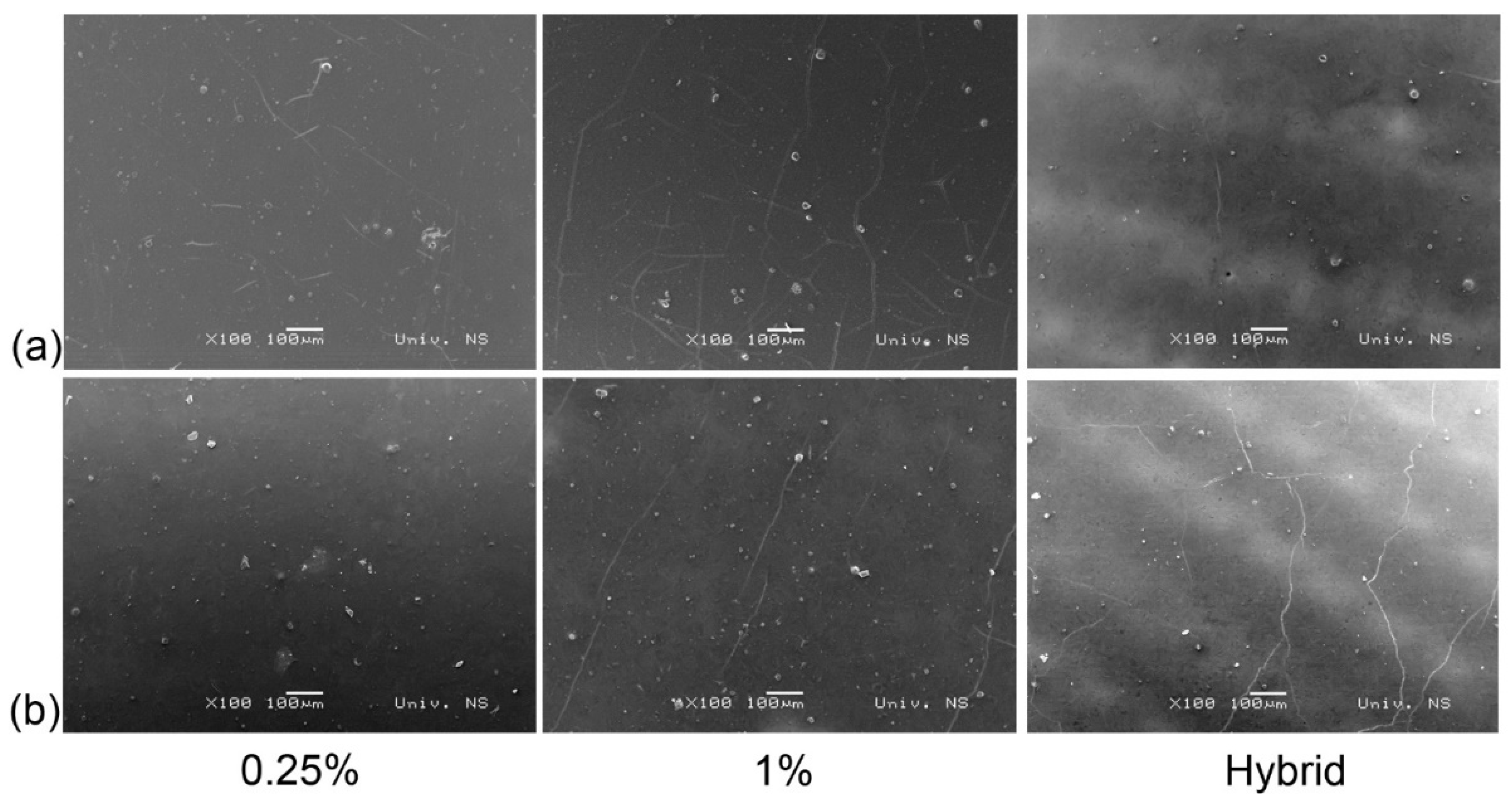
In order to assess if the dispersion of nanoparticles in the applied varnish was satisfactory, SEM images were acquired. Figure 2 shows a combined SEM image of ZnO nanoclusters and TiO2 nanoclusters. The cracks visible in all micrographs are the consequence of cutting the sample for the acquisition of the SEM image.
As can be seen on all micrographs presented in Figure 2, the samples on the surface feature some agglomerates of irregular shape which cannot be related to the weight ratio of the added nanoparticles; i.e., regardless of the nanoparticle’s weight ratio, the number of agglomerates is similar. To further investigate the agglomerates, EDS analysis was performed.
As evident from Figure 3, the EDS spectra detect various compounds including calcium (Ca) which indicates that the visible agglomerates on the micrographs originate from an anti-setoff powder. The anti-setoff powder is often used in offset printing to avoid wet ink film transfer onto the back side of the print. These powders are often composed of calcium carbonate or calcium sulfate [39]. Other compounds (natrium or magnesium) most probably originate from paper production.

3.2. SFE Analysis

The surface free energy (total, polar, and dispersive parts) of the samples was calculated from the measured contact angles.
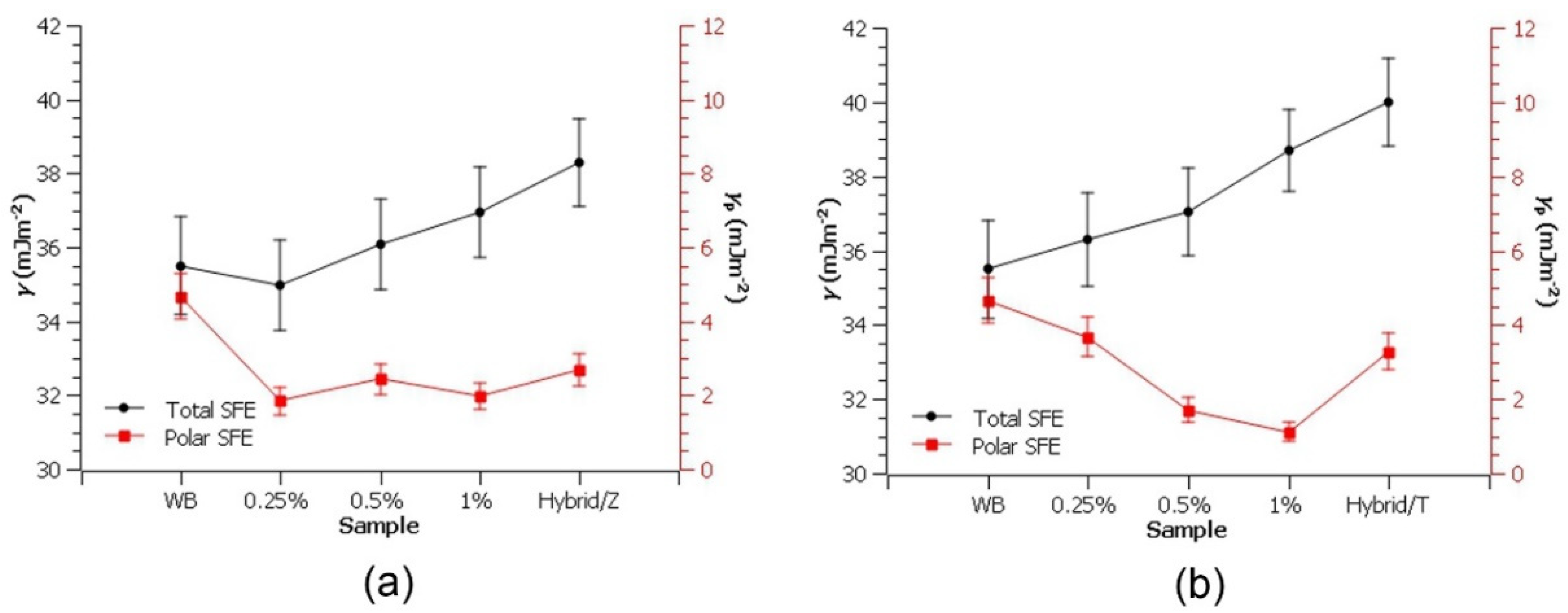
Figure 4 provides the calculated SFE (total and polar) values of the presented modulations. An increase is evident in the total SFE with an increase in the weight ratio of the nanocomposite. On the other hand, the WB has the highest polar component (γp = 5.08 mJm−2), and with the incorporation of both nanoparticles, the polar SFE values drop significantly. Although the polar component of SFE drops for both particles, it can be noted that with increasing the ZnO weight ratio, the polar component of SFE decreases, while with increasing the weight ratio of TiO2, the polar component of SFE decreases. As this research included rutile TiO2, this behavior could be expected as previous researchers showed the use of rutile TiO2 in creating hydrophobic surfaces [40]. As for the ZnO in the nanoscale, it is known that they turn hydrophobic if forming a structure, and a wire/rod can form a superhydrophobic surface [41,42]. Nevertheless, in this case, it is more likely that compounds in the WB interacted with ZnO leading to a decrease in the polar component of SFE. In addition, it could be noted that the addition of SiO2 (Hybrid/Z and Hybrid/T) in the coating increases the polar component in comparison to the samples coated with the nanocomposite containing the same amount of ZnO or TiO2, which is in line with the used hydrophilic SiO2.

3.3. Microbiological Analyses

The results of the microbiological analyses of the samples coated with ZnO or TiO2 and water-based varnish nanocomposites are presented in Table 3. The data show the differences in the growth inhibition of the inoculated pathogens S. aureus and L. monocytogenes depending on the nanocomposite formulations used; i.e., the lower the number, the higher the inhibition capacity. For aerobic mesophilic bacteria, the data show differences in bacterial contamination between the control and experimental samples exposed to environmental conditions (air pollution, room temperature). The lower number of aerobic mesophilic bacteria indicates a higher antimicrobial capacity of the nanocomposite formulation.
When comparing the water-based varnish samples with the nanoparticle-coated samples, the lower aerobic mesophilic bacteria counts were observed by applying ZnO and Hybrid/Z; however, the differences were not significant (p > 0.05).
The nanocomposite coatings did not upgrade the water-based varnish’s antimicrobial mechanism against S. aureus ATCC 25923 (p > 0.05). However, the commercial water-based varnish itself affected the growth of S. aureus by reducing its number by 0.8 log. On the other hand, the opposite was noted in the case of the growth of L. monocytogenes ATCC 7644. Additionally, the antimicrobial properties of the water-based varnish were significantly improved by applying nanocomposite coatings containing different concentrations of ZnO and Hybrid/Z. The highest reduction rate of L. monocytogenes was found with both 0.25 and 1% of ZnO applied (p < 0.05), as well as in the case of Hybrid/Z (1 log reduction, p < 0.05). In the case of TiO2 nanocomposite, the antilisterial capacity of the commercial varnish was improved by applying the concentration of 0.5 % (1 log reduction, p < 0.05). Opposite to the Hybrid/Z, the Hybrid/T did not show any inhibitory effect on the growth of L. monocytogenes. Other studies showed the low antimicrobial potential of TiO2, and when implemented by the standard method in humid conditions, it resulted in the increased growth of Listeria [43]. On the other hand, the results of the antimicrobial activity showed that the Hybrid/Z has the strongest antimicrobial potential. This can be attributed to the fumed SiO2 used for this research that was combined with ZnO nanoparticles that also showed strong antimicrobial potential. In the research by Liu et al., fumed SiO2 showed that the compound has a very strong antimicrobial potential where it absorbs the bacteria and inhibits its growth [44]. As mentioned before, water is required for the bacteria growth and migration where Hybrid/Z showed higher hydrophilic properties than Hybrid/T.
In addition to the antilisterial properties, the Hybrid/Z nanocoating showed the highest reduction in C. freundii (by 1.7 log) compared to other formulations. In the case of the artificial inoculation of Y. enterocolitica and Penicillium spp. on the control and experimental samples, no growth was observed. This clearly shows that the assessment of the antimicrobial properties of nanocomposites depends on applied microbes and their (non) ability to grow on specific materials [23,45,46]. S. aureus and L. monocytogenes can grow on different types of surfaces by biofilm formation, thus presenting the best indicator microorganisms for testing the antimicrobial properties of applied nanocomposites.

4. Conclusions

The growing and expanding packaging market demands new solutions that can cope with the rising needs of consumers. In the recent COVID-19 pandemic, consumers’ behavior has drastically changed, leaving a “gap” in printed packaging production that needs to be closed, which also leaves a possibly great marketing tool.
This research showed that the varnish can be upgraded with selected nanosized compounds that have the desired protective effect. The SEM micrographs combined with the EDS analysis show that the procedure of composing nanocomposites and their application on the printed surface is adequate. The SFE of the samples showed that adding nanoparticles will increase the total SFE almost linearly to the increase in the nanoparticle weight ratio (for both ZnO and TiO2). The weight ratio of the ZnO nanoparticles in the coating has no influence on the polar SFE, although even the smallest used nanoparticle’s concentration decreases the polar SFE while increasing the weight ratio of TiO2 will decrease the polar SFE. These results indicate a lower tendency to be wetted by water, i.e., lower hydrophilic behavior. According to the results of antimicrobial activity, Hybrid/Z achieved the best overall results from all the nanocomposites presented. Additionally, ZnO nanocomposites provided higher antimicrobial activity than the TiO2 ones.
To conclude, adding nanoparticles will enhance the properties of the coating, but further research is necessary to optimize the composition of the nanocomposites and to investigate the antimicrobial activity against other microorganisms which could come in contact with the packaging.

Author Contributions

Conceptualization, T.H. and T.C.; methodology, T.H., T.C. and N.Z.; validation, T.H., T.C., N.Z. and M.K.; formal analysis, T.H. and M.K.; investigation, T.H. and N.Z.; resources, T.C. and N.Z.; writing—original draft preparation, T.H. and M.K.; writing—review and editing, T.C. and N.Z.; visualization, T.H. and M.K.; funding acquisition, T.C. All authors have read and agreed to the published version of the manuscript.

Funding

This research was funded by Croatian Science Foundation, grant number UIP-2017-05-4081.

Data Availability Statement

Not applicable.

Acknowledgments

The authors would like to thank Milos Bokorov for the SEM and EDS spectra analysis.

Conflicts of Interest

The authors declare no conflict of interest.

References

  1. Rastogi, V.K.; Samyn, P. Bio-Based Coatings for Paper Applications. Coatings 2015, 5, 887–930. [Google Scholar] [CrossRef] [Green Version]
  2. Soroka, W. Fundamentals of Packaging Technology. In Paper and Paperboard; Institute of Packaging Professionals: Herndon, VA, USA, 2002; pp. 109–127. [Google Scholar]
  3. Rawdkuen, S.; Punbusayakul, N.; Lee, D.S. Antimicrobial Packaging for Meat Products. In Antimicrobial Food Packaging; Academic Press: Cambridge, MA, USA, 2016; pp. 229–241. [Google Scholar] [CrossRef]
  4. Jung, J.; Zhao, Y. Antimicrobial Packaging for Fresh and Minimally Processed Fruits and Vegetables. In Antimicrobial Food Packaging; Academic Press: Cambridge, MA, USA, 2016; pp. 243–256. [Google Scholar] [CrossRef]
  5. Malhotra, B.; Keshwani, A.; Kharkwal, H. Antimicrobial Food Packaging: Potential and Pitfalls. Front. Microbiol. 2015, 6, 611. [Google Scholar] [CrossRef] [PubMed] [Green Version]
  6. Koelsch Sand, C. Antimicrobial Packaging on the Rise Again. Food Technol. 2020, 74, 80–82. [Google Scholar]
  7. Moradi, M.; Guimarães, J.T.; Sahin, S. Current Applications of Exopolysaccharides from Lactic Acid Bacteria in the Development of Food Active Edible Packaging. Curr. Opin. Food Sci. 2021, 40, 33–39. [Google Scholar] [CrossRef]
  8. Salathé, M.; Kazandjieva, M.; Lee, J.W.; Levis, P.; Feldman, M.W.; Jones, J.H. A High-Resolution Human Contact Network for Infectious Disease Transmission. Proc. Natl. Acad. Sci. USA 2010, 107, 22020–22025. [Google Scholar] [CrossRef] [Green Version]
  9. Coronavirus Disease (COVID-19): How Is It Transmitted? Available online: https://www.who.int/news-room/questions-and-answers/item/coronavirus-disease-covid-19-how-is-it-transmitted (accessed on 12 September 2022).
  10. Hudika, T.; Cigula, T.; Vukoje, M. Antimicrobial Properties of TiO2 Nanocomposite Coating. In Proceedings of the Proceedings 13th International Conference on Nanomaterials—Research & Application, Brno, Czech Republic, 20–22 October 2021; pp. 351–358. [Google Scholar]
  11. Cigula, T.; Hudika, T.; Vukoje, M. Modulation of Water Based Commercial Varnish by Adding ZnO and SO2 Nanoparticles to Enhance Protective Function on Printed Packaging. In Proceedings of the 2nd International Circular Packaging Conference, ICP, FTPO Slovenia, Ljubljana, Slovenia, 9–10 September 2021; pp. 249–260. [Google Scholar]
  12. The Importance of Secondary Packaging. Available online: https://www.alcaminow.com/blog/the-importance-of-secondary-packaging (accessed on 25 January 2022).
  13. Zvekic, D.; Srdic, V.; Karaman, M.; Matavulj, M. Antimicrobial Properties of ZnO Nanoparticles Incorporated in Polyurethane Varnish. Process. Appl. Ceram. 2011, 5, 41–45. [Google Scholar] [CrossRef]
  14. Al-Tayyar, N.A.; Youssef, A.M.; Al-Hindi, R.R. Antimicrobial Packaging Efficiency of ZnO-SiO2 Nanocomposites Infused into PVA/CS Film for Enhancing the Shelf Life of Food Products. Food Packag. Shelf. Life 2020, 25, 100523. [Google Scholar] [CrossRef]
  15. Kubacka, A.; Diez, M.S.; Rojo, D.; Bargiela, R.; Ciordia, S.; Zapico, I.; Albar, J.P.; Barbas, C.; Martins Dos Santos, V.A.P.; Fernández-García, M.; et al. Understanding the Antimicrobial Mechanism of TiO2-Based Nanocomposite Films in a Pathogenic Bacterium. Sci. Rep. 2014, 4, 4134. [Google Scholar] [CrossRef] [Green Version]
  16. Kumar, R.; Umar, A.; Kumar, G.; Nalwa, H.S. Antimicrobial Properties of ZnO Nanomaterials: A Review. Ceram. Int. 2017, 43, 3940–3961. [Google Scholar] [CrossRef]
  17. Meng, D.; Liu, X.; Xie, Y.; Du, Y.; Yang, Y.; Xiao, C. Antibacterial Activity of Visible Light-Activated TiO2 Thin Films with Low Level of Fe Doping. Adv. Mater. Sci. Eng. 2019, 2019, 5819805. [Google Scholar] [CrossRef] [Green Version]
  18. Espitia, P.J.P.; Otoni, C.G.; Soares, N.F.F. Zinc Oxide Nanoparticles for Food Packaging Applications. In Antimicrobial Food Packaging; Academic Press: Cambridge, MA, USA, 2016; pp. 425–431. [Google Scholar] [CrossRef]
  19. Ghule, K.; Ghule, A.V.; Chen, B.J.; Ling, Y.C. Preparation and Characterization of ZnO Nanoparticles Coated Paper and Its Antibacterial Activity Study. Green Chem. 2006, 8, 1034–1041. [Google Scholar] [CrossRef] [Green Version]
  20. Petković, G.; Vukoje, M.; Bota, J.; Pasanec Preprotić, S. Enhancement of Polyvinyl Acetate (PVAc) Adhesion Performance by SiO2 and TiO2 Nanoparticles. Coatings 2019, 9, 707. [Google Scholar] [CrossRef] [Green Version]
  21. Vukoje, M.; Kulčar, R.; Itrić Ivanda, K.; Bota, J.; Cigula, T. Improvement in Thermochromic Offset Print UV Stability by Applying PCL Nanocomposite Coatings. Polymers 2022, 14, 1484. [Google Scholar] [CrossRef]
  22. Pietrzyk, B.; Porȩbska, K.; Jakubowski, W.; Miszczak, S. Antibacterial Properties of Zn Doped Hydrophobic SiO2 Coatings Produced by Sol-Gel Method. Coatings 2019, 9, 362. [Google Scholar] [CrossRef] [Green Version]
  23. Kadhum, S.A. The Effect of Two Types of Nano-Particles (ZnO & SiO2) on Different Types of Bacterial Growth. Biomed. Pharmacol. J. 2017, 10, 1701–1708. [Google Scholar] [CrossRef]
  24. Yang, D.L.; Cui, Y.N.; Sun, Q.; Liu, M.; Niu, H.; Wang, J.X. Antibacterial Activity and Reinforcing Effect of SiO2-ZnO Complex Cluster Fillers for Dental Resin Composites. Biomater. Sci. 2021, 9, 1795–1804. [Google Scholar] [CrossRef]
  25. Bota, J.; Hrnjak-Murgić, Z.; Brozović, M. The Effect of Film Thickness and Concentration of SIO2 Nanoparticles in PCL Coatings on Color Change of Tonal Value Increase. Acta Graph. 2016, 27, 15–22. [Google Scholar]
  26. Nguyen, T.V.; Nguyen, T.A.; Nguyen, T.H. The Synergistic Effects of SiO2 Nanoparticles and Organic Photostabilizers for Enhanced Weathering Resistance of Acrylic Polyurethane Coating. J. Compos. Sci. 2020, 4, 23. [Google Scholar] [CrossRef] [Green Version]
  27. Mills, D.J.; Jamali, S.S.; Paprocka, K. Investigation into the Effect of Nano-Silica on the Protective Properties of Polyurethane Coatings. Surf. Coat. Technol. 2012, 209, 137–142. [Google Scholar] [CrossRef]
  28. Malaki, M.; Hashemzadeh, Y.; Karevan, M. Effect of Nano-Silica on the Mechanical Properties of Acrylic Polyurethane Coatings. Prog. Org. Coat. 2016, 101, 477–485. [Google Scholar] [CrossRef]
  29. Wang, F.; Feng, L.; Ma, H.; Zhai, Z.; Liu, Z. Influence of Nano-SiO 2 on the Bonding Strength and Wear Resistance Properties of Polyurethane Coating. Sci. Eng. Compos. Mater. 2019, 26, 77–83. [Google Scholar] [CrossRef]
  30. Downey, A. COVID-19: Do I Need to Wash My Shopping and Groceries? | Patient. Available online: https://patient.info/news-and-features/covid-19-do-i-need-to-wash-my-shopping-and-groceries (accessed on 25 January 2022).
  31. Gloves Won’t Reduce Your Risk of COVID-19 at the Grocery Store. Available online: https://www.healthline.com/health-news/wearing-gloves-while-grocery-shopping-doesnt-prevent-covid-19#Why-gloves-arent-medically-necessary (accessed on 14 September 2021).
  32. Antimicrobial Packaging Market—Growth, Trends, COVID-19 Impact, and Forecasts (2021–2026). Available online: https://www.mordorintelligence.com/industry-reports/antimicrobial-packaging-market (accessed on 15 April 2021).
  33. King, A. More Bacteria Are Becoming Resistant to Antibiotics—Here’s How Viruses and Vaccines Could Help | Research and Innovation. Available online: https://ec.europa.eu/research-and-innovation/en/horizon-magazine/more-bacteria-are-becoming-resistant-antibiotics-heres-how-viruses-and-vaccines-could-help (accessed on 25 January 2022).
  34. Russell, C.D.; Fairfield, C.J. Co-Infections, Secondary Infections, and Antimicrobial Use in Patients Hospitalised with COVID-19 during the First Pandemic Wave from the ISARIC WHO CCP-UK Study: A Multicentre, Prospective Cohort Study. Lancet Microbe 2021, 2, e354–e365. [Google Scholar] [CrossRef]
  35. Guzińska, K.; Owczarek, M.; Dymel, M. Investigation in the Microbiological Purity of Paper and Board Packaging Intended for Contact with Food. Fibres Text. East. Eur. 2012, 96, 186–190. [Google Scholar]
  36. Skrivanek, T. Determination of the Surface Free Energy of Electronic Components. Metal Finish. 2003, 101, 87. [Google Scholar] [CrossRef]
  37. van Oss, C.J.; Giese, R.F.; Wu, W. On the Predominant Electron-Donicity of Polar Solid Surfaces. J. Adhes. 1997, 63, 71–88. [Google Scholar] [CrossRef]
  38. Zdolec, N.; Kiš, M.; Jankuloski, D.; Blagoevska, K.; Kazazić, S.; Pavlak, M.; Blagojević, B.; Antić, D.; Fredriksson-Ahomaa, M.; Pažin, V. Prevalence and Persistence of Multidrug-Resistant Yersinia enterocolitica 4/O:3 in Tonsils of Slaughter Pigs from Different Housing Systems in Croatia. Foods 2022, 11, 1459. [Google Scholar] [CrossRef]
  39. Bernd Schwegmann GmbH & Co. Anti Set-off Powders for the Printing Industry. Available online: https://www.schwegmannnet.de/PDF/Broschueren/Druck/EN/Wirbelwind_Broschuere_EN.pdf (accessed on 9 September 2022).
  40. Shamsudin, S.; Ahmad, M.K.; Aziz, A.N.; Fakhriah, R.; Mohamad, F.; Ahmad, N.; Nafarizal, N.; Soon, C.F.; Ameruddin, A.S.; Faridah, A.B.; et al. Hydrophobic Rutile Phase TiO2 Nanostructure and Its Properties for Self-Cleaning Application. AIP Conf. Proc. 2017, 1883, 020030. [Google Scholar]
  41. Nuruddin, A.; Yuliarto, B.; Kurniasih, S.; Setiyanto, H.; Ramelan, A. Preparation of Superhydrophobic Zinc Oxide Nanorods Coating on Stainless Steel via Chemical Bath Deposition. IOP Conf. Ser. Mater. Sci. Eng. 2019, 547, 012052. [Google Scholar] [CrossRef] [Green Version]
  42. Mardosaitė, R.; Jurkevičiū Tė, A.; Račkauskas, S. Superhydrophobic ZnO Nanowires: Wettability Mechanisms and Functional Applications. Cryst. Growth Des. 2021, 21, 4765–4779. [Google Scholar] [CrossRef]
  43. Akhidime, I.D.; Saubade, F.; Benson, P.S.; Butler, J.A.; Olivier, S.; Kelly, P.; Verran, J.; Whitehead, K.A. The Antimicrobial Effect of Metal Substrates on Food Pathogens. Food Bioprod. Process. 2019, 113, 68–76. [Google Scholar] [CrossRef]
  44. Liu, C.; Tao, Y.; Gout, J.; Qin, D. Antimicrobial Characteristic and Mechanisam of Nano-Fumed Silica Salt Grafted N,N-Dimethly-n-Tetradecylamine. Life Sci. J. 2009, 6, 52–54. [Google Scholar]
  45. Yeo, S.Y.; Lee, H.J.; Jeong, S.H. Preparation of Nanocomposite Fibers for Permanent Antibacterial Effect. J. Mater. Sci. 2003, 38, 2143–2147. [Google Scholar] [CrossRef]
  46. Jiang, J.; Oberdörster, G.; Elder, A.; Gelein, R.; Mercer, P.; Biswas, P. Does Nanoparticle Activity Depend upon Size and Crystal Phase? Nanotoxicology 2008, 2, 33–42. [Google Scholar] [CrossRef] [PubMed]
Figure 1. Experiment setup for this research.
Processes 10 02285 g001
Figure 2. SEM micrographs of samples coated by nanocomposite containing (a) ZnO and (b) TiO2.
Processes 10 02285 g002
Figure 3. Position on the micrograph (red arrow) and EDS spectra on the samples: (a) 1% ZnO nanoclusters, (b) 1% TiO2 nanoclusters, (c) 1% TiO2/SiO2 nanoclusters.
Processes 10 02285 g003
Figure 4. SFE of investigated samples coated with nanocomposite including (a) ZnO and (b) TiO2.
Processes 10 02285 g004
Table 1. Composition, homogenization time, and viscosity of prepared nanocomposites.
NanoparticleWeight Ratio (%)Homogenization Time (min)Viscosity (mPa·s)Denomination
Pure WB--105WB
ZnO0.25151680.25% ZnO nanoclusters
ZnO0.5202720.5% ZnO nanoclusters
ZnO1303761% ZnO nanoclusters
TiO20.25152130.25% TiO2 nanoclusters
TiO20.5203100.5% TiO2 nanoclusters
TiO21303571% TiO2 nanoclusters
SiO2 + TiO20.5 + 0.540690Hybrid/T
SiO2 + ZnO0.5 + 0.540710Hybrid/Z
Table 2. Liquids for surface free energy calculation and their properties.
LiquidSurface Tension (mNm−1)Dispersive Part (mNm−1)Polar Part (mNm−1)Author
Water γ = 2.0 µS cm−172.821.851Strӧm et al. [36]
Glycerol603451Strӧm et al. [36]
Diiodomethane50.850.80Strӧm et al. [36]
Formamide583919Van Oss et al. [37]
Table 3. Results of testing the growth inhibition of artificially inoculated pathogens and environmentally contaminated cardboard samples by nanocomposite coatings.
Added Weight Ratio of NPStaphylococcus aureus ATCC 25923Listeria monocytogenes ATCC 7644 (Serogroup 1/2c)Aerobic Mesophilic
Bacteria
Cardboard without coating 16.12 ± 0.425.77 ± 0.34 ABC2.45 ± 0.04 ABCDE
Pure WB 25.23 ± 0.946.59 ± 0.17 ADEFG2.00 ± 0.05 AG
1% TiO25.56 ± 0.457.00 ± 0.08 Bab2.39 ± 0.03
0.5% TiO25.09 ± 0.295.3 ± 0.26 Dac2.10 ± 0.11 a
0.25% TiO24.97 ± 0.486.21 ± 0.27 bc2.53 ± 0.05 Ga
1% ZnO5.65 ± 0.615.89 ± 0.09 F1.95 ± 0.04 D
0.5% ZnO5.06 ± 0.616.17 ± 0.181.99 ± 0.09 B
0.25% ZnO5.71 ± 0.125.49 ± 0.07 E1.68 ± 0.10 C
0.5%TiO2 + 0.5% SiO2
(Hybrid/T)
5.74 ± 0.276.71 ± 0.36 Cd2.20 ± 0.20 b
0.5% ZnO + 0.5% SiO2
(Hybrid/Z)
5.00 ± 0.095.56 ± 0.23 Gd1.73 ± 0.32 Eb
1,2 control groups, A–G statistically significant differences (p < 0.05) between certain nanocomposite coatings in regard to control groups (1 and 2), a–d statistically significant differences (p < 0.05) between different percentages of nanoparticles within each group of nanocomposite coatings.
Publisher’s Note: MDPI stays neutral with regard to jurisdictional claims in published maps and institutional affiliations.