Article

Human Cytochrome P450 2C9 and Its Polymorphic Modifications: Electroanalysis, Catalytic Properties, and Approaches to the Regulation of Enzymatic Activity

by
Victoria V. Shumyantseva
1,2,*,
Tatiana V. Bulko
1,
Polina I. Koroleva
1,
Evgeniya V. Shikh
3,
Anna A. Makhova
3,
Maryia S. Kisel
4,
Irina V. Haidukevich
4 and
Andrei A. Gilep
1,4
1
Institute of Biomedical Chemistry, Pogodinskaya Street, 10, Build 8, 119121 Moscow, Russia
2
Faculty of Biomedicine, Pirogov Russian National Research Medical University, Ostrovitianov Street, 1, 117997 Moscow, Russia
3
Department of Clinical Pharmacology and Propaedeutics of Internal Diseases, I.M. Sechenov First Moscow Medical State University (Sechenov University), Trubetskaya Str., 8/2, 119991 Moscow, Russia
4
Institute of Bioorganic Chemistry of the National Academy of Sciences of Belarus, 220141 Minsk, Belarus
*
Author to whom correspondence should be addressed.
Processes 2022, 10(2), 383; https://doi.org/10.3390/pr10020383
Submission received: 29 January 2022 / Revised: 9 February 2022 / Accepted: 15 February 2022 / Published: 17 February 2022
(This article belongs to the Section Catalysis Enhanced Processes)

Abstract

:
The electrochemical properties of cytochrome P450 2C9 (CYP2C9) and polymorphic modifications P450 2C9*2 (CYP2C9*2) and P450 2C9*3 (CYP2C9*3) were studied. To analyze the comparative electrochemical and electrocatalytic activity, the enzymes were immobilized on electrodes modified with a membrane-like synthetic surfactant (didodecyldimethylammonium bromide (DDAB)). An adequate choice of the type of modified electrode was confirmed by cyclic voltammetry of cytochromes P450 under anaerobic conditions, demonstrating well-defined peaks of reduction and oxidation of the heme iron. The midpoint potential, Emid, of cytochrome P450 2C9 is −0.318 ± 0.01 V, and Emid = −0.324 ± 0.01 V, and Emid = −0.318 ± 0.03 V for allelic variant 2C9*2 and allelic variant 2C9*3, respectively. In the presence of substrate diclofenac under aerobic conditions, cytochrome P450 2C9 and its polymorphic modifications P450 2C9*2 and P450 2C9*3 exhibit catalytic properties. Stimulation of the metabolism of diclofenac by cytochrome P450 2C9 in the presence of antioxidant medications mexidol and taurine was shown.

1. Introduction

Polymorphism of drug-metabolizing enzymes, cytochromes P450, affects the biotransformation of medications and drug–drug interactions [1]. Cytochrome P450 2C9 (CYP2C9), which accounts for ~20% of the total content of cytochromes P450 in the liver, has 60 known polymorphisms [2,3]. CYP2C9 is responsible for the metabolic clearance of 15–20% of all drugs undergoing metabolism Phase I, including acenocoumarol, candesartan, celecoxib, fluvastatin, glyburide, ketamine, methadone, phenytoin, tolbutamide, testosterone, phenobarbital, S-warfarin, piroxicam, losartan, tamoxifen, and many nonsteroidal anti-inflammatory drugs such as diclofenac and ibuprofen [4,5,6]. Cytochromes P450 2C9*2 and P450 2C9*3 are the most widely represented among the polymorphic variants P450 2C9. Cytochrome P450 2C9*2 is characterized by a C-T substitution in the corresponding gene, which leads to the Arg144Cys mutation. Cytochrome P450 2C9*3 is characterized by A-T substitution, leading to the Ile359Leu mutation [7,8,9,10]. The catalytic activities of cytochrome P450 2C9 and its polymorphic modifications P450 2C9*2 and P450 2C9*3 are different with respect to different substrates [8,9,10]. Decreased catalytic activity in the formation of 7-hydroxy-S-warfarin catalyzed by cytochromes P450 2C9*2 and P450 2C9*3 (warfarin 7-hydroxylation) was noted [11,12]. However, the metabolic transformations of a synthetic derivative of psychotropic cannabinoid drug JWH-018 (synthetic cannabinoid JWH-018 ((1-pentyl-1H-indol-3-yl) -1-naphthalenyl-methanone), catalyzed by cytochrome P450 2C9*2, were characterized by an increased activity (by 3.6 times) compared to the wild-type P450 2C9 [5].
A wide range of drugs metabolized by cytochrome P450 2C9 and its allelic variants, as well as different catalytic activity with respect to substrates/medications, requires the creation of systems and development of various methods for conducting comparative pharmacokinetics and pharmacogenomics studies. To identify the unique features of cytochrome P450 2C9 pharmacokinetics and the role of amino acids in the active center of allelic variants, the crystal structures of cytochrome P450 2C9, 2C9*3 ((I359L) and *30 (A477T) in complex with the antihypertensive drug losartan were studied [11]. The molecular dynamic simulation was also used to characterize the complexes of tamoxifen prodrug and polymorphic modifications CYP2C9 R144C (*2), I359L (*3), D360E (*5), R150H (*8), R335W (*11), and L90P (*13) [3].
The catalytic mechanism of cytochrome P450 functionality is complex, requires participation of service proteins in the electron transfer reaction (protein redox partners such as NADPH-dependent flavoprotein reductase) and NADPH as an electron source for the realization of catalysis towards two substrates: oxygen and organic compounds. Since the catalytic cycle of cytochromes P450 is associated with electron transfer, the use of electrochemical systems has found its practical application for modeling catalysis of this class of hemoproteins. However, redox partners are not obligatory upon the electrochemical reduction of the P450 family hemoproteins, so the catalytic system is essentially simplified. Electrochemical systems execute a dual function: substitute partner proteins and serve as a source of electrons for redox enzymes. The information parameters of electroanalysis enable both calculating the kinetic parameters of enzymatic processes and characterizing the bioelectrochemical system and electrode processes. From the medical and biochemical viewpoint, bioelectrocatalysis with P450s is a promising technique for conducting clinical medicine studies, analyzing drug–drug interactions, and performing substrate/inhibitor studies.
Electrochemical cytochrome P450 systems are a new biotechnological approach in enzymology. Cytochrome P450 bioelectrodes are indispensable adequate experimental models for the development of new drugs, the search for their pharmacological targets, and the study of drug metabolism characteristics by polymorphic variants of enzymes from the standpoint of pharmacogenomics and personalized therapy [13,14]. In this work, comparative electroanalytical and electrocatalytic parameters of cytochromes P450 2C9, 2C9*2, and 2C9*3 were investigated using screen-printed electrodes (SPEs), modified by membrane-like didodecyldimethylammonium bromide (DDAB) (SPE/DDAB).
Hereditary genetic variability of genes of drug-metabolizing enzymes, namely cytochromes P450, affects the biotransformation of drugs and drug–drug interactions [1]. However, it is important to emphasize that cytochrome P450 2C9 and its allelic variants exhibit different catalytic activity towards different substrates [5,8,9,10]. This may be due to both the different structure of the drug molecule and the fact that the active centers of polymorphic forms have differences associated with amino acid substitutions of the polypeptide chain of the enzyme [3]. The rational search for activators/modulators and inhibitors of drug biocatalysis is an urgent task. The regulation of the catalytic activity of enzymes in systems in vitro can proceed by various mechanisms: incorporation into membranes, interaction with redox partner proteins, chemical modification, effect of detergents, and allosteric mechanisms [15]. For targeted regulation of drug biotransformation in vivo, an approach based on correction of the actual activity of the enzyme can be implemented. In this regard, it is important to regulate the enzymatic activity using compounds that are not substrates of this hemoprotein but affect individual stages of catalysis. Previously, antioxidant metabolic drugs and antioxidant vitamins were studied as the correctors of the cytochrome P450 catalytic processes. It has been shown that such drugs stimulate the process of heme iron electrochemical reduction, which is registered by an increase in the amplitude of the cathodic current at the reduction potential of cytochromes P450 3A4, P450 2C9, and P450 2D6 [16,17,18,19,20,21].
This work is aimed at studying the effect of the antioxidant metabolic drug mexidol and vitamin-like antioxidant compound taurine on the metabolic transformations of diclofenac catalyzed by cytochrome P450 2C9. Mexidol and taurine stimulated not only the stage of cytochrome P450 2C9 electrochemical reduction but also demonstrated an activating effect on the catalytic activity of cytochrome P450 2C9 in relation to diclofenac by 1.7 times and 1.5 times, respectively.

2. Materials and Methods

2.1. Materials

Cloning, expression, and purification of cytochrome P450 2C9, 2C9*2, and 2C9*3 were performed as described [22]. The enzyme concentration was determined spectrophotometrically by complex formation of a reduced form with carbon monoxide; absorption coefficient ε450–490 = 91 mM−1cm−1 [23]. The following reagents were used in this work: didodecyldimethylammonium bromide (DDAB) (Sigma-Aldrich, St. Louis, Missouri, USA), taurine (PicPharma, Moscow, Russia). diclofenac (2-(2,6-dichloroaniline) phenylacetic acid) (Novartis, Moscow, Russia), mexidol (2-ethyl-6-methyl-3-hydroxypyridine succinate (50 mg/mL, Farmasoft, Moscow, Russia)).

2.2. Electrochemical Procedures

Electrochemical measurements were carried out using AutoLab 12 potentiostat/galvanostat (Metrohm AutoLab, Utrecht, Netherlands) equipped with the GPES software (version 4.9.7).
In this work, we used three-pronged electrodes obtained by screen-printing electrodes (SPEs) with graphite working and auxiliary electrodes and silver/silver chloride (Ag/AgCl) the reference electrode (Color Electronics, Moscow, Russia). The diameter of the working electrode was 0.2 cm (area = 0.0314 cm2).

2.3. Preparation of Electrochemical Sensors

For the preparation of modified electrodes, the SPEs were modified by 1 µL 0.1 M DDAB in chloroform and incubated for 10 min. Then, 2 µL 175 µM P450 2C9 (or 1 µL 320 µM P450 2C9*2, or 1 µL 250 µM P450 2C9*3) was placed on the electrode. To prevent the complete drying of the electrodes, they were kept for 12 h at 4 °C in a humid chamber.
In the electrochemical experiments, freshly prepared solutions of 10 mM diclofenac in water were used. Electrochemical measurements were performed at room temperature in 0.1 M potassium phosphate buffer (pH 7.4) containing 0.05 M NaCl. Cyclic voltammograms (CV) were registered at a scan rate of 0.01–0.1 V/s and potential range from 0 to −0.8 V (vs. Ag/AgCl). All potentials were referred to the Ag/AgCl electrode. Direct electrochemistry of CYP3A4 was achieved by noncovalent immobilization of the protein on the SPEs modified with 1 mL 0.1 M DDAB in chloroform. For the investigation of diclofenac conversion, we used differential pulse voltammetry (DPV). DPV (oxidation) experiments were carried out in a 60 μL drop applied onto the SPEs to cover all three electrodes using the planar regimen of electrode mode. The following experimental DPV parameters were used: potential range of 0.2–1.2 V, pulse amplitude of 0.025 V, potential step of 0.005 V, pulse duration of 50 ms, and modulation amplitude of 0.05 V. All experiments were performed in triplicate. The data are presented as average values ± standard deviations.

2.4. Electrocatalysis of P450 2C9, 2C9*2, and 2C9* in the Presence of Diclofenac

Electrocatalytic conversion of the diclofenac by CYP3A4 was carried out by electrolysis of the drug or drug in the presence of antioxidant mexidol or taurine at controlled potential of E = −0.55 V for 20 min in an electrochemical cell supplied by SPE/DDAB/CYP3A4 electrode. For the analysis of the residual diclofenac concentration during CYP3A4-electrocatalysis, 60 mL aliquots were taken from the reaction mixture in the first electrochemical cell after 20 min of electrolysis, and the residual drug concentration was analyzed by SPEs in a separate cell in the planar regimen of the electrode.
For the analysis of diclofenac by the DPV in the potential range of 0.2–1.2 V, unmodified SPEs were used.
The experimental data were processed using Origin 8.1 software.

3. Results and Discussion

Investigation of CYP enzyme catalytic activity involves different approaches, such as in silico, in vivo, in vitro model systems. Since the catalytic cycle of cytochromes P450 is associated with electron transfer, the use of electrochemical systems has found its practical application for modeling catalysis of this class of hemoproteins.
Cytochrome P450-based electrochemical systems are effective noninvasive models for studying the substrate specificity of this class of hemoproteins, the search for new drugs, and analysis of drug–drug interactions [12,13,14,24,25,26,27,28,29].

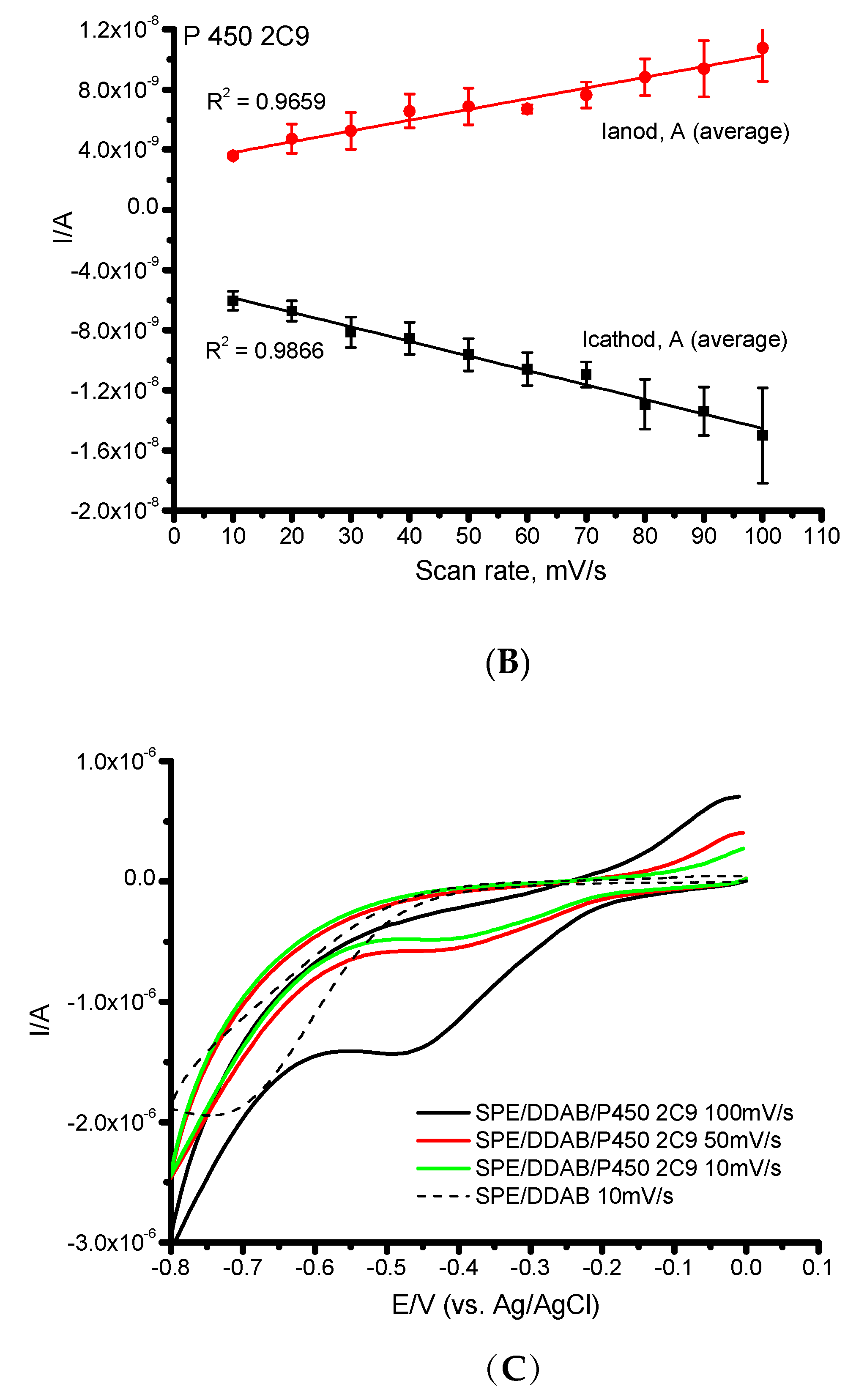
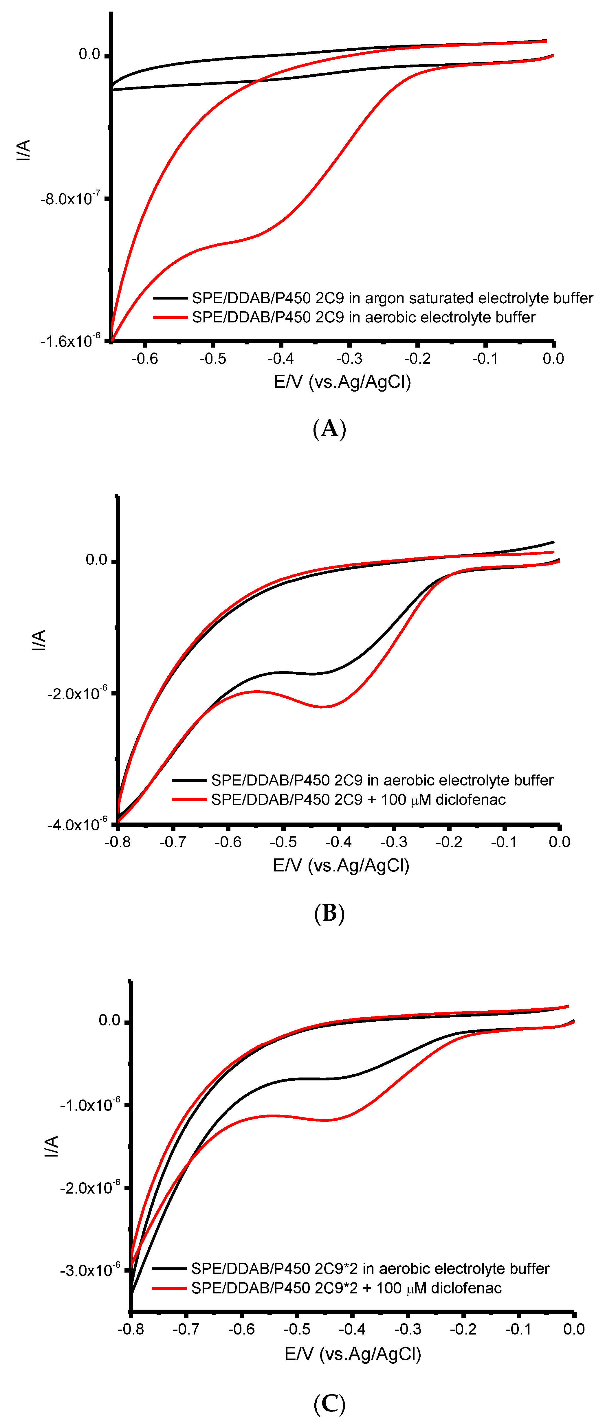
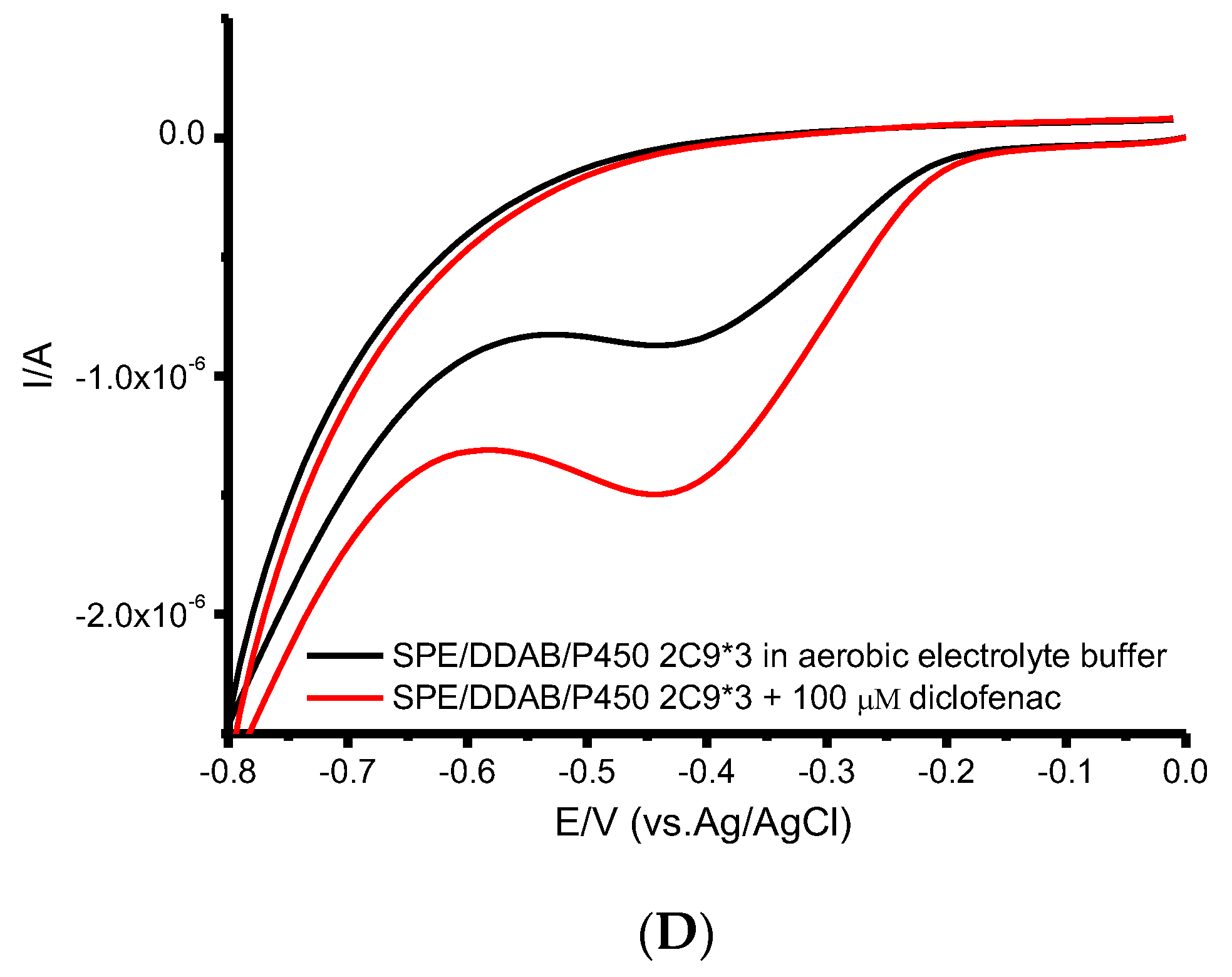
3.1. Electrochemical Characteristics and Electrocatalytic Properties of Cytochrome P450 2C9 and Polymorphic Modifications P450 2C9*2 and P450 2C9*3

The electrochemical characteristics of cytochrome P450 2C9 and polymorphic modifications P450 2C9*2 and P450 2C9*3 immobilized on the graphite electrodes modified with a synthetic lipid-like compound didodecyldimethylammonium bromide (DDAB) were studied both under aerobic and anaerobic electrolyte buffer. Figure 1A shows the cyclic voltammograms (CV) of cytochrome P450 2C9 at scan rates of 10–100 mV/s in argon-saturated electrolyte buffer, which characterize the nearly reversible redox process CYP-Fe(III) + ē (from the electrode) ⇄ CYP-Fe(II) for heme redox center, where CYP-Fe(III) is the oxidized (ferri) form, and CYP-Fe(II) is the reduced (ferro) form of cytochrome P450.
The linear dependence of the maximum amplitudes of cathodic and anodic currents on the scan rates is characteristic of the processes occurring on the electrode surface so-called “protein film voltammetry” and not controlled by diffusion [30,31] (Figure 1B). The electroanalytical characteristics of cytochrome P450 2C9, 2C9*2, and 2C9*3 are presented in Table 1. The midpoint potentials E0’ of cytochromes P450s were calculated from the values of anodic and cathodic peak potentials by the equation E0’ = (Epc + Epa)/2. Cytochrome P450 2C9 and its allelic variants 2C9*2 and 2C9*3 demonstrated very close midpoint potential values as Emid = −0.318 ± 0.01 V, −0.324 ± 0.01 V, and −0.318 ± 0.03 V, respectively (Figures S1 and S2).
The heterogeneous electron transfer rate constant (ks) values for the electron transfer between the cytochrome P450 and SPE/DDAB at the scan rate 100 mV s−1 and using a transfer coefficient α of 0.5 were determined according to Laviron’s model [32]. Heterogeneous electron transfer rate constants ks for ΔE < 200 mV were 0.54 ± 0.02, 0.48 ± 0.01, and 0.50 ± 0.04 s−1 for cytochromes P450 2C9, 2C9*2, and 2C9*3 (Table 1). The amount of electroactive protein was calculated according to Faraday’s law by Equation (1).
Γ 0 = Q nFA
where Γ0 is the surface coverage or surface concentration of electroactive protein, mol cm−2; Q is the electric charge calculated from integration of voltammogram peaks, C; n is the number of electrons transferred (n = 1 for cytochrome P450s); F is the Faraday constant, 96485 C mol−1; and A is the surface area of the working electrode, cm−2 [30,33].
Table 1 summarizes the electrochemical parameters experimentally calculated for cytochrome P450 2C9 and allelic variants 2C9*2 and 2C9*3.
The electrocatalytic properties of cytochromes P450 2C9, 2C9*2, and 2C9*3 in aerobic buffer towards oxygen as a cosubstrate of this class of hemoproteins were investigated. Under aerobic conditions, the reduced Fe(II) ion actively interacts with oxygen, and the cathodic reduction peak is recorded in accordance with the irreversible reaction CYP-Fe (II) + O2 → CYP-Fe (II)O2, which increases with an increase in the scan rate (Figure 1C and Figure 2A). Reduction potentials Ered (2C9) = −0.443 ± 0.020 V, −0.393 ± 0.020 V (2C9*2), and −0.395 ± 0.020 V (2C9*3) were registered at a scan rate of 100 mV/s (Figure 2B–D; Table 2). The reduction potentials of allelic variants of cytochromes P450 2C9*2 and P450 2C9*3 in an aerobic buffer is characterized by a shift to the positive anodic potential compared to cytochrome P450 2C9 with ΔE~50 mV. This shift characterizes a more active electron transfer process to the heme redox center of allelic variants. The experimental electroanalytical characteristics of the cytochrome P450 2C9 family are in accordance with the previously published values of both midpoint potentials, Emid, and reduction potentials, Ered, in the presence of oxygen [13,25,26,27,34].
An effective electrochemical system made it possible to further study the internals and details of the interaction of cytochromes P450 2C9, 2C9*2, and 2C9*3 with diclofenac as a substrate of these enzymes from the cytochrome P450 family.
Diclofenac (2- (2,6-dichloroaniline) phenylacetic acid) refers to nonsteroidal anti-inflammatory drugs (NSAIDs, NSAIDs) and has analgesic, anti-inflammatory, and antipyretic effects, as well as anticancer properties [35,36]. Diclofenac is metabolized by cytochrome P450 2C9 and its polymorphic modifications 2C9*2 and 2C9*3 with the formation of 4-hydroxydiclofenac (Scheme 1) [1,7,10].
A unique feature of cytochromes P450 is the participation in the catalytic cycle with two substrates—oxygen and an organic compound [37]. The ratio of the maximum amplitude of the catalytic current in the presence of 100 μM diclofenac at the potential of catalysis, Ecat = −0.393 V, and the reduction current under aerobic conditions IO2 at the potential of heme reduction, Ered = −0.443 V (IDF/IO2), is 1.80 ± 0.06 for the P450 2C9 isoform (at a scan rate of 100 mV/s). This ratio reflects the redistribution of the electron flow and can serve as an indicator of efficiency (catalytic index) in the catalysis of cytochrome P450, as it indicates the metabolism and participation in the catalytic cycle of both oxygen and organic substrate (diclofenac). The catalytic index as IDF/IO2 ratio also reflects the distribution/redistribution of the electrons between two substrates—oxygen and an organic compound (in this example, a drug). The analytical sensitivity of bioelectrode SPE/DDAB/P450 2C9 in relation to diclofenac as a substrate is 0.0045 Ampere (A)/mole (M), (S/N = 3). The potential for the start of catalysis of diclofenac Eonset in the electrochemical system for the P450 2C9 is −0.212 V and is shifted to the anodic positive potential region by 180 mV compared to the potential of catalysis, Ecat (Table 2 and Figure 2B). Similar tendencies were observed for allelic variants: Eonset for the P450 2C9*2 form = −0.211 V, and for P450 2C9*3, Eonset = −0.207 V.
The catalytic indexes as IDF/IO2 ratio for 2C9*2 and 2C9*3 are 1.33 ± 0.07 and 2.18 ± 0.06, respectively. The analytical sensitivity for diclofenac is 0.0043 A/M and 0.015 A/M (S/N = 3) for the bioelectrodes SPE/DDAB/P450 2C9*2 and SPE/DDAB/P450 2C9*3, respectively. It is necessary to emphasize that SPE/DDAB/P450 2C9*3 is 3.5 times more sensitive for diclofenac in comparison with 2C9 and 2C9*2 forms.
Analysis of the electrocatalytic activity of cytochrome P450 2C9, 2C9*2, and 2C9*3 in the presence of diclofenac showed a concentration-dependent increase in the reduction current of cytochrome P450 2C9 (inherently catalytic current), which is indicative of substrates of this class of hemoproteins (Figure 3). Similar dependencies were obtained for polymorphic cytochromes P450 2C9*2 and 2C9*3. The Michaelis constants, KM, of cytochrome P450 2C9 and its polymorphic modifications were calculated according to Equation (2) [26,31,33,34,38] from the dependences of the catalytic current as a measure of reaction rate on concentration using nonlinear regression analysis.
Icat = I cat   max · [ S ] K M app + [ S ]
where Icat is the catalytic current at a certain substrate concentration, A; Imax is the maximum catalytic current at full saturation of the enzyme, A; [S] is the concentration of the substrate, M; and KM is the electrochemical Michaelis constant, M.
The Michaelis constants KM of diclofenac for cytochromes P450 2C9, P450 2C9*2, and P450 2C9*3, expressed in yeast microsomes, were determined in the microsomal system as 4.3 ± 2.4, 2.5 ± 0.3, and 11.2 ± 2.3 μM, respectively, according to [7]. Cytochrome P450 2C9*3 is characterized by a higher value (KM = 11.2 ± 2.3 μM) compared to P450 2C9 and 2C9*2. However, the catalytic constants of 4-hydroxylation of diclofenac in microsomes with P450 isoforms 2C9, 2C9*2, and 2C9*3 do not differ dramatically and are quite comparable: kcat 12.5 ± 0.2, 11.2 ± 0.4, 8.1 ± 1.3 nmol/min/nmol P450 [7].
Based on the analysis of the dependences of the catalytic current on the concentration of diclofenac for bioelectrode for the P450 enzymes 2C9, 2C9*2, and 2C9*3, the following values of the Michaelis constants KM were obtained: 45 ± 5, 68 ± 2, and 62 ± 2 μM, respectively (Figure 3). The Michaelis constants determined in electrochemical systems, as a rule, have higher values in comparison with the microsomal system containing redox partner proteins, conducting the best structural conformation for substrate binding [7,39,40].
Cytochrome P450 2C9 and its polymorphic modifications catalyze metabolic transformations of the anticoagulant S-warfarin with the formation of 7-hydroxy-warfarin [1,13]. In contrast to diclofenac, the analysis of the catalytic activity of warfarin demonstrated a significant difference in the activity of cytochrome P450 2C9 and allelic variants 2C9*2 and 2C9*3 [6,12,34]. Thus, P450 2C9*3 was approximately 6 times less active in the 7-hydroxylation reaction of warfarin compared to the “wild type” [12,39,40].
Analysis of the catalytic activity of cytochrome P450 2C9 and its polymorphic modifications 2C9*2 and 2C9*3, expressed in baculosomes, to a synthetic compound with psychotropic activity (1-pentyl-1H-indol-3-yl)-1-naphthalenyl-methanone, JWH-018) showed that the allelic variant 2C9*2 was 3.6 times more active in the hydroxylation reaction of this compound compared to the wild type. The values of Michaelis constants KM showed the highest affinity for variant 2C9*2 (0.48 µM) and 0.90 and 0.68 µM for P450 2C9 and 2C9*3, respectively [5]. 2C9*13 is an allele identified in a Chinese poor metabolizer of lornoxicam, which has a Leu90Pro amino acid substitution. 2C9*3 and 2C*13 variants produced far lower luminescence than the 2C9 variant in luciferin H metabolism. P450 2C9*13 exhibited an 11-fold increase in Km but no change in Vmax with tolbutamide as the substrate, a five-fold increase in Km, and an 88.8% reduction in Vmax with diclofenac [41].
The main point that can be made when comparing the kinetic parameters of metabolic transformations of an extensive repertoire of various substrates of cytochrome P450 2C9 and its allelic variants is that the chemical structure and physicochemical properties of the molecules of these compounds make a significant contribution to their metabolism. Warfarin is a hydrophobic molecule composed of two aromatic rings; diclofenac is a phenylacetic acid derivative that additionally resembles a polar pyramidal ammonium-like molecule. The synthetic psychotropic drug JWH-018 consists of a naphthalene ring linked by a carbonyl bridge to an indole ring [5]. Based on the foregoing concerning the metabolic activity of allelic cytochrome P450 2C9 variants, studies are needed for each drug exhibiting substrate activity with respect to these isoenzymes.

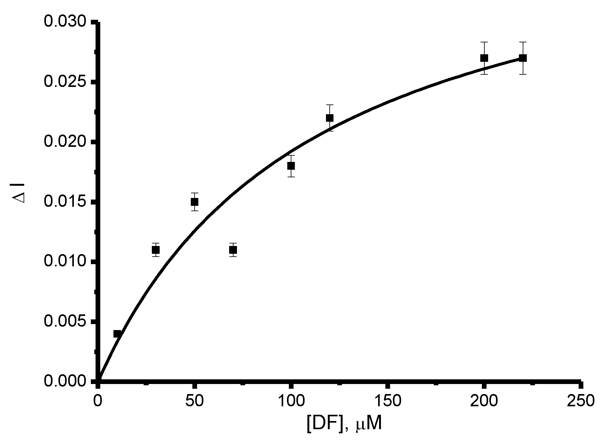
3.2. Influence of the Antioxidants on the Catalytic Activity of Cytochrome P450 2C9 and Polymorphic Modifications P450 2C9*2 and P450 2C9*3

To analyze the electrocatalytic activity of cytochrome P450 2C9, we used the previously developed method for the analysis of diclofenac based on the electrochemical oxidation of this drug [42]. In our experiments, we used an unmodified screen-printed graphite electrode (SPE) due to the low background current and appropriate sensitivity of the system towards diclofenac electrooxidation. Differential pulse voltammetry (DPV) technique was implemented for diclofenac registration. The electrochemical behavior of diclofenac makes it possible to use electrooxidation to measure the drug residual concentration in the probe after cytochrome P450 2C9-dependent electrocatalysis.
The comparative activity of cytochrome P450 2C9 to diclofenac is shown in Figure 4. Analysis of the signal intensity of electrooxidation of diclofenac before (0 min of electrolysis) and after electrolysis at a controlled potential of −0.55 V, for 20 min, allows calculating the catalytic activity of the system, which corresponds to 19 ± 3% of the reacted substrate.
We previously studied the effect of antioxidant metabolic preparations on the catalytic activity of cytochromes P450 [16,17,18]. Antioxidant therapy now is well known and used in medicinal practice. It was shown that melatonin as an antioxidant protects the cytochrome P450 system [43]. Cytochrome c and vitamin C, possessing antioxidant properties, serve as nonspecific agents for redox relay and can enhance the electron transfer in reactions mediating redox processes for heme peroxidases such as chloroperoxidase and horseradish peroxidase [44]. It was also shown that vitamin C, being a strong antioxidant, is capable of scavenging reactive oxygen species in low concentration ranges and possesses prooxidant capacity in high concentrations [45].
Inspired by this, we took this approach for our attempts to modulate the catalytic activity of cytochrome P450 2C9 using diclofenac as a widely used nonsteroidal anti-inflammatory drug.
Mexidol (2-ethyl-6-methyl-3-hydroxypyridine succinate), a metabolic antioxidant drug with antihypoxic, membrane-protective, nootropic, anticonvulsant, and anxiolytic action, is widely used in cerebrovascular accidents. Mexidol itself does not exhibit substrate properties of the cytochrome P450 superfamily; however, it stimulates the electrochemical reduction of CYPs, as has been shown earlier [17]. Its metabolic transformations are associated with the enzyme glucuronyltransferase (UDP), and it is excreted from the body mainly in the glucuronoconjugated form. In electrochemical experiments, antioxidants serve as modulating and/or stimulating additives with respect to the electrochemical activity of CYP due to their free-radical scavenging, antihypoxant, and/or electron mediator properties [16,17].
In the presence of mexidol, a significant increase in the catalytic current of diclofenac is recorded (Figure 5A,B, red and green lines). The stimulating factor in the presence of mexidol alone was 2.5. The ratio IDF/IO2 with diclofenac and mexidol is 4.0 (based on CV experiments, Figure 5A,B, green line). The mechanism of the stimulating effect of antioxidants on the stage of heme electrochemical reduction is based on the properties of these compounds as “traps” of the reactive oxygen species generated in the catalytic cycle of cytochromes P450. This phenomenon leads to stabilization of the enzyme and a local increase in the concentration of oxygen as a cosubstrate of cytochromes P450 [16,17,43,44,45,46].
Analysis of the diclofenac electrooxidation intensity before and after electrolysis at a controlled potential of E = −0.55 V for 20 min in the presence of 98 μM mexidol showed an increase in the activity of up to 33 ± 5% of the substrate reacted. Based on the foregoing, diclofenac metabolism in the presence of mexidol can lead to an acceleration of diclofenac elimination and a change in its interaction with potential molecular targets (phosphoinositol-3 kinase) [35,36].
Taurine (2-aminoethanesulfonic acid) is the most abundant free amino acid in the human body and plays an important role in biological processes such as conjugation of bile acids, maintenance of calcium homeostasis, osmoregulation, and membrane stabilization. In clinical practice, taurine is used as an antioxidant, membrane-stabilizing agent, hepatic-protector, anti-inflammatory, and antiapoptotic compound [47]. Taurine is also effective for the treatment of rheumatoid arthritis [48] as a cholesterol-reducing agent [49]. Thus, the addition of taurine to COVID-19 patients may be useful for treatment protocols [50].
It was previously shown that taurine (50 μM) stimulated both aerobic heme iron reduction and metabolic reactions of erythromycin N-demethylation catalyzed by cytochrome P450 3A4 (an increase in the catalytic constant, kcat, by 16% was recorded) [17,20].
Cytochrome P450 2C9-electrode was implemented to monitor the influence of taurine on the catalytic conversion of diclofenac. In the presence of 100 μM taurine, the catalytic index as IDF/IO2 ratio was 2.0 ± 0.04 (Figure 5A,B, blue line) in comparison with the 1.80 ± 0.06 index without the antioxidant. Analysis of the signal intensity of diclofenac electrooxidation before and after electrolysis at a controlled potential of −0.55 V for 20 min in the presence of 100 μM taurine showed an increase in catalytic activity by 29 ± 3% compared to the experiments without the antioxidant (19 ± 3%).
Analysis of the electrocatalytic conversion of diclofenac in the presence of mexidol and taurine revealed the influence of these antioxidants on drug metabolism of CYP2C9*2 and CYP2C9*3 allelic variants (Table 3). Using the same approach to registering diclofenac conversion [42], we have shown that the impact of mexidol on diclofenac metabolism catalyzed by CYP2C9 and CYP2C9*2 is comparable. However, CYP2C9*3 response on mexidol addition was significantly less and corresponded to 8 ± 3%. Taurine affects the catalytic activity of 2C9*2 and 2C9*3 allelic variants in the range of 16 ± 3% and 19 ± 4%, respectively.
SPE/DDAB/P450 2C9, SPE/DDAB/P450 2C9*2, and SPE/DDAB P450 2C9*3 were used, applied potential of electrolysis was E = −0.55 V (20 min), and 0.1 M potassium phosphate buffer, pH 7.4, containing 0.05 M NaCl, was used as supporting electrolyte.
It was shown that Ser 365 and Phe 114 play a decisive role in the catalytic reactions of cytochrome P450 2C9-hydroxylation of diclofenac, and Phe 476 is a crucial residue in the binding of diclofenac [10]. Cytochrome P450 2C9*2 is an Arg144Cys mutant, and cytochrome P450 2C9*3 is an Ile359Leu mutant compared to the “wild” type P450 2C9; therefore, we can assume a comparable catalytic activity of these isoforms in relation to the 4-hydroxylation reaction of diclofenac.

4. Conclusions

Electrochemically driven cytochrome P450 catalysis represents a new biotechnology trend and is an alternative model system for pharmacological and comparative pharmacokinetic and pharmacogenomic research. Characteristic cyclic voltammograms of polymorphic enzymes P450 2C9, P450 2C9*2, and P450 2C9*3 with heme iron reduction potentials in the range of –0.3 ÷ −0.5 V were carried out on electrodes modified with a membrane-like didodecyldimethylammonium bromide (DDAB) (SPE/DDAB). The catalytic activity of cytochromes P450 2C9, P450 2C9*2, and P450 2C9*3 in the presence of diclofenac was shown, expressed in the registration of the catalytic current of the “wild-type” enzyme and both allelic isoforms.
Recently, new approaches for modulating cytochrome P450 activity were investigated. The influence of small molecules (drugs, such as rifampicin, dhurrin, cyclophosphamide) on cytochrome P450 oxidoreductase was studied from the point of view of cytochrome P450-mediated metabolism [51].
Antioxidant intake can lead to a change in the catalytic efficiency of cytochrome P450 enzymes. Antioxidants could influence the direct electron transfer of human CYP enzymes immobilized on electrode surfaces. In electrochemical experiments, drug-antioxidants served as modulating and/or stimulating additives with respect to P450 electrochemical activity due to their free-radical scavenging and antihypoxant properties. Based on the foregoing, it is possible to propose antioxidants as promising agents for modulation/stimulation of cytochrome P450 catalysis. The stimulating role of antioxidant drug mexidol and taurine in concentrations comparable to those of the substrates of these isoforms on the metabolic reactions of diclofenac catalyzed by cytochromes P450 2C9, P450 2C9*2, and P450 2C9*3 has been shown.

Supplementary Materials

The following supporting information can be downloaded at: https://www.mdpi.com/article/10.3390/pr10020383/s1, Figure S1: Cyclic voltammograms of SPE/DDAB/P450 2C9*2. Measurement parameters: potential sweep rate from 10 to 100 mV/s, potential range from −0.8 to 0 V (vs. Ag/AgCl). The measurements were carried out in 1 ml of electrolyte buffer, pH 7.4, saturated with argon (anaerobic conditions); Figure S2: Cyclic voltammograms of SPE/DDAB/P450 2C9*3. Measurement parameters: potential sweep rate from 10 to 100 mV/s, potential range from −0.8 to 0 V (vs. Ag/AgCl). The measurements were carried out in 1 ml of electrolyte buffer, pH 7.4, saturated with argon (anaerobic conditions); Figure S3: The dependence of the catalytic current on the concentration of diclofenac in the concentration range of 10–250 μM for SPE/DDAB/P450 2C9*2, applied potential of electrolysis is E = −0.55 V; Figure S4: The dependence of the catalytic current on the concentration of diclofenac in the concentration range of 10–250 μM for SPE/DDAB/P450 2C9*3, applied potential of electrolysis is E = −0.55 V.

Author Contributions

V.V.S. developed the concept, conceptualization, and methodology; V.V.S. and T.V.B. designed the experiments and analyzed the data obtained, T.V.B., V.V.S., and P.I.K. performed the electrochemical experiments, E.V.S. and A.A.M. designed the experiments with drugs and analyzed the data obtained, V.V.S. prepared an original draft, carried out a review and editing. A.A.G., M.S.K., and I.V.H. prepared and characterized proteins. All authors have read and agreed to the published version of the manuscript.

Funding

This work was financed by the Ministry of Science and Higher Education of the Russian Federation within the framework of state support for the creation and development of World-Class Research Centers “Digital biodesign and personalized healthcare” No. 75-15-2020-913.

Institutional Review Board Statement

In this section, please add the Institutional Review Board Statement and approval number for studies involving humans or animals.

Informed Consent Statement

Not applicable.

Acknowledgments

In this section, you can acknowledge any support given which is not covered by the author contribution or funding sections. This may include administrative and technical support, or donations in kind (e.g., materials used for experiments).

Conflicts of Interest

The authors declare that they have no conflict of interest.

References

  1. Zanger, U.M.; Schwab, M. Cytochrome P450 enzymes in drug metabolism: Regulation of gene expression, enzyme activities, and impact of genetic variation. Pharmacol. Ther. 2013, 138, 103–141. [Google Scholar] [CrossRef] [PubMed]
  2. Isvoran, A.; Louet, M.; Vladoiu, D.L.; Craciun, D.; Loriot, M.-A.; Villoutreix, B.O.; Miteva, M.A. Pharmacogenomics of the cytochrome P450 2C family: Impacts of amino acid variations on drug metabolism. Drug. Discov. Today. 2017, 22, 366–376. [Google Scholar] [CrossRef] [PubMed]
  3. Manish, M.; Andrew, M.L.; Smriti, M. Cytochrome P450 2C9 polymorphism: Effect of amino acid substitutions on protein flexibility in the presence of tamoxifen. Comput. Biol. Chem. 2020, 84, 107166. [Google Scholar] [CrossRef] [PubMed]
  4. Zhou, S.-F.; Zhou, Z.-W.; Huang, M. Polymorphisms of human cytochrome P450 2C9 and the functional relevance. Toxicology 2010, 278, 165–188. [Google Scholar] [CrossRef]
  5. Patton, A.L.; Seely, K.A.; Yarbrough, A.L.; Fantegrossi, W.; James, L.P.; McCain, K.R.; Fujiwara, R.; Prather, P.L.; Moran, J.H.; Radominska-Pandya, A. Altered metabolism of synthetic cannabinoid JWH-018 by human cytochrome P450 2C9 and variants. Biochem. Biophys. Res. Commun. 2018, 498, 597–602. [Google Scholar] [CrossRef]
  6. Jarrar, Y.B.; Lee, S.J. Molecular functionality of CYP2C9 polymorphisms and their influence on drug therapy. Drug Metabol. Drug Interact. 2014, 29, 211–220. [Google Scholar] [CrossRef]
  7. Yamazaki, H.; Inoue, K.; Chiba, K.; Ozawa, N.; Kawai, T.; Suzuki, Y.; Goldstein, J.A.; Guengerich, F.P.; Shimada, T. Comparative Studies on the Catalytic Roles of Cytochrome P450 2C9 and Its Cys- and Leu-Variants in the Oxidation of Warfarin, Flurbiprofen, and Diclofenac by Human Liver Microsomes. Biochem. Pharmacol. 1998, 56, 243–251. [Google Scholar] [CrossRef]
  8. McBride, G.M.; Soo, J.Y.; Varcoe, T.; Morrison, J.L.; Wiese, M.D. Development of a method to determine cytochrome P450 1A2, 2C9, 2D6 and 3A4 activity sheep hepatic microsomes. J. Pharmacol. Toxicol. Methods. 2020, 106, 106934. [Google Scholar] [CrossRef]
  9. Yamazaki, H.; Inoue, K.; Shimada, T. Roles of two allelic 642 variants (Arg144Cys and Ile359Leu) of cytochrome P450 2C9 in the oxidation of tolbutamide and warfarin by human liver microsomes. Xenobiotica 1998, 28, 103–115. [Google Scholar] [CrossRef]
  10. Melet, A.; Assrir, N.; Jean, P.; Lopez-Garcia, M.P.; Marques-Soares, C.; Jaouen, M.; Dansette, P.M.; Sari, M.-A.; Mansuy, D. Substrate selectivity of human cytochrome P450 2C9: Importance of residues 476, 365, and 114 in recognition of diclofenac and sulfaphenazole and in mechanism-based inactivation by tienilic acid. Arch. Biochem. Biophys. 2003, 409, 80–91. [Google Scholar] [CrossRef]
  11. Maekawa, K.; Adachi, M.; Matsuzawa, Y.; Zhan, Q.; Kuroki, R.; Saito, Y.; Shah, M.B. Structural Basis of Single Nucleotide Polymorphisms in Cytochrome P450 2C9. Biochemistry 2017, 56, 5476–5480. [Google Scholar] [CrossRef] [PubMed]
  12. Mie, Y.; Tateyama, E.; Komatsu, Y. P-Aminothiophenol modification on gold surface improves stability for electrochemically driven cytochrome P450 microsome activity. Electrochim. Acta. 2014, 115, 364–369. [Google Scholar] [CrossRef]
  13. Panicco, P.; Castrignanò, S.; Sadeghi, S.J.; Di Nardo, G.; Gilardi, G. Engineered human CYP2C9 and its main polymorphic variants for bioelectrochemical measurements of catalytic response. Bioelectrochemistry 2021, 138, 107729. [Google Scholar] [CrossRef] [PubMed]
  14. Shumyantseva, V.V.; Makhova, A.A.; Bulko, T.V.; Kuzikov, A.V.; Masamrekh, R.A.; Shkel, T.; Usanov, S.; Gilep, A.; Archakov, A.I. Bioelectrochemical systems as technologies for studying drug interactions related to cytochrome P450. BioNanoScience 2018, 9, 79–86. [Google Scholar] [CrossRef]
  15. Im, S.C.; Waskell, L. The interaction of microsomal cytochrome P450 2B4 with its redox partners, cytochrome P450 reductase and cytochrome b5. Arch. Biochem. Biophys. 2011, 507, 144–153. [Google Scholar] [CrossRef] [Green Version]
  16. Shumyantseva, V.V.; Makhova, A.A.; Bulko, T.V.; Kuzikov, A.V.; Shich, E.V.; Suprun, E.V.; Kukes, V.; Usanov, S.; Archakov, A.I. The dose-dependent influence of antioxidant vitamins on electrochemically-driven cytochrome P450 3A4 catalysis. Oxid. Antioxid. Med. Sci. 2013, 2, 113–117. [Google Scholar] [CrossRef]
  17. Shumyantseva, V.V.; Makhova, A.A.; Bulko, T.V.; Kuzikov, A.V.; Shich, E.V.; Kukes, V.; Archakov, A.I. Electrocatalytic cycle of P450 cytochromes: The protective and stimulating roles of antioxidants. RSC Adv. 2015, 5, 71306. [Google Scholar] [CrossRef]
  18. Makhova, A.A.; Shikh, E.V.; Bulko, T.V.; Sizova, Z.M.; Shumyantseva, V.V. The influence of taurine and L-carnitine on 6 β-hydroxycortisol/cortisol ratio in human urine of healthy volunteers. Drug Metabol. Personal. Ther. 2019, 20190013. [Google Scholar] [CrossRef]
  19. Makhova, A.A.; Shumyantseva, V.V.; Shich, E.V.; Bulko, T.V.; Kukes, V.G.; Sizova, O.S.; Ramenskaya, G.V.; Usanov, S.A.; Archakov, A.I. Electroanalysis of cytochrome P450 3A4 catalytic properties with nanostructured electrodes: The influence of vitamin B group on diclofenac metabolism. BioNanoScience 2011, 1, 46–52. [Google Scholar] [CrossRef]
  20. Shumyantseva, V.V.; Makhova, A.A.; Bulko, T.V.; Bernhardt, R.; Kuzikov, A.V.; Shich, E.V.; Kukes, V.G.; Archakov, A.I. Taurine modulates catalytic activity of cytochrome P450 3A4. Biochemistry 2015, 80, 366–373. [Google Scholar] [CrossRef]
  21. Shumyantseva, V.V.; Shich, E.V.; Makhova, A.A.; Bulko, T.V.; Kukes, V.G.; Sizova, O.S.; Ramenskaya, G.V.; Usanov, S.A.; Archakov, A.I. The Influence of B-Group Vitamins on Monooxygenase Activity of Cytochrome P450 3A4: Pharmacokinetics and Electro Analysis of the Catalytic Properties. Biochem. Suppl. Ser. B Biomed. Chem. 2012, 6, 87–93. [Google Scholar] [CrossRef]
  22. Pechurskaya, T.A.; Lukashevich, O.P.; Gilep, A.A.; Usanov, S.A. Engineering, expression, and purification of “soluble” human cytochrome P45017alpha and its functional characterization. Biochemistry 2008, 73, 806–811. [Google Scholar] [CrossRef] [PubMed]
  23. Omura, T.; Sato, R. The carbon monoxide-binding pigment of liver microsomes. II. Solubilization, purification, and properties. J. Biol. Chem. 1964, 239, 2379–2385. [Google Scholar] [CrossRef]
  24. Shumyantseva, V.V.; Bulko, T.V.; Archakov, A.I. Electrochemical reduction of cytochrome P450 as an approach to the construction of biosensors and bioreactors. J. Inorg. Biochem. 2005, 99, 1051–1063. [Google Scholar] [CrossRef]
  25. Schneider, E.; Clark, D.S. Cytochrome P450 (CYP) enzymes and the development of CYP biosensors. Biosens. Bioelectron. 2013, 39, 1–13. [Google Scholar] [CrossRef]
  26. Johnson, D.L.; Lewis, B.C.; Elliot, D.J.; Miners, J.O.; Martin, L.L. Electrochemical characterization of the human cytochrome P450 CYP2C9. Biochem. Pharmacol. 2005, 69, 1533–1541. [Google Scholar] [CrossRef]
  27. Lu, J.; Cui, D.; Li, H.; Zhang, Y.; Liu, S. Cytochrome P450 bienzymes assembled on Au/chitosan/reduced grapheme oxide nanosheets for electrochemically-driven drug cascade metabolism. Electrochim. Acta 2015, 165, 36–44. [Google Scholar] [CrossRef]
  28. Shumyantseva, V.V.; Kuzikov, A.V.; Masamrekh, R.A.; Bulko, T.V.; Archakov, A.I. From electrochemistry to enzyme kinetics of cytochrome P450. Biosens. Bioelectron. 2018, 121, 192–204. [Google Scholar] [CrossRef]
  29. Mi, L.; He, F.; Jiang, L.; Shangguan, L.; Zhang, X.; Ding, T.; Liu, A.; Zhang, Y.; Liu, S. Electrochemically-driven benzo [a] pyrene metabolism via human cytochrome P450 1A1 with reductase coated nitrogen-doped graphene nano- composites. J. Electroanal. Chem. 2017, 804, 23–28. [Google Scholar] [CrossRef]
  30. Murray, R.W.; Bard, A.J. Electroanalytical Chemistry; Marcel Dekker: New York, NY, USA, 2001; p. 191. [Google Scholar]
  31. Rusling, J.F.; Wang, B.; Yun, S. Electrochemistry of redox enzymes. In Bioelectrochemistry: Fundametals, Experimental Techniques and Applications; Bartlett, P.N., Ed.; John Wiley & Sons, Ltd.: New Jersey, NJ, USA, 2008; pp. 39–85. [Google Scholar] [CrossRef]
  32. Laviron, E. General expression of the linear potential sweep voltammogram in the case of diffusionless electrochemical systems. J. Electroanal. Chem. 1979, 101, 19–28. [Google Scholar] [CrossRef]
  33. Sadeghi, S.J.; Ferrero, S.; Di Nardo, G.; Gilardi, G. Drug-drug interactions and cooperative effects detected in electrochemically driven human cytochrome P450 3A4. Bioelectrochem. 2012, 86, 87–91. [Google Scholar] [CrossRef] [PubMed]
  34. Sun, P.; Wu, Y. An amperometric biosensor based on human cytochrome P450 2C9 in polyacrylamide hydrogel films for bisphenol A determination. Sens. Actuators B. Chem. 2013, 178, 113–118. [Google Scholar] [CrossRef]
  35. Pandey, S.K.; Yadav, S.; Goel, Y.; Temre, M.K.; Singh, V.K.; Singh, S.M. Molecular docking of anti-inflammatory drug diclofenac with metabolic targets: Potential applications in cancer therapeutics. J. Theor. Biol. 2019, 465, 117–125. [Google Scholar] [CrossRef]
  36. Arisan, E.D.; Akar, R.O.; Rencuzogullari, O.; Yerlikaya, P.O.; Gurkan, A.C.; Akın, B.; Dener, E.; Kayhan, E.; Unsal, N.P. The molecular targets of diclofenac differs from ibuprofen to induce apoptosis and epithelial mesenchymal transition due to alternation on oxidative stress management p53 independently in PC3 prostate cancer cells. Prostate Int. 2019, 7, 156–165. [Google Scholar] [CrossRef] [PubMed]
  37. Wu, J.; Guan, X.; Dai, Z.; He, R.; Ding, X.; Yang, L.; Ge, G. Molecular probes for human cytochrome P450 enzymes: Recent progress and future perspectives. Coord. Chem. Rev. 2021, 421, 213600. [Google Scholar] [CrossRef]
  38. Wang, J. Analytical Electrochemistry, 3rd ed.; Wiley-VCH: Weinheim, Germany, 2006; p. 32. [Google Scholar]
  39. Kaminsky, L.S.; Zhang, Z.Y. Human P450 metabolism of warfarin. Pharmacol. Ther. 1997, 73, 67–74. [Google Scholar] [CrossRef]
  40. Haining, R.L.; Hunter, A.P.; Veronese, M.E.; Trager, W.F.; Rettie, A.E. Allelic variants of human cytochrome P450 2C9: Baculovirus-mediated expression, purification, structural characterization, substrate stereoselectivity, and prochiral selectivity of the wild-type and I359L mutant forms. Arch. Biochem. Biophys. 1996, 333, 447–458. [Google Scholar] [CrossRef] [PubMed]
  41. Guo, Y.; Wang, Y.; Si, D.; Fawcett, P.J.; Zhong, D.; Zhou, H. Catalytic activities of human cytochrome P450 2C9*1, 2C9*3 and 2C9*13. Xenobiotica 2005, 35, 853–861. [Google Scholar] [CrossRef]
  42. Shumyantseva, V.V.; Bulko, T.V.; Kuzikov, A.V.; Masamrekh, R.A.; Konyakhina, A.Y.; Romanenko, I.; Max, J.B.; Köhlere, M.; Gilep, A.A.; Usanov, S.A.; et al. All-electrochemical nanocomposite two-electrode setup for quantification of drugs and study of their electrocatalytical conversion by cytochromes P450. Electrochim. Acta. 2020, 336, 135579. [Google Scholar] [CrossRef]
  43. Letelier, M.A.; Jara-Sandoval, J.; Molina-Berríos, A.; Faúndez, M.; Aracena-Parks, P.; Aguilera, F. Melatonin protects the cytochrome P450 system through a novel antioxidant mechanism. Chem. Biol. Interact. 2010, 185, 208–214. [Google Scholar] [CrossRef]
  44. Gade, S.K.; Bhattacharya, S.; Manoj, K.M. Redox active molecules cytochrome c and vitamin C enhance heme-enzyme peroxidations by serving as non-specific agents for redox relay. Biochem. Biophys. Res. Commun. 2012, 419, 211–214. [Google Scholar] [CrossRef] [PubMed]
  45. Bian, C.; Xiong, H.; Zhang, X.; Ye, Y.; Gu, H.; Wang, S. Electrochemical detection of BSA damage induced by Fenton reagents in room temperature ionic liquid. Sens. Actuators 2012, 169, 368–373. [Google Scholar] [CrossRef]
  46. de Montellano, P.R.O. Cytochrome P450: Structure, Mechanism, and Biochemistry; Springer: New York, NY, USA, 2015; pp. 523–785. [Google Scholar] [CrossRef]
  47. Samadi, M.; Haghi-Aminjan, H.; Sattari, M.; Shayesteh, M.R.H.; Bameri, B.; Armandeh, M.; Naddafi, M.; Eghbal, M.A.; Abdollahi, M. The role of taurine on chemotherapy-induced cardiotoxicity: A systematic review of non-clinical study. Life Sci. 2021, 265, 118813. [Google Scholar] [CrossRef] [PubMed]
  48. Mahdavi, A.M.; Javadivala, Z. A systematic review of preclinical studies on the efficacy of taurine for the treatment of rheumatoid arthritis. Amino Acids. 2021, 53, 783–800. [Google Scholar] [CrossRef] [PubMed]
  49. Miyata, M.; Tanaka, T.; Takahashi, K.; Funaki, A.; Sugiura, Y. Cholesterol-lowering effects of taurine through the reduction of ileal FXR signaling due to the alteration of ileal bile acid composition. Amino Acids. 2021, 53, 1523–1532. [Google Scholar] [CrossRef]
  50. Atila, A.; Alay, H.; Yaman, M.E.; Akman, ·T.C.; Cadirci, E.; Bayrak, ·B.; Celik, ·S.; Atila, ·N.E.; Yaganoglu, A.M.; Kadioglu, Y.; et al. The serum amino acid profile in COVID-19. Amino Acids 2021, 53, 1569–1588. [Google Scholar] [CrossRef]
  51. Jensen, S.B.; Thodberg, S.; Parween, S.; Moses, M.E.; Hansen, C.C.; Thomsen, J.; Sletfjerding, M.B.; Knudsen, C.; Giudice, R.D.; Lund, P.M.; et al. Biased cytochrome P450-mediated metabolism via small-molecule ligands binding P450 oxidoreductase. Nat. Commun. 2021, 12, 2260. [Google Scholar] [CrossRef]
Figure 1. (A) Cyclic voltammogram of SPE/DDAB/P450 2C9. Measurement parameters: potential sweep rate from 10 to 100 mV/s, potential range from −0.8 to 0 V (vs. Ag/AgCl). The measurements were carried out in 1 mL electrolyte buffer, pH 7.4, saturated with argon (anaerobic conditions). (B) Dependences of the reduction (Ic) and oxidation (Ia) currents on the potential scan rate. (C) Cyclic voltammograms of SPE/DDAB/P450 2C9 under aerobic conditions at a scan rate of 10 (-), 50 (-), 100 mV/s (-), SPE/DDAB (…), 10 mV/s.
Processes 10 00383 g001aProcesses 10 00383 g001b
Figure 2. (A) Cyclic voltammogram of P450 2C9 immobilized on SPE/DDAB (SPE/DDAB/P450 2C9) in argon-saturated (-) and aerobic (-) electrolyte buffer, scan rate 100 mV/s. (B) Cyclic voltammograms of SPE/DDAB/P450 2C9 in aerobic (-) electrolyte buffer and in the presence of 100 μM diclofenac (-). (C) Cyclic voltammograms of SPE/DDAB/P450 2C9*2 in aerobic saturated (-) electrolyte buffer and in the presence of 100 μM diclofenac (-). (D) Cyclic voltammograms of SPE/DDAB/P450 2C9*3 in aerobic (-) electrolyte buffer and in the presence of 100 μM diclofenac (-).
Processes 10 00383 g002aProcesses 10 00383 g002b
Scheme 1. Diclofenac 4-hydroxylation catalyzed by cytochrome P450 2C9 (CYP2C9) [1,7,10].
Processes 10 00383 sch001
Figure 3. Dependence of the catalytic current on the concentration of diclofenac in the concentration range of 10–200 μM for SPE/DDAB/P450 2C9, applied potential of electrolysis is E = −0.55 V.
Processes 10 00383 g003
Figure 4. Differential pulse voltammetry (DPV) of diclofenac (100 μM) before (-) and after electrolysis (-). SPEs were used for diclofenac registration (…). Electrolysis of diclofenac was performed on SPE/DDAB/P450 2C9, at the potential of electrolysis E = −0.55 V, 20 min.
Processes 10 00383 g004
Figure 5. (A) Cyclic voltammograms of SPE/DDAB/P450 2C9 (-), in the presence of 100 μM diclofenac (-), in the presence of 98 μM mexidol and 100 μM diclofenac (-), in the presence of 100 μM taurine and 100 μM diclofenac (-), at a scan rate of 100 mV/s. (B) DPV of SPE/DDAB/P450 2C9 (-), in the presence of 100 μM diclofenac (-), in the presence of 98 μM mexidol and 100 μM diclofenac (-), in the presence of 100 μM taurine and 100 μM diclofenac (-).
Processes 10 00383 g005
Table 1. Electroanalytical parameters of cytochromes P450 P450 2C9, 2C9*2, and 2C9*3 on SPE/DDAB saturated with argon (anaerobic conditions, 0.1 M potassium phosphate buffer, pH 7.4, containing 0.05 M NaCl).
CYPEc, VEa, VEmid, V∆E, Vks, s−1Γ0, mol/cm2Electroactive, %
CYP2C9−0.383 ± 0.01−0.252 ± 0.006−0.318 ± 0.010.1310.54 ± 0.02(5.9 ± 1.1)
10−12
0.2
CYP2C9*2−0.396 ± 0.01−0.252 ± 0.006−0.324 ± 0.010.1440.48 ± 0.01(4.7 ± 3.5)
10−12
0.2
CYP2C9*3−0.386 ± 0.02−0.249 ± 0.006−0.318 ± 0.030.1380.50 ± 0.04(5.7 ± 2.2)
10−12
0.2
Table 2. Electrocatalytic parameters of cytochromes P450 2C9, 2C9*2, and 2C9*3 immobilized on SPE/DDAB under aerobic conditions (0.1 M potassium phosphate buffer, pH 7.4, containing 0.05 M NaCl). The parameters were calculated at a scan rate of 100 mV/s, the concentration of diclofenac is 100 μM. Ered is the potential of cytochrome P450 reduction, Ecat is the potential of cytochrome P450 reduction in the presence of substrate (diclofenac), Eonset is the potential of the start of diclofenac electrocatalysis.
CYPEred, V (in the Presence of Oxygen)Ecat, V (in the Presence of Diclofenac)Eonset, VIDF/IO2
CYP2C9−0.443 ± 0.020−0.393 ± 0.020−0.212 ± 0.0021.80 ± 0.06
CYP2C9*2−0.393 ± 0.030−0.361 ± 0.020−0.211 ± 0.0021.33 ± 0.07
CYP2C9*3−0.395 ± 0.020−0.406 ± 0.020−0.207 ± 0.0012.18 ± 0.06
Table 3. Influence of the antioxidants mexidol and taurine on the electrocatalytic conversion of diclofenac catalyzed by cytochrome P450 2C9 and its allelic variants.
CYPKm (DF), μM100 μM DF
Conversion
100 μM DF +
98 μM Mexidol
100 μM DF Conversion + 100 μM Taurine
CYP2C945 ± 519 ± 3%33 ± 5%29 ± 3%
CYP2C9*268 ± 211 ± 2%29 ± 3%16 ± 3%
CYP2C9*362 ± 217 ± 3%8 ± 3%19 ± 4%
Publisher’s Note: MDPI stays neutral with regard to jurisdictional claims in published maps and institutional affiliations.

