Article

Improvement of the Gut Microbiota In Vivo by a Short-Chain Fatty Acids-Producing Strain Lactococcus garvieae CF11

1
NMPA Key Laboratory for Safety Evaluation of Cosmetics, Guangdong Provincial Key Laboratory of Tropical Disease Research, Department of Environmental Health, School of Public Health, Southern Medical University, Guangzhou 510515, China
2
Guangdong-Hong Kong-Macao Joint Laboratory for Contaminants Exposure and Health, Guangzhou 510006, China
*
Author to whom correspondence should be addressed.
These authors contributed equally to this work.
Processes 2022, 10(3), 604; https://doi.org/10.3390/pr10030604
Submission received: 28 February 2022 / Revised: 15 March 2022 / Accepted: 15 March 2022 / Published: 20 March 2022
(This article belongs to the Special Issue Nitrogen Cycling Processes in Coastal Ecosystems)

Abstract

:
Gut microbiota has strong connections with health. Regulating and enhancing gut microbiota and increasing the population of beneficial microorganisms constitutes a new approach to increasing the efficiency of health status. Although it has been shown that Lactococcus can adjust gut microbiota and be beneficial for the host, little is known about whether strains of Lactococcus petauri can improve the gut microbiota. This study focused on the influence of Lactococcus petauri CF11 on the gut microbiome composition and the levels of short-chain fatty acids (SCFAs) in vivo in healthy Sprague Dawley rats. The present results showed that strain CF11 was able to induce a higher amount of fecal acetic acid and propionic acid and enhance species richness. Moreover, strain CF11 improved the gut microbiota community structure. In the experimental group, the genera Oscillospira, Coprococcus, and Ruminococcus, which are reported to be able to produce SCFAs, are significantly increased when compared with the control group (p < 0.05). Finally, the functions of genes revealed that 180 pathways were upregulated or downregulated in comparison with the control group. Among them, the top-five clearly enriched pathways regarding metabolism included porphyrin and chlorophyll metabolism; C5-Branched dibasic acid metabolism; valine, leucine, and isoleucine biosynthesis; phenylalanine, tyrosine, and tryptophan biosynthesis; and ascorbate and aldarate metabolism. Our data suggest that the SCFAs-producing strain CF11 is a potential probiotic.

1. Introduction

Gut microbiota is a complicated ecosystem that develops in close parallel with the whole body and depends on the host’s physiological environment [1]. The human gut microbiota has a microbial-cell-to-human-cell ratio of approximately 1:1. Along with its by-products, it has been proven to be fundamental to the host’s health and metabolism [2]. More and more evidence has emphasized that intestinal microbes play a very important role in metabolism, nutrition, immune, and other physiological functions, which contribute to the survival and thriving of humanity [3,4]. There are three major groups of microbiotas in the human gut, namely beneficial bacteria (probiotics), neutral bacteria, and pathogens. Normally, they maintain a mutually beneficial relationship with the host. Once this homeostasis is disrupted, gut microbial dysbacteriosis may lead to many kinds of diseases [5,6]. The beneficial regulation or alteration of intestinal flora can alleviate many chronic and metabolic diseases. Thus, the intestinal microbiome, as a potential treatment target, is becoming a new research hotspot [7].
Probiotics have strong clinically demonstrated effects and can induce various health benefits in a great number of ways, both as a treatment and as a supplement to maintain fitness. Short-chain fatty acids (SCFAs), mostly including acetate, propionate, and butyrate, are of benefit to the health and well-being of humans, if sufficient quantities are available. SCFAs (also known as volatile fatty acids) produced in microbial metabolic processes have been confirmed to have positive effects on the energy metabolism of mammals. Over and above glucose, these SCFAs are also utilized as a metabolic fuel in mammals. SCFAs have been described as an “umbrella” against cancer. It is of great importance to search for and develop bacterial inhabitants that restore or stimulate the amounts of SCFA producers in the intestine, which can be a promising way to apply probiotics to ameliorating various human diseases [7]. Therefore, specific probiotic strains can be used to treat maladaptive intestinal flora. These reports have led to the widespread use of probiotic supplements over the past decade, expanding the consumer market for probiotics. Clostridium tyrobutyricum, a gut bacteria producing the SCFA butyrate and upregulating tight-junction proteins, has been shown to improve the integrity of the blood–brain barrier in germ-free mice [8]. However, probiotics have often been given to animal models of disease states or human subjects, and relatively limited studies investigate the role of probiotics in healthy environments [9]. SCFAs are mostly produced by indigestible carbohydrates (such as resistant starches, dietary fiber, and oligosaccharides) fermented by anaerobic bacteria in different ways, while a small amount comes from protein degradation and amino acid fermentation [2]. The increase in SCFAs in feces was not consistent with the ability to produce SCFAs in vitro. Therefore, the total amount and proportion of SCFAs are different when using different fermentation substrates, which may be caused by the different carbon sources in the diets of SD rats.
Lactococcus petauri (L. petauri) is a species of the genus Lactococcus. As members of streptococci, the species of this genus were famous for producing lactic acid in the past [10]. They are not pathogenic for humans, especially Lactococcus lactis and Lactococcus garvieae (L. garvieae), which are capable of producing antimicrobial substances [11]. They are considered an antagonist in place of traditional chemicals to inhibit pathogenic bacteria such as S. aureus [12]. Moreover, it has been claimed that Lactococcus garvieae is a member of natural microorganisms in many different kinds of dairy products made from raw milk [13,14]. L. petauri was reported as a new species of the Lactococcus genus in 2017 for the first time and was confirmed as the closest relative of L. garvieae [15]. As a representative of a novel species, L. petauri 159469T was isolated from a facial abscess in a kind of marsupial named the sugar glider in the USA. In another study, a new strain of L. petauri, L. petauri strain LG_SAV_20, was described as a causative agent of Lactococcosis, which was isolated from a rainbow trout suffering from Lactococcosis [16]. There are no studies on L. Petauri isolated from healthy human fecal, considered to be a potential probiotic that ferments carbohydrates to produce SCFAs.
L. petauri CF11 produced high levels of acetic acid, propionate acid, and butyric acid, perhaps representing a strong candidate probiotic strain. In previous work, we have performed complete genome sequencing of L. petauri CF11. In this study, Sprague Dawley rats were used as a healthy host model to investigate the effect of L. petauri CF11 in altering the gut microbiota in vivo by using 16S rRNA gene high-throughput sequencing. We further analyze this strain’s effect in the functional profiling of microbial communities.

2. Materials and Methods

Animals and Strain

All the procedures of animal care and animal experiments were conducted according to the Guide for the Care and Use of Laboratory Animals edited by the laboratory animal center of Southern Medical University. The protocol was approved by the Southern Medical University Ethics Review Board (Ethics no, L2018198). In this study, Sprague Dawley (SD) rats (11–12 weeks old) were used and required to experience a 1-week acclimation before conducting the experiments. In this model, rats (n = 8) with similar body weights were randomly divided into 2 groups: An experimental group (4 rats) given CF11 suspension (OD600 = 1.0) every day and a control group (4 rats) given the same volume of PBS (phosphate buffer saline). All rats were arranged to go through a twelve-hour light/dark cycle under a temperature of 22–24 °C [17]. Strain CF11 was obtained from the department of environmental health of southern medical university. It was first isolated from the gut of healthy humans. Feces collected from healthy persons were suspended in PBS buffer and coated evenly on MRS solid medium, then placed in anaerobic cultivation at 37 °C for 24–48 h. After separation and purification, the single colony that grew fastest was named CF11. Then DNA of CF11 was extracted and the complete genome sequencing was also implemented. The nucleotide sequence can be accessed in the GenBank with the numbers MH986337.

3. Fecal Sample Collection and DNA Extraction

We collected the fresh fecal samples of rats individually and had them frozen and stored at −80 °C until the next experiments. After the thawing of fecal samples, DNA was extracted using 100 μL of the TE buffer solution (1 M Tris-HCl: Tris alkali 6.06 g was added to ultra-pure water; 0.5 M EDTA: EDTA-Na2 9.306 g was dissolved in ultra-pure water, and NaOH was adjusted to pH 8.0 with a constant volume of 50 mL; TE buffer: 1 mL of Tris-HCl and 0.2 mL of 0.5 M EDTA were mixed, then ultrapure water added to 100 mL), and the solution was mixed for 10 s in Vortex mixer. Then, 3 μL lysozyme (10 mg/mL dissolve with TE buffer) was added, and 37 °C baths were conducted for 30 min. Furthermore, 300 μL n-dodecyl creatine sodium was added (Dissolve with TE buffer), mixed for 10 s with an oscillator, and placed in a 60 °C metal bath for 30 min. After removal, 400 μL Tris saturated phenol was added and mixed for 10 s by oscillation. After being centrifugated at 14,000 rpm under 4 °C for 12 min, the supernatant was quickly collected and transferred to a new aseptic 1.5 mL Eppendorf tube, which was mixed with 400 μL of chloroform for 10 s using an oscillator. Then, it was centrifuged at 14,000 rpm for 8 min at a temperature of 4 °C. After centrifugation, the supernatant was quickly collected and transferred to a new 1.5 mL Eppendorf tube. Then, 400 μL of isopropanol and 40 μL, 3 M sodium acetate (pH5.8) were added. The supernatant was carefully decanted after 8-min centrifugation at 14,000 rpm. Then, 500 μL70% ethanol was added, followed by centrifugation at 14000 rpm for 8 min, and the supernatant was abandoned. After drying, the TE buffer was added to dissolve the solution.

4. Real-Time qPCR

ABI 7500 Fast Real-Time PCR system was utilized to perform the quantification of DNA. The species-specific primers are CAU12F, 5′-ACTCGTGCTATCCTT-3′ and CAU15R, 5′-TGGGTACTCCCAACTTCC-3′. The process was continued in a total volume of 20 μL comprising 1 μL of each template DNA, 0.5 μL of upstream primers and 0.5 μL of downstream primers, 8 μL of nuclease-free water, and 10 μL of 2 × SYBR Green master mix (Takara Bio Inc., Dalian, China). The reaction started with an initial denaturation step of 3 min under 95 °C, then a degeneration step of 40 cycles at 95 °C for 10 s, followed by 56 °C for 60 s and 72 °C lasting 30 s, with a final extension step of 5 min at a temperature of 72 °C. After PCR amplification, the dissociation curve was generated by continuously collecting the fluorescence signals at each step at 0.2 °C/ s with a decreased temperature, from 95 to 55 °C [18]. To construct the standard curve in RT analysis, the genomic DNA from the 16S rRNA gene of CF11 was diluted 10×gradient gradually, with 1.0 × 101–1.0 × 10⁶ copies finally obtained from the PCR products. All quantifications were performed three times, and the maximum variation was guaranteed to be less than 0.5 Ct. Mean Ct was utilized for the calculation. Thus, these samples were given a value in correspondence with the detection limit of the RT PCR assay. Log10 values of per gram wet-weight feces were used to express bacterial counts.

5. 16S rRNA High-Throughput Sequencing

The V4 region of the 16S rRNA gene was amplified using barcoded V4 primers 514F (5-GTGCCAGCMGCCGCGGTAA-3) and 805R (5-GGACTACHVGGGTWTCTAAT-3). The protocol was derived from Chen and modified slightly. The PCR reaction was conducted in a system with a total volume of 25 μL, including 2 μL of the DNA template, 0.25 μL of Taq DNA polymerase (5U μL−1, TaKaRa, Shiga, Janpa), 2 μL of the dNTPs mix (2.5 mM, TaKaRa), 1.5 μL of MgCl2 (25 mM, TaKaRa), 2.5 μL of 10×Buffer (TaKaRa), 15.75 μL ddH2O, 0.5 μL of the forward primer (10 μM, Sangon Biotech, Shanghai, China), and 0.5 μL of the reverse primer (10 μM, Sangon Biotech). The PCR procedure began with a 2 min DNA heat denaturation under 94 °C, then 25 cycles under 94 °C for 30 s, followed by 52 °C for 30 s, 72 °C for 45 s, and eventually, 72 °C for 5 min. Then the sequencing was performed according to the Illumina Hiseq PE250 sequencing strategy from Guangdong Magigene Biotechnology Co., Ltd., China [19]. After electrophoresis on 1.0% agarose, the detection of single bands was used to validate the integrality of PCR products. The content of bacterial metagenome was predicted by the microbial composition of the 16S rRNA gene, and inferences relevant to functions were acquired from the Kyoto Encyclopedia of Gene and Genomes (KEGG) catalog, via the online PICRUSt algorithm on http://huttenhower.sph.harvard.edu/galaxy (14 March 2022) or the software regarding PICRUSt genome prediction obtained from http://picrust.github.io/picrust/ (14 March 2022). There were a total of 262 KEGG functional pathways, among which pathways with p > 0.05 were thrown away, leaving 180 KEGG pathways for analysis [20].

6. Determination of SCFAs in Feces

One gram of feces was collected from SD rats individually, then was mixed with 0.2 mL of 50% H2SO4 and 1 mL of ether in a 1.5 mL sterile Eppendorf tube for 30 s, at shaking of 250 r·min−1 for 45 min, then 3000 r·min−1 at 4 °C for 5 min. Another 1.5 mL sterile Eppendorf tube was prepared to contain the transferred supernatant fluid. Then we added anhydrous CaCl2 after dehydration, performed supernatant (about 50–100 μL) filtering in the bottle, and added 800 μL of the ether constant volume to 1 mL with the internal standard 2-ethyl butyrate (10 mg/mL) at 5 μL; the sample could then be measured. The calibration curve standards were structured for acetate, propionate, isobutyrate, butyrate, isovalerate, valerate, and hexanoic with concentrations of 10, 20, 40, 60, 80, and 100 μg/mL, which were used to quantify the samples.

7. SCFAs Analysis

The Agilent gas chromatography-mass spectrometry system (5977B GC/MSD) with the mass selective detector and matching part no. 19091f-433, with an HP-FFAP 30 m × 0.25 mm × 0.25 μm chromatographic column, were purchased from the Agilent company, and helium was used as the carrier gas. The ionic source was EI, with a temperature of 230 °C. The airflow velocity and the column flow both remained at 1.0 mL/min, along with the column shunt ratio of 30:1. Using temperature programming, the initial temperature was set to 90 °C, the column temperature lasted 1 min, the temperature rose at a rate of about 15 °C·min1 to 120 °C and remained for 0.5 min, then the temperature rose at a rate of about 15 °C·min−1 to 150 °C for the last 0.5 min, and finally, at 15 °C·min−1 the rate rose to 180 °C, which lasted 1 min. The total running time was 9 min, and after the operating temperature reached 230 °C, a 1 μL sample under test sample quantity was maintained for 0.5 min, with an acquisition type of SIM. We collected the GC-MS spectra and compared the standard spectra of compounds in the database, and the peak of the similarity was greater than 90% of the specimens, the peak, and the molecular ion peaks of different short-chain fatty acids, according to the ratio of qualitative to quantitative software ion mass charge as a quantitative ion, using the highest peak as a qualitative example to determine the ion. According to the standard curve of each acid, the contents of seven kinds of SCFAs in the sample were calculated.

8. PICRUSt Analysis

Phylogenetic Investigation of Communities by Reconstruction of Unobserved States (PICRUSt) on the principle of the bacterial genome measured the length of the 16s rRNA sequence and was able to deduce their common ancestor of gene function, construct the whole spectrum of bacteria domain gene function prediction, and finally, obtain the flora composition of the sequencing “map” to the database, to forecast the flora function. As the best instrument for metagenome functional prediction of a microbial community, PICRUSt uses KEGG (Kyoto Encyclopedia of Genes and Genomes), COG (Clusters of Orthologs Groups), and Orthology (KO) [21]. Some scholars have predicted the potential function of the microbial community using PICRUSt [22,23], which might lay a foundation for developing and utilizing microbial resources.

9. Statistical Analysis

All data were expressed as means and standard deviations. Operational Taxonomic Units (OTUs) with abundances higher than 1% proceeded to subsequent steps of the analysis. QIIME was used to compute the taxonomy assignment and diversity analyses to compare the richness of bacterial species between groups. Several indices, with the Shannon index, Observed_OTUs index, PD_whole_tree index, and chao1 included, were used to describe the alpha-diversity of the samples. Based on weighted and unweighted Unifrac distance matrices, beta-diversity was generated to represent the differences in the microbial community structures among samples. The Kruskal–Wallis test, Dunn’s posthoc analysis, and the Wilcoxon rank-sum test were utilized to calculate statistical differences between different groups. An unpaired t-test was employed to compare the SCFAs among groups. SigmaPlot (version12.0; http://sigmaplot.com or http://www.systat.com (14 March 2022) was used to conduct all statistical analyses. Unless otherwise specified, statistical significance was believed to exist with a two-sided p-value <0.05.

10. Results

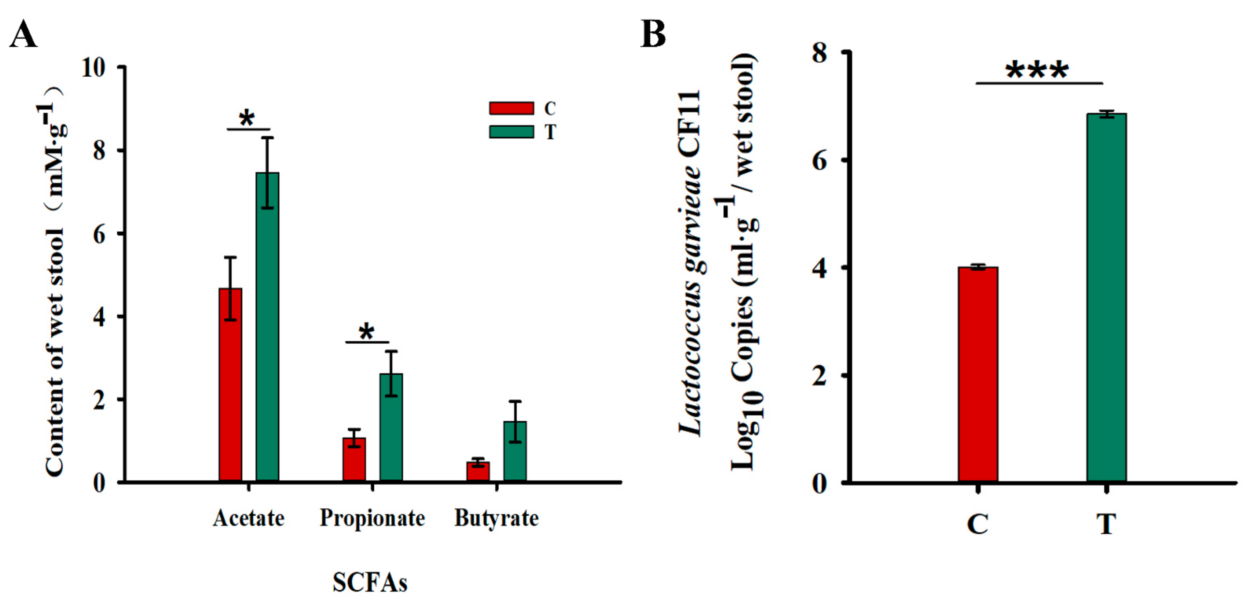
Increased in the Fecal Levels of Acetic Acid and Propionic Acids

To detect the potential impact of CF11 on the gut microbiome and their ability to produce SCFAs, we tested SCFA levels in the feces of SD rats. The average content of acetic, propionic, and butyric acids per gram of feces was 7.45 ± 0.84, 2.61 ± 0.54, and 1.45 ± 0.49 mM in the T (experimental) group and 4.67 ± 0.75, 1.07 ± 0.21, and 0.48 ± 0.09 mM in the C (control) group, respectively. CF11 significantly augmented the population of SCFAs in feces (Figure 1A). The increase in SCFAs can affect human health in several ways, including energy metabolism and antioxidant damage in the body. Furthermore, the population of L. petauri CF11 in the feces of SD rats was determined using qPCR. In each gram of wet stool, the population of CF11 Log10 16s RNA gene copies was 4.01 ± 0.04 (CT value: 25.92 ± 0.12) in the control group and 6.85 ± 0.06 (CT value:16.56 ± 0.19) in the experimental group (Figure 1B). It was indicated that the experimental group had a significantly higher content of CF11 in feces of SD rats after CF11 intervention, compared with the control group (p < 0.001).

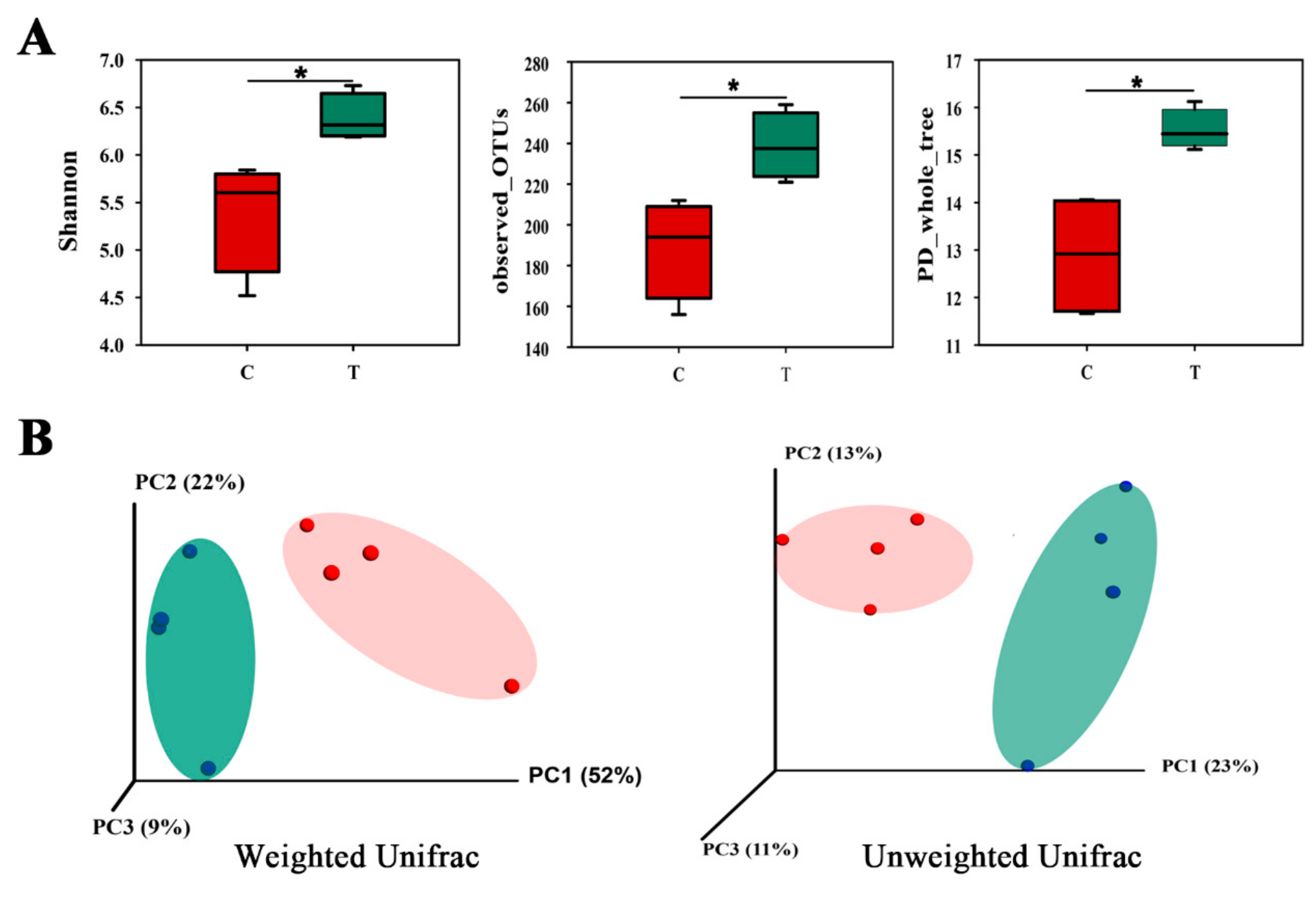
11. Gut Microbial Community in Two Groups

Our study revealed that the experimental group had prominently higher diversity values and richness estimates in comparison with the control group (Figure 2A). Observed_OTUs, PD_whole_tree, and Shannon index values have p values of 0.014, 0.029, and 0.021, respectively. The results signified that CF11 interference had an obvious effect on improving the abundance and diversity of intestinal flora. We performed principal component analysis (PCA) of the Unifrac distance by QIIME, and clustering results of the two groups were obtained (Figure 3B). Each point in the figure represented a sample, and two groups of samples were divided into two clusters at the Weighted_Unifrac distance, suggesting that there were differences between the experimental group and the control group. Changes in the composition and increased diversity of the intestinal flora may directly or indirectly cause the altered production and metabolism of SCFAs.
In light of the taxonomy profiles at the phylum level, it was shown how, in the experimental group, Bacteroidetes, Firmicutes, and Proteobacteria dominated the intestinal bacteria community with an average of 42.3%, 48.1%, and 7.8%, respectively, while in the control group, it was dominated by Bacteroidetes, Firmicutes, and Proteobacteria with means of 40.9%, 52.7%, and 3.7%. In contrast, Proteobacteria decreased by 4.1% and Firmicutes increased by 4.6% (Table 1). There were continuous changes toward the increased Bacteroidetes and Proteobacteria, but oppositely, the Phylum Firmicutes decreased between the control and experimental groups, revealing how CF11 influenced the gut microbiota. Major changes in the bacteria family occurred after 21 days of the CF11 intervention, and the percentages of Lactobacillaceae, Streptococcaceae, Ruminococcaceae, and Desulfovibrionaceae were significantly different (Figure 3A). The Ward method was used to analyze heatmap clustering of the two groups of fecal flora structures. The present results indicated that Oscillospira, Coprococcus, Ruminococcus, Desulfovibrio, and Dorea significantly increased, while Lactobacillus, Butyricimonas, and Sneathia decreased in the experimental group (Figure 3B).

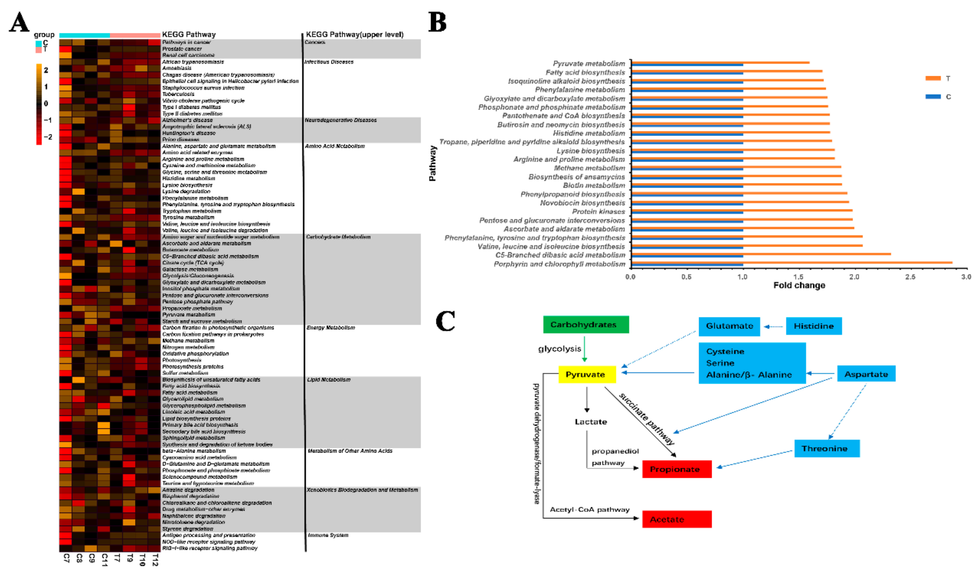
12. Genes Related to SCFA Generation

We used the PICRUSt algorithm to explore the function of the microbiome with the inferred metagenomes. Among the tested 262 KEGG pathways, there were differences in the abundance of 180 pathways between the experimental group and the control group with p < 0.05; these included pathways relating to cell growth and death, cell motility, transport, and catabolism, signaling molecules and interaction, replication and repair, transcription, cancers, infectious diseases, metabolic diseases, amino acid metabolism, carbohydrate metabolism, energy metabolism, and so on. Interestingly, in the experimental group, pathways associated with infectious diseases and the biosynthesis of other secondary metabolites were depleted, while lipid and energy metabolism was enriched. Many of these pathways were involved in the bacterial genus altered in CF11 gavages in Sprague Dawley rats (Figure 4A). For example, the experimental group that underwent the CF11 intervention may positively be associated with fatty acid biosynthesis and Citrate cycle (TCA cycle) pathways. Furthermore, the top-five clearly enriched pathways regarding metabolism included porphyrin and chlorophyll metabolism; C5-Branched dibasic acid metabolism; valine, leucine, and isoleucine biosynthesis; phenylalanine, tyrosine, and tryptophan biosynthesis; and ascorbate and aldarate metabolism (Figure 4B). The above results show that CF11 can cause secondary changes in the metabolic status of the body by altering the composition of the intestinal flora, thus affecting the disease and health status of the body.

13. Discussion

Microbial production of SCFAs is regarded to benefit the host, for the reason that these compounds prevent pathogens and lower cholesterol synthesis [24], stimulate a cellular immune response [25], provide muscle energy to enhance contraction [26], and serve as an umbrella to protect the colon against cancer development [27]. In C57BL/6 J mice, several original human probiotics, such as the Lactobacillus and Enterococcus strains, can significantly increase propionate and butyrate, and the production of acetate and propionate remarkably increased during fecal slurry fermentation compared with the intervention without probiotics [7]. Different carbohydrates were selected as substrates for fermentation, resulting in different acid production capacities. It was found that rhamnose had the greatest capacity of producing propionic acid, and α-and β-glucans gave rise to higher butyrate production [28]. In this study, the content of acetic acid and propionic acid in the feces of the experimental group was significantly higher than that of the control group. The result indicated that CF11 may modulate metabolic activity and improve the production of SCFAs in the gut, which is beneficial to the host.
As a result of the difference in fecal levels of acetic acid and propionic acid, certain shifts of the gut microbial community were observed in the experimental group. Interestingly, we found enhanced Oscillospira, Coprococcus, Ruminococcus, Lactobacillaceae, and Streptococcaceae in rats, among which Oscillospira, Coprococcus, and Ruminococcus can produce short-chain fatty acids [29,30]. Lactobacillaceae comprises well-known probiotic bacteria that are considered safe and well suited for the intestinal environment [31]. Lactobacillaceae is regarded as a machine in humans and animals that continuously generates energy by way of increasing SCFAs, especially acetate, propionate, and butyrate in the large intestine [32]. Streptococcaceae has been reported to possess the capacity of producing acetic acid [33]. Increased Bacteroidetes were also found in the experimental group. It has been previously confirmed that bacteria from Bacteroidetes can fermentate fiber into acetates and propionates with high efficiency in the gut [34,35,36]. Thus, our study suggested that CF11 treatment can improve the abundance of some SCFA producers. One possible reason is that some metabolites from CF11 could be used by those producers. Another one is that the intervention of CF11 changed the intestinal environment of rats, making it more suitable for the growth of SCFA producers. Detailed mechanisms should be further investigated in future studies.
PICRUSt provides an expedient method for the prediction of the function of metagenomes with a reference genome database. It helps researchers to delve into the predicted functional potential of the studied microbiome and form function-based assumptions that lay a solid foundation for future research. It has been emphasized that microbial communities are of great importance in human disease, the biosynthesis and degradation of natural products, and other metabolism processes [3]. In our study, PICRUSt was utilized to predict the population and abundances of functional genes of microbial communities in SD rats with a CF11 intervention, and a few appealing genes were found related to (1) human diseases, (2) amino acid metabolism, (3) carbohydrate metabolism, (4) energy metabolism, (5) lipid metabolism, (6) the metabolism of other amino acids, (7) xenobiotics biodegradation and metabolism, and (8) the immune system. We also ascertained that in the course of the TCA cycle, carbohydrate metabolism, fatty acid biosynthesis and metabolism, and amino acid metabolism, there are many enriched pathways associated with SCFAs (mainly acetate and propionate acids) [29,33,37,38]. Based on our pathway enrichment analysis, we can conclude that amino acid fermentation and carbohydrate glycolysis fermentation could strengthen pyruvate metabolism and biosynthesis, which might enhance the production of acetyl-CoA and improve the conversion from acetyl-CoA to acetic acid. This process may be strengthened through the enriched pathway of “acetate generation from acetyl-CoA I”, and then enrich the pathway of succinate and propanediol to produce propionate (Figure 4C). Then, the accelerated conversion from pyruvate to acetic acid or propionic acid would make the concentration of these two SCFAs much higher in the intestines of rats [39]. Considering that the enriched genera of rats’ gut microbiota had a close relationship with SCFAs production, this dramatic increase in metabolic potential in the production of SCFAs may be due to changes in the microbial community.
In animal studies, some L. garvieae strains can bring about Lactococcosis in marine or freshwater fish [40], mastitis in cows [41,42], and pneumonia in pigs [43]. In human studies, Lactococcus spp. has been isolated from healthy people, and many researchers have confirmed that Lactococcus spp. is probiotic [44,45]. Lactococcus spp. are commonly used to manufacture varieties of cheeses and fermented dairy products in the dairy industry [14,46]. Ref. [22] identified that Lactococcus garvieae A1 isolated from the soil is a probiotic bacterium. In previous studies, Lactococcus petauri strains were isolated from an abscess of a sugar glider (Goodman et al., 2017) and a rainbow trout suffering from Lactococcosis [16]. However, no studies have investigated L. petauri isolated from the gut microbiota of healthy people and confirmed its beneficial function and ability to alter gut microbiota. In our study, CF11 could enhance microbial diversity and the production of SCFAs. In line with the WHO/FAO, a probiotic is a kind of living microorganism that is good for the host when presented in sufficient amounts [47]. In this sense, Lactococcus petauri CF11 could be a potential probiotic.

14. Conclusions

The present study suggests that L. petauri CF11 significantly modifies the composition of rats’ gut microbial community by enhancing SCFA producers, which also leads to increased production of total SCFAs, acetic acids, and propionic acids. The PICRUSt analysis further confirmed the vital role of the gut microbiota in SCFA synthesis and metabolism. We can conclude from these results that L. petauri CF11 is a potential probiotic. Further study should be performed to investigate whether L. petauri CF11 is safe for the host.

Author Contributions

Conceptualization, S.F. and G.Z.; investigation, S.F., T.Q. and T.Y.; methodology, G.Z.; supervision, G.Z.; writing—review and editing, S.F. and T.Q. All authors have read and agreed to the published version of the manuscript.

Funding

This research was funded by the National Science Foundation of China (NSFC 31500076 and 82173476), the Science and Technology Program of Guangzhou, China (201904010161), and the Guangzhou Municipal Science and Technology Project (20191A011063). The authors would like to express their appreciation to Weide Zhang in Southern Medical University for helping in the SCFAs detection.

Institutional Review Board Statement

The study was conducted in accordance with the Declaration of Helsinki, and approved by the Ethical Committee of Southern Medical University, Guangzhou for studies involving animals. The protocol was approved by the Southern Medical University Ethics Review Board (Ethics no, L2018198).

Informed Consent Statement

Not applicable.

Data Availability Statement

Informed consent was obtained from all subjects involved in the study. The datasets generated for this study can be found in GenBank. The 16S rRNA gene sequence of strain CF11 has been deposited in the GenBank database under the accession number MH986337. The data reported in this paper have been deposited in the Sequence Read Archive (SRA) database, www.ncbi.nlm.nih.gov/sra (accession no. PRJEB30830 (accessed on 14 March 2022)).

Conflicts of Interest

The authors declare no conflict of interest.

References

  1. Sun, J.; Kato, I. Gut microbiota, inflammation and colorectal cancer. Genes Dis. 2016, 3, 130–143. [Google Scholar] [CrossRef] [Green Version]
  2. Tan, J.; McKenzie, C.; Potamitis, M.; Thorburn, A.N.; Mackay, C.R.; Macia, L. The Role of Short-Chain Fatty Acids in Health and Disease. Adv. Immunol. 2014, 121, 91–119. [Google Scholar] [CrossRef] [PubMed]
  3. Jiang, H.-Y.; Zhang, X.; Yu, Z.-H.; Zhang, Z.; Deng, M.; Zhao, J.-H.; Ruan, B. Altered gut microbiota profile in patients with generalized anxiety disorder. J. Psychiatr. Res. 2018, 104, 130–136. [Google Scholar] [CrossRef] [PubMed]
  4. Zhai, S.; Zhu, L.; Qin, S.; Li, L. Effect of lactulose intervention on gut microbiota and short chain fatty acid composition of C57 BL/6J mice. MicrobiologyOpen 2018, 7, e00612. [Google Scholar] [CrossRef] [PubMed] [Green Version]
  5. Melbye, P.; Olsson, A.; Hansen, T.H.; Søndergaard, H.B.; Oturai, A.B. Short-chain fatty acids and gut microbiota in multiple sclerosis. Acta Neurol. Scand. 2018, 139, 208–219. [Google Scholar] [CrossRef] [PubMed]
  6. Murphy, P.; Bs, J.H.; Ang, B.; Wolfson, T.; Gamst, A.; Bydder, M.; Middleton, M.; Behling, C.; Loomba, R.; Sirlin, C. Associations between histologic features of nonalcoholic fatty liver disease (NAFLD) and quantitative diffusion-weighted MRI measurements in adults. J. Magn. Reson. Imaging 2014, 41, 1629–1638. [Google Scholar] [CrossRef] [PubMed] [Green Version]
  7. Nagpal, R.; Wang, S.; Ahmadi, S.; Hayes, J.; Gagliano, J.; Subashchandrabose, S.; Kitzman, D.W.; Becton, T.; Read, R.; Yadav, H. Human-origin probiotic cocktail increases short-chain fatty acid production via modulation of mice and human gut microbiome. Sci. Rep. 2018, 8, 12649. [Google Scholar] [CrossRef] [PubMed] [Green Version]
  8. Braniste, V.; Al-Asmakh, M.; Kowal, C.; Anuar, F.; Abbaspour, A.; Tóth, M.; Korecka, A.; Bakocevic, N.; Ng, L.G.; Kundu, P.; et al. The gut microbiota influences blood-brain barrier permeability in mice. Sci. Transl. Med. 2014, 6, 263ra158. [Google Scholar] [CrossRef] [Green Version]
  9. Nikbakht, E.; Khalesi, S.; Singh, I.; Williams, L.T.; West, N.P.; Colson, N. Effect of probiotics and synbiotics on blood glucose: A systematic review and meta-analysis of controlled trials. Eur. J. Nutr. 2016, 57, 95–106. [Google Scholar] [CrossRef]
  10. Varsha, K.K.; Nampoothiri, K. Lactococcus garvieae subsp. bovis subsp. nov., lactic acid bacteria isolated from wild gaur (Bos gaurus) dung, and description of Lactococcus garvieae subsp. garvieae subsp. nov. Int. J. Syst. Evol. Microbiol. 2016, 66, 3805–3809. [Google Scholar] [CrossRef] [PubMed]
  11. Meyburgh, C.M.; Bragg, R.R.; Boucher, C.E. Lactococcus garvieae: An emerging bacterial pathogen of fish. Dis. Aquat. Org. 2017, 123, 67–79. [Google Scholar] [CrossRef]
  12. López-Campos, G.; Aguado-Urda, M.; Blanco, M.M.; Gibello, A.; Cutuli, M.T.; López-Alonso, V.; Martín-Sánchez, F.; Fernández-Garayzábal, J.F. Lactococcus garvieae: A small bacteria and a big data world. Health Inf. Sci. Syst. 2015, 3, S5. [Google Scholar] [CrossRef] [Green Version]
  13. Abdelfatah, E.N.; Mahboub, H.H.H. Studies on the effect ofLactococcus garvieaeof dairy origin on both cheese and Nile tilapia (O. niloticus). Int. J. Vet. Sci. Med. 2018, 6, 201–207. [Google Scholar] [CrossRef] [Green Version]
  14. Moumene, M.; Drissi, F.; Croce, O.; Djebbari, B.; Robert, C.; Angelakis, E.; Benouareth, D.; Raoult, D.; Merhej, V. Complete genome sequence and description of Lactococcus garvieae M14 isolated from Algerian fermented milk. New Microbes New Infect. 2016, 10, 122–131. [Google Scholar] [CrossRef] [Green Version]
  15. Goodman, L.B.; Lawton, M.R.; Franklin-Guild, R.J.; Anderson, R.R.; Schaan, L.; Thachil, A.J.; Wiedmann, M.; Miller, C.B.; Alcaine, S.D.; Kovač, J. Lactococcus petauri sp. nov., isolated from an abscess of a sugar glider. Int. J. Syst. Evol. Microbiol. 2017, 67, 4397–4404. [Google Scholar] [CrossRef] [Green Version]
  16. Kotzamanidis, C.; Malousi, A.; Bitchava, K.; Vafeas, G.; Chatzidimitriou, D.; Skoura, L.; Papadimitriou, E.; Chatzopoulou, F.; Zdragas, A. First Report of Isolation and Genome Sequence of L. petauri Strain from a Rainbow Trout Lactococcosis Outbreak. Curr. Microbiol. 2020, 77, 1089–1096. [Google Scholar] [CrossRef]
  17. Tang, W.H.W.; Wang, Z.; Levison, B.S.; Koeth, R.A.; Britt, E.B.; Fu, X.; Wu, Y.; Hazen, S.L. Intestinal Microbial Metabolism of Phosphatidylcholine and Cardiovascular Risk. N. Engl. J. Med. 2013, 368, 1575–1584. [Google Scholar] [CrossRef] [Green Version]
  18. Jung, M.; Chang, Y.-H.; Kim, W. A real-time PCR assay for detection and quantification ofLactococcus garvieae. J. Appl. Microbiol. 2010, 108, 1694–1701. [Google Scholar] [CrossRef]
  19. Chen, Y.-R.; Fang, S.-T.; Liu, H.-Y.; Zheng, H.-M.; He, Y.; Chen, Z.-W.; Chen, M.-X.; Zhang, G.-X.; Zhou, H.-W. Degradation of trimethylamine in vitro and in vivo by Enterococcus faecalis isolated from healthy human gut. Int. Biodeterior. Biodegrad. 2018, 135, 24–32. [Google Scholar] [CrossRef]
  20. Wu, J.; Peters, A.B.; Dominianni, C.; Zhang, Y.; Pei, Z.; Yang, L.; Ma, Y.; Purdue, M.P.; Jacobs, E.J.; Gapstur, S.M.; et al. Cigarette smoking and the oral microbiome in a large study of American adults. ISME J. 2016, 10, 2435–2446. [Google Scholar] [CrossRef]
  21. Langille, M.G.I.; Zaneveld, J.; Caporaso, J.G.; McDonald, D.; Knights, D.; Reyes, J.A.; Clemente, J.C.; Burkepile, D.E.; Vega Thurber, R.L.; Knight, R.; et al. Predictive functional profiling of microbial communities using 16S rRNA marker gene sequences. Nat. Biotechnol. 2013, 31, 814–821. [Google Scholar] [CrossRef] [PubMed]
  22. Altın, G.; Nikerel, E.; Şahin, F. Draft Genome Sequence of Magnesium-Dissolving Lactococcus garvieae A1, Isolated from Soil. Genome Announc. 2017, 5, e00386-17. [Google Scholar] [CrossRef] [Green Version]
  23. Wang, F.; Men, X.; Zhang, G.; Liang, K.; Xin, Y.; Wang, J.; Li, A.; Zhang, H.; Liu, H.; Wu, L. Assessment of 16S rRNA gene primers for studying bacterial community structure and function of aging flue-cured tobaccos. AMB Express 2018, 8, 182. [Google Scholar] [CrossRef] [PubMed]
  24. Sivieri, K.; Morales, M.L.V.; Adorno, M.A.T.; Sakamoto, I.K.; Saad, S.M.I.; Rossi, A.E. Lactobacillus acidophilus CRL 1014 improved “gut health” in the SHIME®reactor. BMC Gastroenterol. 2013, 13, 100. [Google Scholar] [CrossRef] [PubMed]
  25. Bhutia, Y.D.; Ganapathy, V. Short, but Smart: SCFAs Train T Cells in the Gut to Fight Autoimmunity in the Brain. Immunity 2015, 43, 629–631. [Google Scholar] [CrossRef] [PubMed] [Green Version]
  26. Den Besten, G.; van Eunen, K.; Groen, A.K.; Venema, K.; Reijngoud, D.-J.; Bakker, B.M. The role of short-chain fatty acids in the interplay between diet, gut microbiota, and host energy metabolism. J. Lipid Res. 2013, 54, 2325–2340. [Google Scholar] [CrossRef] [PubMed] [Green Version]
  27. Fellows, R.; Denizot, J.; Stellato, C.; Cuomo, A.; Jain, P.; Stoyanova, E.; Balázsi, S.; Hajnády, Z.; Liebert, A.; Kazakevych, J.; et al. Microbiota derived short chain fatty acids promote histone crotonylation in the colon through histone deacetylases. Nat. Commun. 2018, 9, 105. [Google Scholar] [CrossRef] [PubMed]
  28. Reichardt, N.; Vollmer, M.; Holtrop, G.; Farquharson, F.M.; Wefers, D.; Bunzel, M.; Duncan, S.H.; Drew, J.E.; Williams, L.M.; Milligan, G.; et al. Specific substrate-driven changes in human faecal microbiota composition contrast with functional redundancy in short-chain fatty acid production. ISME J. 2018, 12, 610–622. [Google Scholar] [CrossRef] [PubMed]
  29. Louis, P.; Flint, H.J. Formation of propionate and butyrate by the human colonic microbiota. Environ. Microbiol. 2016, 19, 29–41. [Google Scholar] [CrossRef] [PubMed] [Green Version]
  30. Bernalier-Donadille, A. Fermentative metabolism by the human gut microbiota. Gastroenterol. Clin. Biol. 2010, 34, S16–S22. [Google Scholar] [CrossRef]
  31. Etzold, S.; Kober, O.I.; MacKenzie, D.A.; Tailford, L.E.; Gunning, A.P.; Walshaw, J.; Hemmings, A.M.; Juge, N. Structural basis for adaptation of lactobacilli to gastrointestinal mucus. Environ. Microbiol. 2014, 16, 888–903. [Google Scholar] [CrossRef] [PubMed]
  32. Guerra-Ordaz, A.A.; González-Ortiz, G.; La Ragione, R.M.; Woodward, M.J.; Collins, J.W.; Pérez, J.F.; Martín-Orúe, S.M. Lactulose and Lactobacillus plantarum, a Potential Complementary Synbiotic To Control Postweaning Colibacillosis in Piglets. Appl. Environ. Microbiol. 2014, 80, 4879–4886. [Google Scholar] [CrossRef] [PubMed] [Green Version]
  33. Koh, A.; De Vadder, F.; Kovatcheva-Datchary, P.; Bäckhed, F. From Dietary Fiber to Host Physiology: Short-Chain Fatty Acids as Key Bacterial Metabolites. Cell 2016, 165, 1332–1345. [Google Scholar] [CrossRef] [PubMed] [Green Version]
  34. Flint, H.J.; Duncan, S.; Scott, K.P.; Louis, P. Interactions and competition within the microbial community of the human colon: Links between diet and health. Environ. Microbiol. 2007, 9, 1101–1111. [Google Scholar] [CrossRef] [PubMed]
  35. Louis, P.; Scott, K.P.; Duncan, S.H.; Flint, H.J. Understanding the effects of diet on bacterial metabolism in the large intestine. J. Appl. Microbiol. 2007, 102, 1197–1208. [Google Scholar] [CrossRef] [PubMed]
  36. Pryde, S.E.; Duncan, S.H.; Hold, G.L.; Stewart, C.S.; Flint, H.J. The microbiology of butyrate formation in the human colon. FEMS Microbiol. Lett. 2002, 217, 133–139. [Google Scholar] [CrossRef] [PubMed]
  37. Chan, S.H.J.; Nørregaard, L.; Solem, C.; Jensen, P.R. Acetate Kinase Isozymes Confer Robustness in Acetate Metabolism. PLoS ONE 2014, 9, e92256. [Google Scholar] [CrossRef]
  38. Duncan, S.H.; Barcenilla, A.; Stewart, C.S.; Pryde, S.E.; Flint, H.J. Acetate Utilization and Butyryl Coenzyme A (CoA):Acetate-CoA Transferase in Butyrate-Producing Bacteria from the Human Large Intestine. Appl. Environ. Microbiol. 2002, 68, 5186–5190. [Google Scholar] [CrossRef] [PubMed] [Green Version]
  39. Li, J.; Sung, C.Y.J.; Lee, N.; Ni, Y.; Pihlajamäki, J.; Panagiotou, G.; El-Nezami, H. Probiotics modulated gut microbiota suppresses hepatocellular carcinoma growth in mice. Proc. Natl. Acad. Sci. USA 2016, 113, E1306–E1315. [Google Scholar] [CrossRef] [PubMed] [Green Version]
  40. Tandel, K.; Bhatt, P.; Ranjan, P.; Rathi, K. Meningitis caused by Lactococcus garvieae. Med. J. Armed Forces India 2015, 73, 94–96. [Google Scholar] [CrossRef] [PubMed] [Green Version]
  41. Teixeira, L.M.; Merquior, V.L.C.; Vianni, M.D.C.E.; Carvalho, M.D.G.S.; Fracalanzza, S.E.L.; Steigerwalt, A.G.; Brenner, D.J.; Facklam, R.R. Phenotypic and Genotypic Characterization of Atypical Lactococcus garvieae Strains Isolated from Water Buffalos with Subclinical Mastitis and Confirmation of L. garvieae as a Senior Subjective Synonym of Enterococcus seriolicida. Int. J. Syst. Bacteriol. 1996, 46, 664–668. [Google Scholar] [CrossRef] [Green Version]
  42. Tymoszewska, A.; Diep, D.B.; Aleksandrzak-Piekarczyk, T. The extracellular loop of Man-PTS subunit IID is responsible for the sensitivity of Lactococcus garvieae to garvicins A, B and C. Sci. Rep. 2018, 8, 15790. [Google Scholar] [CrossRef] [Green Version]
  43. Tejedor, J.; Vela, A.; Gibello, A.; Casamayor, A.; Domínguez, L.; Fernandez-Garayzabal, J.F. A genetic comparison of pig, cow and trout isolates of Lactococcus garvieae by PFGE analysis. Lett. Appl. Microbiol. 2011, 53, 614–619. [Google Scholar] [CrossRef]
  44. Kumar, A.; Kundu, S.; Debnath, M. Effects of the probiotics Lactococcus lacttis (MTCC-440) on Salmonella enteric serovar Typhi in co-culture study. Microb. Pathog. 2018, 120, 42–46. [Google Scholar] [CrossRef]
  45. Shin, H.-S.; Baek, D.-H.; Lee, S.-H. Inhibitory effect of Lactococcus lactis on the bioactivity of periodontopathogens. J. Gen. Appl. Microbiol. 2018, 64, 55–61. [Google Scholar] [CrossRef] [Green Version]
  46. Haghshenas, B.; Abdullah, N.; Nami, Y.; Radiah, D.; Rosli, R.; Khosroushahi, A.Y. Different effects of two newly-isolated probiotic Lactobacillus plantarum 15HN and Lactococcus lactis subsp. Lactis 44Lac strains from traditional dairy products on cancer cell lines. Anaerobe 2014, 30, 51–59. [Google Scholar] [CrossRef]
  47. Leblanc, J.G.; Chain, F.; Martín, R.; Bermúdez-Humarán, L.G.; Courau, S.; Langella, P. Beneficial effects on host energy metabolism of short-chain fatty acids and vitamins produced by commensal and probiotic bacteria. Microb. Cell Fact. 2017, 16, 79. [Google Scholar] [CrossRef] [Green Version]
Figure 1. (A) Content of SCFAs in fecal feces of SD rats. (B) The population of CF11 Log10 16s rRNA gene copies. Rats in the experimental group (T) were given the mixed CF11 every day. The control group (C) rats were given the same volume of PBS (phosphate buffer saline) every day. Each group n = 4; * p value < 0.05; *** p value < 0.001.
Processes 10 00604 g001
Figure 2. Alpha-diversity and Beta-diversity of Sprague Dawley Rats after CF11 intervention for 21 days. (A) Alpha-diversity index (Shannon index, observed_OTUs, and PD_whole_tree) of the gut microbiome in Sprague Dawley Rats, CF11 intervention for 21 days. (B) Weighted and unweighted Unifrac distance. Microbial richness estimates and diversity indices were measured at OTUs definition of >97% identity. C (control group, day 21), T (experimental group, day 21), n = 4 each group; Data were analyzed by one-way ANOVA. * p Value < 0.05.
Processes 10 00604 g002
Figure 3. Changes in microbial community compositions of the gut between two groups. (A) Major changes in bacteria family after CF11 intervention for 21 days. (C: C7, C8, C9, and C11 control group, day 21; T: T7, T9, T10, and T12, experimental group, day 21). (B) Heatmap of the percentage abundance of microbial taxa found in the fecal microbial communities of 8 samples.
Processes 10 00604 g003
Figure 4. Gene functional pathways and metabolic pathways related to CF11 intervention. (A) Bacterial taxa associated with CF11 intervention are related to several gene functional pathways. Bacterial gene functions were predicted from 16S rRNA gene-based microbial compositions using the PICRUSt algorithm to make inferences from KEGG-annotated databases. KEGG pathway counts were normalized for DESeq2 size factors and adjusted for the dataset using the ‘remove Batch Effect’ function. Only KEGG pathways relating to diseases, amino acid metabolism, carbohydrate metabolism, energy metabolism, lipid metabolism, metabolism of other amino acids, xenobiotics biodegradation and metabolism, and immune system are included in the heatmap; full lists of genera and KEGG pathways associated with smoking can be found in tables. (B) Significantly enriched pathways regarding metabolism; control group, the fold-change would be set to 1. (C) Enriched pathways related to pyruvate fermentation and SCFAs. Amino acid fermentation pathways in light blue, carbohydrate fermentation to pyruvate via glycolysis is shown in green. Dotted lines indicate that several intermediate steps are involved.
Processes 10 00604 g004
Table 1. Relative abundance of the major phyla in the experimental group and control group.
PhylumC (Control Group) %StdT (Experimental Group) %Std
Bacteriodetes40.90.1242.30.09
Firmicutes52.70.1248.10.07
Proteobacteria3.70.027.80.03
others2.70.011.80.01
Publisher’s Note: MDPI stays neutral with regard to jurisdictional claims in published maps and institutional affiliations.

