Article

Adjusting the Structure of a Peptide Nucleic Acid (PNA) Molecular Beacon and Promoting Its DNA Detection by a Hybrid with Quencher-Modified DNA

1
Health and Medical Research Institute, National Institute of Advanced Industrial Science and Technology (AIST), 2217-14 Hayashi-cho, Takamatsu 761-0395, Japan
2
Department of Applied Chemistry, Kindai University, 3-4-1 Kowakae, Higashi-Osaka 577-8502, Japan
3
Department of Interdisciplinary Science and Engineering in Health Systems, Okayama University, 3-1-1 Tsushimanaka, Okayama 700-8530, Japan
*
Author to whom correspondence should be addressed.
Processes 2022, 10(4), 722; https://doi.org/10.3390/pr10040722
Submission received: 18 March 2022 / Revised: 3 April 2022 / Accepted: 6 April 2022 / Published: 8 April 2022
(This article belongs to the Special Issue The Amazing World of Peptide Engineering)

Abstract

:
In this study, we performed an elaborate adjustment of the structure of peptide nucleic acid (PNA) molecular beacons as probes for detecting nucleic acids. We synthesized the PNA beacons with various numbers of Glu, Lys, and dabcyl (Dab) quenchers in them, and we investigated their fluorescence changes (F1/1/F0) with and without full-match DNA. As the numbers of Glu/Lys or Dab increased, the F1/1/F0 tended to decrease. Among the different beacons, the PNA beacon with one Glu and one Lys (P1Q1) showed the largest F1/1/F0. On the other hand, a relatively large F1/1/F0 was obtained when the number of Glu/Lys and the number of Dab were the same, and the balance between the numbers of Glu/Lys and Dab seemed to affect the F1/1/F0. We also investigated the DNA detection by the prehybrid of P1Q1, which consists of the T790M base sequence, [P1Q1(T790M)], with quencher-modified DNA (Q-DNA). We examined the DNA detection with single-base mismatch by P1Q1(T790M), and we clarified that there was difficulty in detecting the sequence with P1Q1 alone, but that the sequence was successfully detected by the prehybrid of P1Q1 with the Q-DNA.

1. Introduction

Peptide nucleic acid (PNA) [1] is a nucleic acid surrogate that forms a stable-base sequence-specific hybrid with DNA and RNA [2]. Since PNA consists of an uncharged amide backbone, PNA/DNA and PNA/RNA hybrids do not exhibit electrostatic repulsion, in contrast to DNA/DNA and DNA/RNA hybrids, which results in greater stability. Therefore, it is expected that PNA can be used as a probe for detecting nucleic acids, and colorimetric detection using dye [3,4,5,6,7,8,9,10] and mass-based [11,12], visual [13], electrochemical [11,14,15,16,17,18,19,20], and optical [18,21] detection, among others, has been reported. Furthermore, the detection of nucleic acids on the basis of fluorescence by conjugating PNA and fluorescent dyes is plausible as a highly sensitive detection method, and numerous studies that use these conjugates as probes to detect DNA and RNA have been reported [22].
Among these reported approaches, PNA molecular beacons (PNA beacons), in which the PNA is modified by a fluorescent dye and a quencher, are useful as illuminating probes for detecting DNA and RNA. A typical PNA beacon contains one fluorescent dye (Fam) and one Dab quencher at both termini, and it contains, adjacent to these groups, one negatively (Glu) and one positively (Lys) charged amino acid residue [23,24,25,26,27,28,29,30,31,32]. Before the PNA beacon hybridizes with DNA or RNA, the Glu and the Lys are close to each other because of the intramolecular electrostatic interaction; as a result, the fluorescent dye and the quencher are also close to each other, and the PNA beacon is quenched. On the other hand, as the PNA beacon forms a hybrid with the target DNA and RNA, thereby relieving the interaction between the Glu and the Lys, the fluorescent dye and the quencher become more distant from each other, and the PNA beacon emits fluorescence.
In this work, we consider that the adjustment of the intramolecular electrostatic interaction by varying the numbers of Glu and Lys in the PNA beacon is important in order to obtain a better performance of the beacon. However, to the best of our knowledge, no discussion has yet been conducted on the numbers of Glu/Lys in this context. Therefore, we prepared PNA beacons that contain various numbers of Glu/Lys, while also varying the number of Dab quenchers, in order to obtain a PNA beacon that fluoresces more effectively when hybridized with nucleic acid. We also assessed the fluorescence characteristics in this study. In addition, we recently reported a system in which a hybrid of Fam-modified PNA (Fl-PNA) and Dab-modified DNA (Q-DNA) detected a target nucleic acid by emitting light from the Fl-PNA via strand exchange with the target nucleic acid [33,34,35,36,37]. In this report, we describe that the use of the PNA beacon, instead of the Fl-PNA, further improves nucleic acid detection.

2. Materials and Methods

2.1. Materials

9-Fluorenylmethyloxycarbonyl group (Fmoc)-derivatized amino acids, Fmoc-derivatized super acid labile-poly(ethylene)glycol (Fmoc-NH-SAL-PEG) resin, piperidine, O-(1H-benzotriazol-1-yl)-N,N,N′, N′-tetramethyluronium hexafluorophosphate (HBTU), N-methylmorpholine (NMM), trifluoroacetic acid (TFA), and triisopropylsilane (TIPS) were purchased from Watanabe Chemicals (Hiroshima, Japan). Fmoc-derivatized PNA monomers [Fmoc-A(Bhoc)-OH, Fmoc-T-OH, Fmoc-G(Bhoc)-OH, and Fmoc-C(Bhoc)-OH] were purchased from Panagene (Daejeon, Korea). Fluorescent dye (5(6)-carboxyfluorescein (Fam)), and Fmoc-Lys(dabcyl)-OH as a Dab quencher, were purchased from AAT Bioquest (Sunnyvale, CA, USA). Fmoc-derivatized ethylene glycol linker, containing six ethylene glycol units ((Fmoc-amino)-PEG6-carboxylic acid), was purchased from the Tokyo Chemical Industry (Tokyo, Japan). N,N′-Dimethylformamide (DMF), N-methyl-2-pyrrolidinone (NMP), diethyl ether, and acetonitrile were purchased from the FUJIFILM Wako Pure Chemical Corporation (Osaka, Japan). Phosphate-buffered saline (PBS) (100 mM; pH: 7.0) was purchased from Nacalai Tesque (Kyoto, Japan). DNA oligomers were purchased from Thermo Fisher Scientific (Waltham, MA, USA).

2.2. Peptide Synthesis

All PNA beacons [PnQms, P1Q1(T790M), P1Q1(L858R), and P1Q1(exon19del); Figure 1] were synthesized on Fmoc-NH-SAL-PEG resin, containing 7.2 μmol Fmoc on its surface, by using standard Fmoc protection chemistry. The deprotection and coupling processes were carried out at room temperature. The Fmoc group was removed by using 20% piperidine in DMF for 7 min. Each coupling process included: 4 parts Fmoc-derivatized amino acids; Fmoc-derivatized PNA monomers; an Fmoc-derivatized ethylene glycol linker; a Fam or Dab quencher; 3.6 parts HBTU; and 11.5 90 min at room temperature. Crude peptides were precipitated in diethyl ether and were washed twice with diethyl ether until a neutral pH was reached. Peptides were then dried by parts, and NMM were dissolved in DMF/NMP and were added to the resin. The reaction mixture was then shaken for 45 min. Peptides on the resin were by treatment with 95:2.5:2.5 (v/v) TFA/TIPS/water, and they were dissolved in 50:50 (v/v) 0.1% aqueous TFA in water/acetonitrile. Peptides were purified using reverse-phase high-pressure liquid chromatography (HPLC) on a C18 preparative column (Cadenza 5CD-C18; Imtakt, Kyoto, Japan). The final product was identified by using matrix-assisted laser desorption/ionization–time-of-flight (MALDI-ToF) mass spectrometry (Shimadzu AXIMA Confidence) (Supplementary Material, Figures S1–S17) and HPLC on a C18 analytical column (Cadenza CD-C18; Imtakt) (Supplementary Material, Figures S18–S34).

2.3. Melting Curves of Mixtures of PNA Beacon and DNA

Molar concentrations of PNA beacons and DNAs were estimated from the absorbance at 260 nm and were measured by using a UV-vis spectrometer (JASCO V-560) with molar extinction coefficients of the nucleobases. The UV melting curves of the equimolar mixtures of P1Q1 or P4Q1, with and without full-match DNA (fmDNA: 5′-TCTGCTGGGT-3′) or scrambled DNA (scrDNA: 5′-CGTGGTTCTG-3′), in aqueous buffer (100 mM PBS; pH: 7.0), were measured by using a Shimadzu TMSPC-8 Tm Analysis System that was equipped with a UV-visible spectrometer (UV-1700) and an eight-position Peltier temperature controller. Each concentration of the PNA beacons was 5.0 μM, and the quartz cell length was 1 cm. The melting curves were recorded with the cooling of the solution by 0.5 °C/0.5 min, from 80 to 10 °C, while measuring the absorbance at 260 nm. The observed absorbance was normalized to that at 80 °C. The melting temperature, at which 50% of the strands remained hybridized (Tm), was obtained by using a TMSPC-8 with Tm analysis software.

2.4. Fluorescence Titration Curves of PNA Beacon and DNA

The fluorescence spectra of the 500 nM PnQm and DNA (fmDNA and scrDNA) mixtures, in aqueous buffer (100 mM PBS; pH: 7.0) at 25 °C, were measured at an excitation wavelength of 495 nm, and at emission wavelengths from 500 to 700 nm, by using a JASCO FP-8200 fluorescence spectrometer and a 1 cm quartz cell. In the case of the titration curves, the concentration of PnQm was maintained at 500 nM, while the concentrations of DNA were varied in order to create PnQm/DNA ratios of 0, 0.2, 0.4, 0.6, 0.8, 1.0, 1.2, 1.4, 1.6, 1.8, or 2.0. The fluorescence spectra were measured after the PnQm was incubated with DNA for 15 min. The fluorescence titration curves were recorded on the basis of the fluorescence intensities at 525 nm. The assessment of the DNA detection by PnQm was conducted by measuring the ratio (F/F0) of the fluorescence intensity (at 525 nm) of the PNA beacon without DNA (F0) to that of the PNA beacon with DNA (F). In the case of the fluorescence spectra of P1Q1(T790M), P1Q1(L858R), and P1Q1(exon19del) with DNA, we used T790M: 5′-CATCATGCAG-3′ L858R: 5′-GGGCGGGCCA-3′, and exon19del: 5′-ATCAAAACAT-3′ as the DNAs.

2.5. Relative Fluorescence of PNA Beacon and PNA Beacon/Q-DNA Hybrid with Target DNA

P1Q1(T790M) and 8-mer DNA containing a Dab quencher at the 3′-end (Q-DNA; 5′-TCATGCAG-Dab-3′) were mixed in PBS (each 500 nM). The mixture was incubated at 95 °C for 5 min and was then slowly cooled to 25 °C to form the hybrid. Then, the fluorescence spectra of the P1Q1(T790M)/Q-DNA hybrid were measured at 37 °C by using an excitation wavelength of 480 nm, and emission wavelengths from 500 to 700 nm. Next, a complementary DNA (5′-CAC CGT GCA ACT CAT CAT GCA GCT CAT GCC CTT C-3′) and one-base-mismatched DNA (5′-CAC CGT GCA ACT CAT CAC GCA GCT CAT GCC CTT C-3′) were mixed at 1:1 with the P1Q1(T790M)/Q-DNA hybrid, and fluorescence spectra were measured at 37 °C. Each concentration of DNA and the hybrid was 500 nM. The fluorescence spectrum of the P1Q1(T790M), instead of the P1Q1(T790M)/Q-DNA hybrid, was also measured in the same manner as described above. The DNA detection by P1Q1(T790M) and P1Q1(T790M)/Q-DNA was assessed by the same protocol, as described in Section 2.4.

3. Results

3.1. Hybrid Formation of DNA with PNA Beacon and Fluorescence Detection of DNA by PNA Beacon

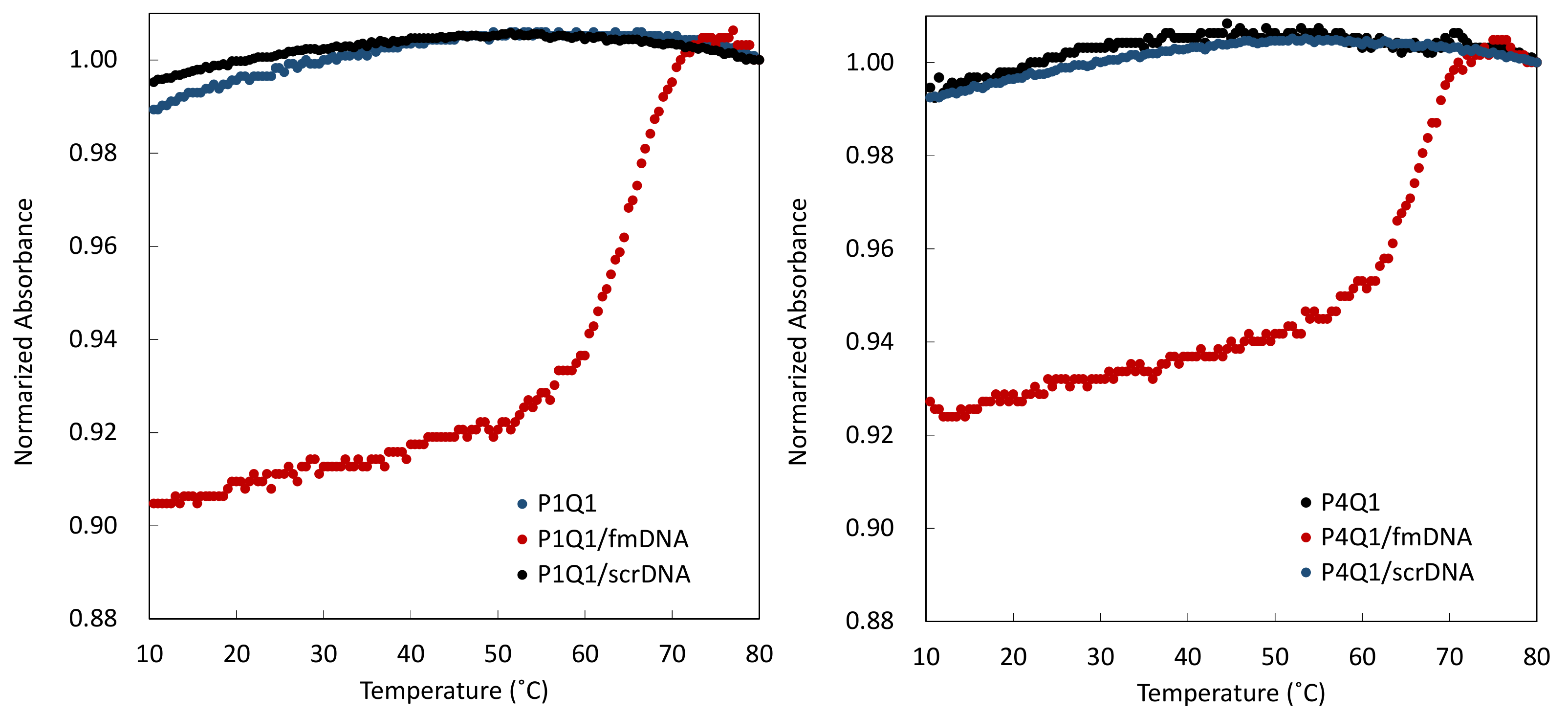
To confirm that the synthesized PNA beacons (PnQms) formed hybrids with the DNA, we measured the UV melting curves of a 1:1 mixture of the P1Q1 and DNAs (left panel in Figure 2). A clear sigmoid curve was observed for the mixture of P1Q1 and full-match DNA (fmDNA), which indicates that they formed a hybrid (red-filled circles: Tm = 65 °C). On the other hand, for P1Q1 alone, and for P1Q1 with scrambled DNA (scrDNA), the absorption changed linearly under the applied conditions, which indicates that a hybrid did not form (blue- and black-filled circles, respectively). The UV melting curve of a 1:1 mixture of P4Q1 with and without DNAs also showed similar results (right panel in Figure 2; Tm = 66 °C for P4Q1/fmDNA). From these results, we confirmed that the PNA beacons and DNA sufficiently form a sequence-specific hybrid under the conditions of 25 °C and 37 °C, and we subsequently performed the fluorescence assessment.
To confirm that PnQms detected the target DNA, we measured the fluorescence spectra of P1Q1 (as a typical PNA beacon) and P1Q0 (not modified by a Dab quencher), as a control, with and without an equimolar amount of fmDNA. As shown in Figure 3, P1Q0 showed fluorescence that was derived from a Fam group at 520 nm (black solid line); however, the fluorescence intensity was less than that of Fam [5(6)-carboxyfluorescein] (green solid line). This indicates that the peptide chain affects the fluorescence properties of the Fam group. The intensity of P1Q0 with fmDNA (black broken line) was lower than that of P1Q0. On the other hand, P1Q1 showed almost no fluorescence under these conditions (red solid line), whereas, in the case of P1Q1 with fmDNA, the fluorescence was observed at 530 nm (red broken line). This fluorescence indicates that, when P1Q1 forms a hybrid with fmDNA, the Fam group moves away from the Dab quencher in the PNA beacon, and the fluorescence that is derived from the Fam group is reactivated. In other words, P1Q1 is well folded in the molecule before forming the hybrid with fmDNA, and it is suggested that Dab quenches the fluorescence of the Fam group. The intensity of P1Q1 with fmDNA cannot be restored to that of P1Q0, which indicates that the chain length of the PNA beacon, which is 10-mer, does not keep the Fam group far enough away from the Dab quencher.
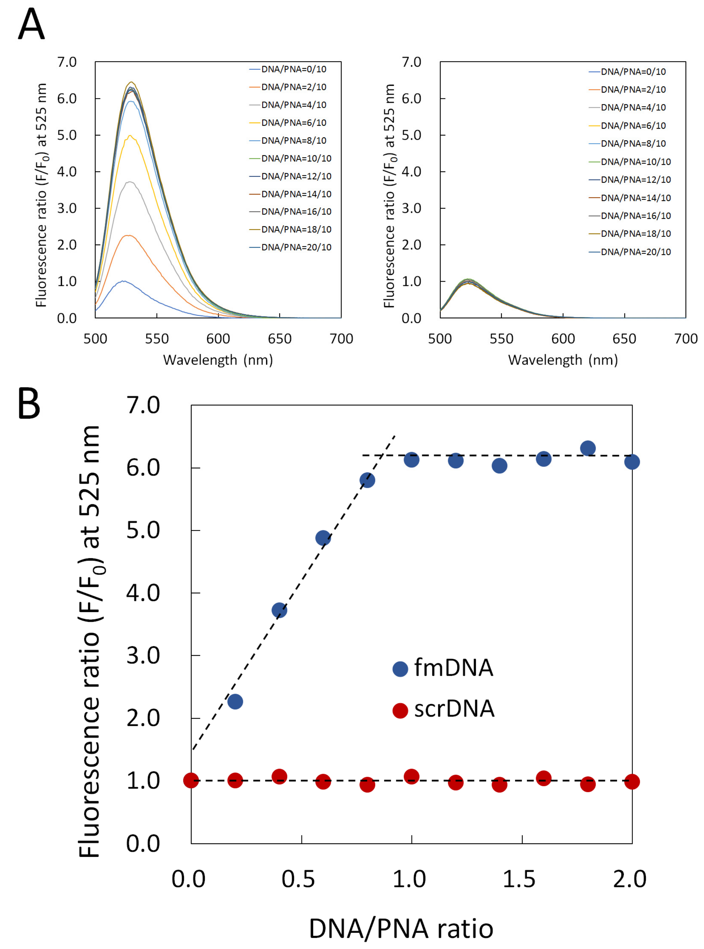
To examine the fluorescence characteristics of the P1Q1 with DNAs, we measured fluorescence spectra for various concentrations of DNAs and created their titration curves (Figure 4). As shown in the left panel in Figure 4A, the fluorescence spectra of P1Q1 shows that the fluorescence intensity at 525 nm increased as the fmDNA was gradually added. On the other hand, the addition of scrDNA, as shown in the right panel in Figure 4A, shows almost no change at 525 nm. This indicates that the PNA beacon forms a sequence-specific hybrid with the DNAs. Next, we plotted the fluorescence intensity (F/F0) at 525 nm for each DNA/P1Q1 ratio on the basis of the fluorescence spectra in Figure 4A,B. As the fmDNA was added from a 0 to 1 equivalent, the F/F0 increased linearly, and, above a 1 equivalent, the F/F0 did not change (blue-filled circles). On the other hand, when scrDNA was added, the P1Q1 hardly emitted fluorescence under these conditions (red-filled circles). These results show that P1Q1 forms a 1:1 hybrid with full-match DNA.

3.2. Elaborate Adjustment of PnQms for Fluorescence Detection of DNA

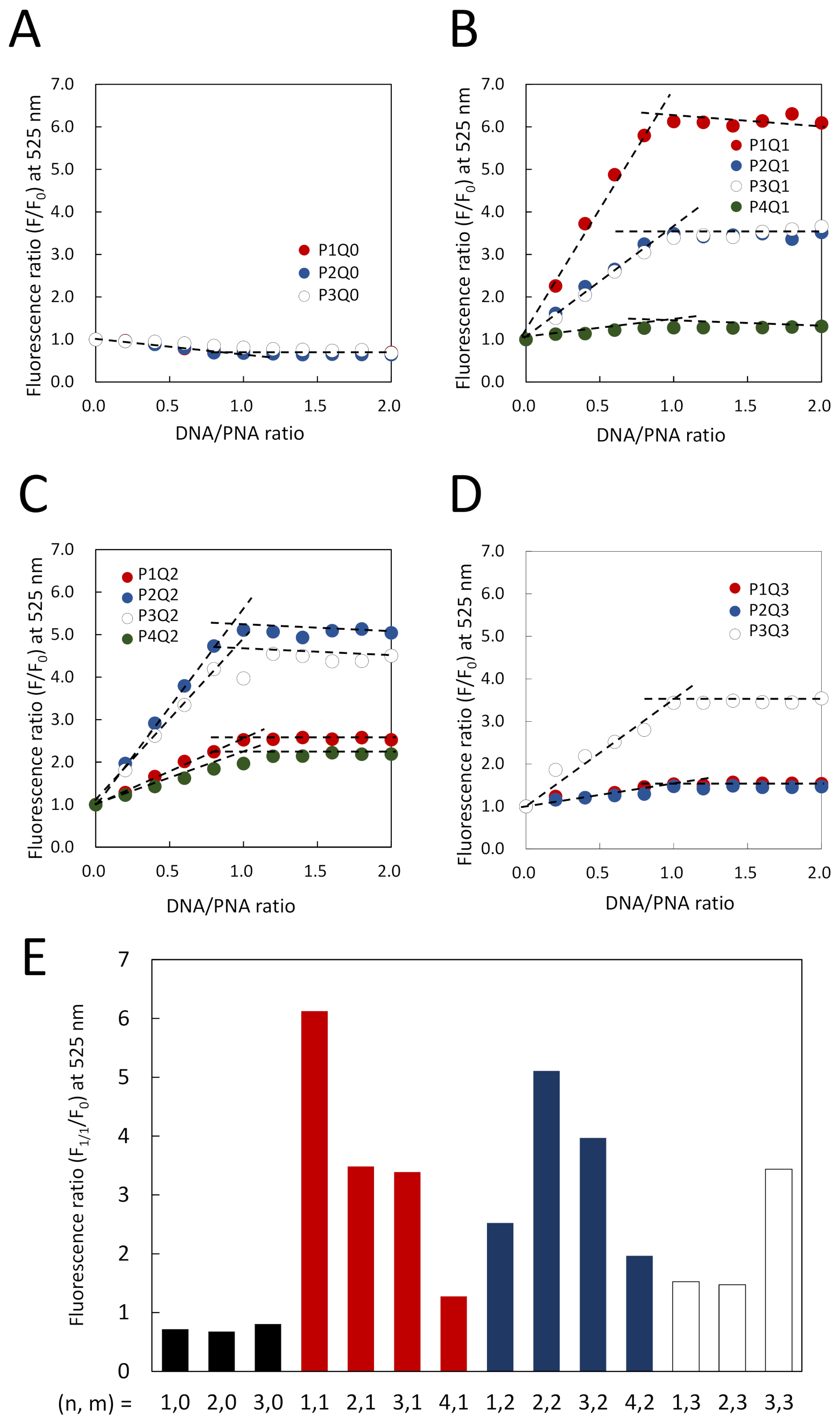
The structure of the PNA beacon, which is reported by Kuhn et al. (P1Q1 in this study), is used as a typical PNA beacon [23]. We are interested in whether this PNA beacon is the optimal structure for detecting nucleic acids. Therefore, we used PnQms, in which the numbers of Glu/Lys and Dab quenchers in the PNA beacon were changed, and we assessed these levels of DNA detection by the fluorescence titration curves for the DNA, in the same manner as in Figure 4. PnQ0, which does not contain the Dab quencher, showed a slight decrease in fluorescence upon the addition of fmDNA, despite varying the number of Glu/Lys from 1 to 3, which indicates that it is difficult for PnQ0 to detect DNA by fluorescence (Figure 5A and Supplementary Material, Figures S35–S37). On the other hand, for PnQ1, a clear 1:1 correspondence of the increase in the fluorescence upon the addition of fmDNA was observed, and the response was further influenced by the number of Glu/Lys (Figure 5B and Supplementary Material, Figures S38–S41). The F1/1/F0 value, which is the fluorescence ratio of the 1:1 mixtures of the PNA beacon with fmDNA to the PNA beacon alone, showed higher fluorescence levels in the order of: P1Q1 > P2Q1 = P3Q1 > P4Q1. It was confirmed that the F1/1/F0 values of PnQ2 and PnQ3 also depend on the number of Glu/Lys: P2Q2P3Q2 > P1Q2P4Q2 (Figure 5C and Supplementary Material, Figures S42–S45) and P3Q3 > P1Q3 = P2Q3 (Figure 5D and Supplementary Material, Figures S46–S48).
As a summary of Figure 5A–D, each F1/1/F0 of PnQms is shown in Figure 5E. This indicates that the numbers of Glu/Lys (n) and Dab quenchers (m) show a relatively large F1/1/F0 when n = 1 and m = 1 (F1/1/F0 = 6.1). When the number of m is 3, the F1/1/F0 level decreased, which indicates that the quenching effect of Dab against the Fam group was too strong. Regarding the number of n, the F1/1/F0 level tended to decrease from a smaller number to a larger number. We assume that, as the number of Glu/Lys increases, the electrostatic interaction between Glu and Lys may inhibit the hybrid formation of PNA and DNA. On the other hand, a relatively high F1/1/F0 level was observed when the numbers, n and m, were the same (F1/1/F0 = 6.1, 5.1, and 3.4 for n; m = 1.1, 2.2, and 3.3, respectively). This shows that not only the numbers of n and m, but also the balance of the numbers of n and m, affect the F1/1/F0 value. Overall, we determined that, among these PNA beacons, the PNA beacon with the highest relative fluorescence is P1Q1. This PNA beacon is consistent with the PNA beacon that is reported by Kuhn et al.
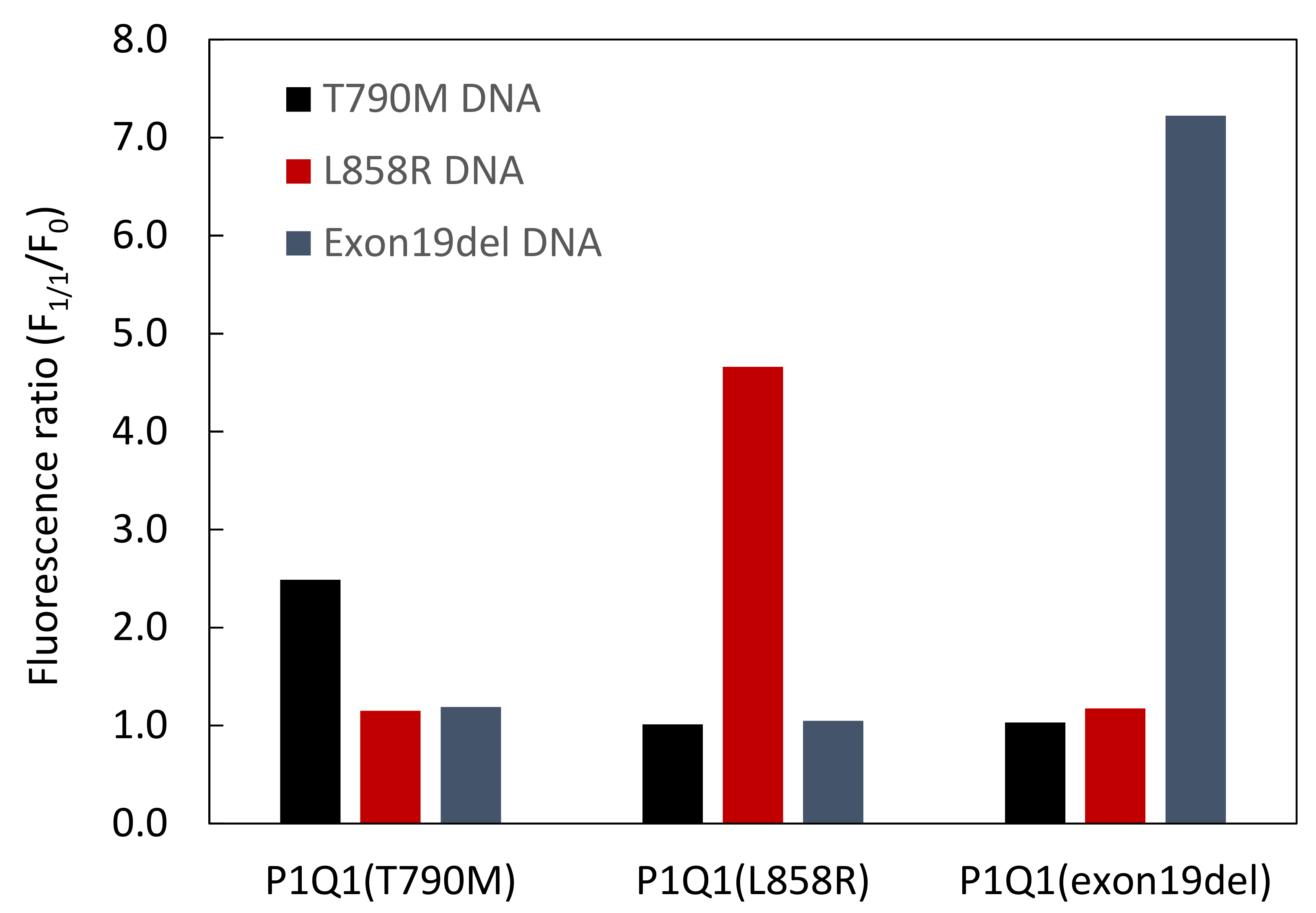
To investigate whether P1Q1 detects various DNA sequences, we used three P1Q1s with different PNA sequences: P1Q1(T790M): N′-CTGCATGATG-C′; P1Q1(L858R): N′-TGGCCCGCCC-C′; and P1Q1(exon19del): N′-ATGTTTTGAT-C′. The F1/1/F0 ratio was estimated from the fluorescence spectra before and after the addition of equivalent amounts of full-match DNAs (T790M: 5′-CATCATGCAG-3′; L858R: 5′-GGGCGGGCCA-3′; and exon19del: 5′-ATCAAAACAT-3′) to each of the P1Q1s. As shown in Figure 6 and in the Supplementary Material, Figures S49–S51, the F1/1/F0 values of P1Q1(T790M), P1Q1(L858R), and P1Q1(exon19del) for full-match DNA were 2.5, 4.7, and 7.2, respectively, whereas these values for noncomplementary DNA were about 1.0–1.2. Among these sequences, the higher F1/1/F0 of P1Q1(exon19del) may be influenced by the self-complementary sequences of the two nucleobases at both ends. These indicate that the series of P1Q1 recognizes full-match DNA, although this depends on the PNA sequence.

3.3. Comparison of Fluorescence Detection for Target DNA of PNA Beacon and PNA Beacon/Quencher-Modified DNA Hybrid

In previous studies, we reported that a probe that prehybridized Fam-modified PNA (Fl-PNA) with a Dab quencher-modified DNA (Q-DNA) successfully detected target DNA [35,36], and that a probe that prehybridized the PNA beacon with Q-DNA detected target miRNA [37]. To investigate whether the prehybridized Q-DNA and PNA beacon in this study detect target DNA more effectively than the PNA beacon alone, we assessed the F1/1/F0 value of the hybrid of P1Q1(T790M) with Q-DNA for target DNA (Figure 7 and Supplementary Material, Figure S52). The values of P1Q1(T790M) mixed with one-base-mismatched T790M DNA and P1Q1(T790M) alone were almost 1.0, whereas the value of P1Q1(T790M) mixed with full-match T790M DNA was 3.0. This was almost the same as the value shown by P1Q1(T790M) to T790M DNA in Figure 6 (F1/1/F0 = 2.5). In other words, P1Q1(T790M) detected the target DNA, but the recognition of the PNA beacon for DNA was not sufficient. On the other hand, the value of the P1Q1(T790M)/Q-DNA hybrid mixed with the one-base-mismatched DNA, and the value of the hybrid only, were almost 1.0, whereas the value of the hybrid mixed with the complementary DNA was 8.0. In a previous study, when the complementary T790M DNA was detected with the Fl-PNA/Q-DNA hybrid, the F1/1/F0 value was 1.5–2.0 [35]. These results indicate that the PNA beacon/Q-DNA hybrid detects the target DNA more effectively than the Fl-PNA/Q-DNA hybrid or the PNA beacon alone. From these results, we demonstrated that a synergistic or additive effect by the Dab quenchers contained in the PNA beacon and the Q-DNA was effectively exerted on the fluorescence detection of DNA in this study.

4. Conclusions

In this study, we optimized the number of Glu/Lys and the number of Dab quenchers in the PNA beacons (PnQms). As a result, we found that the structure of the conventional PNA beacon, P1Q1, is the best for the fluorescence detection of target nucleic acids. In other words, it was not necessary to increase the number of Glu/Lys and the number of Dab quenchers to improve the DNA detection of PNA beacons. However, we also obtained new findings that suggest that the balance between the number of Glu/Lys and the number of Dab quenchers affects the DNA detection of PNA beacons. This result is expected to be valuable to the optimization of the structures of other versions of PNA beacons. P1Q1s were shown to achieve detection, depending on the sequence of the target DNAs, but the hybrid of P1Q1, with quencher-modified DNA, conferred a more effective detection to the PNA sequences, with a relatively low F1/1/F0 value. This indicates that the combination of this PNA beacon and the PNA beacon/quencher-modified DNA hybrid probe is effective when detecting multiple DNA sequences. In the future, we will investigate by using a longer PNA than the PNA (10-mer) that was used in this study in order to develop the PNA beacon that targets RNA, and we plan to use the hybrid probe to detect nucleic acids in cells.

Supplementary Materials

The following are available online at https://www.mdpi.com/article/10.3390/pr10040722/s1, Supplementary Material, Figures S1–S17: MALDI-ToF mass spectra of all peptides; Supplementary Material, Figures S18–S34: HPLC chromatograms of all peptides; Supplementary Material, Figures S35–S52: Fluorescence spectra of all peptides with DNAs.

Author Contributions

Conceptualization, T.O.; methodology, T.O. and S.Y.; software, M.K.; validation, M.K.; formal analysis, H.S., T.K. and K.I.; investigation, H.S., T.K. and K.I.; resources, M.K. and S.Y.; data curation, M.K. and H.S.; writing—original draft preparation, M.K.; writing—review and editing, T.O., S.Y. and H.S.; visualization, M.K.; supervision, M.K. and S.Y.; project administration, M.K.; funding acquisition, M.K. and S.Y. All authors have read and agreed to the published version of the manuscript.

Funding

This research received no external funding.

Institutional Review Board Statement

Not applicable.

Informed Consent Statement

Not applicable.

Data Availability Statement

The data that support the findings of this study are available from the corresponding author upon reasonable request.

Acknowledgments

We thank Mei Okamoto and Miya Mukai (Kindai University) for providing technical support.

Conflicts of Interest

The authors declare no conflict of interest.

References

  1. Nielsen, P.E.; Egholm, M.; Berg, R.H.; Buchardt, O. Sequence-Selective Recognition of DNA by Strand Displacement with a Thymine-Substituted Polyamide. Science 1991, 254, 1497–1500. [Google Scholar] [CrossRef]
  2. Egholm, M.; Buchardt, O.; Christensen, L.; Behrens, C.; Freier, S.M.; Driver, D.A.; Berg, R.H.; Kim, S.K.; Norden, B.; Nielsen, P.E. PNA Hybridizes to Complementary Oligonucleotides Obeying the Watson-Crick Hydrogen Bonding Rules. Nature 1993, 365, 566–568. [Google Scholar] [CrossRef]
  3. Smith, J.O.; Olson, D.A.; Armitage, B.A. Molecular Recognition of PNA-Containing Hybrids: Spontaneous Assembly of Helical Cyanine Dye Aggregates on PNA Templates. J. Am. Chem. Soc. 1999, 121, 2686–2695. [Google Scholar] [CrossRef]
  4. Datta, B.; Armitage, B.A. Hybridization of PNA to Structured DNA Targets: Quadruplex Invasion and the Overhang Effect. J. Am. Chem. Soc. 2001, 123, 9612–9619. [Google Scholar] [CrossRef]
  5. Kushon, S.A.; Jordan, J.P.; Seifert, J.L.; Nielsen, H.; Nielsen, P.E.; Armitage, B.A. Effect of Secondary Structure on the Thermodynamics and Kinetics of PNA Hybridization to DNA Hairpins. J. Am. Chem. Soc. 2001, 123, 10805–10813. [Google Scholar] [CrossRef]
  6. Renikuntla, B.R.; Armitage, B.A. Role Reversal in a Supramolecular Assembly: A Chiral Cyanine Dye Controls the Helicity of a Peptide-Nucleic Acid Duplex. Langmuir 2005, 21, 5362–5366. [Google Scholar] [CrossRef]
  7. Kanjanawarut, R.; Su, X. Colorimetric Detection of DNA Using Unmodified Metallic Nanoparticles and Peptide Nucleic Acid Probes. Anal. Chem. 2009, 81, 6122–6129. [Google Scholar] [CrossRef]
  8. Su, X.; Kanjanawarut, R. Control of Metal Nanoparticles Aggregation and Dispersion by PNA and PNA-DNA Complexes, and its Application for Colorimetric DNA Detection. ACS Nano 2009, 3, 2751–2759. [Google Scholar] [CrossRef]
  9. Ali, M.M.; Li, Y. Colorimetric Sensing by Using Allosteric-DNAzyme-Coupled Rolling Circle Amplification and a Peptide Nucleic Acid–Organic Dye Probe. Angew. Chem. Int. Ed. 2009, 48, 3512–3515. [Google Scholar] [CrossRef]
  10. Xu, M.; Xing, S.; Zhao, Y.; Zhao, C. Peptide Nucleic Acid-Assisted Colorimetric Detection of Single-nucleotide Polymorphisms Based on the Intrinsic Peroxidase-like Activity of Hemin-Carbon Nanotube Nanocomposites. Talanta 2021, 232, 122420–122427. [Google Scholar] [CrossRef]
  11. Wang, J.; Nielsen, P.E.; Jiang, M.; Cai, X.; Fernandes, J.R.; Grant, D.H.; Ozsoz, M.; Beglieter, A.; Mowat, M. Mismatch-Sensitive Hybridization Detection by Peptide Nucleic Acids Immobilized on a Quartz Crystal Microbalance. Anal. Chem. 1997, 69, 5200–5202. [Google Scholar] [CrossRef]
  12. Ross, P.L.; Lee, K.; Belgrader, P. Discrimination of Single-Nucleotide Polymorphisms in Human DNA Using Peptide Nucleic Acid Probes Detected by MALDI-TOF Mass Spectrometry. Anal. Chem. 1997, 69, 4197–4202. [Google Scholar] [CrossRef]
  13. Chakrabarti, R.; Klibanov, A.M. Nanocrystals Modified with Peptide Nucleic Acids (PNAs) for Selective Self-Assembly and DNA Detection. J. Am. Chem. Soc. 2003, 125, 12531–12540. [Google Scholar] [CrossRef]
  14. Wang, J.; Palecek, E.; Nielsen, P.E.; Rivas, G.; Cai, X.; Shiraishi, H.; Dontha, N.; Luo, D.; Farias, P.A.M. Peptide Nucleic Acid Probes for Sequence-Specific DNA Biosensors. J. Am. Chem. Soc. 1996, 118, 7667–7670. [Google Scholar] [CrossRef]
  15. Cai, B.; Wang, S.; Huang, L.; Ning, Y.; Zhang, Z.; Zhang, G.-J. Ultrasensitive Label-Free Detection of PNA-DNA Hybridization by Reduced Graphene Oxide Field-Effect Transistor Biosensor. ACS Nano 2014, 8, 2632–2638. [Google Scholar] [CrossRef]
  16. Ali, M.; Neumann, R.; Ensinger, W. Sequence-Specific Recognition of DNA Oligomer Using Peptide Nucleic Acid (PNA)-Modified Synthetic Ion Channels: PNA/DNA Hybridization in Nanoconfined Environment. ACS Nano 2010, 4, 7267–7274. [Google Scholar] [CrossRef]
  17. Xuan, F.; Fan, T.W.; Hsing, I.-M. Electrochemical Interrogation of Kinetically-Controlled Dendritic DNA/PNA Assembly for Immobilization-Free and Enzyme-Free Nucleic Acids Sensing. ACS Nano 2015, 9, 5027–5033. [Google Scholar] [CrossRef]
  18. Brandt, O.; Hoheisel, J.D. Peptide Nucleic Acids on Microarrays and Other Biosensors. Trends Biotechnol. 2004, 22, 617–622. [Google Scholar] [CrossRef] [Green Version]
  19. Lai, Q.; Chen, W.; Zhang, Y.; Liu, Z. Application Strategies of Peptide Nucleic Acids toward Electrochemical Nucleic Acid Sensors. Analyst 2021, 146, 5822–5835. [Google Scholar] [CrossRef]
  20. Moccia, M.; Antonacci, A.; Saviano, M.; Caratelli, V.; Arduini, F.; Scognamiglio, V. Emerging Technologies in the Design of Peptide Nucleic Acids (PNAs) Based Biosensors. Trends Analyt. Chem. 2020, 132, 116062–116076. [Google Scholar] [CrossRef]
  21. Endo, T.; Kerman, K.; Nagatani, N.; Takamura, Y.; Tamiya, E. Label-Free Detection of Peptide Nucleic Acid-DNA Hybridization Using Localized Surface Plasmon Resonance Based Optical Biosensor. Anal. Chem. 2005, 77, 6976–6984. [Google Scholar] [CrossRef] [PubMed]
  22. Vilaivan, T. Fluorogenic PNA Probes. Beilstein J. Org. Chem. 2018, 14, 253–281. [Google Scholar] [CrossRef] [PubMed] [Green Version]
  23. Kuhn, H.; Demidov, V.V.; Coull, J.M.; Fiandaca, M.J.; Gildea, B.D.; Frank-Kamenetskii, M.D. Hybridization of DNA and PNA Molecular Beacons to Single-Stranded and Double-Stranded DNA Targets. J. Am. Chem. Soc. 2002, 124, 1097–1103. [Google Scholar] [CrossRef] [PubMed]
  24. Xi, C.; Balberg, M.; Boppart, S.A.; Raskin, L. Use of DNA and Peptide Nucleic Acid Molecular Beacons for Detection and Quantification of rRNA in Solution and in Whole Cells. Appl. Environ. Microbiol. 2003, 69, 5673–5678. [Google Scholar] [CrossRef] [PubMed] [Green Version]
  25. Smolina, I.V.; Demidov, V.V.; Soldatenkov, V.A.; Chasovskikh, S.G.; Frank-Kamenetskii, M.D. End Invasion of Peptide Nucleic Acids (PNAs) with Mixed-Base Composition into Linear DNA Duplexes. Nucleic Acids Res. 2005, 33, e146. [Google Scholar] [CrossRef] [Green Version]
  26. Xi, C.; Raskin, L.; Boppart, S.A. Evaluation of Microfluidic Biosensor Development Using Microscopic Analysis of Molecular Beacon Hybridization Kinetics. Biomed. Microdevices 2005, 7, 7–12. [Google Scholar] [CrossRef] [Green Version]
  27. Ye, S.; Miyajima, Y.; Ohnishi, T.; Yamamoto, Y.; Komiyama, M. Combination of Peptide Nucleic Acid Beacon and Nuclease S1 for Clear-Cut Genotyping of Single Nucleotide Polymorphisms. Anal. Biochem. 2007, 363, 300–302. [Google Scholar] [CrossRef]
  28. Smolina, I.V.; Kuhn, H.; Lee, C.; Frank-Kamenetskii, M.D. Fluorescence-Based Detection of Short DNA Sequences under Non-Denaturing Conditions. Bioorg. Med. Chem. 2008, 16, 84–93. [Google Scholar] [CrossRef]
  29. Li, X.; Morgenroth, E.; Raskin, L. Quantitative rRNA-Targeted Solution-Based Hybridization Assay Using Peptide Nucleic Acid Molecular Beacons. Appl. Environ. Microbiol. 2008, 74, 7297–7305. [Google Scholar] [CrossRef] [Green Version]
  30. Totsingan, F.; Rossi, S.; Corradini, R.; Tedeschi, T.; Sforza, S.; Juris, A.; Scaravelli, E.; Marchelli, R. Label-Free Selective DNA Detection with High Mismatch Recognition by PNA Beacons and Ion Exchange HPLC. Org. Biomol. Chem. 2008, 6, 1232–1237. [Google Scholar] [CrossRef]
  31. Totsingan, F.; Tedeschi, T.; Sforza, S.; Corradini, R.; Marchelli, R. Highly Selective Single Nucleotide Polymorphism Recognition by a Chiral (5S) PNA Beacon. Chirality 2009, 21, 245–253. [Google Scholar] [CrossRef] [PubMed]
  32. Zanoli, L.M.; Licciardello, M.; D’Agata, R.; Lantano, C.; Calabretta, A.; Corradini, R.; Marchelli, R.; Spoto, G. Peptide Nucleic Acid Molecular Beacons for the Detection of PCR Amplicons in Droplet-Based Microfluidic Devices. Anal. Bioanal. Chem. 2013, 405, 615–624. [Google Scholar] [CrossRef] [PubMed]
  33. Matsumoto, K.; Nakata, E.; Tamura, T.; Saito, I.; Aizawa, Y.; Morii, T. A Peptide Nucleic Acid (PNA) Heteroduplex Probe Containing an Inosine-Cytosine Base Pair Discriminates a Single-Nucleotide Difference in RNA. Chem. Eur. J. 2013, 19, 5034–5040. [Google Scholar] [CrossRef]
  34. Mach, K.E.; Kaushik, A.M.; Hsieh, K.; Wong, P.K.; Wang, T.-H.; Liao, J.C. Optimizing Peptide Nucleic Acid Probes for Hybridization-Based Detection and Identification of Bacterial Pathogens. Analyst 2019, 144, 1565–1574. [Google Scholar] [CrossRef]
  35. Shigeto, H.; Ohtsuki, T.; Iizuka, A.; Akiyama, Y.; Yamamura, S. Imaging Analysis of EGFR Mutated Cancer Cells Using Peptide Nucleic Acid (PNA)–DNA Probes. Analyst 2019, 144, 4613–4621. [Google Scholar] [CrossRef]
  36. Shigeto, H.; Yamada, E.; Kitamatsu, M.; Ohtsuki, T.; Iizuka, A.; Akiyama, Y.; Yamamura, S. Analysis of Single Nucleotide-Mutated Single Cancer Cells Using the Combined Technologies of Single-Cell Microarray Chips and Peptide Nucleic Acid-DNA Probes. Micromachines 2020, 11, 628. [Google Scholar] [CrossRef]
  37. Tabara, K.; Watanabe, K.; Shigeto, H.; Yamamura, S.; Kishi, T.; Kitamatsu, M.; Ohtsuki, T. Fluorophore–PNA–Quencher/Quencher–DNA Probe for miRNA Detection. Bioorg. Med. Chem. Lett. 2021, 51, 128359–128363. [Google Scholar] [CrossRef]
Figure 1. (A) Chemical structures of 14 PNA beacons (PnQm (n = 1–4 and m = 0–3)) and three P1Q1s with different PNA sequences: P1Q1(T790M), P1Q1(L858R), and P1Q1(exon19del). The N-terminal and near C-terminal in the PNA beacon were modified with Fam and Dab quenchers, respectively. These PNA beacons were modified with four ethylene glycol linkers in the C-terminal to improve the water solubility. (B) The PNA sequences of PnQms, P1Q1(T790M), P1Q1(L858R), and P1Q1(exon19del).
Processes 10 00722 g001
Figure 2. Temperature dependences of absorption intensity at 260 nm for: (left) 1:1 mixtures of P1Q1/fmDNA (red-filled circles) and P1Q1/scrDNA (black-filled circles), and P1Q1 alone (blue-filled circles); and (right) 1:1 mixtures of P4Q1/fmDNA (red-filled circles) and P4Q1/scrDNA (black-filled circles), and P4Q1 alone (blue-filled circles).
Processes 10 00722 g002
Figure 3. Fluorescence spectra of 500 nM P1Q1 and P1Q0 (red and black solid lines, respectively), and 1:1 mixtures of P1Q1/fmDNA and P1Q0/fmDNA (red and black broken lines, respectively). Fam was also observed as a control (green solid line).
Processes 10 00722 g003
Figure 4. (A) Fluorescence spectra of 500 nM P1Q1 with various concentrations of fmDNA (left) and scrDNA (right). (B) Titration curves of fluorescence intensities at 525 nm for P1Q1/fmDNA (blue-filled circles) and P1Q1/scrDNA (red-filled circles).
Processes 10 00722 g004
Figure 5. Titration curves of fluorescence intensities at 525 nm for: (A) PnQ0 (n = 1–3); (B) PnQ1 (n = 1–4); (C) PnQ2 (n = 1–4); and (D) PnQ3 (n = 1–3), with fmDNA. (E) Fluorescence ratios (F1/1/F0) of the 1:1 mixtures of PnQm and fmDNA.
Processes 10 00722 g005
Figure 6. Fluorescence ratios (F1/1/F0) of the 1:1 mixtures of P1Q1s with different PNA sequences and DNAs.
Processes 10 00722 g006
Figure 7. Fluorescence ratios (F1/1/F0) of 1:1 mixtures of P1Q1(T790M)/Q-DNA hybrid with DNAs, and of P1Q1(T790) with DNAs.
Processes 10 00722 g007
Publisher’s Note: MDPI stays neutral with regard to jurisdictional claims in published maps and institutional affiliations.