Article

Processes and Interactions Impacting the Stability and Compatibility of Vitamin K and Gold Nanoparticles

1
Department of Chemistry, Faculty of Sciences, University of Craiova, Calea Bucuresti 107i, 200478 Craiova, Dolj, Romania
2
Faculty of Medicine, University of Medicine and Pharmacy, Petru Rares 2, 200349 Craiova, Dolj, Romania
*
Author to whom correspondence should be addressed.
Processes 2022, 10(9), 1805; https://doi.org/10.3390/pr10091805
Submission received: 29 June 2022 / Revised: 30 August 2022 / Accepted: 4 September 2022 / Published: 7 September 2022
(This article belongs to the Special Issue Application of Nanomaterials in the Process of Water Electrolysis)

Abstract

:
This study provides evidence on the stability of vitamin K1 (VK) in the form of phytomenadione, in the absence and presence of a therapeutic preparation, as the gold nanoparticles (AuNPs), under the effect of sodium halide ions. The degradation susceptibility of the two compounds was assessed individually and in mixtures by cyclic voltammetry and electrolysis at a constant current density assisted by UV-Vis spectrophotometry. Their interactions with the halide ions differently impact on the electrochemical processes as follows: (i) the fluoride ions weakly affects the VK/AuNP stability and compatibility; (ii) the presence of chloride ions leads to VK/AuNP stability, for a short time and restrictive compatibility; (iii) bromide ions induce instability and incompatibility of the VK/AuNP system; (iv) spontaneous interactions between VK/AuNPs and iodide ions take place, consequently defining as an unstable and incompatible system.

1. Introduction

Vitamin K itself is a family of fat-soluble vitamers that play essential roles in various metabolic biochemical processes. Vitamin K supports the functionality of matrix GLAa (MGP) protein in vascular tissue which has the ability to block calcium accumulation in the walls of arteries as the strongest inhibitor of the calcification of arteries, thus avoiding atherosclerosis and diminishing the risk of cardiovascular disease [1]. It also contributes to maintaining the density of bones and reducing the risk of fractures.
Green leafy vegetables, such as cabbage, spinach, and parsley, contain significant amounts of vitamin K. The best-known K vitamers are vitamin K1 (phylloquinone or phytomenadione), K2 (menaquinone), and vitamin K3 (menadione). Vitamin coagulation has a vital role in normal hemostasis, but is also an essential cofactor for hepatic blood-coagulating proteins (prothrombin, factors II, VII, IX, and X) [2,3,4]. Only the first two vitamins are natural, white the others are synthetic (K3, K4, K5, K6, and K7).
Synthetic vitamin K analogues, such as menadione and its derivatives, are classified as “toxic to reproduction” (reprotoxic category), which is why they are banned by the FDA (Food and Drug Administration). For this reason, synthetic vitamin K analogues have been replaced by warfarin (Coumadin) [5]. All homologues of vitamin K (prenyl side chain) are cofactors for γ-glutamyl carboxylase. This enzyme activates proteins containing glutamate groups by carboxylation of them to γ-carboxyglutamate, which is required for the metabolic activity of these enzymes [6].
The Hordaland Health Study [7] on the administration of vitamins K1 and K2 showed that patients had higher sensitivity to K1 than to K2, while K1 is primarily needed for skeletal effects and secondly for coagulation [8]. In view of all this, several routes of therapeutic administration have therefore been investigated [9,10,11,12]. The solubility and stability of vitamin K, one of the fat-soluble vitamins, has been improved by the formation of complexes with large-ring cycloamylose [9]. The application of K1 in dermal ointments/creams has certain limitations due to its photodegradation. The formulation has been enhanced by the application of nanoemulsion spray [10], topical K1 ester derivatives [11], and nanoliposomes for skin delivery [12].
The development of some methods for vitamin K quantification thereby improving its administration was achieved [13,14,15,16,17,18,19,20]. Vitamin K1 was estimated using cascaded channels fiber optic surface plasmon resonance [13] and high-performance liquid chromatography with UV detection [14]. High-performance liquid chromatography in combination with mass spectrometry was used to determine K1 and its 2,3-epoxide derivative from human plasma [15,16] and fruits and vegetables [17]. Chromatographic methods have been used to determine K1 metabolites in human plasma [18] and urine [19]. At the same time, K1 was determined using the metabolites of antagonistic compounds, such as warfarin metabolites [20]. Electrochemical detection using pencil graphite or glassy carbon electrodes was used due to its simplicity, short analysis times, selectivity, and high sensitivity [21,22]. The interaction between warfarin and vitamin K1 was studied by electrochemical impedance spectroscopy, cyclic voltammetry, and differential pulse voltammetry methods using a carbon paste electrode [23]. The sensitive voltammetric determination of vitamin K2 using a new electrochemical sensor indicates responses proportional to the concentration of VK2 in the range of 0.2–8.0 μg·L−1 (R = 0.9987) and 4–20 μg·L−1 (R = 0.9959) with a limit of detection of 0.009 and 0.012 μg·L−1, respectively [24].
AuNPs have attracted significant attention due to their unique physical, chemical, and especially biological properties [25,26,27]. AuNPs have several applications, such as antimicrobial, antioxidants, hepatoprotector, anticancer therapeutic potential, drug delivery, pollution control, and water purification [25,26,27]. Functionalized AuNPs show selective toxicity to LK-2 and TIG-120 tumor cells. Polymer-functionalized Au-NPs have been used successfully in tumor cell-specific imaging and targeting [25]. I125-labeled AuNPs have also been used in radiotherapy. The specific chemical and electrochemical interactions of AuNPs with various chemical or biological compounds have allowed the development of sensors and biosensors for a specific and accurate determination [27]. Nowadays, we are faced with confusing or contradictory information about the administration of biologically active compounds, diets, or more or less natural supplements. In this context, vitamin K (as K1) and colloidal gold nanoparticles are the subject of this controversy. There is a lot of information and publicity about the compounds we should consume, but there is a lack of information about their stability, reactivity, or the interactions they participate in immediately after administration [28,29,30,31].
To revive and tone the body, as well as in the case of its debilitation caused by different diseases, many people resort to alternative treatments. Thus, gold has beneficial effects on the human body health, contributing to the treatment of rheumatoid arthritis, the prevention of the tumors and blood clotting, the healing of skin conditions, the coordination of the neurovegetative system, etc. [25,26,27]. Many times, these alternative treatments are not compatible with allopathic ones, and the interactions of nutritional supplements, such as AuNPs, with compounds of the basic metabolism can have an unforeseen action on their pharmacokinetics and pharmacodynamics, causing changes in the decomposition rate or half-life [30]. In addition, the presence of some halide anions that are found in the composition of some pharmaceutical preparations or drugs that can be ingested simultaneously with certain nutritional supplements, intensifies or delays the activity of some compounds, such as vitamins [31].
On another hand, the waste waters from the medical system, which contain a multitude of compounds, even if they are in very low concentrations, should be decontaminated through different depollution techniques, which involve the knowledge of secondary reactions or additional interactions. Thus, the approach to these complex phenomena can have industrial applications leading to the appropriate choice of the decontamination methods of some residual waters [31].
Based on the foregoing, our study aims to elucidate some interactions occurring among vitamin K1 (VK) molecules and colloidal gold nanoparticles (AuNPs) under the effect of sodium halide ions, which can be ingested from various drug complexes, food, teas, oral hygiene products, etc.

2. Materials and Methods

2.1. Materials

Vitamin K (VK) was used in this study as phytomenadione (2-methyl-3-[(E,7R,11R)-3,7,11,15-tetramethylhexadec-2-enyl]naphthalene-1,4-dione). It was bottled in a single-use brown glass ampoule with a capacity of 1 mL. Vitamin K1 (phytomenadione) with a concentration of 10 mg·mL−1 was purchased from Terapia SA, Bucharest, Romania. Sodium halides, sulfuric acid, and ethyl alcohol with reagent grade purities were supplied by Merck/Sigma Aldrich, Steinheim, Germany. To prepare all solutions, the distilled water treated time of five minutes with UV-irradiation was used.
Four stock solutions of sodium halides, with concentrations of 1.0 mol·L−1, were prepared. These solutions provided the necessary supporting electrolyte. In addition, halide solutions containing vitamin K in a concentration of 0.1 g·L−1 (2.21 × 10⁻⁴ mol·L−1) were stocked. During the study, all solutions were kept in a closed and dark space.
The gold nanoparticles (AuNPs) with a spherical geometry and average dimensions between 28 and 35 nm were purchased as a nanosuspension (0.48 L brown bottles) stabilized in citrate buffer, from AquaNano (Aghoras Invent srl, Bucharest, Romania), having a concentration of 110 mg·L−1.
For experimental tests, four categories of media were prepared in well-determined concentrations in order to obtain adequate spectrophotometric responses (Figure 1), as follows:
  • 0.1 mol·L−1 NaX, further named as NaX, where X = F, Cl, Br, I);
  • 0.1 mol·L−1 NaX, 6.65 × 10⁻⁵ mol·L−1 VK, noted further as NaX–VK;
  • 0.1 mol·L−1 NaX, 75 mg·L−1 AuNPs hereinafter referred to as NaX–AuNPs;
  • 0.1 mol·L−1 NaX, 6.65 × 10⁻⁵ mol·L−1 VK, 75 mg·L−1 AuNPs referred to below as NaX–VK–AuNPs.
All solutions were freshly prepared and used on the same day.
Please note that the natural pH of the studied media was chosen that is close to the neutral one in order to obtain adequate spectrophotometric responses and to avoid further interactions and/or interference caused by the sensitivity of the compounds in the acidic or alkaline media.
Taking into account vitamin K’s limited water solubility, several concentrations were tested, choosing those for which adequate spectrophotometric responses were obtained. The higher concentrations led to numerous splits and noises of the absorbance maxima, and for lower the ones a too decreased absorbance value was recorded.
Figure 1 shows the vitamin K1 UV-Vis spectra for different concentrations, namely 0.01 g L−1, 0.03 g L−1, 0.05 g L−1, and 0.1 g L−1, respectively, 2.21 × 10⁻⁵ mol L1, 6.65 × 10⁻⁵ mol L−1, 1.1 × 10⁻⁴ mol L−1, and 2.21 × 10⁻⁴ mol L−1, in the aqueous medium as well as its molecular formula. As can be observed, the most eloquent spectrum is displayed for the vitamin K1 concentration of 6.65 × 10⁻⁵ mol·L−1, with the absorption maxima being centered at the wavelengths of 332 nm, 268 nm, and 244 nm.

2.2. Methods and Procedures

2.2.1. Electrochemical Measurements

Before the electrochemical measurements, the surface of the electrodes was ultrasonically cleaned in a sulfuric acid solution. Subsequently, the platinum electrodes (geometric area of 1 cm2, 99.95% purity, and Sigma-Aldrich provenience) were degreased with ethyl alcohol and dried in warm air.
The electrochemical experiments were performed at room temperature, under dynamic regime using a magnetic stirrer running at 300 rpm.
Cyclic voltammetry was carried out on a platinum electrode, in the solutions of sodium halides containing vitamin K, in the absence and presence of gold nanoparticles, between potential values of −2.0 V and +2.0 V with a potential scan rate of 100 mV·s−1. A wide potential window was recorded, in order to detail the nonfaradic zone, where in the specific potential range no charge–transfer reactions appear and adsorption processes on the electrode surface take place, and, consequently, the overlayer formation can be discussed.
The electrode processes over time were investigated, under the same conditions to those of cyclic voltammetry, through electrolysis at a constant current density, timing of 60 min, by applying a value of 50 mA·cm−2 for current density. The optimal operating parameters of the electrolytic process were chosen, which allowed the observation of certain identifiable spectral differences (e.g., absorbance value change), at the sampling times.
A three-electrode system was coupled to an electrochemical system VoltaLab Radiometer Analytical SAS, France with VoltaMaster 4 software (version 7.8.26338.3), namely working and auxiliary electrodes made from platinum and AgǀAgCl KClsat electrodes as a reference.

2.2.2. UV-Vis Spectrophotometry

The media submitted to UV-Vis spectrophotometric analysis consisted of: (a) sodium halides blank solutions, noted further as NaX, where X = F, Cl, Br, I; (b) NaX solutions containing vitamin K1 (VK), named further as NaX–VK; (c) NaX solutions containing gold nanoparticles (AuNPs), referred to below as NaX–AuNPs; (d) finally, NaX solutions containing both compounds, vitamin K1 and gold nanoparticles (AuNPs), hereinafter referred to as NaX–VK–AuNPs.
Three categories of samples were subjected to spectrophotometric analysis, namely initial solutions, the media resulting before and after cyclic voltammetry, and the samples collected every minute, time of one hour, and during electrolysis at a constant current density. Each category consisted of sodium halide blank solutions (NaX), sodium halide solutions containing vitamin K1 (NaX–VK), sodium halide solutions containing gold nanoparticles (NaX–AuNPs), and sodium halide solutions containing both vitamin K1 and gold nanoparticles (NaX–VK–AuNPs). In all cases, X successively represented F, Cl, Br, and I ions. The specific composition of the media was as mentioned above (Section 2.1).
During constant current density electrolysis, for the accuracy of the comments and for a coherent rendering of the processes and phenomena, spectra obtained every minute, or every 10 min, as the case may be, were selected, as specified in Section 3.3 “Results and Discussion”.
The UV-Vis spectra were recorded in the wavelength range between 800 nm and 200 nm using a Varyan Cary 50 UV-Vis spectrophotometer, Varian Inc., Mulgrave, VIC, Australia with CaryWin UV software, version 3. The UV-Vis analysis was performed in a quartz cell with a volume of 4 mL (10 × 10 × 45 mm). A similar methodology and similar devices were used in our previous studies [28,29,30,31,32,33,34,35,36].

3. Results and Discussion

3.1. UV-Vis Spectrophotometry of Initial Media

The UV-Vis spectra of the media mentioned above are displayed in Figure 2.
Similar spectra with those from the literature [37], were recorded for vitamin K1 analyzed in our study. Three absorption maxima of VK, at wavelengths (λ) of 332 nm, 268 nm, and 244 nm, can be observed in the NaF, NaCl, and NaBr solutions (Figure 2a–c; NaX–VK spectra), both in the absence and in the presence of AuNPs, suggesting that there are no instantaneous interactions, at first contact, between nanoparticles and halide ions. Absorption peaks are slightly shifted compared to those reported in the literature for vitamin K1, at 260 nm and 240 nm, respectively [37], due to the effects generated by the environment in which the analysis was performed. In all cases, both in the absence and in the presence of vitamin, the addition of AuNPs (absorption maximum highlighted at λ = 550 nm) [31], leads to the baseline shifting, more obvious in the case of environments based on NaCl and NaBr than in those based on NaF solution (Figure 2a–c).
The UV-Vis spectra of VK in the absence and presence of AuNPs recorded in NaI are very different from those presented above (Figure 2d). The spectrophotometric analysis of the NaI solution displays two absorption maxima centered at 290 nm and 355 nm, corresponding to iodine/iodide species [33] that instantaneously appear in the aqueous solution (Figure 2d). The spectral characteristics of vitamin K are completely changed, and the absorbance drop is significant (Figure 2d) compared to previous cases (Figure 2a–c), indicating that the vitamin is still found in the NaI solution in a very small concentration. In addition, the absorption maximum of gold nanoparticles is poorly visible at 550 nm (Figure 2d; NaI–AuNP spectrum).
Consequently, there were instantaneous interactions, both of the vitamin and of the AuNPs with the iodine/iodide species from the solution, favoring certain transformations and/or the appearance of other compounds. The new chemical species can be generated by cleavage of the bond between the radical R and the naphthoquinone ring (molecular formula inserted in Figure 1) resulting in another form of vitamin K (e.g., K3) or other products, which absorb at the same wavelength.
The sodium iodide solution spectrum containing vitamin K and AuNPs (NaI–VK–AuNPs) shows a peak located at 336 nm, which can result from the interferences between several compounds existing in the environment.
According to the literature data, the vitamin K1 main degradation compounds are quinone, hydroquinone, and its epoxide forms [15,19,38]. Among them, 2,3-epoxide and menaquinone-4 are two of the degradation products, and menaquinone-4 is both a metabolite of K1 in extrahepatic tissue and a member of the vitamin K2 family [15]. This oxide metabolite was identified and determined in human plasma by thin-layer chromatography and mass spectrometry [15,38].
Thus, the I action on vitamin K and nanoparticles leads to instantaneous interactions between species, which change the environment composition, making it difficult to assess the evolution of vitamin K and AuNPs, under these conditions. For this reason, we will further analyze the solutions in which vitamin K and AuNPs have a proven stability state, namely their evolution in NaF, NaCl, and NaBr solutions.

3.2. Evolution of Vitamin K and Gold Nanoparticles in NaX Solutions Studied by Cyclic Voltammetry (CV) Assisted by UV-Vis Spectrophotometry

The cyclic voltammograms were recorded on the platinum electrode, with a potential scan rate of 100 mV·s−1, in the electrolyte solutions containing halide ions, in a concentration of 0.1 mol·L−1, without and with 6.65 × 10⁻⁵ mol·L−1 VK and 75 mg·L−1 AuNPs, which have been successively as well as simultaneously added in supporting electrolytes (Figure 3a,b, Figure 4a,b and Figure 5a,b). In order to study the VK/AuNP stability at high current density values, a large potential range, between −2 V and +2 V, to perform cyclic voltammetry was chosen. In addition, the UV-Vis spectral characteristics of VK and AuNPs, before and after cyclic voltammetry in the presence of F, Cl, and Br ions are displayed in Figure 3c–e, Figure 4c–e and Figure 5c–f.

3.2.1. Vitamin K/Gold Nanoparticle Behavior in the Presence of F Ions

Figure 3a,b show the cyclic voltammograms recorded for the NaF blank solution and NaF containing VK, in the absence and presence of AuNPs.
At potential negative values, the main cathodic process that takes place at the working pH close to neutral is represented by the hydrogen evolution reaction (HER) through the discharge of water molecules, following which next to molecular hydrogen, hydroxyl ions are formed (Reaction (R1)). The additional cathodic process is represented by H+ reduction generating also molecular hydrogen (Reaction (R2)).
2H2O + 2e → H2 + 2OH
2H+ + 2e → H2
Thus, the platinum electrode surface is protected by the adsorption of hydrogen molecules. As the potential shifts to positive values, HER becomes an insignificant competing process that does not influence the adsorption processes of other chemical species from the electrolyte.
For the oxygen evolution reaction (OER), a high overpotential is needed (Reactions (R3) and (R4)).
4OH → 2H2O + O2 + 4e
2H2O → O2 + 4H+ + 4e
In this case, the molecular oxygen is adsorbed on the electrode surface facilitating vitamin K oxidation, especially from the layer developed by the adsorption of its molecules on the platinum surface. In addition, active oxygen species, such as hydroxide and peroxide radicals, are electrochemically generated in this potential range [33].
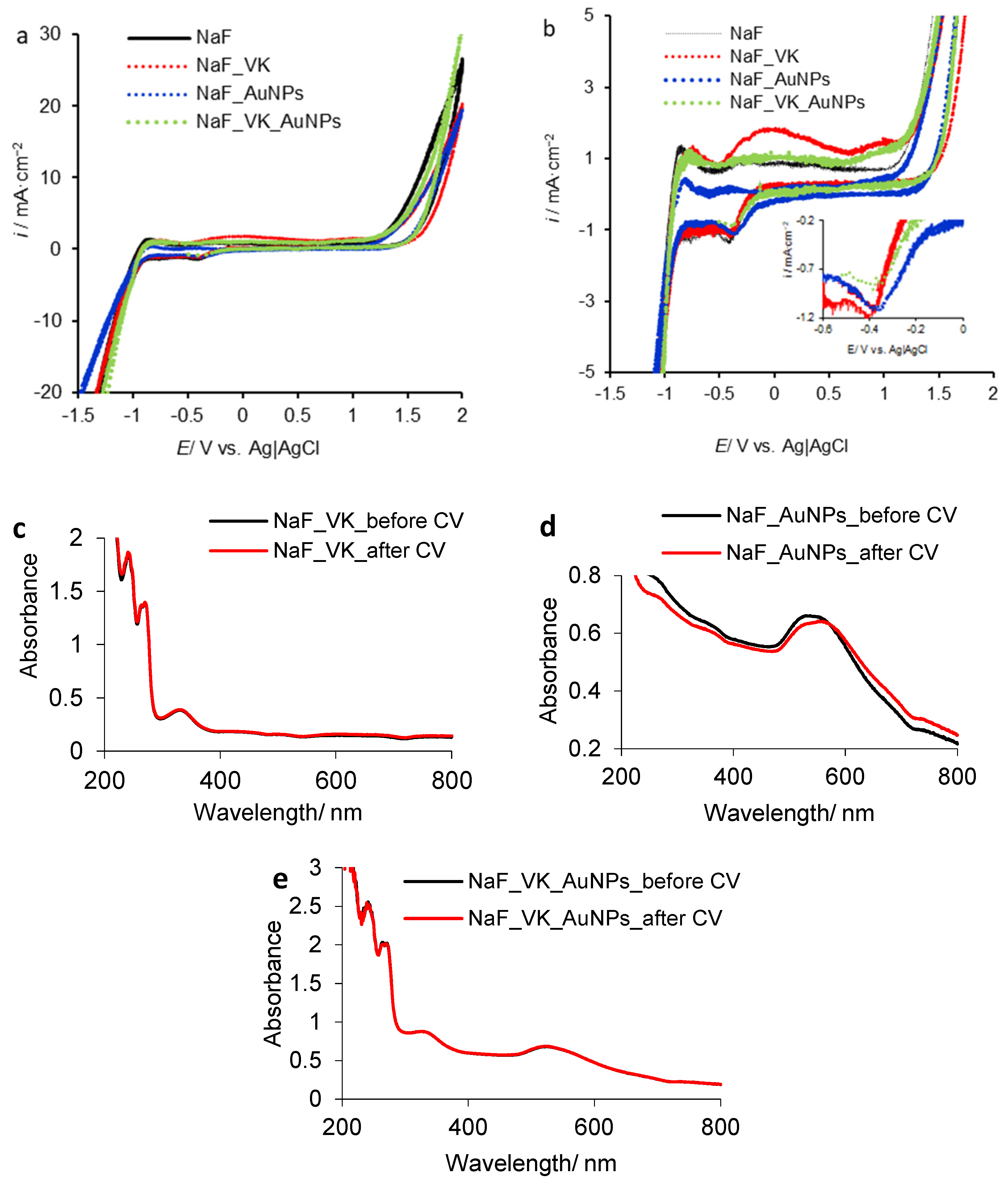
Comparing the four cyclic voltammograms, we observed that the presence of VK and AuNPs (NaF–VK–AuNPs) leads to a charge transfer rate decrease revealed by the lower values of recorded cathodic current density compared to those obtained in the solution containing only VK or AuNPs (NaF–VK and NaF–AuNPs). As shown in the detail inserted in Figure 3b, well-defined cathodic peaks corresponding to the NaF–VK and NaF–AuNP media were obtained compared to the ones recorded in the NaF solution containing both compounds (NaF–VK–AuNPs), indicating a cathodic process suppression, as a result of the oxidation reaction that slowed down, as can be observed on the anodic scan.
Hysteresis maxima located at lower current densities (Figure 3a, NaF–VK and NaF–AuNPs) than those of the blank solution (Figure 3a, NaF) suggest that a thin film was formed on the electrode surface [21,22,24].
The overlayer changes the surface architecture and its characteristics, disfavoring the ion exchange at the platinum/electrolyte interface and default leading to the inhibition of the oxidation processes from that solution.
Based on the molecular structure of vitamin K1 (inserted in Figure 1), its adsorption on a platinum electrode surface can take place by the formation of coordination bonds between the nonparticipating electron pairs of the oxygen atoms from the quinone ring and the vacant platinum orbitals or through hydrogen bonds or halogen bridges. Thus, through the adsorption of vitamin molecules, a thin layer is formed on the platinum surface, which partially blocks the charge transfer at the electrode/electrolyte interface.
In addition, the oxidation processes in the film can lead to secondary products, as shown in Scheme 1. Thus, the oxidation process can take place at the -C=C- bond in position 2 of the quinone ring (Scheme 1). In this case, an oxidic compound results (vitamin 2,3-epoxide) that is mentioned in the literature as the main vitamin oxidation product [15,19,38].
The detail designed for low values of current densities in Figure 3b, highlights, on the NaF–VK voltammogram, an extended oxidation peak, such as a shoulder, followed by a smaller one, inducing the idea that certain oxidation processes took place, but probably especially in the surface overlayer. The oxidation processes at the electrode/medium interface are less noticeable on the other voltammograms shown in Figure 3b, but these could take place with a low intensity. The compounds released in solution should be in a very low concentration (traces) and thus have a minimal impact on the environment composition. Generally, adsorbed thin films change the surface characteristics and attenuate/delay and even block the oxidation processes in the solution [21,22]. Consequently, the NaF solution is an inert electrolyte that does not affect the stability of the vitamin and the nanoparticles, whether these are in a solution as a single compound or in a mixture.
The UV-Vis spectra performed before and after cyclic voltammetry are illustrated in Figure 3c–e.
Note that for the blank NaF and NaCl solutions, the UV-Vis spectra are not shown, because for them, no absorption maxima were recorded at certain wavelengths and/or other spectral changes after cyclic voltammetry.
Figure 3c shows that for the NaF solution containing vitamin K (NaF–VK), the UV-Vis spectrum recorded after CV overlaps with the one initially obtained. Thus, after CV, vitamin K is stable, which proves that the oxidation processes take place mainly on the surface of the thin layer formed by the vitamin’s spontaneous adsorption on the platinum electrode. The AuNP spectrum (NaF–AuNPs) is slightly changed after CV (Figure 3d), noting a baseline shifting and the absorption maximum displacement from 550 nm to 560 nm [31], as well as a small decrease in absorbance, suggesting a certain susceptibility of AuNPs, in the presence of the F ions. Thus, over time and under more drastic conditions (e.g., high current densities), its transformation/degradation could take place. Instead, the VK–AuNP system (Figure 3e) is stable, while the two spectra before and after CV are overlapping. More or less hypothetically, the two compounds, through a synergistic action, block each other’s degradation reactions and do not show incompatibility in the presence of fluoride ions.

3.2.2. Vitamin K/Gold Nanoparticle Behavior in the Presence of Cl Ions

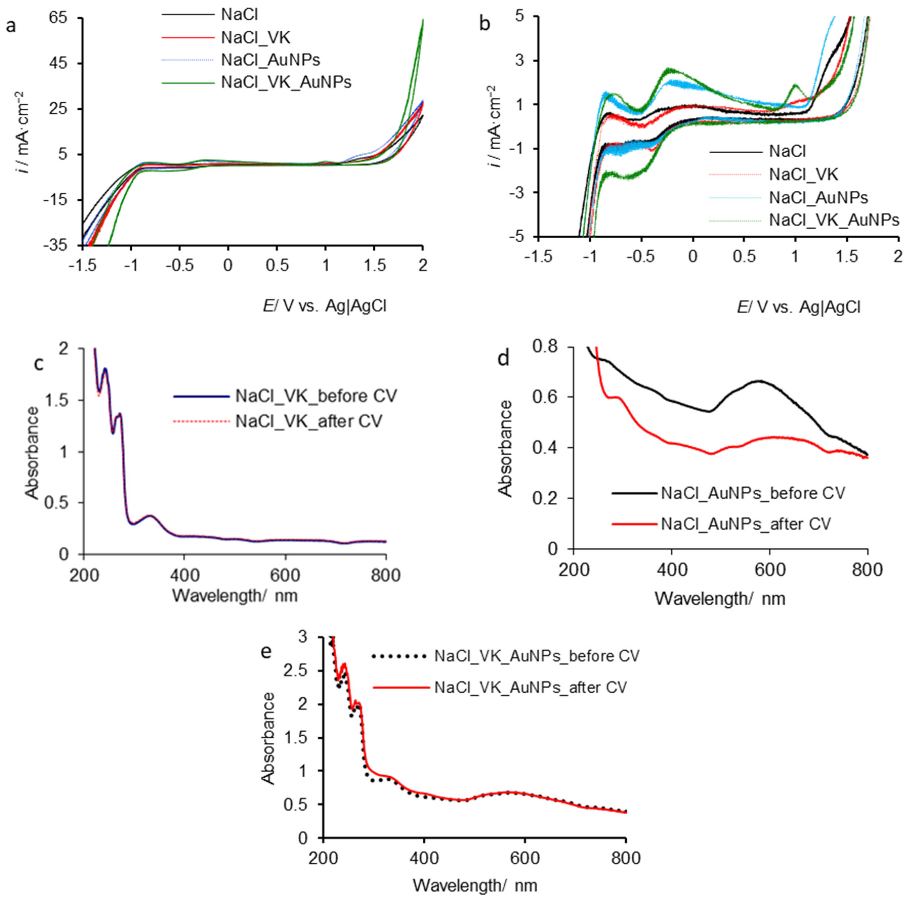
Figure 4a displays apparently similar hysteresis to those discussed above (Figure 3a). The difference consists in the hysteresis maxima, which reach higher current densities (around of 26 mA·cm−2) for NaCl solutions containing vitamin K (NaCl–VK) and gold nanoparticles (NaCl–AuNPs) than those recorded for NaF–VK and NaF–AuNPs, respectively (around of 20 mA·cm−2), and a much higher value (63 mA·cm−2) for NaCl–VK–AuNPs compared to 30 mA·cm−2 achieved for NaF–VK–AuNPs.
Thus, the processes could be similar to those that occurred in the presence of fluoride ions, but more intense, chloride ions acting as activators of oxidation processes [33,34,35]. The detail from Figure 4b displays a voltammogram shape, obtained in NaCl–VK, approximately similar to that recorded in fluoride presence (Figure 3b) and consequently, the vitamin degradation from the solution can be restricted by the adsorbed film on the electrode surface.
In contrast, in the case of NaCl–AuNP and NaCl–VK–AuNP solutions (Figure 4b), the voltammograms have completely different shapes comparative to those obtained in the case of the NaF electrolyte (Figure 3b).
During the anodic scan, pronounced oxidation peaks are observed in both media of NaCl–AuNPs and NaCl–VK–AuNPs. Thus, it is possible that gold nanoparticles are the subject of an irreversible transformation/degradation, especially when they are in the NaCl solution as a single compound (NaCl–AuNPs). In this case, it is not registered as significant reduction processes, during the cathodic pathway, unlike the NaCl solution containing both the compounds (NaCl–VK–AuNPs), which generate an intense cathodic correspondent, namely the peak centered at −0.5 V.
It is difficult to estimate the formation mechanism of some chemical species, as well as the reversibility/irreversibility of oxidation reactions, but it can be suggested that gold nanoparticles are subjected to certain transformations. Therefore, in the NaCl solution containing both VK and AuNPs (NaCl–VK–AuNPs), the vitamin constitutes a protective coating for gold nanoparticles, acting on both the anodic process and the evolution of cathodic reactions.
Figure 4c–e present the UV-Vis spectra recorded before and after cyclic voltammetry (CV). As in the previous case, for the NaCl solution containing only the vitamin (NaCl–VK), superimposed spectra were recorded, suggesting its chemical stability, under the given conditions (Figure 4c). Instead, the gold nanoparticles (Figure 3d) significantly degrade compared to those from the solution containing both the compounds, in terms of NaCl–VK–AuNPs (Figure 4d). As shown in Figure 4e, the acquired AuNP spectra, before and after the CV are overlapped, thus attesting to their stability.
The vitamin spectrum is altered (Figure 4e); the absorption maxima from 244 nm and 268 nm increase, and the one from 332 nm flattens, indicating a certain instability of the vitamin in the presence of gold nanoparticles. Consequently, the vitamin acts as a stability agent for AuNPs, but gold nanoparticles constitute a susceptibility factor regarding vitamin K degradation. It can be that over time, the impact of gold nanoparticles on the vitamin’s stability becomes much more pronounced, activating its decomposition/degradation reaction. Along with polyphenols, alkaloids, or amino acids, vitamins are used for the synthesis of nanoparticles precisely because of the specific interaction between them and their ability to prevent agglomeration [25,26,27].
In addition, over time, the protective action of the vitamin on the gold nanoparticles can diminish, thus facilitating their degradation. Consequently, for a short time, vitamin K and gold nanoparticles have a relative compatibility that, over time, under the action of multitude factors can attenuate or turn into incompatibility.

3.2.3. Vitamin K/Gold Nanoparticle Behavior in the Presence of Br Ions

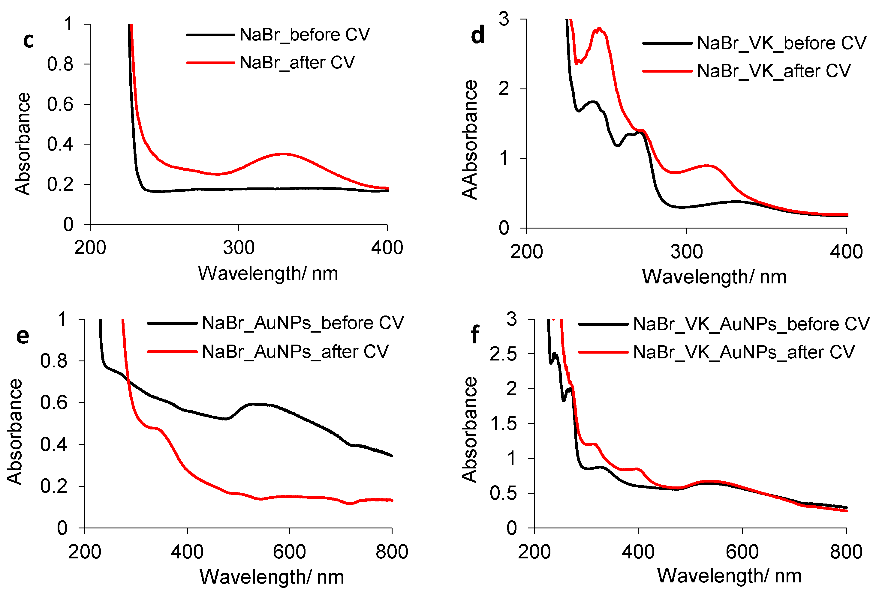
The cyclic voltammograms shown in Figure 5a are completely different from those previously obtained for fluoride and chloride ions. The hysteresis maxima reach high current densities (100 mA·cm−2), except for the one recorded for the NaBr solution containing both compounds (NaBr–VK–AuNPs), whose maximum rises until approximately 50 mA·cm−2. Numerous fluctuations can be observed, especially on the hysteresis obtained for the NaBr blank solution and the NaBr solution containing AuNPs. In these cases, during the anodic and cathodic paths, uncontrolled oxidation/reduction processes probably take place.
The anodic and cathodic scans of the NaBr solution containing the vitamin (NaBr–VK) and of that containing both compounds (NaBr–VK–AuNPs) are apparently smoother than that of the two mentioned above due to the vitamin film development by its adsorption on the platinum electrode [21,22]. The detail designed in Figure 5b shows that at low current densities, there is a relative surface passivation, probably due to the formation of oxybromide species [31,33], which agglomerate at the platinum/electrolyte interface, thus blocking/delaying certain electrode processes. The large peaks highlighted on the cathodic scan and centered around 0.7 V define a response to the oxidation processes that occur around this value, but they are not explicitly visible on anodic scans due to intense fluctuations.
The cathodic peak detected at 0.78 V, in the case of the NaBr solution containing only vitamin K (NaBr–VK), has the lowest intensity, because the thin surface film inhibits the oxidation processes from the solution, and thus the reduced species are small in number.
For the solution containing gold nanoparticles (NaBr–AuNPs), the cathodic peak can correspond to the reduction of some oxidized species of gold consisting of AuBr4 (0.82 V) and AuBr2 (0.96 V).
The extended cathodic peak, displayed for the solution containing both compounds (NaBr–VK–AuNPs), reaches the highest current density, probably due to the interferences of several cathodic processes. In the presence of AuNPs, VK can exercise an electrocatalytic effect on the reduction processes, and it can form a self-assembled protective layer, such as a capsule, around gold nanoparticles.
The displacement of the cathodic peak to 0.7 V is caused by the numerous reduction processes that take place in the NaBr solution, including the reduction of other bromide species [31,33,35].
On the other hand, the cyclic voltammogram of the supporting electrolyte (Figure 5b) is characterized by the higher cathodic current density, than that recorded for the NaBr–VK and NaBr–AuNPs, indicating that HER is favored to a greater extent. The experimental results in this case highlight the shift of the onset potential for HER to more positive values. Unlike fluoride and chlorine anions, in the presence of bromide, the range of potential corresponding to a relative stability is narrowed down, and the onset potential for increasing the charge transfer rate is the smallest. The high value of anodic current density (100 mA·cm−2) is caused by OER overlapping with other electrochemical oxidation processes of bromide with the formation of bromine, hypobromite, bromite, bromate, and perbromate, which are subsequently reduced on the cathodic pathway.
UV-Vis spectra, before and after CV are shown in Figure 5c–f. During cyclic voltammetry, certain species of bromide appear in the NaBr blank solution revealing an absorption maximum located at 334 nm (Figure 5c) that can interfere with the one of vitamin K from 332 nm.
After cyclic voltammetry, the vitamin spectrum recorded in the NaBr–VK solution is completely different from the initial one (Figure 5d), showing two peaks at 319 nm and 248 nm, respectively, which can be attributed to new compounds and/or a subsequent transformation of vitamin K1 into another form.
As shown in Figure 5e, the gold nanoparticles from the NaBr–AuNP electrolyte are imperceptible, and the peak centered at 550 nm is no longer noticeable after cyclic voltammetry. For the NaBr–VK–AuNP system, the AuNP spectra recorded before and after cyclic voltammetry are overlapped, indicating that the gold nanoparticles are stable, but the vitamin K is completely degraded (Figure 5f). Gold nanoparticles show a range of colors, such as brown, orange, red, and purple, in aqueous solution as a function of core size and generally a size-relative absorption peak of at 500 to 550 nm [25,26,27].
Consequently, in NaBr solutions containing only vitamin K (NaBr–VK) or gold nanoparticles (NaBr–AuNPs), both compounds degrade significantly, unlike the NaBr solution where these coexist (NaBr–VK–AuNPs) when the vitamin degrades completely and the gold nanoparticles are stable. Thus, the two compounds show incompatibility in the presence of bromide ions.
Likewise, the stability of citrate (stabilizing agent) is influenced by the supporting electrolyte composition. Thus, in the NaBr solution, citrate has the lowest protection efficiency on colloidal gold nanoparticles (Figure 5d), unlike the fluoride ions, which do not significantly impact on the ligand, and, consequently, the gold nanoparticles have a greater stability (Figure 3d).
In the case of NaX–AuNPs during cyclic voltammetry, the environment color change from pink to intense red, suggesting certain random agglomerations of gold nanoparticles, knowing that nanodispersions containing larger size nanoparticles have more nuanced colors.

3.2.4. The Calculation of the Net Amount of Electric Charge Passed on Electrode Active Area

In order to provide additional information on the competitiveness of the chemical species from the studied electrolytes, the net amount of electric charge (q) passed on the electrode active area was computed based on Equation (1) presented below.
q = idt
where “i” is the current density (A cm−2); “q” is the electric charge (C cm−2), and “t” is the time (s).
Practically, the current density (i) is determined from the cumulative condition of the anodic scan maximum current density (ia) and minimum cathodic one (ic) for the potential range on which cyclic voltammetry hysteresis is designed, respectively, between 1000 mV and 2000 mV (Figure 3a, Figure 4a and Figure 5a). Thus, Equations (2) and (3) are obtained as follows:
q = i a i c dt
q = i a i c · t
The potential conversion over time can be obtained by dividing the potential range applied between anode and cathode by the scan rate. Finally, the net amount of electric charge passed on the electrode active area (q) is obtained in A·s·cm−2 meaning C·cm−2. The results are shown in Table 1.
Analyzing data from Table 1, we observed that the electric charge passed on the electrode active area (q) decreases, both in the presence of vitamin k (NaF–VK) and AuNPs (NaF–AuNPs) and increases in the electrolyte containing the two compounds (NaF–VK–AuNPs), compared to that obtained in the NaF blank solution.
This can be associated with the affinity of the platinum electrode for vitamin K and colloidal gold, with their diffusion at the electrode/electrolyte interface prevailing that of fluoride ions. Thus, the film developed on the platinum surface by the adsorption of vitamin K assures adequate protection and attenuates the platinum electrocatalytic effect on the vitamin K degradation reaction.
In the case of the NaF–AuNP system, the lowest value for q was obtained, which explains the absence of oxidation peaks on the anodic scan from Figure 3b.
The very small dimension of the nanoparticles facilitates their diffusion toward the electrode and, more or less hypothetically, some metal–metal bonds between gold and platinum appear leading to the surface overlayer development.
As in the previous case of the vitamin, the platinum electrocatalytic effect is lowed, inducing the delay of the gold degradation reaction.
As shown in Figure 3c,d, the spectral characteristics of the vitamin and nanoparticles do not show significant changes while maintaining the integrity of the compounds.
In the case of the electrolyte containing both compounds (NaF–VK–AuNPs), the net amount of electric charge (q) increases due to the appearance of random ionic agglomerations in the electric double layer, leading to an uneven surface over-layer with uncontrolled development dynamics. A slightly decrease in the net amount of electric charge takes place without affecting the stability of vitamin K and the gold nanoparticles, as displayed by the UV-Vis spectra from Figure 3e.
The retention of the chloride ions across the platinum is diminished compared to that of the fluoride ions, which leads to a lower electric charge passed on the electrode surface.
At the same time, chloride ions prevent the diffusion to the electrode surface of both vitamin molecules and gold nanoparticles, thus retarding the formation of the surface overlayer. The initiation of certain competitive oxidation processes that are more intense in the case of gold nanoparticles than of vitamin K ones takes place, as confirmed by the oxidation peak highlighted in Figure 4b.
Thus, the relative stability of vitamin K and the sensitivity of gold nanoparticles in a sodium chloride solution are explained, with the results being in good agreement with UV-Vis spectra from Figure 4c,d.
Increasing the net amount of electric charge in the NaCl–VK–AuNP electrolyte impacts more on the vitamin K stability than on that of the gold nanoparticles, which was also demonstrated by UV-Vis spectrophotometry (Figure 4e).
Moreover, the net amount of electric charge passed on the electrode active area in the NaCl solution reaches a higher value than that which was calculated in the NaF solution, influencing the vitamin K decomposition reaction, but ensuring a good protection of gold nanoparticles (Figure 4e).
In the presence of bromide ions, a small variation of the net amount of electric charge is observed in the case of NaBr–VK and NaBr–AuNP systems compared to that obtained for the NaBr blank solution. The numerous ionic species agglomerated at the electrode/electrolyte interface are generated by simultaneous and uncontrolled oxidation processes, as shown in the previous sections. Thus, the instability of the two compounds in the NaBr–VK and NaBr–AuNP systems and the changes in the spectral characteristics after cyclic voltammetry are confirmed (Figure 5d,e).
An unexpected result was obtained for the sodium bromide solution containing both compounds (NaBr–VK–AuNPs) when the electric charge passed on the electrode active area has the lowest value, as opposed to the previous cases.
This is probably due to the competitive adsorption of vitamin degradation products on the platinum surface followed by a protective film formation that inhibits the oxidation of gold nanoparticles (Figure 5f).

3.3. Evolution of Vitamin K and Gold Nanoparticles over Time in NaX Solutions Studied by Constant Current Density Electrolysis Assisted by UV-Vis Spectrophotometry

To study the stability of vitamin K and gold nanoparticles over time, the above-mentioned media were subjected to electrolysis at a constant current density of 50 mA·cm−2. The samples were taken minute by minute and analyzed by UV-Vis spectrophotometry. For the accuracy of the spectrophotograms, the spectra obtained every 10 min were selected. The results are presented in Figure 6, Figure 7 and Figure 8, and these were comparatively discussed by categories as follows: (i) the electrochemical behavior of vitamin K in the presence of the studied anions (NaX–VK; X = F, Cl, Br); (ii) the electrochemical behavior of gold nanoparticles (NaX–AuNPs; X = F, Cl, Br); (iii) finally, the effect of the compounds on one another when both coexist in the halide solutions (NaX–VK–AuNPs; X = F, Cl, Br).

3.3.1. Degradation of VK over Time in NaX Solutions

The UV-Vis spectra recorded in NaX–VK solutions, time one hour, are represented in Figure 6. Throughout the electrolysis, vitamin K is stable, and in the presence of F ions, the spectra collected during the electrolysis are superimposed over the initial one (Figure 6a) and are unstable in the presence of Cl (Figure 6b) and Br ions (Figure 6c).
As shown in Figure 6b, in the NaCl–VK solution, after 30 min, the characteristics of the VK spectrum in the presence of Cl ions are changed, by the appearance of the peak located at 305 nm and the significant increase in the absorption maximum from 244 nm, suggesting the vitamin K1 electrotransformation, in another form, and/or the appearance of other compounds that absorb at the same wavelength. Certainly, after 40 min, the compounds from 244 nm are almost completely degraded, and new decomposition/degradation products, which absorb at 280 nm and around 300 nm, are noticeable. The extended peak of the spectrum, with a shoulder at 300 nm, induces a mixture of decomposition products to form that absorb at a wavelength between 350 nm and 260 nm.
In the presence of Br ions (Figure 6c), the spectral characteristics of the vitamin are completely modified, and its possible traces are highlighted only after a time of 10 min. The wide peak at 336 nm is formed as a result of interferences between the bromide compounds [31,33] and other environmental species that present absorption at a wavelength of 336 nm. After 30 min, the absorption maxima show fluctuations and noises that increase in intensity over time, which means that the composition of the environment is completely changed. The results are in agreement with those obtained from cyclic voltammetry: (i) in the presence of F ions (NaF–VK), vitamin K is stable; (ii) in the NaCl solution (NaCl–VK), vitamin K shows an inertia, time of 10 min, after which it degrades; (iii) in the NaBr–VK solution, vitamin K shows lability, also caused by the byproducts resulting from the degradation of the sodium bromide.
The degradation mechanism of vitamin K was proposed. The vitamin K1 molecule has two double bonds susceptible to oxidation in its structure [15,19,38]. As shown in Scheme 1, vitamin 2,3-epoxide is the main compound resulting from vitamin oxidation at the -C=C- bond from position 2 of the quinone ring [15,19,38]. The second double bond that can be oxidized is located in position 2 of the hexadecane side chain. In this case, the electrooxidation can lead to hydroxyl, ketone, or carboxyl groups (Scheme 2). Their subsequent oxidation to a higher oxidation state can be achieved with chain cleavage and oxidative decarboxylation.

3.3.2. Spectroelectrochemical Behavior of AuNPs over Time in NaX Solutions

The electrochemical behavior of gold nanoparticles in solutions of sodium halides is shown in Figure 7a–c.
In the presence of F ions (Figure 7a), the degradation of gold nanoparticles takes place, “with faster disappearance of those with smallest size” [39], leading to the change of the spectral characteristics, such as the shift of the absorption maximum from 550 nm to 580 nm and a significant baseline alteration. Although cyclic voltammetry showed a slight susceptibility of gold nanoparticles in the NaF solution, in contrast over time and under given conditions, these are instable, but the process can be controlled.
The spectrum from the initial moment undergoes over time a changing of its characteristics (shape and position). Consequently, the absorbance regression over time suggests that the number of particles on volume unit decreases, with their concentration respecting the same tendency. The absorption maximum becomes much wider; if initially it is centered around 550, at the end of the experiment, it stretches over a range of almost 200 nm. This feature is specific to changing the structure of nanoparticles; therefore, the electrolyte system contains nanoparticles with different sizes, and their agglomeration leads to the occurrence of some nanoaggregates.
In the NaCl (Figure 7b) and NaBr (Figure 7c) solutions, the maximum located at 550 nm completely disappears, after 20 min and 10 min, respectively, and other adsorption maxima were formed at wavelengths of 310 nm and 340 nm, respectively, consequently changing the composition of the environments.

3.3.3. Spectroelectrochemical Behavior of VK/AuNP System over Time in NaX Solutions

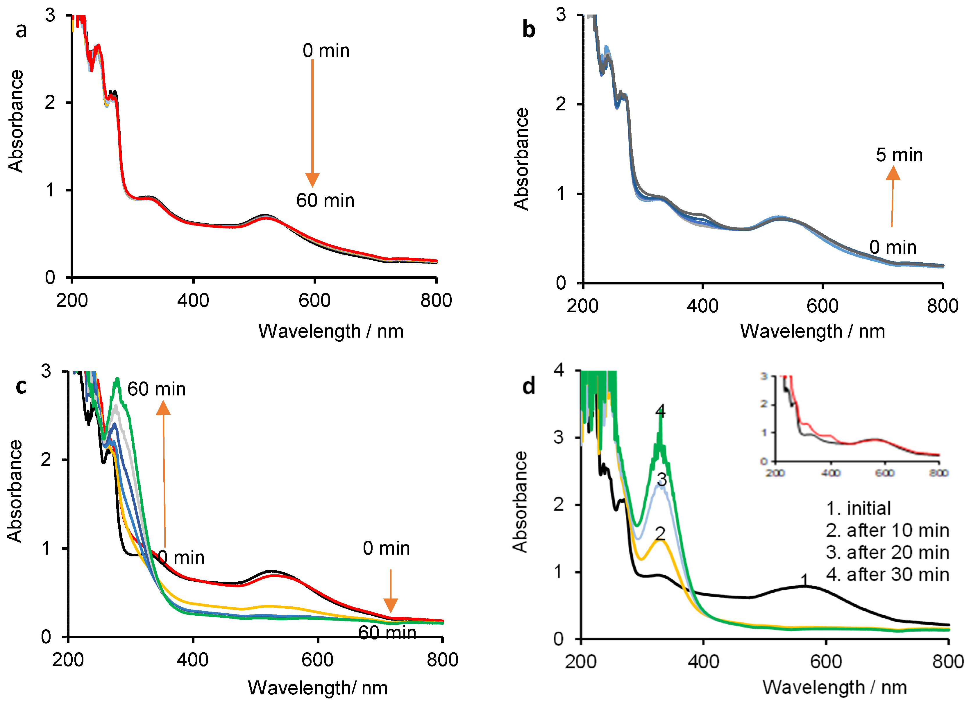
The UV-Vis spectra from Figure 8 illustrate the electrochemical behavior of the system consisting of vitamin K and gold nanoparticles (VK–AuNPs), in sodium halide solutions (NaX–VK–AuNPs), during electrolysis at a constant current density.
In the presence of F ions (Figure 8a), both vitamin K and gold nanoparticles have good stability, reflecting a similar trend to that of cyclic voltammetry (Figure 3e), mutually protecting their stability.
In the presence of Cl ions, for five minutes, the VK–AuNP system is relatively stable (Figure 8b), following the same tendency as in cyclic voltammetry (Figure 4e). As shown in Figure 8c, after 10 min, for both compounds, the instability is amplified similarly to that displayed by the solutions containing VK and AuNPs, as a single compound, NaCl–VK and NaCl–AuNPs, respectively (Figure 6b and Figure 7b), and consequently leading to the solution composition alteration. Thus, the compatibility of the VK–AuNP system in the presence of chloride ions must be viewed with reservations because their synergy, in terms of stability, acts for a short time. The vitamin–gold interactions involve an induced effect in both directions; VK molecules are degraded in the presence of AuNPs, and at the same time, AuNPs are unstable in the presence of VK molecules. The gold nanoparticles are very sensitive due to a cumulative effect of chlorinated and oxychlorinated species, without being able to distinguish between them.
In the presence of Br ions (Figure 8d), the VK–AuNP system maintains a similar tendency to that of cyclic voltammetry (Figure 5f), for two minutes, when the vitamin degrades completely, and the nanoparticles show relative stability (detail inserted in Figure 8d). After ten minutes, both compounds are completely unnoticeable, with the gold nanoparticles respecting the same tendency as in the case of the solution containing it as a single compound (Figure 7c). The bromide oxidation processes to bromine, hypobromite, bromite, bromate, and perbromate have a considerable impact on the electrochemical behavior of the two components, accelerating the vitamin K1 degradation reaction and significantly affecting the stability of AuNPs.
The results of the electrochemical measurements are systematized in Table 2.
The levels of such high potential values are certainly not achievable in the human body, but a multitude of internal and external factors that act on living organisms altering both the biological response and the metabolic breakdown, cause the changes in the chemical structure of various ingested compounds. Biotransformation requires the knowledge of all parameters that can only be determined by in vivo tests, e.g., (i) the way, rate, and absorption degree in living organisms; (ii) metabolic breakdown and elimination rate; (iii) the amount stored in the organs, the cumulative actions, and the sensitivity of the species subjected to the experiment etc.
The investigations in simulated solutions certainly have a completely different approach, but the results can be an indication regarding the stability and incompatibility of chemicals, whose random administration can have a negative impact on human health.
In addition, the administration of alternative treatments must follow a coherent medical protocol, so that there are no implications on the biological activity of other compounds. At the same time, the instantaneous interactions that can occur between chemical compounds can lead to the generation of some toxins that also affect human health.
The mechanism of decomposition reaction of the vitamin can be carried out directly or indirectly. Indirect degradation in the presence of nanoparticles may involve interactions with nanocolloid, halide ions, and/or oxyhalide ions (hypochlorite, chlorite, chlorate, and perchlorate), as well as reactive oxygenated species (e.g., hydroxyl).
The electrotransformation mechanism of nanoparticles in the presence of vitamins can occur in a much more difficult way due to the multitude of species present in the electrolyte solution. Thus, the particles can change their state along with the degradation of the stabilizing agent. This leads to an increase in the oxidation susceptibility of the colloid in the presence of vitamin molecules, intermediate ionic molecular species, oxyhalogenated oxidant ions, or reactive oxygenated species. These processes may be responsible for the partial oxidation of the colloid with the formation of semioxidic particles and/or their agglomeration.

4. Conclusions

The electrochemical behavior of both vitamin K as well as gold nanoparticles, as individual compounds and in mixture, in sodium halide solutions, was investigated by cyclic voltammetry and electrolysis at a constant current density assisted by UV-Vis spectrophotometry.
The motivation of the study is based on the fact that there may be practical situations in which all these species come into contact. Halogen ions are components of daily ingestion, while vitamins and nanoparticles are increasingly indicated for consumption due to their antioxidant and antitumor properties.
The results obtained by association using the spectroscopic method with cyclic voltammetry lead to the following conclusion: (i) the fluorine ion being inert, does not modify the electrooxidation mechanism of vitamin K or nanoparticles. AuNPs have an electrocatalytic effect on the electrooxidation of vitamin molecules; (ii) chloride and bromide ions are electrochemically active, thus leading to the electrogeneration of oxygenated species that have a strong oxidizing character. AuNPs are oxidized in the presence of these ions, while in the presence of vitamin molecules, the oxidation processes of the vitamin predominate. In other words, vitamins have a role in protecting nanoparticles from oxidation; (iii) in the presence of iodine, specific interactions are highlighted that lead to much faster oxidation of AuNPs. Vitamin molecules no longer protect AuNPs.
The UV-Vis-electrolysis study concludes that in the case of fluoride-containing systems, the highest stability of the vitamin is in the two-component system, and the nanoparticles have the highest stability in the three-component system. Chloride ions decrease VK stability and even AuNP stability in both two- and three-component systems. The presence of bromide ions indicates specific vitamin–gold interactions that lead to a degradation of both vitamin and nanoparticles; a delayed effect of the vitamin on the stability of the nanoparticles is no longer observed.
In the presence of fluoride ions, vitamin K and AuNPs show good stability, both individually and in mixture. Chloride ions are more active, restricting their stability over a long time. In the presence of Br ions, the compounds are unstable, both when alone or coexisting in the sodium bromide solution. Iodine ions have the greatest impact on stability, in the sense that their action manifests from the first contact of the studied compounds with the environment.
This study can be indicative, revealing the trend regarding the appearance of interactions among vitamin K, gold nanoparticles, and halide ions, as well as their stability and chemical compatibility.

Author Contributions

All authors had equal contribution to the article along with the first author. Conceptualization, A.S.; methodology, A.S. and B.T.; software, B.T. and B.O.; validation, B.O.; investigation, B.T.; resources, B.O.; writing—original draft preparation, A.S. and B.T.; writing—review and editing, B.O.; supervision, A.S.; funding acquisition, A.S., B.T. and B.O. All authors have read and agreed to the published version of the manuscript.

Funding

This research received no external funding.

Data Availability Statement

Data is contained within the article.

Conflicts of Interest

The authors declare no conflict of interest.

References

  1. Malhotra, R.; Nicholson, C.J.; Wang, D.; Bhambhani, V.; Paniagua, S.; Slocum, C.; Sigurslid, H.H.; Cardenas, C.L.; Li, R.; Boerboom, S.L.; et al. Matrix Gla protein levels are associated with arterial stiffness and incident heart failure with preserved ejection fraction. Arterioscler. Thromb. Vasc. 2022, 42, e61–e73. [Google Scholar] [CrossRef] [PubMed]
  2. Tarento, T.D.C.; McClure, D.D.; Vasiljevski, E.; Schindeler, A.; Dehghani, F.; Kavanagh, J.M. Microalgae as a source of vitamin K1. Algal Res. 2018, 36, 77–87. [Google Scholar] [CrossRef]
  3. Wei, X.; Hang, Y.; Li, X.; Hua, X.; Cong, X.; Yi, W.; Guo, X. Effects of dietary vitamin K3 levels on growth, coagulation, calcium content, and antioxidant capacity in largemouth bass, Micropterus salmoides. Aquac. Fish. 2021, in press. [Google Scholar] [CrossRef]
  4. Giarma, E.; Mpampali, Z.; Lialiaris, T.; Mourelatos, D. Cytoprotective and genotoxic effects of vitamins K1 and B1 on irinotecan in vitro. Mutat. Res./Genet. Toxicol. Environ. Mutagenesis 2019, 837, 60–64. [Google Scholar] [CrossRef]
  5. Chetot, T.; Mouette-Bonnet, M.; Taufana, S.; Fourel, I.; Lefebvre, S.; Benoit, E.; Lattard, V. Differences in teratogenicity of some vitamin K antagonist substances used as human therapeutic or rodenticide are due to major differences in their fate after an oral administration. Toxicol. Lett. 2020, 333, 71–79. [Google Scholar] [CrossRef]
  6. Bar, A.; Kus, K.; Manterys, A.; Proniewski, B.; Sternak, M.; Przyborowski, K.; Moorlag, M.; Sitek, B.; Marczyk, B.; Jasztal, A.; et al. Vitamin K2-MK-7 improves nitric oxide-dependent endothelial function in ApoE/LDLR−/− mice. Vasc. Pharmacol. 2019, 122–123, 106581. [Google Scholar] [CrossRef] [PubMed]
  7. Apalset, E.M.; Gjesdal, C.G.; Eide, G.E.; Tell, G.S. Intake of vitamin K1 and K2 and risk of hip fractures: The Hordaland Health Study. Bone 2011, 49, 990–995. [Google Scholar] [CrossRef]
  8. Moore, A.E.; Kim, E.J.; Dulnoan, D.; Dolan, A.L.; Voong, K.; Ahmad, I.; Gorska, R.; Harrington, D.J.; Hampson, G. Serum vitamin K1 (phylloquinone) is associated with fracture risk and hip strength in post-menopausal osteoporosis: A cross-sectional study. Bone 2020, 141, 115630. [Google Scholar] [CrossRef] [PubMed]
  9. Rho, S.-J.; Kim, Y.-R. Improving solubility and stability of fat-soluble vitamins (A, D, E, and K) using large-ring cycloamylose. LWT Food Sci. Technol. 2022, 153, 112502. [Google Scholar] [CrossRef]
  10. Campani, V.; Biondi, M.; Mayol, L.; Cilurzo, F.; Pitaro, M.; De Rosa, G. Development of nanoemulsions for topical delivery of vitamin K1. Int. J. Pharm. 2016, 511, 170–177. [Google Scholar] [CrossRef] [PubMed]
  11. Goto, S.; Setoguchi, S.; Nagata-Akaho, N.; Terada, K.; Watase, D.; Yamakawa, H.; Toki, E.; Koga, M.; Matsunaga, K.; Karube, Y.; et al. Ester derivatives of phyllohydroquinone effectively deliver the active form of vitamin K1 topically, owing to their non-photosensitivity. Eur. J. Pharm. Sci. 2020, 155, 105519. [Google Scholar] [CrossRef] [PubMed]
  12. Samadi, N.; Azar, P.A.; Husain, S.W.; Maibach, H.I.; Nafisi, S. Experimental design in formulation optimization of vitamin K1 oxide-loaded nanoliposomes for skin delivery. Int. J. Pharm. 2020, 579, 119136. [Google Scholar] [CrossRef] [PubMed]
  13. Tabassum, R.; Gupta, B.D. Simultaneous estimation of vitamin K1 and heparin with low limit of detection using cascaded channels fiber optic surface plasmon resonance. Biosens. Bioelectron. 2016, 86, 48–55. [Google Scholar] [CrossRef] [PubMed]
  14. Cholerton, S.; Park, B.K. Quantitative analysis of pharmacological concentrations of vitamin K1 and vitamin K1 2,3-epoxide in rat liver by high-performance hquid chromatography. J. Chromatogr. Biomed. Appl. 1986, 375, 147–153. [Google Scholar] [CrossRef]
  15. Gentili, A.; Cafolla, A.; Gasperi, T.; Bellante, S.; Caretti, F.; Curini, R.; Pérez Fernández, V. Rapid, high performance method for the determination of vitamin K1, menaquinone-4 and vitamin K1 2,3-epoxide in human serum andplasma using liquid chromatography-hybrid quadrupole linear iontrap mass spectrometry. J. Chromatogr. A 2014, 1338, 102–110. [Google Scholar] [CrossRef]
  16. Zhang, Y.; Chhonker, Y.S.; Bala, V.; Hagg, A.; Snetselaar, L.G.; Wahls, T.L.; Murry, D.J. Reversed phase UPLC/APCI-MS determination of vitamin K1 and menaquinone-4 in human plasma: Application to a clinical study. J. Pharmaceut. Biomed. 2020, 183, 113147. [Google Scholar] [CrossRef] [PubMed]
  17. Japelt, R.B.; Jakobsen, J. Analysis of vitamin K1 in fruits and vegetables using accelerated solvent extraction and liquid chromatography tandem mass spectrometry with atmospheric pressure chemical ionization. Food Chem. 2016, 192, 402–408. [Google Scholar] [CrossRef]
  18. Konieczna, L.; Kazmierska, K.; Roszkowska, A.; Szlagatys-Sidorkiewicz, A.; Baczek, T. The LC–MS method for the simultaneous analysis of selected fat-soluble vitamins and their metabolites in serum samples obtained from pediatric patients with cystic fibrosis. J. Pharmaceut. Biomed. 2016, 124, 374–381. [Google Scholar] [CrossRef] [PubMed]
  19. Shearer, M.J.; Barkhan, P. Studies on the metabolites of phyleoquinone (vitamin K1) in the urine of man. Biochim. Biophys. Acta 1973, 297, 300–312. [Google Scholar] [CrossRef]
  20. Haque, J.A.; McDonald, M.G.; Kulman, J.D.; Rettie, A.E. A cellular system for quantitation of vitamin K cycle activity: Structure-activity effects on vitamin K antagonism by warfarin metabolites. Blood 2014, 123, 582–589. [Google Scholar] [CrossRef] [Green Version]
  21. Rostami-Javanroudi, S.; Babakhanian, A. New electrochemical sensor for direct quantification of vitamin K in human blood serum. Microchem. J. 2021, 163, 105716. [Google Scholar] [CrossRef]
  22. Sýs, M.; Jashari, G.; Švecová, B.; Arbneshi, T.; Metelka, R. Determination of vitamin K1 using square wave adsorptive stripping voltammetry at solid glassy carbon electrode. J. Electroanal. Chem. 2018, 821, 10–15. [Google Scholar] [CrossRef]
  23. Torkashvand, M.; Gholivand, M.B.; (Arman) Taherpour, A.; Boochani, A.; Akhtar, A. Introduction of a carbon paste electrode based on nickel carbide for investigation of interaction between warfarin and vitamin K1. J. Pharmaceut. Biomed. 2017, 139, 156–164. [Google Scholar] [CrossRef] [PubMed]
  24. Jedlinska, K.; Strus, M.; Bas, B. A new electrochemical sensor with the refreshable silver liquid amalgam film multi-electrode for sensitive voltammetric determination of vitamin K2 (menaquinone). Electrochim. Acta 2018, 265, 355–363. [Google Scholar] [CrossRef]
  25. Aljarba, N.H.; Imtiaz, S.; Anwar, N.; Alanazi, I.S.; Alkahtani, S. Anticancer and microbial activities of gold nanoparticles: A mechanistic review. J. King Saud Univ. Sci. 2022, 34, 101907. [Google Scholar] [CrossRef]
  26. Kalimuthu, K.; Cha, B.S.; Kim, S.; Park, K.S. Eco-friendly synthesis and biomedical applications of gold nanoparticles: A review. Microchem. J. 2020, 152, 104296. [Google Scholar] [CrossRef]
  27. Alim, S.; Vejayan, J.; Yusoff, M.M.; Kafi, A.K.M. Recent uses of carbon nanotubes & gold nanoparticles in electrochemistry with application in biosensing: A review. Biosens Bioelectron. 2018, 121, 125–136. [Google Scholar]
  28. Tutunaru, B.; Samide, A.; Neamtu, C.; Tigae, C. Spectroelectrochemical studies of interactions between vitamin A and nanocolloidal silver. Int. J. Electrochem. Sci. 2018, 13, 5850–5859. [Google Scholar] [CrossRef]
  29. Samide, A.; Tutunaru, B. Interactions between vitamin C and nanocolloidal silver particles studied by cyclic voltammetry and UV−Vis spectrophotometry. Electroanalysis 2017, 29, 2498–2506. [Google Scholar] [CrossRef]
  30. Samide, A.; Tutunaru, B.; Varut, R.-M.; Oprea, B.; Iordache, S. Interactions of some chemotherapeutic agents as epirubicin, gemcitabine and paclitaxel in multicomponent systems based on orange essential oil. Pharmaceuticals 2021, 14, 619. [Google Scholar] [CrossRef]
  31. Tutunaru, B.; Samide, A.; Spînu, C.-I.; Tigae, C.; Oprea, B. Induced effect of gold nanoparticles (AuNPs) and halide ions on pyridoxine molecule stability. Appl. Sci. 2021, 11, 9470. [Google Scholar] [CrossRef]
  32. Samide, A.; Tutunaru, B.; Bratulescu, G.; Ionescu, C. Electrochemical synthesis and characterization of new electrodes based on poly-hematoxylin films. J. Appl. Polym. Sci. 2013, 130, 687–697. [Google Scholar] [CrossRef]
  33. Tutunaru, B.; Samide, A.; Iordache, S.; Tigae, C.; Simionescu, A.; Popescu, A. Ceftriaxone degradation in the presence of sodium halides investigated by electrochemical methods assisted by UV−Vis spectrophotometry. Appl. Sci. 2021, 11, 1376. [Google Scholar] [CrossRef]
  34. Samide, A.; Tutunaru, B.; Tigae, C.; Efrem, R.; Moanta, A.; Dragoi, M. Removal of Methylene Blue and Methyl Blue from wastewater by electrochemical degradation. Environ. Prot. Eng. 2014, 40, 93–104. [Google Scholar] [CrossRef]
  35. Tutunaru, B.; Tigae, C.; Spînu, C.; Prunaru, I. Spectrophotometry and electrochemistry of Brilliant Blue FCF in aqueous solution of NaX. Int. J. Electrochem. Sci. 2017, 12, 396–412. [Google Scholar] [CrossRef]
  36. Tutunaru, B.; Samide, A.; Moanta, A.; Ionescu, C.; Tigae, C. Electrochemical study of metribuzin pesticide degradation on bismuth electrode in aqueous solution. Int. J. Electrochem. Sci. 2015, 10, 223–234. [Google Scholar]
  37. Hubicka, U.; Padiasek, A.; Zuromska-Witek, B.; Szlósarczyk, M. Determination of vitamins K1, K2 MK-4, MK-7, MK-9 and D3 in pharmaceutical products and dietary supplements by TLC-densitometry. Processes 2020, 8, 870. [Google Scholar] [CrossRef]
  38. Hua, K.; Li, Y.; Ding, R.; Zhai, Y.; Chen, L.; Qian, W.; Yang, J. A simple, sensitive, and high-throughput LC-APCI-MS/MS method for simultaneous determination of vitamin K1, vitamin K1 2,3-epoxide in human plasma and its application to a clinical pharmacodynamics study of warfarin. J. Pharmaceut. Biomed. 2018, 159, 82–91. [Google Scholar] [CrossRef] [PubMed]
  39. Balfourier, A.; Luciani, N.; Wang, G.; Gerald Lelon, G.; Ersen, O.; Khelfa, A.; Alloyeau, D.; Gazeau, D.; Florent Carn, F. Unexpected intracellular biodegradation and recrystallization of gold nanoparticles. Proc. Natl. Acad. Sci. USA 2020, 117, 103–113. [Google Scholar] [PubMed]
Figure 1. The UV-Vis spectra recorded in aqueous environment for different concentrations of vitamin K.
Processes 10 01805 g001
Figure 2. UV-Vis spectra of vitamin K (VK) in concentration of 6.65 × 10⁻⁵ mol·L−1, in the absence and presence of 75 mg·L−1 AuNPs: (a)—in 0.1 mol·L−1 NaF solution; (b)—in 0.1 mol·L−1 NaCl solution; (c)—in 0.1 mol·L−1 NaBr solution; (d)—in 0.1 mol·L−1 NaI solution.
Processes 10 01805 g002
Figure 3. Cyclic voltammograms recorded on platinum electrode in 0.1 mol·L−1 NaF blank solution and 0.1 mol·L−1 NaF solution containing 6.65 × 10⁻⁵ mol·L−1 VK, in the absence and presence of 75 mg·L−1 AuNPs (a) and their details for low current densities (b); UV-Vis spectra collected before and after CV; (c)—in NaF solution containing VK (NaF–VK); (d)—in NaF solution containing AuNPs (NaF–AuNPs); (e)—in NaF solution containing VK and AuNPs (NaF–VK–AuNPs).
Processes 10 01805 g003
Figure 4. Cyclic voltammograms recorded on platinum electrode in 0.1 mol·L−1 NaCl blank solution and 0.1 mol·L−1 NaCl solution containing 6.65 × 10⁻⁵ mol·L−1 VK, in the absence and presence of 75 mg·L−1 AuNPs (a) and their details for low current densities (b); UV-Vis spectra collected before and after CV; (c)—in NaCl solution containing VK (NaCl–VK); (d)—in NaCl solution containing AuNPs (NaCl–AuNPs); (e)—in NaCl solution containing VK and AuNPs (NaCl–VK–AuNPs).
Processes 10 01805 g004
Figure 5. Cyclic voltammograms recorded on platinum electrode in 0.1 mol·L−1 NaBr blank solution and 0.1 mol·L−1 NaBr solution containing 6.65 × 10⁻⁵ mol·L−1 VK, in the absence and presence of 75 mg·L−1 AuNPs (a) and their details for low current densities (b); UV-Vis spectra collected before and after CV; (c)—in NaBr blank solution (NaBr); (d)—in NaBr solution containing VK (NaBr–VK); (e)—in NaBr solution containing AuNPs (NaBr–AuNPs); (f)—in NaBr solution containing VK and AuNPs (NaBr–VK–AuNPs).
Processes 10 01805 g005aProcesses 10 01805 g005b
Scheme 1. The proposed mechanism for the electrochemical oxidation of the VK molecule with the formation of the epoxy structure.
Processes 10 01805 sch001
Figure 6. UV-Vis spectra recorded before and during constant current density electrolysis, every 10 min, for one hour: (a)—in 0.1 mol L−1 NaF solution containing 6.65 × 10⁻⁵ mol·L−1 VK (NaF–VK); (b)—in 0.1 mol L−1 NaCl solution containing 6.65 × 10⁻⁵ mol·L−1 VK (NaCl–VK); (c)—in 0.1 mol L−1 NaBr solution containing 6.65 × 10⁻⁵ mol·L−1 VK (NaBr–VK).
Processes 10 01805 g006
Figure 7. UV-Vis spectra recorded before and during constant current density electrolysis, every 10 min: (a)—in 0.1 mol L−1 NaF solution containing 75 mg·L−1 AuNPs (NaF–AuNPs); (b)—in 0.1 mol L−1 NaCl solution containing 75 mg·L−1 AuNPs (NaCl–AuNPs); (c)—in 0.1 mol L−1 NaBr solution containing 75 mg·L−1 AuNPs (NaBr–AuNPs).
Processes 10 01805 g007
Figure 8. UV-Vis spectra recorded before and during constant current density electrolysis: (a)—in 0.1 mol L−1 NaF solution containing 6.65 × 10⁻⁵ mol·L−1 VK and 75 mg·L−1 AuNPs (NaF–VK–AuNPs); (b)—in 0.1 mol L−1 NaCl solution containing 6.65 × 10⁻⁵ mol·L−1 VK and 75 mg·L−1 AuNPs (NaCl–VK–AuNPs), every minute—from initial time to five minutes; (c)—in 0.1 mol L−1 NaCl solution containing 6.65 × 10⁻⁵ mol·L−1 VK and 75 mg·L−1 AuNPs (NaCl–VK–AuNPs), every 10 min—from initial time to 60 min; (d)—in 0.1 mol L−1 NaBr solution containing 6.65 × 10⁻⁵ mol·L−1 VK and 75 mg·L−1 AuNPs (NaBr–VK–AuNPs), every 10 min—from initial time to 30 min.
Processes 10 01805 g008
Scheme 2. The proposed mechanism for the electrochemical oxidation of the VK molecule at the side chain.
Processes 10 01805 sch002
Table 1. The net amount of electric charge passed on electrode active area deduced from cyclic voltammetry recorded on platinum electrode in sodium halide solutions in potential range from 1000 mV to 2000 mV, with a scan rate of 100 mV s−1.
Sampleε (mV)Time (s)i × 103 (A cm−2)q (C cm−2)
εa (mV)εc (mV)iaic
NaF200010001026.90.3750.266
NaF–VK20.30.290.200
NaF–AuNPs19.1−0.0180.191
NaF–VK–AuNPs30.10.170.299
NaCl21.90.310.216
NaCl–VK27.00.210.267
NaCl–AuNPs29.10.180.289
NaCl–VK–AuNPs63.40.150.632
NaBr92.59.10.834
NaBr–VK87.38.90.784
NaBr–AuNPs90.87.10.830
NaBr–VK–AuNPs59.67.10.498
Table 2. Systematization of the results obtained from electrochemical measurements regarding the chemical stability and incompatibility of vitamin K (VK) and gold nanoparticles (AuNPs) in sodium halide solutions.
CompoundsIons
FClBrI
StabilityCompatibilityStabilityCompatibilityStabilityCompatibilityStability
VKgood-relative-unstable-unstable from the first contact
AuNPSgood-low-unstable-unstable from the first contact
VK–AuNPSgoodgoodfor a short timelow (restrictive)unstableincompatibleunstable from the first contact/incompatible
Publisher’s Note: MDPI stays neutral with regard to jurisdictional claims in published maps and institutional affiliations.