Article

Proteomics Provide Insight into the Interaction between Selenite and the Microalgae Dunaliella salina

1
School of Marine Sciences, Guangxi University, Nanning 530004, China
2
Guangxi Beibu Gulf Marine Research Center, Guangxi Academy of Science, Nanning 530007, China
3
Green and Low-Carbon Technology Research Institute, Guangxi Institute of Industrial, Nanning 530001, China
4
School of Resources, Environment and Materials, Guangxi University, Nanning 530004, China
*
Authors to whom correspondence should be addressed.
Processes 2023, 11(2), 563; https://doi.org/10.3390/pr11020563
Submission received: 3 January 2023 / Revised: 27 January 2023 / Accepted: 31 January 2023 / Published: 13 February 2023
(This article belongs to the Section Biological Processes and Systems)

Abstract

:
Dunaliella salina is currently one of the most commercially valuable microalgae species in the world. In reponse to selenite, D. salina is a microalgae with a high selenium content, thereby increasing its value, which is crucial for increasing its economic value as a nutrional supplement. However, the effects of selenite on D. salina are still unclear, and its molecular mechanism of the response to selenite stress is also elusive. Here, in order to study the effects of selenite on D. salina and the corresponding regulatory mechanism, we characterized the physiological phenotypes of D. salina under different selenite concentrations and carried out a quantitative proteomic study. The results showed that the effective concentration for 50% growth inhibition (EC50) of the algae was 192.7 mg/L after 11 days of cultivation. When selenite concentration was lower than 100 mg/L, selenite did not hinder the growth of D. salina in the early stage, but shortened the cell growth cycle, although cell growth was significantly inhibited when the concentration of selenium was higher than 250 mg/L. Bioaccumulation experiments showed that the content of intracellular selenium in D. salina cells reached the highest level under the treatment with 50 mg/L selenite, and the contents of total selenium and organic selenium in D. salina cells were 499.77 μg/g and 303.01 μg/g (dry weight), respectively. Proteomic analysis revealed that a series of proteins related to stress responses, amino acid metabolism and energy production pathways were profoundly altered by the selenite treatment. Glutathione peroxidase (GPX7), a selenium-containing protein, was identified in the group given the selenium treatment. Moreover, proteins involved in photoreactions and oxidative phosphorylation were significantly upregulated, indicating that D. salina effectively balanced the energy demand and energy production under selenite stress. This study provides novel insights into the responses to selenite of D. salina, a microalgae candidate as a biological carrier of selenium and would be helpful for the development of industrial strains rich in selenium.

1. Introduction

Selenium (Se) has pleiotropic biological functions in humans and is an essential trace nutrient element. It is required for resistance to viral infections and the regulation of the immune and reproductive systems [1]. A number of epidemiological studies have shown the relationship between selenium deficiency and an increase in various pathological risks, such as Keshan disease, Kashin-Beck diseases, cancers, neurogenerative diseases, etc. [2,3]. Unlike other trace elements that act as cofactors, the physiological functions of Se are usually performed by selenoproteins [4,5]. At present, Se deficiency in the diet is a global health concern. It is estimated that 15% of the global population is Se-deficient [6], especially in China, Africa, India and Eastern Europe. According to the recmmendations of WHO/FAO (26–34 μg/day), 39–61% of Chinese residents have a low daily Se intake [6]. Therefore, artificial Se supplementation in selenium-deficient areas is still of important practical significance. Traditional Se supplements include inorganic Se and organic Se, and its bioavailability also depends on its chemical form, as organic selenium was identified as being most available after ingestion [7]. Hence, improving the bioavailability and bioactivity of Se in food through biofortification is a good way to solve the problem of Se deficiency.
As the primary producers of the aquatic ecosystems, microalgae are the main selenium absorbers in the aquatic environments [8]. Unlike higher plants, Se has been shown to be an essential trace element for the growth of some microalgae [9]. Recently, the interactions between Se and microalgae has been investigated in Chlamydomonas reinhardtii [10], Spirulina platensis [11], Chlorella pyrenoidosa [12], Scenedesmus quadricauda [13], Haematococcus pluvialis [14] and D. salina [15,16]. It was found that some microalgae maintain a strong tolerance to Se. Selenite is usually used as the Se source of selenium-rich algae because it has weaker toxicity and higher enrichment efficiency than selenate [17,18]. Previous studies have shown there are two main mechanisms for Se absorption in C. reinhardtii. One is a specific transport system that is rapidly saturated at low concentrations, and the other is a non-specific transport system that is saturated only at high ambient concentrations [19]. The absorption process is influenced by the temperature, pH value, the morphology of selenium, the specific anion concentration, etc. [20]. Upon uptake, inorganic Se could be directly bound to macromolecules, such as proteins and polysaccharides, or further metabolized into various organic compounds with a low molecular weight containing selenium through the assimilation of sulfur, which is continuously accumulated in cells. In addition, microalgae grow quickly and contain many nutritional components, including proteins, carbohydrates, fatty acids, vitamins and minerals, making it a promising food and feed sources for humans and animals [21]. Thus, microalgae are excellent carriers for organic Se bioenrichment, and the cultures of Se-enriched microalgae are a promising new branch of algal biotechnology.
Dunaliella salina, a unicellular halotolerant green alga, is capable of accumulating massive amounts of β-carotene under stressful conditions such as high salinity, high temperature and bright light. It was reported that D. salina has the highest content of β-carotene in the plant kingdom known so far [22]. Thus, it was considered to among the most promising commercially valuable microalgae [23]. Previous studies have proven that D. salina has a strong tolerance to Se and that the absorbed Se is mainly distributed in its proteins; thus, enrichment of organic Se by D. salina may not only improve the nutritional value of it as a food, but may also have great commercial value. However, the effects of selenite on D. salina are still unclear, and the metabolism of Se and the underlying molecular mechanisms involved in D. salina remain unclear. The aim of this study was to evaluate the toxicity or physiological effects of selenite on D. salina, and to examine the proteome changes caused by theaccumulation of Se in Se-enriched D. salina to fully understand selenite’s phytotoxicity and the mechanism of selenite regulation in D. salina, would be beneficial developing products made from selenium-rich D. salina.

2. Materials and Methods

2.1. Materials and Culture Conditions

D. salina GY-H13 was obtained from the Guangyu Biological Technology Co., Ltd. (Shanghai, China). An f/2 medium was adopted in which 1 Lof a medium contained 75 mg NaNO3, 5.653 mg NaH2PO4·2H2O, 3.15 mg FeCl3·6H2O, 4.26 mg Na2EDTA·2H2O, 0.0098 mg CuSO4·5H2O, 0.022 mg ZnSO4·7H2O, 0.01 mg CoCl·6H2O, 0.18 mg MnCl2·4H2O, 0.0063 mg Na2MoO4·2H2O, 0.1 mg vitamin B1, 0.0005 mg biotin and 0.005 mg vitamin B12. The algae was cultured in a 500 mL Erlenmeyer flask containing 400 mL of the medium under a light intensity of 3500 lux with a photoperiod of 12 h/12 h light/dark and a temperature of 20 ± 2 °C. The initial cell density was about 1 × 10⁵ cells/mL, corresponding to an optical density at 640 nm (OD640) of 0.020–0.025. The duration of the culture was 20 days. Algal cells were incubated with sodium selenite (Alfa, 99.75%) at six concentrations (0, 10, 50, 100, 250 and 500 mg/L), and 0 mg/L was set as the control. Each group contained three replicates, shaken periodically. All chemicals were of analytical grade or higher. All solutions and culturing containers were autoclaved before use.

2.2. Algal Growth and Toxicity Test

The abundance of cells in the culture cell abundance and the cells’ diameter were measured under a microscope (Motic microscope AE 2000, Spain) with a plankton counting board. We exposed D. salina to a certain range of sodium selenite for 11 days to obtain the effective concentration for 50% growth inhibition, that is, the initial concentration of selenite that inhibited 50% growth of organisms [24]. Cell counts were measured every three days, and the data were also used to obtain the EC50 at 11 days.

2.3. Measurement of the Chlorophyll Fluorescence Parameters

Measurements of the chlorophyll fluorescence parameters were made using a portable pulse amplitude modulated fluorometer from Water-PAM (Walz, Effeltrich, Germany). Prior to measurement, each sample was incubated for 15 min in the dark to completely eliminate the electrons which accumulated at PQ in the PS II electron chain. After dark adaptation, the measuring light was turned on and the stable minimum fluorescence (Fo) was recorded. A saturation pulse with a duration of only 0.2–1.5 s was then immediately opened to measure the maximum fluorescence (Fm) and the maximal photochemical efficiency of PSII (Fv/Fm, Fv = Fm − Fo).

2.4. Antioxidant Enzymes Assay

Antioxidant enzyme activity assays were performed on algal cells on the fourth and seventh day or the first day after inoculation. For this 50 mL of homogenized algal cells was collected by centrifugation and washed 3 times with phosphate-buffered saline, and the pellets were resuspended in 2 mL ultrapure water. Each sample was pulverized by an ultrasonic cell grinder (ScientzIID, Scientz, China) at 200 W for 10 mins (2 s pulses and 3 s intervals) and centrifuged at 4 °C for 10 min at 10,000× g. The supernatant was collected and immediately used for analysis of the antioxidant enzymes’ activities. The protein content of the enzyme extract was determined with a BCA assay kit (Nanjing Jiancheng Bio Inst, China). The total superoxide dismutase (SOD) activity was assayed by the Hydroxylamine method (T-SOD assay kit, Nanjing Jiancheng Bio Inst, China) based on its ability to inhibit the oxidation of hydroxylamine through the xanthine–xanthine oxidase system. The activity of glutathione peroxidase was measured by determining of the reduced GSH content in the enzyme extract (GSH-PX assay kit, Nanjing Jiancheng Bio Inst, China), since its activity can be reflected by the reaction rate. All steps of the operation followed the manufacturer’s instructions.

2.5. Determination of Total and Organic Se Contents

Total Se and organic Se contents were determined using an atomic fluorescence spectrometer (Jitian, AFS-9230, China). After 12 days of culture, algal cells were harvested by centrifugation at 5000 rpm for 5 min at 20 °C. The pellets were rinsed three times with phosphate-buffered saline to remove the Se adsorbed to the cell walls, and then dried in a freeze drier. The 40 mg dried algal sample was digested with 6 mL of concentrated nitric acid and 2 mL of hydrogen peroxide in a microwave digestion system (Xinyi, MDS-15, China).
The digested solution was placed on a heating plate followed by the addition of 5 mL of 6 M hydrochloric acid, and continual heating until the solution was clear and colorless. The residual solution was reconstituted to 10 mL with double-distilled water and analyzed for total Se content. For determination of the inorganic Se determination, each samples with weights of 40 mg was added into 10 mL of ultrapure water and crushed by sonication. The algal extract was centrifuged at 5000 rpm for 10 min. The supernatant was collected and poured into a separating funnel, then, 5 mL of cyclohexanol was added and fully mix for 20 min before standing, and the water phase was collected. The aqueous phase contained inorganic selenium, which was digested by a microwave digestion. The subsequent operation was consistent with the process used for determining the total selenium. The organic Se content was calculated from the difference between the total Se concentration and the inorganic Se concentration.

2.6. Protein Extraction and Proteomic Analysis

Proteomic analysis was carried out in the control group and the selenite treatment group (100 mg/L). A large amount of D. salina was cultured for 5 days, using the culture methodology described previously, to acquire ample material for proteomic analysis. Algae cells were collected by centrifugation at 5000 rpm for 5 min at 4 °C, and then rinsed three times with phosphate-buffered saline, then immediately frozen at −80 °C. The iST sample pretreatment kit (PreOmics, Germany) was used for sample pretreatment to complete protein extraction, enzymatic hydrolysis and peptide de-salting. The peptide mixtures thus obtained were resuspended in Buffer A (20 mM ammonium formate in ultrapure water, pH = 10.0), and then separated at a high pH using an ultimate 3000 system (Thermo Fisher Scientific, MA, USA) linked to a reverse phase C18 column (4.6 mm × 250 mm, 5 μm, Waters Corporation, MA, USA). Separation was implemented with a linear gradient from 5% B to 45% Buffer B (20 mM ammonium formate in 80% acetonitrile, pH = 10.0) within 40 min. After 15 min of re-equilibration, the column’s flow rate was kept at 1 mL/min and the column’s temperature was maintained at 30 °C. Six fractions were collected and dried in a vacuum concentrator. The fractions were dissolved in 30 μL of Buffer C (0.1% formic acid in ultrapure water) and analyzed by online nanospray LC-MS/MS on an Orbitrap Fusion Lumos coupled to an EASY-nLC 1200 system (Thermo Fisher Scientific, MA, USA). The primary data from data-independent acquisition (DIA) were processed by Spectronaut X (Biognosys AG) with the default settings to identify the proteins. Functional and pathway analysis of the identified proteins was carried out using Blas2GO software, as well as the Gene Ontology (GO) and Kyoto Encyclopedia of Genes and Genomes (KEGG) databases [25].

2.7. Statistical Analysis

Each experiment was performed with three independent biological replicates. Experimental data are expressed as the mean ± SD. The determination of EC50 was based on a four-parameter logistic function using a dose–response model in Prism 9 software. The results are shown as the mean ± standard deviation or mean with 95% confidence intervals (n = 3). One-way ANOVA analysis with Dunnett’s post hoc tests was performed using SPSS 23.0. The level of statistical significance level was defined as p < 0.05. A t-test was applied to analyze the differentially expressed proteins between two experimental groups at a threshold value of p ≤ 0.05 and a fold change (FC) of >1.2.

3. Results and Discussion

3.1. Toxicity and Effects of Selenite on the Growth Dunaliella salina

To explore the optimal concentration range of selenite for promoting growth, the growth of D. salina cells was monitored and observed. After 11 days of cultivation, the dose–response curves of D. salina were obtained (Figure 1A), and the EC50 was 192.7 mg/L (95% confidence interval, 178.8–228.7 mg/L). It was observed that there was no significant difference in the number of algal cells between the experimental group and the control group during the culture process at a selenite concentration of 10 mg/L. However, at concentration of a 50 to 100 mg/L selenite, the growth of both groups was same, but the inhibitory effect grew stronger with the increase in the concentration in the later stage. However, algal cell numbers decreased by 84–99% at selenite concentrations of 250–500 mg/L, and the growth of algae was severely inhibited (Figure 1B). After 11 days of exposure, the average diameter of cells in the control group was 11.20 μm, while the average diameter of cells exposed to 500 mg/L selenite was 7.37 μm. The average diameter of cells decreased with the increase in the selenite exposure concentration. (Figure 1C and Supplemental Figure S1). Prolonged exposure (>30 d) of D. salina to Se concentrations induced cell shrinkage, decolorization and the formation of amyloplasts. As the selenite concentration increased, the medium in each group turned red and the algal cells were gradually disrupted (Figure 1C and Supplemental Figure S2).
Se is an essential trace element for the growth and metabolism of some algae [26]. Low doses of Se promote the cell growth of some algae, but at high doses, the growth of most algae was inhibited [27]. In this study, it was found that low doses of selenite negatively impacted the growth of D. salina cells, but the toxicity was low, whereas high doses of selenite significantly inhibited the growth of cells. The phenomenon observed here is as same as that previously reported for H. pluvialis [14]. Since the selenium requirements of algae are generally low, concentrations between 0 and 10 mg/L may promote the growth of algal cells without harming the algae. Different algae have different levels of sensitivities to selenite. The EC50 of selenite on D. salina (192.7 mg/L, 11 d) was much higher than that for C. reinhardtii [8] (6.3 mg/L, 4 d), H. pluvialis [14] (24.02 mg/L, 15 d) and wild-type N. oceanica [17] (12.94 mg/L,4 d), which indicated that D. salina has a strong tolerance of selenite. It was reported that selenite treatment would cause the volume of C. rheiniscens cells to become larger [28]. However, we found that the selenite treatment made D. salina cells smaller, suggesting there were adaptive responses to high selenium concentrations. In addition, similar reddening of the medium was also observed in some plants and microorganisms. Several studies have shown that this red substance consists of Se nanoparticles [10,29], suggesting a similar reduction mechanism in the cells of these organisms.

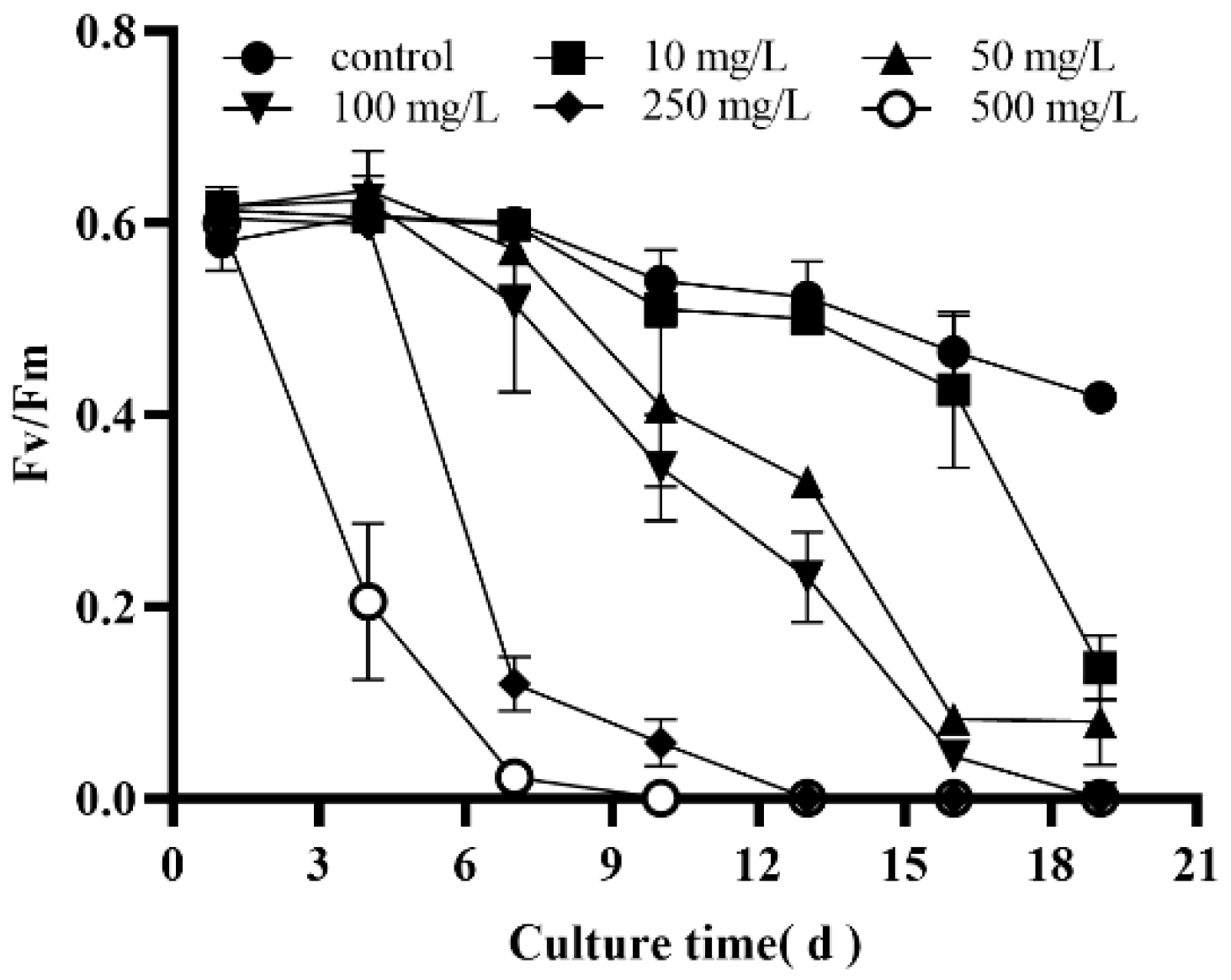
3.2. Effects of Selenite on the Photosynthesis of Dunaliella salina

The effects of selenite on the photosynthesis of D. salina were evaluated by monitoring the photochemical performance of the Photosystem II (PSII) and the content of cellular pigments. The Fv/Fm reflects the potential maximum light energy conversion efficiency of plants and is an important index for studying the effects of photoinhibition or various environmental stresses on photosynthesis. For algae, the Fv/Fm in a healthy physiological state does not have a fixed value (it generally falls between 0.65 and 0.8), because of the large variation in their evolution. In the control group, the Fv/Fm decreased slowly with an increase in the incubation time, which may have been caused by the gradual depletion of nutrients in the culture medium and the aging of the population. At the early stage of culture, there was no difference in the maximum fluorescence yield of PSII (Fv/Fm) between the control group and the low concentration of 10 mg/L selenite (Figure 2). These results indicate that the structure and function of PSII D. salina were affected by selenite even at lower doses, which is consistent with the observed phenomenon, namely that the selenium affected D. salina’s growth. Chloroplasts are important targets of the selenium toxicity mechanism [28]. Under the selenite treatment, the algal cells were discolored, and their chloroplast structures were destroyed, including fingerprint-like thylakoids, granular stroma, autophagic vacuoles, plastoglobules and overproduction of starch [28,30]. Since Se is chemically similar to sulfur, they are metabolized through the same pathways, primarily in the chloroplasts [31]. Selenium interferes with the photosynthetic electron transport from PSII to PSI in the chloroplast vesicular membrane by replacing the sulfur in Cyt b6f ferrithin, thus affecting photosynthesis [28].

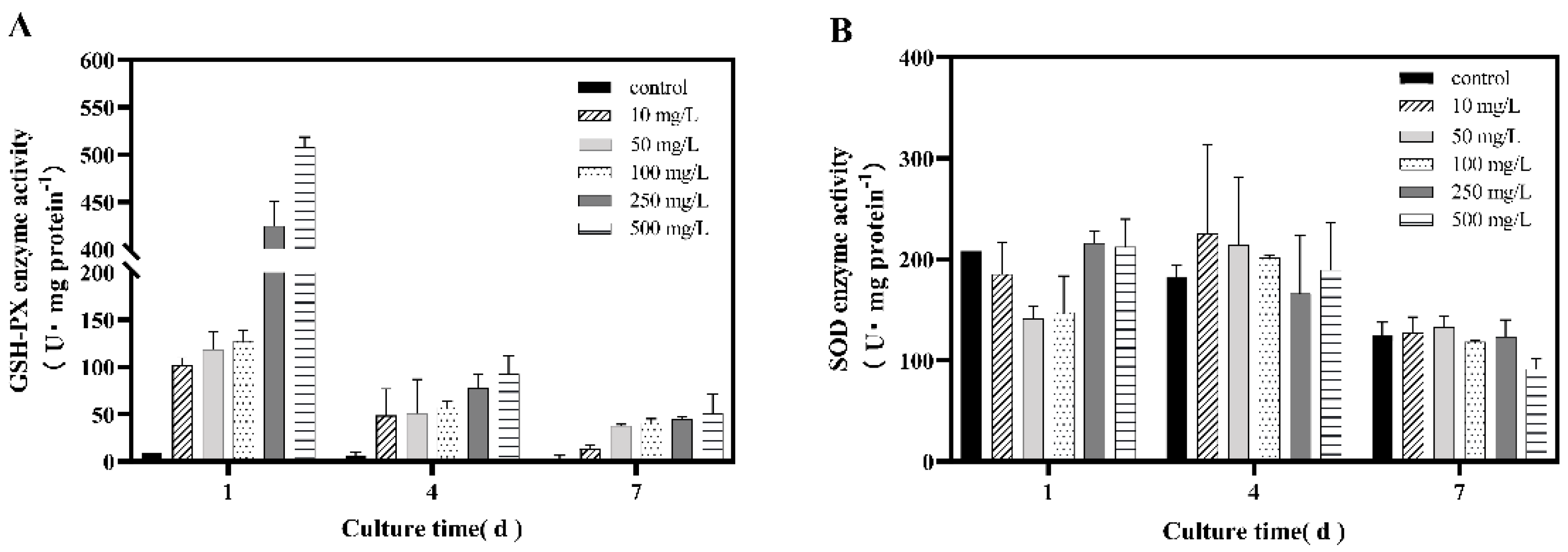
3.3. Effect of Selenite on the Activities of Antioxidant Enzymes in Dunaliella salina

Glutathione peroxidase (GPX) and superoxide dismutase (SOD) are the main antioxidant enzymes in organisms, and can protect organisms from oxidative stress. The activities of GPX and SOD were measured at 1, 4 and 7 days, respectively, under various selenite concentrations. It was found that the GPX activity in the control group was extremely low, while the GPX activity in the 10, 50, 100, 250 and 500 mg/L selenite treatment groups was 11.9, 13.7, 14.8, 49.5 and 59.2 times that of the control group (Figure 3), respectively, indicating that Se may be an inducer of GPX in D. salina. A similar phenomenon was observed in C. rheiniscens, and the enzymic properties of the selenite-induced glutathione peroxidase closely resembled those of animal glutathione peroxidase, whichcontaining selenium [32]. Compared with GPX, the changes in the activity of SOD in D. salina under Se were more complicated. On the first day of incubation, when the selenite concentration was 50~100 mg/L, the activity of SOD was significantly reduced compared the control group, but was not significantly different from the control group at high doses. On the contrary, SOD activity increased at low doses on the fourth day. In previous studies, the results of the effect of Se on SOD activity were different [12,33,34]. In the study with C. pyrenoidosa, SOD activity increased at low selenium concentrations and decreased at high selenium concentrations [12]. In Gracilaria lemaneiformis, the SOD enzymes were negatively correlated with the selenium concentration [33]. The effect of selenium on SOD enzymes in algae is a dynamic process, which may be one of the reasons for the differences. It is possible that low concentrations of Se were integrated into selenoproteins, while high concentrations of selenium were incorporated into other non-specific proteins, thereby leading to abnormal structures in these proteins, which may cause oxidative stress and excessive ROS production.

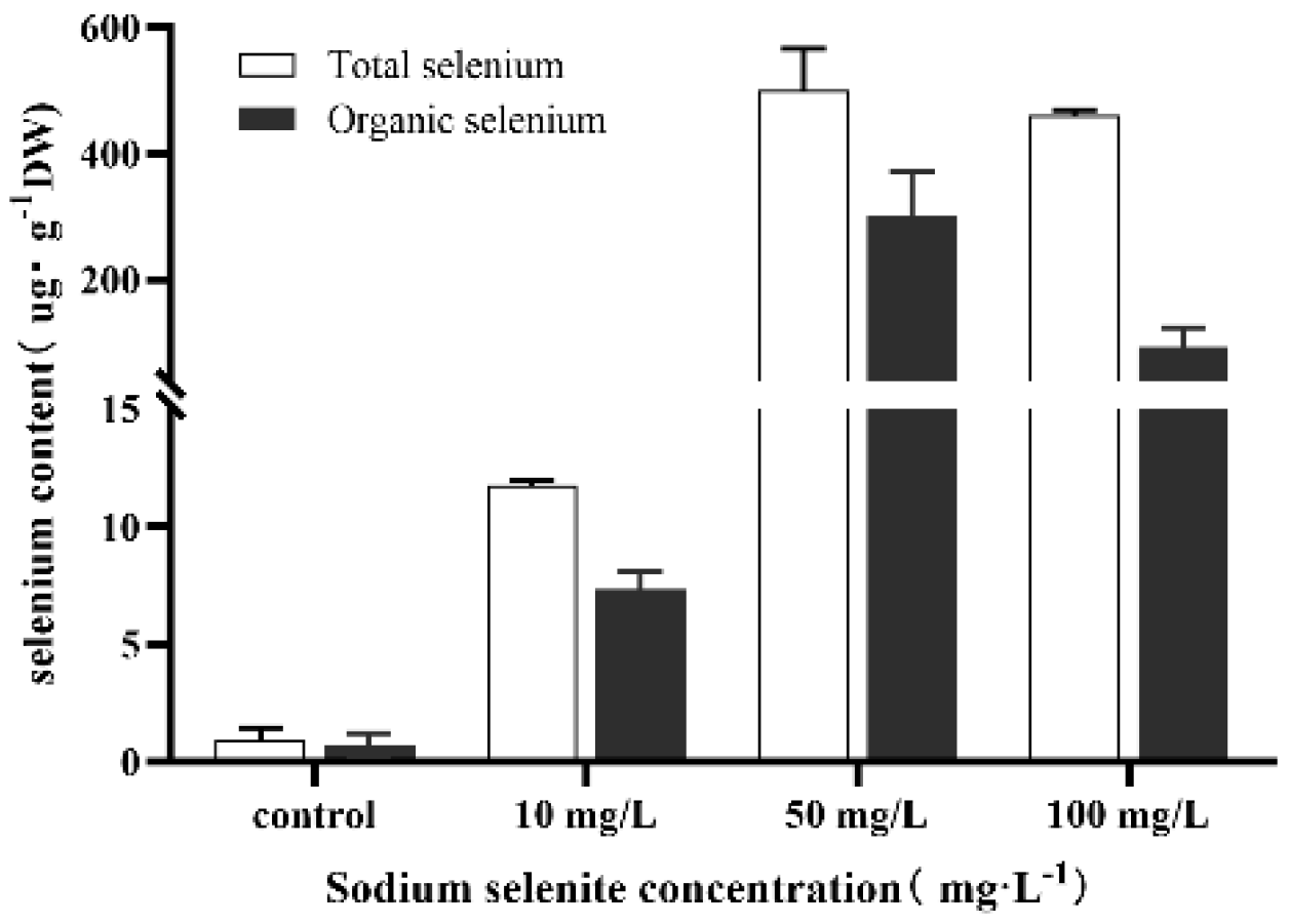
3.4. Intracellular Se Bioaccumulation

The accumulation of Se in D. salina was evaluated after 11 days of treatment with 0 to 100 mg /L of sodium selenite. In the control group without selenite, Se was also present in the cells, but at extremely lowvalues, which may be due to Se impurities in some reagents. When selenite was added into the culture medium, the intracellular selenium content increased significantly compared with the control (Figure 4).However, at a concentration of 50 mg/L, the contents of total Se and organic Se in D. salina cells reached 499.77 μg/g and 303.01 μg/g, which were 539.13 and 414.47 times those in the control group, respectively. At selenite concentrations above 50 mg/L, the content of intracellular organic Se decreased gradually. These results were consistent with the observation that the medium turned red with a decrease in the selenite concentration, indicating that when organic selenium was accumulated to a certain extent, it would be metabolized into Se nanoparticles through some reduction pathway (Supplemental Figure S2). Algae differ in their capacity to accumulate Se in their cells, depending on the algal species. For example, C. pyrenoidosa [12] could accumulate 72 μg/g organic Se under a 2 mg/L selenite treatment. The highest accumulation of Se in Ulva fasciata 36 was 375.6 μg/g at a 800 mg/L concentration [35]. The results in Figure 4 show that D. salina has strong tolerance to Se and agreater accumulation of organic Se. In general, unicellular algae have higher Se accumulation levels than macroalgae. In addition, Se accumulation in microalgae is often influenced by the ambient concentration of Se. When the concentration of sulfate in the environment increased, the intracellular Se levels usually decreased significantly. However, on thecontrary, in U. fasciata, Se bioaccumulation was not altered by a high sulfate concentration in the medium [35]. Se accumulation in algae is closely related to the degree of biotransformation of the inorganic Se forms. Selenocysteine (SeCys), selenomethylocysteine (MeSeCys), selenomethionine (SeMet) and red reduced Se have been detected in some microalgae [10,36]. Several studies have shown that algae, similarly to Se hyperaccumulators plants, can produce large amounts of volatile selenides, such as dimethylselenide (DMSe) and dimethyldiselenide (DMDSe) [37]. These metabolic processes are thought to comprise the mechanism of detoxification in algae and an adaptation to high Se contents in the aquatic environment.

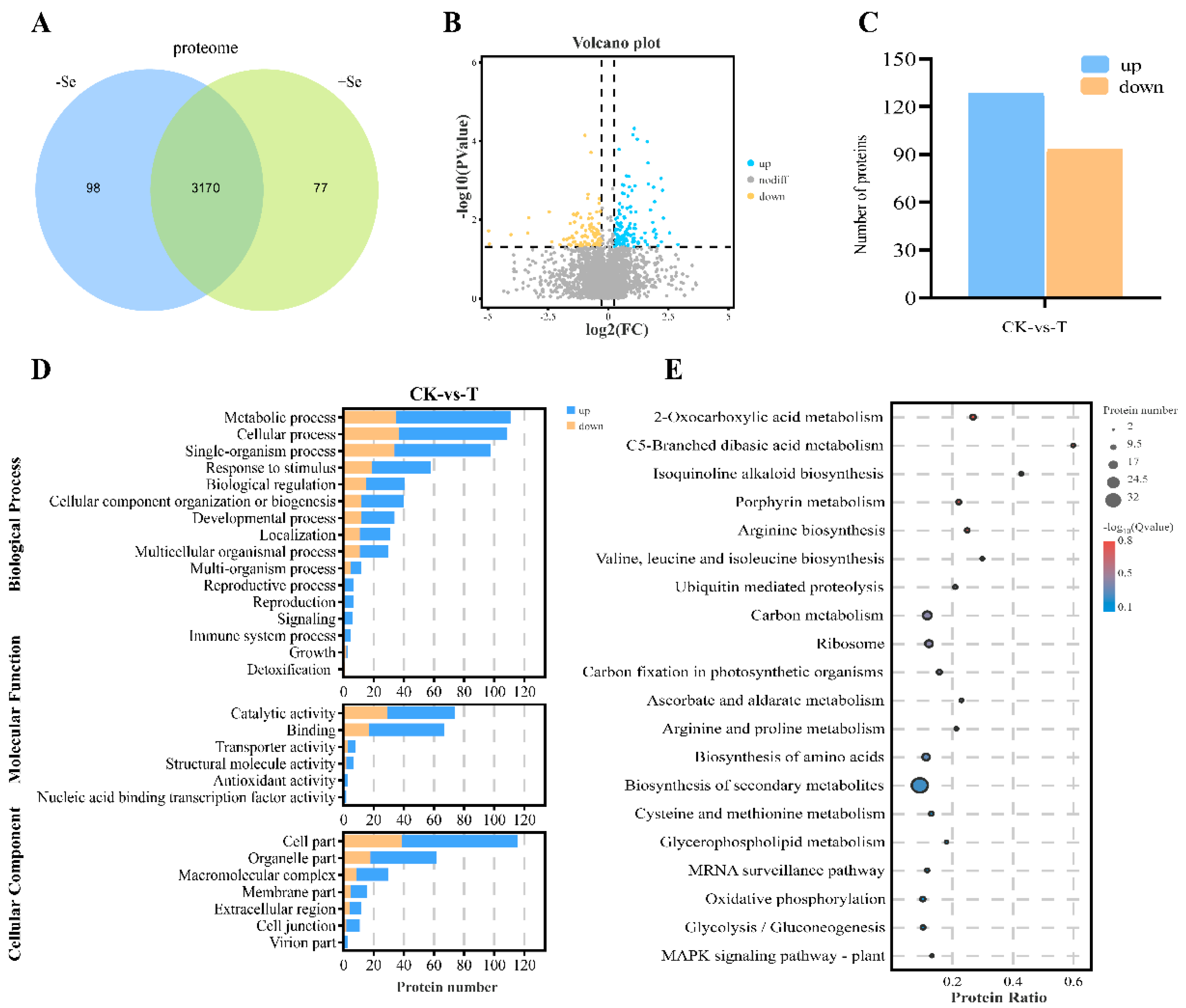
3.5. Quantitative Proteomic Analysis

To further investigate the transformation mechanism of selenite in D. salina, we compared the proteomic profiles of samples treated with or without Na2SeO3. Intotal of 3347 proteins were identified from the results of the raw data in the samples (Figure 5A). Moreover, 221 proteins were identified as differentially expressed proteins (DEPs) in the Na2SeO3 treatment group, of which 128 were downregulated and 93 were upregulated (p < 0.05; FC > 1.2) (Figure 5B,C). The Gene Ontology (GO) database was applied to classify the DEPs. This database describes in three aspects: biological processes, molecular functions and cellular components. GO enrichment of the identified DEPs revealed significant enrichment in some biological processes, such as metabolic processes, cellular processes, single-organism processes, response to stimuli and biological regulation, showing that these processes were related to the response of D. salina to the selenite treatment. In the molecular functions category, the most abundant terms were linked to catalytic activity, binding activity, transporter activity and antioxidant activity (Figure 5D). It was reported that selenite treatment had an analogous effect on the proteome in Providencia rettgeri HF16-A [29]. The top 20 enriched KEGG pathways for DEPs included C5-branched dibasic acid metabolism; ascorbic acid metabolism; ribosome; carbon metabolism; valine; leucine and isoleucine biosynthesis; ubiquitin-mediated proteolysis; oxidative phosphorylation, etc. (Figure 5E).

3.6. Redox and Antistress-Related Proteins in Response to Selenite

Se disrupts cellular redox homeostasis and causes ROS generation in organisms. ROS were slightly induced at lower selenium concentrations to enhance the antioxidant systems of the organism, which may, in turn, have strengthened its defense ability of the organism, whereas theexcessive ROS induced under the high Se treatment damaged the photosystems and caused death [38]. In this study, it was found that the NEET protein was significantly downregulated in the presence of selenite (Figure 6 and Table S1). NEET protein is a conserved 2Fe-2S protein that can bind and release its 2Fe-2S clusters, which plays an important role in regulating the levels of iron and reactive oxygen species in the chloroplasts of plants [39]. Previous studies have shown that disruption of NEET protein’s function impacted the transfer of 2Fe-2S clusters from the chloroplast’s 2Fe-2S biogenic pathway to chloroplast Fe-S proteins and the cytosolic Fe-S biogenic system, which resulted in the overaccunulation of Fe and ROS in the chloroplasts and triggered Fe-deficiency responses in some areas of the cell [40]. The low expression of the NEET protein under the selenite treatment may be related to selenocysteine replacing Cys into the protein. The destructive impacts of ROS have caused D. salina to develop sophisticated redox homeostatic mechanisms to respond to the overproduction of ROS during oxidative stress. For example, nucleoside diphosphate kinase (NDPK), which is involved in the regulatory of MAPK signaling, was upregulated [41]. The overexpression of NDPK may confer an enhanced tolerance to selenite stresses that alleviated ROS accumulation. In addition, the abundance of many antioxidative proteins was altered under the Se treatment compared with the control group, suggesting that Se may also perturb the redox homeostasis of D. salina. Glutathione peroxidase (GPX7) and ascorbate peroxidase (APX8) were upregulated significantly in the presence of selenite; these which are the main peroxide detoxifying enzymes of the chloroplasts. Consistent with the previous results, GPX7, which is a Se-containing protein, was strongly induced by selenite. GPX7 was virtually absent in cells grown in the absence of selenite. Generally, selenoproteins are present in animals and algae, whereas fungi and higher plants lack them. Plants also have cysteine-containing homologs of selenoproteins (hereafter referred to as Cys-homologs). Selenoproteins are more highly resistant to irreversible oxidation than their Cys-homologues [42]. Moreover, monodehydroascorbate reductase (MDAR) was clearly downregulated; This enzyme usually acts with ascorbate peroxidase (APX) to scavenge H2O2 [43]. First, the enzyme APX converts H2O2 into water with the help of ascorbic acid (AsA) as an electron donor, which is also converted into monodehydroascorbate (MDHA).Next, MDHA again regenerates AsA through the activity of MDAR [44]. Our results revealed that selenite leads to the low expression of monodehydroascorbate reductase, which may have reduced the Asc redox state, thus affecting the whole process of AsA recycling. Nevertheless, AsA can be regenerated by other processes. For example, MDHA can rapidly reduce nonenzymatically to produce DHA and AsA. Of these, the DHA produced can be reduced to AsA by dehydroascorbate reductase (DHAR) [44]. As we observed, DHAR was upregulated under the Se treatment, but there was no significant difference, suggesting when ascorbic acid supplementation is insufficient, the algae will replenish the required ascorbic acid by other means to maintain the homeostasis of the organism. Thioredoxin-like protein CDSP32 and 2-Cys peroxiredoxin BAS1-like in D. salina were also significantly upregulated, whichhave been shown to serve as key protectants of the chloroplast against environmental stresses [45]. 2-Cys peroxiredoxin BAS1-like is a thiol-dependent peroxidase capable of reducing hydrogen peroxide through the reactive catalysis of cysteine. At present, studies have confirmed the interaction between thioredoxin-like protein CDSP32 and 2-Cys peroxiredoxin BAS1. Thioredoxin-like protein CDSP32 serves as an electron donor to 2-Cys peroxiredoxin BAS1 and plays a role in antioxidative stress [46]. Notably, to cope with selenite stresses, defense-related tetratricopeptide (TPR) repeat proteins were also significantly upregulated. It has been found that TPRs function as molecular chaperones to regulate protein–protein interactions and assemble as multiprotein complexes to defend against external stresses [47].

3.7. Responses of Amino Acid Synthesis-Related Proteins to Selenite

When subjected to stresses, plants accumulate a mass of metabolites, especially amino acids. As previously mentioned in the current study, the results showed that selenite upregulates the enzymes involved aspartate, arginine and branched-chain amino acids. In addition, the serine-related enzymes of D. salina were also altered under the selenite treatment; serine hydroxymethyltransferase was upregulated and phosphoserine aminotransferase was downregulated (Figure 6 and Table S1). Serine biosynthesis in plants proceeds by two pathways. One associated with photorespiration and the pathway from 3-phosphoglycerate in the plastids [48]. Serine hydroxyltransferase and phosphoserine aminotransferase play important roles in these two pathways, respectively [48], which indicates that the path of serine synthesis was adjusted by selenite in D. salina. When exposed to selenite stress, serine synthesis is more inclined to occur in the pathway from 3-phosphoglycerate. On the other hand, the results showed that the two aspartate aminotransferases were upregulated. Aspartate biosynthesis is mediated by the enzyme aspartate aminotransferase, which catalyzes the reversible transamination between glutamate and oxaloacetate to generate aspartate and 2-oxoglutarate, and overexpression of this enzyme would increase the amount of aspartate in the organism [49]. Aspartate, in addition to constituting proteins, is a precursor leading to the biosynthesis of multiple biomolecules required for plant growth and defense, such as nucleotides, nicotinamide adenine dinucleotide (NAD), organic acids, amino acids and their derived metabolites [50]. A series of studies have shown that a change in Asp content is closely related to stress. The aspartate content observably accumulated in tomato plants grown in soil contaminated with arsenate (As(V)) [51], which indicated that aspartate plays an essential role in stress responses. Similarly, acetolactate synthase and 3-isopropyl malate dehydrogenase were also significantly upregulated under selenite stress; of these, acetolactate synthase is the key enzyme in the branched-chain amino acid (BCAA) biosynthetic pathway [52]. It has been documented that branched-chain amino acids generally accumulate under abiotic stress conditions, but the specific regulatory mechanism is still unclear [53].

3.8. Energy and Carbohydrate Metabolism-Related Proteins in Response to Selenite

Environmental stress certainly affects photosynthesis and carbon metabolism in algae, and this effect has been observed in C. reinhardtii [8], N. oceanica [17] and C. vulgaris [34] under selenite treatment. However, these studies focused on assessing the effects of selenite on photosynthesis in algae by observing chloroplast structure or measuring the chlorophyll fluorescence parameters. In this study, we investigated the effect of selenite on D. salina via a proteomic approach. We observed upregulation of the chlorophyll a-b binding proteins (LHCB), the Photosystem II 22 kDa protein (PSBS1) and the chloroplast oxygen-evolving protein (PSBQ) in algae during exposure to selenite (Figure 6 and Table S1). LHCB are the apoproteins of the light-harvesting complex of Photosystem II and normally act as an antenna complex, absorbing sunlight and transferring the excitation energy to the core complexes of PSII in order to drive photosynthetic electron transport [54]. The PSBS protein of Photosystem II functions in the regulation of photosynthetic light harvesting, which is necessary for photoprotective thermal dissipation (qE) of the excess absorbed light energy in plants [55]. PSBQ is one of the extra-membrane luminal subunits of Photosystem II (PSII), the exact function of which is currently unclear. The upregulation of these proteins would promote the light reaction phase of photosynthesis, accumulating more ATP and NADPH to meet the increasing energy demand during selenite stress. In general, the ATP and NADPH produced in the light phase are used for the dark reaction. However, under selenite stress, we observed downregulation of glyceraldehyde-3-phosphate dehydrogenase and fructose-1,6-bisphosphate aldolase, which are related to the Calvin cycle, suggesting a downregulation in the reduction phase of the Calvin cycle. Instead, phosphoribulokinase (PRK), sedoheptulose-1,7-bisphosphatase, and ribulose phosphate-3-epimerase were upregulated. These proteins each play an important role in the regeneration of ribulose. Sedoheptulose 1,7 bisphosphatase, a light-regulated enzyme, can indirectly regulate the influx of CO2 for this process. Selenite can promote the photoreactive phase of photosynthesis to some extent and inhibit the Calvin cycle, meaning that large amounts of ATP and NADPH will accumulate during photosynthesis. In addition, the proteins involved in oxidative phosphorylation were significantly altered in algae under selenite stress. Among them, soluble inorganic pyrophosphatase (sPPases), F1F0 ATP synthase and succinate dehydrogenase were upregulated, whereas cytochrome c oxidase subunit 6b-1 was downregulated. The sPPases hydrolyzes pyrophosphoric acid (PPi) into two molecular phosphates and generates a large amount of energy in the process, which can function as an alternative energy source in plant cells [56]. Both prokaryotic and eukaryotic sPPases have been shown to be induced under a variety of environmental conditions, limiting the status of cellular energy [57] and revealing that the PPi may serve as an alternative energy source in algae exposed to selenite. Consistent with the above, the upregulation of F1F0 ATP synthase also increased the ATP content in D. salina to meet the energy requirements of the organism. Overall, combined with the results above on the maximum fluorescence yield of PSII (Fv/Fm), selenite basically no influence on the Fv/Fm value at a low concentrations in the early stage (1–7 days) of the selenium treatment, but it decreased rapidly in the later stages. These results indicate that cells rely on monitoring mechanisms to sense deviations in homeostasis and activate the appropriate responses through signaling pathways to readjust the cellular homeostasis. However, when the regulation of various mechanisms in organisms still fails to cause the cells to reach homeostasis, cells will inevitably be perturbed or die.

3.9. Ubiquitin–Proteasome—Related Proteins in Response to Selenite

The ubiquitin–proteasome system (UPS) is an important pathway for the selective degradation of intracellular proteins. The ubiquitin molecule is bound to the target protein through ubiquitin-activating enzymes, ubiquitin-binding enzymes and ubiquitin–protein ligases to form a polyubiquitin chain, which is finally recognized and degraded by the 26S proteasome [58]. The UPS also modulates environmental and endogenous signals in plants, including abiotic and biotic stress responses [59]. In this study, however, the majority of the enzymes involved in the ubiquitinproteasome pathway were significantly downregulated under selenite stress, including E3 ubiquitinprotein ligase, ring box protein, cullin-3, pre-mRNA-processing factor 19 and proteasome subunit beta type (Figure 6 and Table S1). Of these, ring box protein and cullin-3 are both key components of the E3 ubiquitin–protein ligase. This implies that the UPS in D. salina was adversely affected by selenite at the experimental concentrations. The reaction of UPS to selenium stress was time-dependent and dose-dependent. Moderate selenite augmented proteasome activity and protein ubiquitination to remove malformed proteins, while excessive selenite or prolonged selenite treatment decreased proteasome activity and inhibited protein ubiquitination [60]. This disruption and inhibition may be related to the accumulation of ROS. In particular, U-box domain-containing protein 1, which is a highly conserved motif widely found in plants, was significantly upregulated. At present, it has been reported that most U-box proteins have E3 ubiquitin ligase activity [61]. In addition, many experiments have shown that U-box proteins are overexpressed under external stresses.Hence, they are largely considered to be involved in biotic and abiotic stress responses [62]. However, the mechanisms of action still remain to be further explored.

4. Conclusions

In this study, we measured the physiological parameters of D. salina under different selenite concentrations and performed a proteomic analysis to clarify the interaction between selenite and D. salina. We found that selenite affects the structure and function of PSII and the growth of D. salina even at low dosages of selenium in the environment. Selenite did not affect the value of Fv/Fm in the early stage (1~7 days) of selenium treatment, but had inhibitory effects atthe later stage. In addition, the proteomic analysis of DIA revealed 221 proteins with altered abundance, indicating that D. salina manipulates its amino acid metabolism and energy production pathways at the protein level in responses to selenite stress. In order to combat reactive oxygen species, glutathione peroxidase (GPX7), a selenium-containing protein, that plays a role in antioxidative stress, was found in the selenium treatment group. In addition, the bio-accumulation test in this study showed that when the selenium concentration was 50 mg/L, the contents of total selenium and organic selenium in D. salina cells reached 499.77 μg/g and 303.01 μg/g, that is, 539.13 and 414.47 times that of the control group, respectively. This indicated that D. salina has a strong tolerance to selenium and it is likely to be a candidate for carrier cells fororganic selenium enrichment. This study provides a detailed understanding of the physiological and proteomic responses of D. salina to selenite and will be helpful for the development of industrial strains of D. salina that are rich in selenium.

Supplementary Materials

The following supporting information can be downloaded at: https://www.mdpi.com/article/10.3390/pr11020563/s1, Figure S1: Changes in the size of D. salina cultured at different selenite concentrations for 11 days; Figure S2: Changes in the culture medium of D. salina cultured at different selenite concentrations for 30 days; Table S1: Changes in the partial protein abundance of D. salina under selenite treatment.

Author Contributions

Conceptualization, X.J., F.J. and Y.W.; methodology, X.J. and L.Y.; software, X.J., K.P. and L.Y.; validation, X.J., L.Y. and K.P.; formal analysis, X.J., L.Y. and F.J.; investigation, X.J. and K.P.; resources, F.J., Y.W. and J.L.; data curation, X.J.; writing—original draft preparation, X.J.; writing—review and editing, X.J., F.J., L.Y., J.L. and K.P.; visualization, X.J. and J.L.; supervision, F.J., Y.W. and J.L.; project administration, F.J. and Y.W.; funding acquisition, F.J. and Y.W. All authors have read and agreed to the published version of the manuscript.

Funding

The project was supported by the Major Science and Technology Project in Guangxi (Guike AA18242047).

Informed Consent Statement

Not applicable.

Data Availability Statement

All data used in the study appear in the submitted article and Supplementary Materials.

Conflicts of Interest

The authors declare that they have no known competing financial interests or personal relationships that could have appeared to influence the work reported in this paper.

References

  1. Rayman, M.P. Selenium and human health. Lancet 2012, 379, 1256–1268. [Google Scholar] [CrossRef]
  2. Ren, H.; Li, X.; Guo, L.; Wang, L.; Hao, X.; Zeng, J. Integrative Transcriptome and Proteome Analysis Reveals the Absorption and Metabolism of Selenium in Tea Plants [Camellia sinensis (L.) O. Kuntze]. Front. Plant Sci. 2022, 13, 848349. [Google Scholar] [CrossRef]
  3. Hatfield, D.L.; Tsuji, P.A.; Carlson, B.A.; Gladyshev, V.N. Selenium and selenocysteine: Roles in cancer, health, and development. Trends Biochem. Sci. 2014, 39, 112–120. [Google Scholar] [CrossRef]
  4. Vindry, C.; Ohlmann, T.; Chavatte, L. Translation regulation of mammalian selenoproteins. Biochim. Biophys. Acta BBA Gen. Subj. 2018, 1862, 2480–2492. [Google Scholar] [CrossRef]
  5. Guillin, O.M.; Vindry, C.; Ohlmann, T.; Chavatte, L. Selenium, Selenoproteins and Viral Infection. Nutrients 2019, 11, 2101. [Google Scholar] [CrossRef] [PubMed]
  6. Dinh, Q.T.; Cui, Z.; Huang, J.; Tran, T.A.T.; Wang, D.; Yang, W.; Zhou, F.; Wang, M.; Yu, D.; Liang, D. Selenium distribution in the Chinese environment and its relationship with human health: A review. Environ. Int. 2018, 112, 294–309. [Google Scholar] [CrossRef] [PubMed]
  7. Burk, R.F.; Norsworthy, B.K.; Hill, K.E.; Motley, A.K.; Byrne, D.W. Effects of Chemical Form of Selenium on Plasma Biomarkers in a High-Dose Human Supplementation Trial. Cancer Epidemiol. Biomark. Prev. 2006, 15, 804–810. [Google Scholar] [CrossRef]
  8. Morlon, H.; Fortin, C.; Floriani, M.; Adam, C.; Garnier-Laplace, J.; Boudou, A. Toxicity of selenite in the unicellular green alga Chlamydomonas reinhardtii: Comparison between effects at the population and sub-cellular level. Aquat. Toxicol. 2005, 73, 65–78. [Google Scholar] [CrossRef]
  9. Novoselov, S.; Rao, M.; Onoshko, N.V.; Zhi, H.; Kryukov, G.; Xiang, Y.; Weeks, D.P.; Hatfield, D.L.; Gladyshev, V.N. Selenoproteins and selenocysteine insertion system in the model plant cell system, Chlamydomonas reinhardtii. EMBO J. 2002, 21, 3681–3693. [Google Scholar] [CrossRef] [PubMed]
  10. Vriens, B.; Behra, R.; Voegelin, A.; Zupanic, A.; Winkel, L.H.E. Selenium Uptake and Methylation by the Microalga Chlamydomonas reinhardtii. Environ. Sci. Technol. 2016, 50, 711–720. [Google Scholar] [CrossRef]
  11. Wu, H.-L.; Chen, T.-F.; Yin, X.; Zheng, W.-J. Spectrometric characteristics and underlying mechanisms of protective effects of selenium on Spirulina platensis against oxidative stress. Guang Pu Xue Yu Guang Pu Fen Xi 2012, 32, 749–754. [Google Scholar]
  12. Zhao, Y.; Song, X.; Cao, X.; Wang, Y.; Si, Z.; Chen, Y. Toxic effect and bioaccumulation of selenium in green alga Chlorella pyrenoidosa. J. Appl. Phycol. 2019, 31, 1733–1742. [Google Scholar] [CrossRef]
  13. Umysová, D.; Vítová, M.; Doušková, I.; Bišová, K.; Hlavová, M.; Čížková, M.; Machát, J.; Doucha, J.; Zachleder, V. Bioaccumulation and toxicity of selenium compounds in the green alga Scenedesmus quadricauda. BMC Plant Biol. 2009, 9, 58. [Google Scholar] [CrossRef]
  14. Zheng, Y.; Li, Z.; Tao, M.; Li, J.; Hu, Z. Effects of selenite on green microalga Haematococcus pluvialis: Bioaccumulation of selenium and enhancement of astaxanthin production. Aquat. Toxicol. 2017, 183, 21–27. [Google Scholar] [CrossRef] [PubMed]
  15. Constantinescu-Aruxandei, D.; Vlaicu, A.; Marinaș, I.C.; Vintilă, A.C.N.; Dimitriu, L.; Oancea, F. Effect of betaine and selenium on the growth and photosynthetic pigment production in Dunaliella salina as biostimulants. FEMS Microbiol. Lett. 2019, 366, 257. [Google Scholar] [CrossRef] [PubMed]
  16. Wang, D.Z.; Cheng, Z.D.; Li, S.Q.; Gao, Y.H. Toxicity and Accumulation of Selenite in Four Microalgae. Chin. J. Oceanol. Limnol. 2003, 21, 280–285. [Google Scholar]
  17. Guimarães, B.O.; de Boer, K.; Gremmen, P.; Drinkwaard, A.; Wieggers, R.; Wijffels, R.H.; Barbosa, M.J.; D’Adamo, S. Selenium enrichment in the marine microalga Nannochloropsis oceanica. Algal Res. 2021, 59, 102427. [Google Scholar] [CrossRef]
  18. Zou, H.; Huang, J.-C.; Zhou, C.; He, S.; Zhou, W. Mutual effects of selenium and chromium on their removal by Chlorella vulgaris and associated toxicity. Sci. Total Environ. 2020, 724, 138219. [Google Scholar] [CrossRef]
  19. Morlon, H.; Fortin, C.; Adam, C.; Garnier-Laplace, J. Selenite Transport and its Inhibition in the Unicellular Green Alga Chlamydomonas Reinhardtii. Environ. Toxicol. Chem. 2006, 25, 1408–1417. [Google Scholar] [CrossRef]
  20. Ponton, D.E.; Fortin, C.; Hare, L. Organic selenium, selenate, and selenite accumulation by lake plankton and the alga Chlamydomonas reinhardtii at different pH and sulfate concentrations. Environ. Toxicol. Chem. 2018, 37, 2112–2122. [Google Scholar] [CrossRef]
  21. Koyande, A.K.; Chew, K.W.; Rambabu, K.; Tao, Y.; Chu, D.-T.; Show, P.-L. Microalgae: A potential alternative to health supplementation for humans. Food Sci. Hum. Wellness 2019, 8, 16–24. [Google Scholar] [CrossRef]
  22. Mou, C.; Hao, X.; Liu, X.; Chen, X.; Chen, D. Cell Factory of CarotenoidsProgress in Dunaliella Cultivation and Its Research. Adv. Mar. Sci. 2010, 28, 554–562. [Google Scholar]
  23. Soto, J.O. Dunaliella Identification Using DNA Fingerprinting Intron-Sizing Method and Species-Specific Oligonucleotides. Handbook of Marine Microalgae; Academic Press: Cambridge, MA, USA, 2015; pp. 559–568. [Google Scholar] [CrossRef]
  24. Ebenezer, V.; Ki, J.-S. Quantification of the Sub-lethal Toxicity of Metals and Endocrine-disrupting Chemicals to the Marine Green Microalga Tetraselmis suecica. Fish. Aquat. Sci. 2013, 16, 187–194. [Google Scholar] [CrossRef]
  25. Karasinski, J.; Wrobel, K.; Escobosa, A.R.C.; Konopka, A.; Bulska, E.; Wrobel, K. Allium cepa L. Response to Sodium Selenite (Se(IV)) Studied in Plant Roots by a LC-MS-Based Proteomic Approach. J. Agric. Food Chem. 2017, 65, 3995–4004. [Google Scholar] [CrossRef] [PubMed]
  26. Araie, H.; Shiraiwa, Y. Selenium Utilization Strategy by Microalgae. Molecules 2009, 14, 4880–4891. [Google Scholar] [CrossRef]
  27. Schiavon, M.; Ertani, A.; Parrasia, S.; Vecchia, F.D. Selenium accumulation and metabolism in algae. Aquat. Toxicol. 2017, 189, 11. [Google Scholar] [CrossRef]
  28. Geoffroy, L.; Gilbin, R.; Simon, O.; Floriani, M.; Adam, C.; Pradines, C.; Cournac, L.; Garnier-Laplace, J. Effect of selenate on growth and photosynthesis of Chlamydomonas reinhardtii. Aquat. Toxicol. 2007, 83, 149–158. [Google Scholar] [CrossRef]
  29. Huang, S.; Wang, Y.; Tang, C.; Jia, H.; Wu, L. Speeding up selenite bioremediation using the highly selenite-tolerant strain Providencia rettgeri HF16-A novel mechanism of selenite reduction based on proteomic analysis. J. Hazard. Mater. 2020, 406, 124690. [Google Scholar] [CrossRef]
  30. Vítová, M.; Bišová, K.; Hlavová, M.; Zachleder, V.; Rucki, M.; Čížková, M. Glutathione peroxidase activity in the selenium-treated alga Scenedesmus quadricauda. Aquat. Toxicol. 2011, 102, 87–94. [Google Scholar] [CrossRef]
  31. Pilonsmits, E.; Pilon, M. Sulfur metabolism in plastids. Advances in Photosynthesis and Respiration. In Advances in Photosynthesis and Respiration; Wise, R.R., Hoober, J.K., Eds.; Springer: Dordrecht, The Netherlands, 2007; pp. 387–402. [Google Scholar]
  32. Yokota, A.; Shigeoka, S.; Onishi, T.; Kitaoka, S. Selenium as Inducer of Glutathione Peroxidase in low-CO2-Grown Chlamydomonas reinhardtii. Plant Physiol. 1988, 86, 649–651. [Google Scholar] [CrossRef] [PubMed]
  33. Wang, Q.; Zuo, Y.; Chen, T.; Zheng, W.; Yang, Y. Effects of selenium on antioxidant enzymes and photosynthesis in the edible seaweed Gracilaria lemaneiformis. J. Appl. Phycol. 2018, 31, 1303–1310. [Google Scholar] [CrossRef]
  34. Sun, X.; Zhong, Y.; Huang, Z.; Yang, Y. Selenium Accumulation in Unicellular Green Alga Chlorella vulgaris and Its Effects on Antioxidant Enzymes and Content of Photosynthetic Pigments. PLoS ONE 2014, 9, e112270. [Google Scholar] [CrossRef] [Green Version]
  35. Schiavon, M.; Dall’acqua, S.; Mietto, A.; Pilon-Smits, E.A.; Sambo, P.M.A. Selenium fertilization alters the chemical composition and antioxidant constituents of tomato (Solanumlycopersicon L.). Aquat. Toxicol. 2012, 61, 10542–10545. [Google Scholar] [CrossRef]
  36. Gómez-Jacinto, V.; García-Barrera, T.; Garbayo, I.; Vílchez, C.; Gómez-Ariza, J.L. Metallomic study of selenium biomolecules metabolized by the microalgae Chlorella sorkiniana in the biotechnological production of functional foods enriched in selenium. Pure Appl. Chem. 2012, 84, 269–280. [Google Scholar] [CrossRef]
  37. Neumann, P.M.; DE Souza, M.P.; Pickering, I.J.; Terry, N. Rapid microalgal metabolism of selenate to volatile dimethylselenide. Plant Cell Environ. 2003, 26, 897–905. [Google Scholar] [CrossRef]
  38. Wang, Y.-D.; Wang, X.; Ngai, S.-M.; Wong, Y.-S. Comparative Proteomics Analysis of Selenium Responses in Selenium-Enriched Rice Grains. J. Proteome Res. 2013, 12, 808–820. [Google Scholar] [CrossRef] [PubMed]
  39. Zandalinas, S.I.; Song, L.; Nechushtai, R.; Mendoza-Cozatl, D.G.; Mittler, R. The Cluster Transfer Function of AtNEET Supports the Ferredoxin–Thioredoxin Network of Plant Cells. Antioxidants 2022, 11, 1533. [Google Scholar] [CrossRef]
  40. Zandalinas, S.I.; Song, L.; Sengupta, S.; McInturf, S.A.; Grant, D.G.; Marjault, H.; Castro-Guerrero, N.A.; Burks, D.; Azad, R.K.; Cozatl, D.G.M.; et al. Expression of a dominant-negative AtNEET-H89C protein disrupts iron–sulfur metabolism and iron homeostasis in Arabidopsis. Plant J. 2019, 101, 1152–1169. [Google Scholar] [CrossRef]
  41. Moon, H.; Lee, B.; Choi, G.; Shin, D.; Prasad, D.T.; Lee, O.; Kwak, S.-S.; Kim, D.H.; Nam, J.; Bahk, J.; et al. NDP kinase 2 interacts with two oxidative stress-activated MAPKs to regulate cellular redox state and enhances multiple stress tolerance in transgenic plants. Proc. Natl. Acad. Sci. USA 2002, 100, 358–363. [Google Scholar] [CrossRef]
  42. Ghuge, S.A.; Kadam, U.S.; Hong, J.C. Selenoprotein: Potential Player in Redox Regulation in Chlamydomonas reinhardtii. Antioxidants 2022, 11, 1630. [Google Scholar] [CrossRef]
  43. Jimenez, A.; Hernandez, J.A.; del Rio, L.A.; Sevilla, F. Evidence for the Presence of the Ascorbate-Glutathione Cycle in Mitochondria and Peroxisomes of Pea Leaves. Plant Physiol. 1997, 114, 275–284. [Google Scholar] [CrossRef]
  44. Hasanuzzaman, M.; Bhuyan, M.H.M.B.; Anee, T.I.; Parvin, K.; Nahar, K.; Mahmud, J.A.; Fujita, M. Regulation of Ascorbate-Glutathione Pathway in Mitigating Oxidative Damage in Plants under Abiotic Stress. Antioxidants 2019, 8, 384. [Google Scholar] [CrossRef] [Green Version]
  45. Kim, S.Y.; Jang, H.H.; Lee, J.R.; Sung, N.R.; Bin Lee, H.; Lee, D.H.; Park, D.-J.; Kang, C.H.; Chung, W.S.; Lim, C.O.; et al. Oligomerization and chaperone activity of a plant 2-Cys peroxiredoxin in response to oxidative stress. Plant Sci. 2009, 177, 227–232. [Google Scholar] [CrossRef]
  46. Rey, P.; Cuiné, S.; Eymery, F.; Garin, J.; Court, M.; Jacquot, J.-P.; Rouhier, N.; Broin, M. Analysis of the proteins targeted by CDSP32, a plastidic thioredoxin participating in oxidative stress responses. Plant J. 2004, 41, 31–42. [Google Scholar] [CrossRef] [PubMed]
  47. Paeng, S.K.; Kang, C.H.; Chi, Y.H.; Chae, H.B.; Lee, E.S.; Park, J.H.; Wi, S.D.; Bin Bae, S.; Phan, K.A.T.; Lee, S.Y. AtTPR10 Containing Multiple ANK and TPR Domains Exhibits Chaperone Activity and Heat-Shock Dependent Structural Switching. Appl. Sci. 2020, 10, 1265. [Google Scholar] [CrossRef]
  48. Ho, C.-L.; Saito, K. Molecular biology of the plastidic phosphorylated serine biosynthetic pathway in Arabidopsis thaliana. Amino Acids 2001, 20, 243–259. [Google Scholar] [CrossRef] [PubMed]
  49. DE LA Torre, F.; Cañas, R.A.; Pascual, M.B.; Avila, C.; Cánovas, F.M. Plastidic aspartate aminotransferases and the biosynthesis of essential amino acids in plants. J. Exp. Bot. 2014, 65, 5527–5534. [Google Scholar] [CrossRef]
  50. Han, M.; Zhang, C.; Suglo, P.; Sun, S.; Wang, M.; Su, T. L-Aspartate: An Essential Metabolite for Plant Growth and Stress Acclimation. Molecules 2021, 26, 1887. [Google Scholar] [CrossRef]
  51. Okunev, R.V. Free Amino Acid Accumulation in Soil and Tomato Plants (Solanum lycopersicum L.) Associated with Arsenic Stress. Water Air Soil Pollut. 2019, 230, 253. [Google Scholar] [CrossRef]
  52. Garcia, M.D.; Nouwens, A.; Lonhienne, T.G.; Guddat, L.W. Comprehensive understanding of acetohydroxyacid synthase inhibition by different herbicide families. Proc. Natl. Acad. Sci. USA 2017, 114, E1091–E1100. [Google Scholar] [CrossRef] [PubMed]
  53. Obata, T.; Fernie, A.R. The use of metabolomics to dissect plant responses to abiotic stresses. Cell. Mol. Life Sci. 2012, 69, 3225–3243. [Google Scholar] [CrossRef] [PubMed]
  54. Liu, R.; Xu, Y.-H.; Jiang, S.-C.; Lu, K.; Lu, Y.-F.; Feng, X.-J.; Wu, Z.; Liang, S.; Yu, Y.-T.; Wang, X.-F.; et al. Light-harvesting chlorophyll a/b-binding proteins, positively involved in abscisic acid signalling, require a transcription repressor, WRKY40, to balance their function. J. Exp. Bot. 2013, 64, 5443–5456. [Google Scholar] [CrossRef] [PubMed] [Green Version]
  55. Niyogi, K.K.; Li, X.-P.; Rosenberg, V.; Jung, H.-S. Is PsbS the site of non-photochemical quenching in photosynthesis? J. Exp. Bot. 2004, 56, 375–382. [Google Scholar] [CrossRef]
  56. Xian, J.; Zhang, M.; Sun, C.; Wang, Y.; Wang, K.; Chen, J.; Zhao, M.; Wang, Y. Research Progress of Soluble Pyrophosphatase. Genom. Appl. Biol. 2019, 38, 4030–4035. [Google Scholar]
  57. Castineira, J.R.P.; Serrano, A. The H+-Translocating Inorganic Pyrophosphatase from Arabidopsis thaliana Is More Sensitive to Sodium Than Its Na+-Translocating Counterpart from Methanosarcina mazei. Front. Plant Sci. 2020, 11, 1240. [Google Scholar] [CrossRef] [PubMed]
  58. Xu, F.-Q.; Xue, H.-W. The ubiquitin-proteasome system in plant responses to environments. Plant Cell Environ. 2019, 42, 2931–2944. [Google Scholar] [CrossRef]
  59. Sharma, S.; Prasad, A.; Sharma, N.; Prasad, M. Role of ubiquitination enzymes in abiotic environmental interactions with plants. Int. J. Biol. Macromol. 2021, 181, 494–507. [Google Scholar] [CrossRef]
  60. Vallentine, P.; Hung, C.-Y.; Xie, J.; Van Hoewyk, D. The ubiquitin-proteasome pathway protects Chlamydomonas reinhardtii against selenite toxicity, but is impaired as reactive oxygen species accumulate. AoB Plants 2014, 6, plu062. [Google Scholar] [CrossRef]
  61. Wiborg, J.; O’Shea, C.; Skriver, K. Biochemical function of typical and variant Arabidopsis thaliana U-box E3 ubiquitin-protein ligases. Biochem. J. 2008, 413, 447–457. [Google Scholar] [CrossRef]
  62. Li, Q.-S.; Lin, X.-M.; Qiao, R.-Y.; Zheng, X.-Q.; Lu, J.-L.; Ye, J.-H.; Liang, Y.-R. Effect of fluoride treatment on gene expression in tea plant (Camellia sinensis). Sci. Rep. 2017, 7, 1–8. [Google Scholar] [CrossRef] [Green Version]
Figure 1. Effect of different concentrations of selenite on the growth of D. salina. (A) Dose-response curve of the different selenite concentrations according to the final cell concentration reached after 11 days. (B) Effect of different concentrations of selenite on the growth curves of D. salina. (C) Size of D. salina cells at different selenite concentrations. Different low case letters above columns indicate statistical differences at p < 0.05.
Processes 11 00563 g001
Figure 2. Effect of different concentrations of selenite on the chlorophyll fluorescence parameters Fv/Fm.
Processes 11 00563 g002
Figure 3. Effect of different selenite concentrations on antioxidant enzyme activities. (A) Effect of different selenite concentrations on GSH-PX enzyme activities. (B) Effect of different selenite concentrations on SOD enzyme activities.
Processes 11 00563 g003
Figure 4. Total selenium and organic selenium content in D. salina treated with selenite.
Processes 11 00563 g004
Figure 5. Analysis of the differentially expressed proteins (DEPs) in response to selenite treatments. (A) Venn diagram showing the overlap in the numbers of proteins between the Na2SeO3 treatment and the control. (B) Volcano plot of DEPs under Na2SeO3 stress. (C) D. salina DEPs under Na2SeO3 stress. (D) The GO functional classifications of DEPs in response to Na2SeO3. (E) KEGG enrichment analysis of DEPs in response to Na2SeO3.
Processes 11 00563 g005
Figure 6. Changes in the protein abundance of D. salina under the selenite treatments. Negative values indicate downregulation and positive values indicate upregulation of the proteins.
Processes 11 00563 g006
Disclaimer/Publisher’s Note: The statements, opinions and data contained in all publications are solely those of the individual author(s) and contributor(s) and not of MDPI and/or the editor(s). MDPI and/or the editor(s) disclaim responsibility for any injury to people or property resulting from any ideas, methods, instructions or products referred to in the content.

