Article

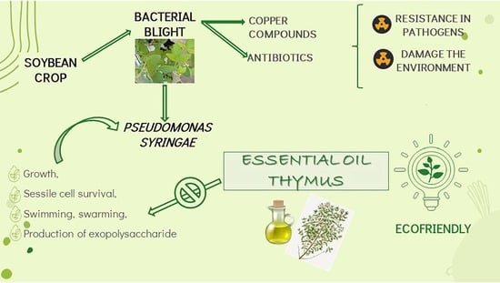
Inhibitory Potential of Thymus vulgaris Essential Oil against Growth, Biofilm Formation, Swarming, and Swimming in Pseudomonas syringae Isolates

by
María Evangelina Carezzano
1,2,3,
María Fernanda Paletti Rovey
1,3,
Jesica P. Sotelo
1,3,
Melina Giordano
1,3,
Pablo Bogino
1,2,
María de las Mercedes Oliva
1,3,* and
Walter Giordano
1,2,*
1
Instituto de Biotecnología Ambiental y Salud, INBIAS-CONICET, Río Cuarto X5800, Argentina
2
Departamento de Biología Molecular, Universidad Nacional de Río Cuarto (UNRC), Río Cuarto X5800, Argentina
3
Departamento de Microbiología e Inmunología, Universidad Nacional de Río Cuarto (UNRC), Río Cuarto X5800, Argentina
*
Authors to whom correspondence should be addressed.
Processes 2023, 11(3), 933; https://doi.org/10.3390/pr11030933
Submission received: 21 February 2023 / Revised: 14 March 2023 / Accepted: 17 March 2023 / Published: 18 March 2023

Abstract

:
As a follow-up to previous studies, the effects of Thymus vulgaris essential oil on selected virulence factors (growth, sessile cell survival, swimming, swarming, and exopolysaccharide production) were evaluated in phytopathogenic Pseudomonas syringae strains isolated from soybean fields in Argentina; reference strains Pseudomonas savastanoi pv. glycinea B076 and Pseudomonas aeruginosa PAO1. P. syringae are responsible for bacterial blight, a disease that affects crops worldwide. Plant bacterioses are usually treated with antibiotics and copper compounds, which may contribute to the development of resistance in pathogens and damage the environment. For these reasons, eco-friendly alternatives are necessary. Although aromatic plants are a natural source of antimicrobial substances, the effects of these substances on phytopathogenic bacteria remain largely unexplored. Subinhibitory concentrations of the oil significantly reduced the slope and rate of bacterial growth. In addition, biofilm and exopolysaccharide (EPS) production were inhibited, with swimming and swarming motility patterns being affected at all of the oil concentrations tested. Therefore, TEO could potentially be a highly efficient antipseudomonal agent for treating plant infections caused by P. syringae.

Graphical Abstract

1. Introduction

The human population is estimated to reach 9600 million by 2050 globally. The associated increase in food demand will make it necessary to boost crop production by up to 110% [1]. To this end, various crop species have been improved and fertilizers and pesticides are extensively applied [2]. Although the unregulated use of agrochemicals has led to better yields in recent years, it has come with a series of negative consequences: the degradation of natural resources, high environmental toxicity, and the emergence of resistant phytopathogenic microorganisms. To minimize the harmful effects of these products on crops and humans, standard protocols for their use must be rigorously enforced, or new ones put in place where necessary. Moreover, more sustainable practices could go a long way in guaranteeing environmental, ecological, and social safety, as they may help reduce biodiversity loss, protect health, and build secure food systems over the long term [3].
Crop diseases caused by phytopathogenic microorganisms are responsible for at least 10% of yield losses worldwide [4]. Pseudomonas syringae, a Gram-negative rod-shaped bacterium with aerobic metabolism, is the causative agent of bacterial blight, which affects many crop species. Pseudomonas spp. produce yellow–green fluorescent pigments that act as siderophores, which have a large number of plasmids containing inducible operons for the synthesis of specific enzymes. This makes them metabolically versatile since it allows them to use many substrates as sources of carbon and thus colonize a wide range of niches, including nonagricultural habitats such as rivers and snow [5,6,7].
P. syringae can live saprophytically as an epiphyte on leaf surfaces, or become pathogenic when it enters the leaf apoplast. During its epiphytic phase, it maintains an inoculum for subsequent infections. For this to occur, the bacterial population must grow sufficiently, to the point where it becomes able to recognize “preferred sites” for invasion, such as glandular trichomes or epidermal cell junctions, which provide protection against extreme environmental conditions and are likely sources of water and nutrients. However, the leaf surface can often become a hostile environment for bacteria, due to desiccation, ultraviolet radiation, and limited nutrients [8]. One way in which phytopathogens such as P. syringae offset these adverse conditions is by forming complex aggregated structures on plant tissues, known as biofilms. These are embedded in a self-produced extracellular matrix made up of polysaccharides, proteins, and extracellular DNA, and feature inner channels through which water and nutrients circulate [9,10]. Biofilms increase pathogenicity and virulence, and, therefore, significantly affect agrifood systems [11]. The communal lifestyle present within biofilms also improves resistance to phagocytosis, predation, desiccation, antibacterial substances, and adverse environmental conditions [12,13]. The increased resistance to antimicrobial agents has been attributed to changes in gene expression, slower growth, low metabolic activity, the formation of persistent cells, and specific responses to stress, among other mechanisms [4,14].
Bacteria within the genus Pseudomonas are able to move individually or collectively by swimming or swarming as a result of one or more polar flagella. Motility favours the cells in different ways: it helps them to explore the leaf surface in search of resources, uptake of nutrients, in competing with other microorganisms, in evading toxic substances and other stressful situations, in locating preferred hosts and optimal sites for colonization within them and to help them disperse into the environment during transmission. It also facilitates the formation of biofilms [10,15].
P. syringae pathovars can infect more than 50 plant species worldwide, including apple, pepper, cabbage, wheat, and soybean [16]. In addition to leaf or grain blight, they can cause leaf spots and cankers [4,17]. Bacteriosis is usually treated with antibiotics and copper compounds that generate resistance and/or toxicity, which is why alternative management strategies are increasingly being explored, such as bacteriophages, growth-promoting bacteria, essential oils (EOs), and aromatic plant extracts [18]. These new antimicrobials may act on the whole bacterial cell, on cell structures related to pathogenesis, on virulence factors, and/or on their biosynthetic pathways [14]. Any substance able to interfere with the expression of these processes (including biofilm formation) rather than directly killing bacteria could be a promising candidate for pathogen control [19]. This is because as they exert less selection pressure, they make it less likely for microorganisms to develop resistance to them [20].
Several studies have described the activity of EOs against phytopathogenic bacteria [21,22,23]. Many have focused on their inhibitory capacity or their ability to arrest some of the synthesis pathways involved in pathogenesis, which is regulated by quorum sensing (QS). Thyme (Thymus vulgaris (L)) essential oil (TEO) has been studied for its inhibitory activity against plant pathogenic bacteria and their virulence factors, such as biofilms and phytotoxins, even at low concentrations [6]. Sotelo et al. (2021) [24] demonstrated that nonphytotoxic concentrations of TEO were able to reduce the phytopathogenic load of P. syringae on soybean seeds. The main terpene components of TEO are thymol and carvacrol, both of which have a hydroxyl group in their structure that gives them strong insecticidal, antifungal, antibacterial, and antioxidant properties. This structure disrupts the membrane of Gram-negative bacteria and the citrate pathway. It also destabilizes the concentration of inorganic ions and consequently alters the pH within the cytoplasm [25]. In addition, carvacrol has been observed to control biofilm formation, motility, and adherence in phytopathogenic bacteria [26].
This study aimed to investigate the inhibitory effects of TEO on growth, sessile cell survival, swimming, swarming, and EPS production in phytopathogenic P. syringae strains isolated from soybean fields in Argentina.

2. Materials and Methods

2.1. Bacterial Strains, Media, and Culture Conditions

The P. syringae strains used here were isolated from soybean fields in Argentina and identified in the Laboratory of Microbiology at the Universidad Nacional de Río Cuarto [23]. The reference strains used were P. savastanoi pv. glycinea B076 and Pseudomonas aeruginosa PAO1. The strains were grown at 28 °C in the case of P. syringae (Table 1) and at 37 °C for P. aeruginosa in solid or liquid King’s B medium for the time indicated below in the descriptions for each experiment.

2.2. Essential Oil

The essential oil of T. vulgaris was prepared at Los Molles, a farm in the San Luis province (Argentina). One hundred g of dry thyme leaves were weighed and placed in the extraction column of a Clevenger-like apparatus for performing hydrodistillation by steam stripping. The EO was stored at 20 °C with anhydrous sodium sulphate [27]. Thirty-six of its components had been previously identified and quantified by gas chromatography–mass spectrometry (GC-MS). The main ones detected were carvacrol, p-cymene, and γ-terpinene. The percentages of thymol were observed to be low [23] (Table 2).

2.3. Effect of TEO on the Growth of P. syringae

The minimum inhibitory concentration (MIC) and the minimum bactericidal concentration (MBC) of TEO against the phytopathogenic strains had been previously determined through a microdilution assay. These values ranged from 1.4 to 11.5 mg/mL [6,23] (Table 3).
Growth curves were constructed by monitoring the reference strain P. savastanoi pv. glycinea B076 and P. syringae Q. The latter was selected because it is representative of the virulence characteristics (e.g., toxin and biofilm production) exhibited by all of the isolates listed in 2.1. [6]. A colony of each strain (grown on King’s B agar (KBA) at 28 °C) was resuspended in 10 mL of King’s B broth (KBB) and incubated for two days at 28 °C on a rotary shaker (280 rpm). Then, 100 µL aliquots were placed into 9900 µL of KBB. Subinhibitory concentrations of TEO (2.9 mg/mL) were added, and the tubes were incubated at 28 °C on a rotary shaker. Bacterial growth was monitored every 8–12 h for 100 h by measuring the optical density (OD) at 620 nm. The control consisted of tubes prepared in the same way without TEO. All of the experiments were carried out in triplicate.

2.4. Effect of TEO on Sessile Cell Survival

Earlier results confirmed that all of the phytopathogenic P. syringae strains used in this study were able to form biofilms and that the MICs of TEO could inhibit this production [6].
The effect of TEO on the survival of cells within a biofilm was tested through a microplate-based assay. The EO was diluted in dimethyl sulphoxide (DMSO) (1:8 v/v) to obtain final concentrations ranging from 0.022 mg/mL to 46 mg/mL. The phytopatogenic P. syringae strains and P. aeruginosa PAO1 were grown in KBB for 24 h at 28 °C and 37 °C, respectively, with rotary shaking at 150 rpm. Then, the cultures were diluted in KBB until reaching a concentration of 106 CFU/mL, and 180 µL aliquots of each culture were added into each well. These wells had been previously filled with an appropriate concentration (MIC) of the EO. After the plate was incubated for 24 h at 28 °C, the planktonic cells were removed and the remaining biofilm was gently rinsed three times with PBS. Then, 200 µL of fresh KBB was added to each well, and the optical density was measured immediately afterwards at 600 nm with a microplate reader (T0). The plate was then incubated for 24 h at 28 °C. After this second incubation, the optical density was measured again (Tf) and the difference between the final OD and the initial OD was calculated in order to assess changes in growth. Controls were performed by adding DMSO to the inocula instead of the EO [28].

2.5. Effect of TEO on the Production of Extracellular Polysaccharides

The effect of TEO on the production of cellulose, an EPS, was analysed using the static microcosms technique by Ude et al. (2006) [29], with some modifications. For biofilm formation, the P. syringae strains and P. aeruginosa PAO1 were grown aerobically without shaking at 28 °C for 7 days in 3 mL of KBB containing different dilutions of TEO (ranging from 2.9 to 23 mg/mL). The biofilm material was subsequently recovered with a wire loop and transferred onto a glass slide inside a Petri dish. This slide was then covered with 3 mL of KBB, and the dishes were statically incubated for 24 h at 28 °C. The KBB was then removed, fresh broth was added, and the plates were incubated again. This process was repeated three times. Calcofluor (Sigma Aldrich), a fluorescent stain with an affinity for polysaccharides, was added for the third incubation. Afterwards, the slides were observed under a fluorescence microscope (Optic Microscope Axiophot, Carl Zeiss, Germany). Images were taken with a digital 7.1-megapixel Powershot camera (Canon, Japan), and processed in AxioVision 6.3.1 (Carl Zeiss, Germany). Controls were performed by adding DMSO to the inocula instead of the EO.

2.6. Effect of TEO on Swimming and Swarming Motility

Swimming and swarming were monitored in the isolate P. syringae Q and in the reference strains P. savastanoi pv. glycinea B076 and P. aeruginosa PAO1. The latter has a well-studied QS, swimming, and swarming system, and is highly resistant to commonly used antibiotics [30].
For the swimming assay, 5 µL of inocula were spotted on the centre of Petri dishes containing KBA 0.3% and different concentrations of the EO (23, 11.5, 5.7, and 2.9 mg/mL). These dishes were incubated at 28 °C (isolate) or 37 °C (reference strains) for 48 h. The diameters of the swimming halos were measured daily and compared with the control (plates without EO) [31].
For the swarming assay, different concentrations of the oil (23, 11.5, 5.7, and 2.9 mg/mL) were mixed with molten KB medium containing 0.5% of agar–agar. A drop of an inoculum grown in KBB for 2 days at 28 °C (5 µL) was placed on the centre of the agar surface and incubated at 28 °C or 37 °C (depending on the strain, as described above) for 48 h. The swarming extent was determined by measuring the swarming area and comparing it with that of the control lacking TEO [20,32].

2.7. Statistical Analysis

The data were expressed as the mean ± standard error of the mean (SEM). Data distribution was analysed with the Shapiro–Wilks test, and the homogeneity of variance was determined with the Levene test. A nonparametric Kruskal–Wallis analysis of variance was performed to determine differences in growth curves, swimming, and swarming motility, and sessile cell survival. All of the analyses and graphs were made using InfoStat software (Grupo InfoStat, FCA, Universidad Nacional de Córdoba, Córdoba, Argentina). Statistical significance was attributed to values of p ≤ 0.05.

3. Results

3.1. Effect of TEO on the Growth of P. Syringae

The effect of TEO at a subinhibitory concentration (2.9 mg/mL) was assessed on the growth of the phytopathogenic bacteria P. syringae Q and P. savastanoi pv. glycinea B076 for 100 h. The initial inoculum concentration was 106 CFU/mL. In the absence of the oil (optimal growth), both bacteria showed a very short lag phase for the first 5 h and then began to grow continuously. Exponential growth was maintained for 45 h, at which time the slope began to stabilize. Towards the end of the experiment (around 100 h), a difference was observed between the two strains: the OD values remained constant for P. syringae Q but decreased for P. savastanoi pv. glycinea B076. This could be attributed to a slight lytic effect at the onset of growth. When TEO was added at a subinhibitory concentration at the beginning of the assay, both strains grew exponentially for 22–25 h, with significantly lower OD values observed than the control (p = 0.0006 for P. syringae Q and p = 0.0110 for P. savastanoi pv. glycinea B076, according to a Kruskal–Wallis test). After this time, growth seemed to stop and the OD values began to fall, as in the control curves. These results are shown in Figure 1 and Figure 2.

3.2. Effects of TEO on Sessile Cell Survival

The effect of TEO on the survival of cells within a biofilm (sessile cells) was also evaluated. At all of the oil concentrations tested, the OD values for all of the phytopathogenic isolates and P. aeruginosa PAO1 were lower than for the control (Figure 3). In other words, there was a reduction in cell viability in all cases. Growth was remarkably inhibited, even at TEO concentrations below the MIC. In all of the strains, a subinhibitory concentration (2.9 mg/mL) revealed statistically significant differences in growth compared to the control (without TEO).
The percentages of sessile cell survival remained constantly below 25% for all of the strains when they were treated with 2.9 mg/mL of TEO. These values were statistically significant compared to the control. Once again, these results indicated that the oil was able to inhibit the growth of all of the strains tested, even at concentrations below the MIC, which might in turn affect biofilm production (Figure 4).

3.3. Effect of TEO on the Production of Extracellular Polysaccharides

For pathogenesis to be successful, bacteria must be able to adhere to plant surfaces, tolerate stress, and survive epiphytically. During the epiphytic phase, bacteria interact directly with the host surface through extracellular components in the cell envelope, such as lipo-and exopolysaccharides (LPS and EPS). The ability of P. syringae strains to produce cellulose, an EPS, was evaluated in this study. Cellulose, the main component of the biofilm matrix produced by several bacteria (including P. syringae pv. tomato), is made up of β-d-glucose monomers. It is involved in changes that occur during the epiphytic and pathogenic phases on the leaf and which make infection possible [33].
All of the biofilm-forming P. syringae strains were capable of cellulose production, at levels that were detectable through Calcofluor staining. Large amounts of EPS were generally observed in the form of intensely fluorescent groups of crystals of different sizes and shapes (Figure 5a). Several samples also featured viscous, nonfluorescent material, which may have corresponded to other matrix components. Interestingly, P. aeruginosa PAO1 produced crystals as well, in contrast with the fibres described by other researchers for this species [29].
After treatment with TEO, fluorescence was markedly reduced and there were fewer and smaller crystals. Their structure was altered and they were more dispersed across the field. These effects were observed for all of the strains (Figure 5b).

3.4. Effect of TEO on Swimming and Swarming Motility

Swimming and swarming ability were monitored in P. syringae Q, P. savastanoi pv. Glycinea B076, and P. aeruginosa PAO1. All of these showed both types of motilities on plates containing 0.3% agar (swimming) or 0.5% agar (swarming). These results were expressed qualitatively with crosses, and quantitatively in mm.
Swimming was observed in all of the strains as a rounded growth that expanded from the centre of the plate towards the edges, with P syringae Q and P. aeruginosa PAOI having the best motility, which was considered to be completely inhibited when no movement or development was registered. According to this criterion, all of the assayed concentrations of TEO were effective at inhibiting swimming (Table 4 and Figure 6).
Figure 7 shows the inhibitory effect of different TEO concentrations on swimming, with statistically significant differences observed compared to the control for all three strains: p = 0.0184 for P. syringae Q, p = 0.0317 for P. aeruginosa PAO1, and p = 0.0128 for P. savastanoi pv. glycinea B076.
Swarming was analysed phenotypically by observing the irregular growth which is characteristic of this type of motility (Table 5 and Figure 8). When TEO was added to the medium at inhibitory and subinhibitory concentrations, swarming decreased significantly in all three strains compared to the control (p = 0.0095 for P. savastanoi pv. glycinea B076, p = 0.0176 for P. syringae Q, and p = 0.0113 for P. aeruginosa PAO1) (Figure 9).

4. Discussion

Many reports have described the antimicrobial activity of essential oils (EOs) from aromatic plants against different pathogenic bacteria, including phytopathogens. These EOs and their major components could be promising eco-friendly alternatives for the protection of plants against disease, and might also be suitable replacements for the antibiotics used in animal production [22,34,35,36,37]. Compounds with bacteriostatic abilities, moreover, can protect plants by activating the pathways responsible for the host’s own immune response [38].
These natural compounds such as EOs, in addition to being considered safe due to their low toxicity, act on multiple cellular targets, which reduces the ability of microorganisms to generate resistance to them [6].
The EO and its components alter the plasmatic membrane of the cells since the lipophilic components of the EO are intercalated between the phospholipid chains of the bacteria and cause greater permeability. Consequently, fluidity is also increased, with the regulation and function of membrane-bound enzymes being altered, and with the inhibition of electron transport, protein translocation, and phosphorylation steps, and the synthesis of wall polysaccharides-cell and morphogenesis of cell growth, among other effects. The structure and chemical substituents of the terpene components of EOs are closely related to antimicrobial activity. Several authors have demonstrated this action mechanism of destabilization of the cell membrane; for example, when treating Staphylococcus aureus with EO from Aloysia triphylla, the integrity of the plasma membrane was interrupted and lost, altering cell structure and function until death resulted. Another investigation reported the treatment of pathogenic Candidas species with the same EO, which led to the destabilization of the membrane, the formation of large vacuoles within the cell cytoplasm, loss of cytoplasmic material, and also resulted in the death of the yeasts.
Kokoskova et al. (2011) [36] found that T. vulgaris and Origanum compactum had significant antimicrobial activity against Erwinia amylovora and P. syringae pv. syringae. These were even more effective than streptomycin, the antibiotic used to control these phytopathogenic bacteria and which has led to the development of resistance. Previous research carried out in our laboratory [23] determined the minimum inhibitory concentration (MIC) and the minimum bactericidal concentration (MBC) of T. vulgaris EO against P. syringae strains. A microdilution technique and a redox indicator (rezasurin) were used and inhibitory activity was observed at MIC values between 5.7 and 11.5 mg/mL. No bactericidal effects were recorded, but this might have been due to factors such as the characteristics of the technique, the inoculum, the incubation conditions, etc.
The disruptive action of a given EO against a microorganism can be studied over time by constructing growth curves, which make it possible to visualize alterations in growth (compared to normal conditions) that may be ascribed to the presence of oil [38,39]. Othman (2016) [40] used a growth curve to demonstrate the effectivity of honey against some Gram-positive and Gram-negative bacteria. The study tested the antibacterial properties of five natural substances from different geographical origins through a well-known diffusion method and determined their MIC and MBC through a broth microdilution method. A growth curve also showed that concentrations between 0.1 mg/mL and 1 mg/mL of the hexane extract from Luma apiculata (DC) leaves inhibited Gram-positive bacteria in a bacteriostatic way [41]. Similar results were obtained when Escherichia coli and S. aureus were treated with different concentrations of carvacrol, a terpene present in the EO of T. vulgaris, with the corresponding curves revealing an extended lag phase and a decrease in the growth rate [42].
The present study monitored the growth of two Pseudomonas strains in the presence and absence of TEO for more than 80 h. When the EO was added at subinhibitory concentrations from the beginning, the lag phase was the same as without the oil. However, a difference in cell density was observed in the exponential phase. Absorbance values dropped progressively from the middle of this phase onwards (25 h), which means that the EO might have been causing lysis. There is incomplete knowledge about the growth phases of P. syringae or about how they may be affected by the activity of antimicrobial substances. Ni et al. (2020) [18] studied the effect of carvacrol in combination with a phage on the growth and biofilm formation of Pseudomonas syringae pv. actinidae, a phytopathogen of kiwi. These authors determined a MIC value of 2 mg/mL, at which growth was fully inhibited. These results are roughly in agreement with those obtained in the present study, in which a subinhibitory concentration of TEO (2.9 mg/mL) affected bacterial growth. Previous research by our group had already shown that carvacrol is the main constituent of TEO [23], which lends further support to the idea that this terpene could be responsible for the oil’s antimicrobial activity.
The inhibition of biofilm formation and other structures related to pathogenesis has been gaining attention as a new target for the control of phytopathogens. For this reason, this study sought to explore the structures or processes in P. syringae that were altered by the inhibitory activity of TEO and the mechanisms behind that activity. Pathogenic processes in bacteria are mainly mediated by a QS system. Signalling molecules trigger cascades of effector molecules, which activate the genes that code for several molecules related to virulence (toxins, EPS, etc.). Here, the effects of the oil were assessed on QS-dependent processes, such as the survival of sessile cells (i.e., cells living within biofilm), EPS production, and motility (swarming and swimming).
Earlier experiments carried out in our laboratory demonstrated that subinhibitory concentrations of TEO were able to inhibit biofilm formation and phytotoxin production by P. syringae strains [6]. According to the complementary results presented here, sessile cells were either unable to develop or developed poorly after being exposed to different TEO concentrations from the beginning of the assays. In another study, exposure to sulphur compounds isolated from garlic also resulted in reduced cell viability within P. aeruginosa PAO1 biofilms [28]. A likely explanation is that TEO made it difficult for bacterial cells to initially adhere to the surface and thus disrupted growth and biofilm production, at all of the concentrations tested. Alternatively, the oil might have gone through the biopolymer and prevented the cells within it from developing, even at subinhibitory concentrations. However, more studies are needed to ascertain the direct effects of the oil on preformed biofilms and the cell survival within them.
Sessile cells are much more difficult to eradicate than planktonic cells since the biofilms in which they live offer them enhanced protection against antimicrobial agents [43,44]. Biofilm formed by phytopathogenic bacteria on leaf surfaces can also safeguard bacteria from stressful conditions [45]. In the initial phase of biofilm formation, adhesion is reversible and the synthesis of extracellular polymeric substances is minimal. Carvacrol might interfere with this phase by inserting itself in the cytoplasmic membrane and destabilizing protein receptors. In P. aeruginosa, inhibitory concentrations of the terpene reduced the CFUs of embedded cells and the viability of planktonic cells, and also antagonized the production of a toxin called pyocyanin [46,47]. Subinhibitory concentrations reduced biofilm formation by Pectobacterium carotovorum, a phytopathogen responsible for soft rot. This effect was attributed to disruptions in swimming, adhesion potential, and EPS synthesis [26]. Another study carried out with EO made from T. vulgaris from Sardinia (Italy) determined chromatographically that its main component was not carvacrol but thymol, the isomeric form of carvacrol. Nevertheless, the anti-QS activity observed by the authors was similar to the one described in the present report: there was a reduction in biofilm formation and swimming motility in Pseudomonas fluorescens KM121 [48].
Other natural substances and their components appear to have comparable abilities against Pseudomonas. Subinhibitory concentrations of curcumin, a compound obtained from turmeric (Curcuma longa), inhibited swarming and swimming in urogenital pathogens such as P. aeruginosa PAO1. This led to a reduction in biomass and microcolony formation, and the biofilm structure was accordingly altered. Moreover, the production of EPS and biosurfactants was also impacted [49]. Cinnamon bark oil had strong anti-QS activity against the same reference strain: it inhibited growth, biofilm production, and swarming motility [50]. Biofilm formation, cell dispersion, and swarming were likewise disrupted in P. aeruginosa by subinhibitory concentrations of cinnamaldehyde, the main terpenic component of cinnamon [20]. In our study, swimming and swarming were inhibited by TEO in three Pseudomonas strains (the phytopathogenic isolate P. syringae Q and the reference strains P. savastanoi pv. glycinea B076 and P. aeruginosa PAO1), which is further evidence of the oil’s antimicrobial potential.
Swimming and swarming are individual and collective cell motility, respectively, and they seem to be crucial for biofilm formation [49,51]. This is because motility, which depends on flagella, fimbriae, and pili, allows cells to move and adhere to appropriate host surfaces. In turn, this facilitates colonization [26]. Mutations in the serine residues of the flagellin protein in Pseudomonas syringae pv. tabaci rendered the strain unable to cause disease in tobacco plants and increased its sensitivity to antibiotics [52].
The EPS in the extracellular matrix surrounding biofilm can act as a protective barrier against pH changes, nutrient shortages, and antibiotics [49,53]. For instance, EPS production and the aggregative processes it enables were found to favour the survival of Pseudomonas syringae pv. theae under dry conditions on the leaf surface [54]. Inhibiting polysaccharide secretion could allow antimicrobial substances to go through the biofilm and target sessile cells more directly to disturb growth. As evidenced by Calcofluor staining in our assay, TEO strongly inhibited the production of cellulose, an EPS, by the phytopatogenic strains. In another study, EPS biosynthesis was inhibited in P. carotovorum by subinhibitory concentrations of carvacrol. This compound was posited to act directly on glucosyltransferase, an enzyme involved in sugar polymerization, or on any QS intermediary [26].
Aromatic plant essential oils are among the most promising candidates for new antimicrobial strategies. However, their activity against phytopathogenic microorganisms, especially bacteria, has not been sufficiently investigated. This is the first report on the inhibitory activity of T. vulgaris EO against P. syringae, in terms of cell survival within biofilms, EPS production, and motility. Although the results in the present study are conclusive, it is, for now, difficult to confirm its effects in general due to the limitations in the size of the samples and as few strains were tested. Furthermore, more research is needed, for example, to analyse its ability to inhibit QS. Nevertheless, these findings indicate that the EO of T. vulgaris could be suitable for the development of new environmentally friendly pesticide products or to achieve the optimization of existing ones.

Author Contributions

Conceptualization, M.E.C., M.d.l.M.O. and W.G.; methodology, M.E.C., M.F.P.R., J.P.S., M.G., P.B. and M.d.l.M.O.; formal analysis, M.E.C., M.F.P.R., J.P.S., M.G. and P.B.; investigation, M.E.C., M.F.P.R., J.P.S., M.G. and P.B.; writing, reviewing, and editing, M.E.C., M.d.l.M.O. and W.G.; supervision, M.d.l.M.O. and W.G.; funding acquisition, M.E.C., P.B., M.d.l.M.O. and W.G. All authors have read and agreed to the published version of the manuscript.

Funding

This research was funded by Consejo Nacional de Investigaciones Científicas y Técnicas (CONICET) Grant Number PIP 11220200100867CO. M.E.C. and J.P.S. have a fellowship from CONICET. P.B. and M.d.l.M.O. and W.G. are Career Members of CONICET.

Institutional Review Board Statement

Not applicable.

Informed Consent Statement

Not applicable.

Data Availability Statement

All of the data are included in the manuscript.

Acknowledgments

The authors wish to thank the Electronic Microscopy Area at the UNRC; Andrea Cristoffolini for her valuable collaboration with fluorescence images; Walter Ferrari for his expert help with the illustrations; Florencia Sgarlatta for proofreading the English of the manuscript; and Paul Hobson, a native English speaker, for editorial assistance.

Conflicts of Interest

The authors declare no conflict of interest.

References

  1. Buttimer, C.; McAuliffe, O.; Ross, R.; Hill, C.; O’Mahony, J.; Coffey, A. Bacteriophages and Bacterial Plant Diseases. Front. Microbiol. 2017, 8, 34. [Google Scholar] [CrossRef] [Green Version]
  2. Gnanaprakasam, P.D.; Vanisree, A.J. Recurring detrimental impact of agrochemicals on the ecosystem and a look at organic agriculture as a possible rescue. Environ. Sci. Pollut. Res. 2022, 29, 75103–75112. [Google Scholar] [CrossRef]
  3. FAO. The Future of Food and Agriculture-Trends and Challenges. Rome. 2017. Available online: https://efaidnbmnnnibpcajpcglclefindmkaj/https://www.fao.org/3/i6583e/i6583e.pdf (accessed on 7 December 2022).
  4. Martins, P.; Merfa, M.; Takita, M.; De Souza, A. Persistence in Phytopathogenic Bacteria: Do We Know Enough? Front. Microbiol. 2018, 9, 1099. [Google Scholar] [CrossRef] [PubMed]
  5. Baltrus, D.A.; Nishimura, M.T.; Romanchuk, A.; Chang, J.H.; Mukhtar, M.S.; Cherkis, K.; Roach, J.; Grant, S.R.; Jones, C.J.; Dangl, J.L. Dynamic Evolution of Pathogenicity Revealed by Sequencing and Comparative Genomics of 19 Pseudomonas syringae Isolates. PLoS Pathog. 2011, 7, 7. [Google Scholar] [CrossRef]
  6. Beoletto, V.G.; De Las Mercedes Oliva, M.; Marioli, J.M.; Carezzano, M.E.; Demo, M.S. Antimicrobial natural products against bacterial biofilms. In Antibiotic Resistance: Mechanisms and New Antimicrobial Approaches; Kon, K., Rai, M., Eds.; Elsevier: London, UK, 2016; pp. 290–307. [Google Scholar]
  7. Carezzano, E.; Sotelo, J.; Primo, E.; Reinoso, E.; Paletti Rovey, M.F.; Demo, M.; Giordano, W.; Oliva, M.D.L.M. Inhibitory effect of Thymus vulgaris and Origanum vulgare EO on virulence factors of phytopathogenic Pseudomonas syringae strains. Plant Biol. 2017, 19, 599–607. [Google Scholar] [CrossRef] [PubMed]
  8. Ruinelli, M.; Blom, J.; Smits, T.H.M.; Pothier, J.F. Comparative genomics and pathogenicity potential of members of the Pseudomonas syringae species complex on Prunus spp. BMC Genom. 2019, 20, 172. [Google Scholar] [CrossRef] [PubMed] [Green Version]
  9. Hockett, K.L.; Burch, A.Y.; Lindow, S.E. Thermo-Regulation of Genes Mediating Motility and Plant Interactions in Pseudomonas syringae. PLoS ONE 2013, 8, e59850. [Google Scholar] [CrossRef] [Green Version]
  10. Karimi, A.; Karig, D.; Kumar, A.; Ardekani, A.M. Interplay of Physical Mechanisms and Biofilm Processes: Review of Microfluidic Methods. Lab Chip. 2015, 15, 23–42. [Google Scholar] [CrossRef] [PubMed] [Green Version]
  11. Bai, X.; Nakatsu, C.H.; Bhunia, A.K. Bacterial Biofilms and Their Implications in Pathogenesis and Food Safety. Foods 2021, 10, 2117. [Google Scholar] [CrossRef]
  12. Bogino, P.; Abod, A.; Nievas, F.; Giordano, W. Water-limiting conditions alter the structure and biofilm-forming ability of bacterial multispecies communities in the alfalfa rhizosphere. PLoS ONE 2013, 8, e79614. [Google Scholar] [CrossRef] [Green Version]
  13. Primo, E.; Bogino, P.; Cossovich, S.; Foresto, E.; Nievas, F.; Giordano, W. Exopolysaccharide II is relevant for the survival of Sinorhizobium meliloti under water deficiency and salinity stress. Molecules 2020, 25, 4876. [Google Scholar] [CrossRef]
  14. Gebreyohannes, G.; Nyerere, A.; Bii, C.; Sbhatu, D.B. Challenges of intervention, treatment, and antibiotic resistance of biofilm-forming microorganisms. Heliyon 2019, 5, e02192. [Google Scholar] [CrossRef] [PubMed] [Green Version]
  15. Bouteiller, M.; Dupont, C.; Bourigault, Y.; Latour, X.; Barbey, C.; Konto-Ghiorghi, Y.; Merieau, A. Pseudomonas Flagella: Generalities and Specificities. Int. J. Mol. Sci. 2021, 22, 3337. [Google Scholar] [CrossRef]
  16. Xin, X.F.; Kvitko, B.; He, S.Y. Pseudomonas syringae: What it takes to be a pathogen. Nat. Rev. Microbiol. 2018, 16, 316–328. [Google Scholar] [CrossRef]
  17. Schaad, N.W.; Jones, J.B.; Chun, W. Laboratory Guide for Identification of Plant Pathogenic Bacteria, 2nd ed.; The American Phytopathological Society Press: St. Paul, MN, USA, 2001. [Google Scholar]
  18. Ni, P.; Wang, L.; Deng, B.; Jiu, S.; Ma, C.; Zhang, C.; Almeida, A.; Wang, D.; Xu, W.; Wang, S. Combined application of bacteriophages and carvacrol in the control of Pseudomonas syringae pv actinidiae planktonic and biofilm forms. Microorganisms 2020, 8, 837. [Google Scholar] [CrossRef]
  19. Masak, J.; Cejková, A.; Schreiverová, O.; Rezanka, T. Pseudomonas biofilms: Possibilities of their control. FEMS Microbiol. Ecol. 2014, 89, 1–14. [Google Scholar] [CrossRef] [Green Version]
  20. Topa, S.; Subramoni, S.; Palombo, E.; Kingshott, P.; Rice, S.; Blackall, L. Cinnamaldehyde disrupts biofilm formation and swarming motility of Pseudomonas aeruginosa. Microbiology 2018, 164, 1087–1097. [Google Scholar] [CrossRef]
  21. Soylu, S.; Soylu, E.M.; Bozkurt, A.; Kaya, A.D. Antibacterial activities of EO from oregano, thyme, rosemary and lavender plants against Pseudomonas savastanoi pv phaseolicola, the causal agent of halo blight of bean. Ovidius Univ. Ann. Med. Sci. -Pharm. 2003, 1, 40–44. [Google Scholar]
  22. Vasinauskiene, M.; Radusiene, J.; Zitikaite, I.; Surviliene, E. Antibacterial activities of EO from aromatic and medicinal plants against growth of phytopathogenic bacteria. Agron. Res. 2006, 4, 437–440. [Google Scholar]
  23. Oliva, M.M.; Carezzano, M.E.; Giuliano, M.; Daghero, J.; Zygadlo, J.; Bogino, P.; Giordano, W.; Demo, M. Antimicrobial activity of EO of Thymus vulgaris and Origanum vulgare on phytopathogenic strains isolated from soybean. Plant Biol. 2014, 17, 758–765. [Google Scholar] [CrossRef]
  24. Sotelo, J.P.; Oddino, C.; Giordano, D.F.; Carezzano, M.E.; Oliva, M.D.L.M. Effect of Thymus vulgaris essential oil on soybeans seeds infected with Pseudomonas syringae. Physiol. Mol. Plant Pathol. 2021, 116, 101735. [Google Scholar] [CrossRef]
  25. Wu, J.E.; Lin, J.; Zhong, Q. Physical and antimicrobial characteristics of thyme oil emulsified with soluble soybean polysaccharide. Food Hydrocoll. 2019, 39, 144–150. [Google Scholar] [CrossRef]
  26. Gutierrez-Pacheco, M.M.; Gonzalez-Aguilar, G.A.; Martinez-Tellez, M.A.; Lizardi-Mendoza, J.; Madera-Santana, T.J.; Bernal-Mercado, A.T.; Vazquez-Armenta, F.J.; Ayala-Zavala, J.F. Carvacrol inhibits biofilm formation and production of extracellular polymeric substances of Pectobacterium carotovorum subsp. carotovorum. Food Control. 2018, 89, 210–218. [Google Scholar] [CrossRef]
  27. Aumeeruddy-Elalfi, Z.; Gurib-Fakim, A.; Mahomoodally, M.F. Kinetic studies of tyrosinase inhibitory activity of 19 essential oils extracted from endemic and exotic medicinal plants. South Afr. J. Bot. 2016, 103, 89–94. [Google Scholar] [CrossRef]
  28. Cady, N.C.; McKean, K.A.; Behnke, J.; Kubec, R.; Mosier, A.P.; Kasper, S.H.; Burz, D.S.; Musah, R.A. Inhibition of Biofilm Formation, Quorum Sensing and Infection in Pseudomonas aeruginosa by Natural Products-Inspired Organosulfur Compounds. PLoS ONE 2012, 7, e38492. [Google Scholar] [CrossRef] [Green Version]
  29. Ude, S.; Arnold, D.L.; Moon, C.D.; Timms-Wilson, T.; Spiers, A.J. Biofilm formation and cellulose expression among diverse environmental Pseudomonas isolates. Environ. Microbiol. 2006, 8, 1997–2011. [Google Scholar] [CrossRef]
  30. Grace, A.; Sahu, R.; Owen, D.R.; Dennis, V.A. Pseudomonas aeruginosa reference strains PAO1 and PA14: A genomic, phenotypic, and therapeutic review. Front. Microbiol. 2022, 13, 1023523. [Google Scholar] [CrossRef] [PubMed]
  31. Perez-Mendoza, D.; Aragon, I.; Ramirez, H.; Jimenez, L.; Ramos, C.; Gallegos, M.; Sanjuan, J. Responses to Elevated c-di-GMP Levels in Mutualistic and Pathogenic Plant-Interacting Bacteria. PLoS ONE 2014, 9, e91645. [Google Scholar] [CrossRef] [Green Version]
  32. Quiñones, B.; Dulla, G.; Lindow, S. Quorum sensing regulates exopolysaccharide production, motility, and virulence in Pseudomonas syringae. MPMI 2005, 18, 682–693. [Google Scholar] [CrossRef] [Green Version]
  33. Krishna, P.S.; Woodcock, S.D.; Pfeilmeier, S.; Bornemann, S.; Zipfel, C.; Malone, J.G. Pseudomonas syringae addresses distinct environmental challenges during plant infection through the coordinated deployment of polysaccharides. J. Exp. Bot. 2022, 73, 2206–2221. [Google Scholar] [CrossRef]
  34. Dorman, H.J.D.; Deans, S.G. Antimicrobial agents from plants: Antibacterial activity of plant volatile oils. J. Appl. Microbiol. 2000, 88, 308–316. [Google Scholar] [CrossRef]
  35. Lo Cantore, P.; Iacobellis, N.; De Marco, N.; Capasso, F.; Senatore, F. Antibacterial activity of Coriandrum sativum L. and Foeniculum vulgare Miller Var. vulgare (Miller) EO. J. Agric. Food Chem. 2004, 52, 7862–7866. [Google Scholar] [CrossRef] [PubMed]
  36. Kokoskova, B.; Pouvova, D.; Pavela, R. Effectiveness of plant EO against Erwinia amylovora, Pseudomona syringae pv syringae and associated saprophytic bacteria on/in host plants. J. Plant Pathol. 2011, 93, 133–139. [Google Scholar]
  37. Zhang, L.; Gao, F.; Ge, J.; Li, H.; Xia, F.; Bai, H.; Piao, X.; Shi, L. Potential of aromatic plant-derived essential oils for the control of foodborne bacteria and antibiotic resistance in animal production: A review. Antibiotics 2022, 11, 1673. [Google Scholar] [CrossRef] [PubMed]
  38. Rudilla, H.; Merlos, A.; Sans-Serramitjana, E.; Fuste, E.; Sierra, J.M.; Zalacain, A.; Vinuesa, T.; Vinas, M. New and old tools to evaluate new antimicrobial peptides. AIMS Microbiol. 2018, 4, 522–540. [Google Scholar] [CrossRef]
  39. Yourassowsky, E.; Van Der Linden, M.P.; Lismont, M.J.; Crokaert, F.; Glupczynski, Y. Correlation between growth curve and killing curve of Escherichia coli after a brief exposure to suprainhibitory concentrations of ampicillin and piperacillin. Antimicrob. Agents Chemother. 1985, 28, 756–760. [Google Scholar] [CrossRef] [Green Version]
  40. Othman, A.S. Determination of the antibacterial effect of some natural products against some gram-positive and gram-negative bacteria. Egypt. Pharm. J. 2016, 15, 10–16. [Google Scholar] [CrossRef]
  41. Araya-Contreras, T.; Veas, R.; Escobar, C.A.; Machuca, P.; Bittner, M. Antibacterial effect of Luma apiculata (DC.) burret extracts in clinically important bacteria. Int. J. Microbiol. 2019, 10, 7803726. [Google Scholar] [CrossRef] [Green Version]
  42. Veldhuizen, T.J.A.; Tjeerdsma-van Bokhoven, J.L.M.; Zweijtzer, C.; Burt, S.A.; Haagsman, H.P. Structural requirements for the antimicrobial activity of carvacrol. J. Agric. Food Chem. 2006, 54, 1874–1879. [Google Scholar] [CrossRef]
  43. Donlan, R.D.; Costerton, J.W. Biofilms: Survival mechanisms of clinically relevant microorganisms. Clin. Microbiol. Rev. 2002, 15, 167–193. [Google Scholar] [CrossRef] [Green Version]
  44. Funari, R.; Shen, A.Q. Detection and characterization of bacterial biofilms and biofilm-based sensors. ACS Sensors 2022, 7, 347–357. [Google Scholar] [CrossRef]
  45. Monier, J.M.; Lindow, S.E. Aggregates of resident bacteria facilitate survival of immigrant bacteria on leaf surfaces. Microb. Ecol. 2005, 49, 343–352. [Google Scholar] [CrossRef]
  46. Vu, B.; Chen, M.; Crawford, R.J.; Ivanova, E.P. Bacterial extracellular polysaccharides involved in biofilm formation. Molecules 2009, 14, 2535–2554. [Google Scholar] [CrossRef]
  47. Tapia-Rodriguez, M.R.; Hernandez-Mendoza, A.; Gonzalez-Aguilar, G.A.; Martinez-Tellez, M.A.; Martins, C.M.; Ayala-Zavala, J.F. Carvacrol as potential quorum sensing inhibitor of Pseudomonas aeruginosa and biofilm production on stainless steel surfaces. Food Control. 2017, 75, 255–261. [Google Scholar] [CrossRef]
  48. Myszka, K.; Schmidt, M.T.; Majcher, M.; Juzwa, W.; Olkowicz, M.; Czaczyk, K. Inhibition of quorum sensing-related biofilm of Pseudomonas fluorescens KM121 by Thymus vulgare EO and its major bioactive compounds. Int. Biodeterior. Biodegrad. 2016, 114, 252–259. [Google Scholar] [CrossRef]
  49. Packiavathy, I.A.S.V.; Priya, S.; Pandian, S.K.; Ravi, A.V. Inhibition of biofilm development of uropathogens by curcumin-An anti-quorum sensing agent from Curcuma longa. Food Chem. 2014, 148, 453–460. [Google Scholar] [CrossRef] [PubMed]
  50. Kalia, M.; Yadav, V.K.; Singh, P.K.; Sharma, D.; Pandey, H.; Narvi, S.S.; Agarwal, V. Effect of cinnamon oil on quorum sensing-controlled virulence factors and biofilm formation in Pseudomonas aeruginosa. PLoS ONE 2015, 10, e0135495. [Google Scholar] [CrossRef] [Green Version]
  51. Ariel, G.; Rabani, A.; Benisty, S.; Partridge, J.D.; Harshey, R.M.; Be’er, A. Swarming bacteria migrate by Lévy Walk. Nat. Commun. 2015, 6, 8396. [Google Scholar] [CrossRef] [Green Version]
  52. Taguchi, F.; Yamamoto, M.; Ohnishi-Kameyama, M.; Iwaki, M.; Yoshida, M.; Ishii, T.; Konishi, T.; Ichinose, Y. Defects in flagellin glycosylation affect the virulence of Pseudomonas syringae pv tabaci 6605. Microbiology 2010, 156, 72–80. [Google Scholar] [CrossRef] [PubMed] [Green Version]
  53. Singh, S.; Datta, S.; Narayanan, K.B.; Narayanan Rajnish, K. Bacterial exo-polysaccharides in biofilms: Role in antimicrobial resistance and treatments. J. Genet. Eng. Biotechnol. 2021, 19, 140. [Google Scholar] [CrossRef]
  54. Tomihama, T.; Nishi, Y.; Arai, K. Biofilm formation and resistance to bactericides of Pseudomonas syringae pv theae. J. Gen. Plant Pathol. 2006, 73, 193–196. [Google Scholar] [CrossRef]
Figure 1. Growth curve for P. syringae Q in the presence of TEO. EO (∇): 2.9 mg/mL; C (■): control. The results are expressed as the mean ± standard error (SEM) of the OD measured at 620 nm. Means with different letters indicate statistically significant differences (p < 0.05). Lowercase letters compare the control (0 mg/mL) and strain treated with TEO. In all cases, significant differences with respect to the control were determined using the Kruskal–Wallis test (p ≤ 0.05).
Processes 11 00933 g001
Figure 2. Growth curve for P. savastanoi pv. glycinea B076 in the presence of TEO. TEO (∇): 2.9 mg/mL; C (■): control. The results are expressed as the mean ± standard error (SEM) of the OD measured at 620 nm. Means with different letters indicate statistically significant differences (p < 0.05). Lowercase letters compare the control (0 mg/mL) and strain treated with TEO. In all cases, significant differences with respect to the control were determined using the Kruskal–Wallis test (p ≤ 0.05).
Processes 11 00933 g002
Figure 3. Growth of sessile cells within a biofilm in the presence of TEO (OD 600 nm), as observed in the phytopatogenic strains P. syringae C13, P. syringae EM1, P. syringae LS3, P. syringae Q, P. savastanoi pv. glycinea BO76, and P. aeruginosa PAO1. The results are expressed as the mean ± standard error (SEM) of OD measured at 600 nm. Means with different letters indicate statistically significant differences (p < 0.05). Lowercase letters compare the control (0 mg/mL) and the strains treated with different concentrations of TEO.
Processes 11 00933 g003
Figure 4. The effect of different concentrations of TEO on sessile cell survival (%), as observed in P. syringae C13, P. syringae EM1, P. syringae LS3, P. syringae Q, P. savastanoi pv. glycinea BO76, and P. aeruginosa PAO1. The results are expressed as the mean ± standard error (SEM) of survival (%). Means with different letters indicate statistically significant differences (p < 0.05). Lowercase letters compare the control (0 mg/mL) and the strains treated with different concentrations of TEO.
Processes 11 00933 g004
Figure 5. Calcofluor staining of EPS in biofilm formed by phytopathogenic P. syringae strains and P. aeruginosa PAO1. (a) Untreated control; and (b) treatment with TEO.
Processes 11 00933 g005
Figure 6. Inhibition of swimming after treatment with 5.7 mg/mL of TEO. (a) P. savastanoi pv. glycinea B076; and (b) P. syringae Q.
Processes 11 00933 g006
Figure 7. The effect of different concentrations of TEO on swimming motility (0.3% agar) in P. syringae Q, P. savastanoi pv. glycinea BO76 and P. aeruginosa PAO1, observed after 48 h of incubation. The results are expressed as the mean ± standard error (SEM) of the swimming diameter in millimetres (mm). Means with different letters indicate statistically significant differences (p < 0.05). Lowercase letters compare the control (0 mg/mL) and the strains treated with different concentrations of TEO.
Processes 11 00933 g007
Figure 8. Inhibition of swarming after treatment with 2.9 mg/mL of TEO. (a) P. savastanoi pv. glycinea B076; and (b) P. syringae Q.
Processes 11 00933 g008
Figure 9. The effect of different concentrations of TEO on swarming motility (0.5% agar) in P. syringae Q, P. savastanoi pv. glycinea BO76, and in P. aeruginosa PAO1, observed after 48 h of incubation. The results are expressed as the mean ± standard error (SEM) of the swarm diameter in millimetres (mm). Means with different letters indicate statistically significant differences (p < 0.05). Lowercase letters compare the control (0 mg/mL) and the strains treated with different concentrations of TEO.
Processes 11 00933 g009
Table 1. Pseudomonas strains isolated from soybean fields in Argentina.
StrainsSourceAccession N°Reference
Pseudomonas syringae C13LSSoybeanKJ569375[23]
Pseudomonas syringae EM1SoybeanKJ569377[23]
Pseudomonas syringae LS3SoybeanKJ569373[23]
Pseudomonas syringae QSoybeanKJ569372[23]
Table 2. Components identified in the essential oil of Thymus vulgaris (%) by GC-MS (Table extracted from Oliva et al. 2014 [23]).
CompoundsThymus vulgaris EO
α thujene1.7
α pinene1.6
α fenchene0.8
β pinene1.1
myrcene1.8
3-octanolTr
α phellandreneTr
3-careneTr
α terpinene1.7
p-cymene31.5
1,8-cineole2.4
γ terpinene11.3
terpinolene1.5
para-cymeneneTr
linalool3.5
cis sabinene hydrateTr
camphorTr
borneol1
4-terpineol1.2
p-cymen-8-olTr
α terpineolTr
thymol methyl ether1.7
geraniolTr
geranialTr
thymol1
carvacrol29.5
isobornyl acetateTr
α copaeneTr
β bourboneneTr
longifolene3.6
α cadinene1.1
γ muuroleneTr
γ cadineneTr
δ cadineneTr
cis calamenene 1STr
oxide caryophyllene1.3
99.3
Ref.: Tr: trace. Taken from Oliva et al. (2014) [23] (Reproduced with permission from “Antimicrobial activity of the essential oils of Thymus vulgaris and Origanum vulgare on phytopatogenic strains isolated from soybean”, Oliva, M.; Carezzano, E.; Giuliano, M.; Daghero, J.; Zygadlo, J.; Bogino, P.; Giordano, W.; Demo, M., Plant Biology 17/758–765, © 2014 German Botanical Society and The Royal Botanical Society of the Netherlands).
Table 3. Minimum inhibitory (MIC) and minimum bactericidal (MBC) concentrations of TEO against P. syringae strains (mg/mL).
StrainsSourceT. vulgaris (0.022–45.99 mg/mL)
MICMBC
P. syringae C13LSa Soybean 11.5 *5.7
P. syringae EM1Soybean 11.5 *0.71
P. syringae LS3 Soybean 11.5 *0.17
P. syringae Q Soybean 11.5 *0.71
P. savastanoi pv. glycinea B076Soybean5.8 **0.17
P. aeruginosa PAO1 11.523
Ref.: *: Taken from Oliva et al. (2014) [23] (Reproduced with permission from “Antimicrobial activity of the essential oils of Thymus vulgaris and Origanum vulgare on phytopatogenic strains isolated from soybean”, Oliva, M.; Carezzano, E.; Giuliano, M.; Daghero, J.; Zygadlo, J.; Bogino, P.; Giordano, W.; Demo, M., Plant Biology 17/758–765, © 2014 German Botanical Society and The Royal Botanical Society of the Netherlands); **: Taken from Carezzano et al. (2017) [6] (Reproduced with permission from “Inhibitory effect of Thymus vulgaris and Origanum vulgare essential oils on virulence factors of phytopathogenic Pseudomonas syringae strains”, Carezzano, ME; Sotelo, JP; Primo, E; Reinoso, E; Palleti Rovey, M. F; Demo, M; Giordano, W; Oliva, M., Plant Biology 19/599–607, © 2017 German Botanical Society and The Royal Botanical Society of the Netherlands).
Table 4. Effect of TEO (mg/mL) on the swimming of P. syringae strains and P. aeruginosa PAO1.
StrainsControl2311.55.72.9
P. savastanoi pv. glycinea B076++-+++++
P. syringae Q+++-+++++
P. aeruginosa PAO1++++++++++++
Ref.: (-) no development, (+) minimal development, (++) normal development, and (+++) maximum development.
Table 5. Effect of TEO (mg/mL) on the swarming of P. syringae strains and P. aeruginosa PAO1.
StrainsControl2311.55.72.9
P. savastanoi pv. glycinea B076+++-+++++
P. syringae Q++++++++
P. aeruginosa PAO1+++++++++
Ref.: (-) no development, (+) minimal development, (++) normal development, and (+++) maximum development.
Disclaimer/Publisher’s Note: The statements, opinions and data contained in all publications are solely those of the individual author(s) and contributor(s) and not of MDPI and/or the editor(s). MDPI and/or the editor(s) disclaim responsibility for any injury to people or property resulting from any ideas, methods, instructions or products referred to in the content.

