Article

Physicochemical Analysis and Wound Healing Activity of Azadirachta indica (A. Juss) Fruits

1
Process Engineering Laboratory for Sustainable Development and Health Products, University Constantine 3 Salah Boubnider, Constantine 25000, Algeria
2
Institute of Applied Sciences and Techniques, Faculty of Sciences, University of Oum EL Bouaghi, Oum EL Bouaghi 04000, Algeria
3
Departement NTAA, Faculty of Nature and Life Sciences, Agrobiotechnology and Nutrition Laboratory in Semi-Arid and Arid Zones, Ibn Khaldoun University of Tiaret, Tiaret 14000, Algeria
4
Department of Natural and Life Sciences, Faculty of Sciences, University of M’sila, M’sila 28000, Algeria
5
Laboratory of Ethnobotany and Natural Substances, Department of Natural Sciences, Ecole Normale Superieure (ENS), Algiers 16000, Algeria
6
Department of Biology, Faculty of Biology, “Alexandru Ioan Cuza” University, Bvd. Carol I, No. 20A, 700505 Iasi, Romania
7
Biology Department, College of Science, Imam Mohammad Ibn Saud Islamic University (IMSIU), Riyadh 11623, Saudi Arabia
8
Physics & Astronomy Department, Science College, King Saud University, Riyadh 11451, Saudi Arabia
*
Author to whom correspondence should be addressed.
Processes 2023, 11(6), 1692; https://doi.org/10.3390/pr11061692
Submission received: 16 March 2023 / Revised: 25 May 2023 / Accepted: 28 May 2023 / Published: 1 June 2023
(This article belongs to the Section Pharmaceutical Processes)

Abstract

:
(1) Background: The neem tree, Azadirachta indica (A. Juss), is a well-known species used in traditional medicine for the treatment of skin conditions such as irritation, eczema, burns, and wounds. The objective of the current study was to investigate the physicochemical and wound-healing properties of A. indica fruits. (2) Methods: Gas chromatography coupled with mass spectrometry (GC-MS) was used to determine the content of fatty acids and phytosterols, and high-performance liquid chromatography (HPLC) was used to determine the tocopherol content. The wound-healing activity was observed on the backs of six adult male rabbits. (3) Results: The results showed that the plant contained fatty acids, as well as oleic (46.05%), palmitic (23.94%), linoleic (11.39%), and stearic (7.87%) acids. The phytosterols were three major compounds; in decreasing order of importance, they were β-sitosterol (97.26 mg/100 g), campesterol (18.94 mg/100 g), and stigmasterol (12.05 mg/100 g). There were also traces of cholesterol. The neem was a moderate source of vitamin E (α-tocopherol, 14.86 mg/100 g). The hexane extracts showed no acute toxicity and low eye and skin irritability and had reasonable safety and important wound-healing activity. (4) Conclusions: The results confirmed that A. indica is a valuable source of fatty acids, phytosterols, and vitamin E and is efficient in the process of wound healing.

1. Introduction

Despite the accelerated advances made in modern medicine worldwide, an important demographic sector, comprising the population from developing countries, still relies on traditional medicine and herbal remedies for their basic care. There is also interest from the general public in industrialized countries in natural therapies, mainly herbal medicines [1]. As a result, the potential for new drugs extracted from plants is very promising [2]. Nowadays, traditional therapies still rely on plants and plant products, usually for the treatment of minor problems [3]. Minor wounds and burns are the main focus of herbal medicines such as Madecassol.
The Algerian phytopharmacopeia has underexplored therapeutic potential. Several plants were traditionally used in the local treatment of uninfected wounds or burns [4]. This is the case for the neem tree, Azadirachta indica (A. Juss; family Meliaceae); its roots, stems, bark, leaves, seeds, flowers, and fruits have chemically bioactive substances, and among these, the seeds are listed as one of the most important sources of bioactive compounds [5]. These compounds are well known in traditional medicine for their fast and efficient healing of wounds, burns, and other skin conditions such as irritation, inflammation, and eczema [6,7]. A. indica (A. Juss) is an evergreen tree native to India but now is widely found in tropical, subtropical, semi-arid, and wet-tropical regions of the world [8,9,10]. It is estimated that 60% of the world’s neem trees are found in India, and the rest are found in Africa, Central America, Malaysia, Thailand, Sri Lanka, Indonesia, Singapore, the Philippines, and the Caribbean islands [11].
The Algerian Aurès massif, located between the high plains of Constantine and the Sahara, is one important exotic region in which the neem tree is found, and from it, several biologically active compounds are extracted [9]. All parts of the tree have practical applications for various illnesses, such as throat inflammation and digestive upsets, and they also have analgesic, anthelminthic, antibacterial, antifungal, antiviral, insecticidal, diuretic, antipyretic, antitumor, and immunomodulating properties. The extracts are also used to heal wounds and burns, skin inflammation, and scorpion and snake bites [6], as well as in the pharmaceutical and cosmetic industries [7]. Their fatty acids, phytosterols, and tocopherols are involved in the metabolic and physiological functions of the human body [12]; they were previously reported to be major components of the Meliaceae family and play a crucial role in human health [13].
According to Kaushik [14], neem oil contains four important fatty acids. They are palmitic acid and stearic acid, which are saturated acids; oleic acid, which is a monounsaturated acid; and linoleic acid, which is a polyunsaturated acid.
These compounds differ significantly in their chemical composition, physiological action, and nutritional importance in the human diet [15]. Despite the wide recognition of the effectiveness of this plant in traditional medicine, scientific information on its healing properties is scarce. Controlled preclinical and clinical studies are needed to study these compounds. The current study aimed to characterize this medicinal plant from a physicochemical, pharmacological, and toxicological point of view, given its large-scale use as a healing agent for wounds in animals.

2. Materials and Methods

2.1. Plant Materials and Their Preparation

The fruits of A. indica were collected in June 2014 from trees in the Aurès massif in the northeast region of Biskra, Algeria. The samples were dried in shadow shade at room temperature (19 ± 3 °C) for 14 days before being stored in paper bags for detailed chemical and biological analyses.

2.2. Extractions

2.2.1. Chemicals and Instruments

The chemical compositions of the fatty acids were determined using gas-chromatography–mass-spectrometry (GC-MS) and gas chromatography–flame-ionization-detector (GC-FID) instruments (Shimadzu, Kyoto, Japan). A Shimadzu UV spectrophotometer and high-performance liquid-chromatography (HPLC) apparatus (analytical instrument 908) equipped with two detectors, one with diode arrays (diode array detector 280, 325, and 365 nm), was used for α-tocopherol analysis. Hexane, hydrochloric acid, ethyl ether, potassium hydroxide, anhydrous sodium sulfate, diethyl oxide, ethanol, potassium hydroxide, phenolphthalein solution, hydrochloric acid, and α-tocopherol (Sigma-Aldrich, Schnelldorf, Germany, ≥97%) were also used. Sterols were identified based on their retention times relative to those of commercially available sterols (Stigmast-5-en-3β-ol 95%, Campest-5-en-3β-ol, Campest-5-en-3β-ol 98%, (22 E)-stigmasta-5,22-dien-3β-ol 95 %, and Cholest-5-en-3β-ol 99% provided by Sigma-Aldrich, Burghausen, Germany).

2.2.2. Animals

Healthy adult male rabbits (i.e., albinos from New Zealand) were used in the current experiment. Animals 7 to 12 months old and weighing between 1.85 and 2.3 kg were selected. The rabbits were acclimatized in the standard environment (i.e., room temperature 20 ± 2 °C, 12 h light–dark cycle) for 2 weeks prior to testing. The nimals had access to water and were fed ad libitum with pellets. The animals were transferred to cages for 72 h before the start of the experiment, and groups of 6 animals were considered. White mice of Swiss strain Mus musculus, aged 8 to 13 weeks and weighing 20 to 32 g, were also used for testing. All experimental procedures were adopted in accordance with the international guidelines for the protection of animals. Animal studies were authorized by the Institutional Ethics Committee (registration number PRFU D00L05UN280120220001), and all procedures were performed according to the International Council for Laboratory Animal Science.

2.2.3. Herbal-Extraction Methods

The fruits were treated with hexane for the extraction of lipophilic components. The dried fruits were crushed using a blender until a fine homogeneous powder was obtained. The extraction operation was carried out immediately with hexane in a Soxhlet-type extractor until exhaustion (3 × 8 h). The obtained hexane extract of A. indica (known hereafter as HEAI) was filtered and then evaporated completely under vacuum conditions using a HeidolphG3 rotary evaporator (Heidolph Instruments, Schwabach, Germany). The goal was to obtain yellow residues with an oily consistency. The extracted oil was stored in brown bottles at approximately 6 °C until further analysis. The physicochemical properties of the oily products were analyzed, as well as the concentrations of fatty acids, phytosterols, and tocopherols. The biological activity was assessed based on the healing activity and safety (i.e., lethal dose (LD50) shown on eye and skin irritation tests) for animals.

2.2.4. Preparation of the Free-Fatty-Acid Fraction

The determination of fatty acids in HEAI was done following their acidification and esterification according to the French Association of Normalization [16]. In practice, HEAI was acidified to pH = 1 by the addition of a dilute aqueous solution of hydrochloric acid. The released fatty acids were then extracted with an aliquot (3 × 100 mL) of ethyl ether in a separating funnel. After filtration and evaporation under a vacuum of the solvent in a rotary evaporator at a low temperature (<40 °C), a pasty residue was obtained. The fraction containing the free fatty acids was designated below by FFA.

2.2.5. Preparation of the Non-Saponifiable Fraction

The saponification of HEAI was done based on Lagarda [17]. A mass of 20 g of the oily product was poured into a 500 mL flask containing 250 mL of an alcoholic solution of potassium hydroxide (1 N). The reaction was carried out under reflux for 2 h. After cooling, the reaction mixture was concentrated in a vacuum to remove the alcohol, then extracted with an aliquot (3 × 100 mL) of ethyl ether in a separating funnel. The three ether extracts were combined, and the final ethereal solution was treated with anhydrous sodium sulfate (anhydrous Na2SO4). Following filtration, the filtrate was concentrated to dryness under a vacuum in a rotary evaporator to provide a dry residue, hereafter referred to as unsaponifiable (UNSAP).

2.3. Analysis Methods

2.3.1. Physicochemical Analyses

The herbal drug (HD) and its oily product (HEAI) were measured for the following parameters:
For the herbal drug (HD), the parameters measured were as follows:
  • Water content:
Five test samples with a mass between 1 and 2 g as powder were introduced into five weighed crucibles and placed in an oven at 105 °C for 24 h. After cooling in a desiccator, the samples were weighed and the mass loss observed corresponded to the mass of water loss was noted. The mean was calculated afterward and related to 100 g [18].
  • Dosage of ash rate:
The ash-rate test samples were moved to a furnace and calcinated at 600 °C for 6 h. After cooling in a desiccator, the quantity of ash obtained was weighed and the mean was reported to be 100 g of the test sample [19].
  • Total protein content:
The total protein content was measured as per Sosulski and Imafidonx [18]. The rate of total protein in fruits was measured as nitrogen content and corrected with the conversion coefficient multiplied by 5.68 ± 0.3 [18]. The total nitrogen (N total) was measured based on the Kjeldahl method. In practice, a subsample of 5 µg of dry and crushed vegetable matter was combusted in an elemental analyzer.
  • Determination of relative density:
The determination of the relative density of the oily preparation (HEAI) was done as per Boukeloua and Belkhiri [20] with slight modifications. A pycnometer with a capacity of at least 25 mL was calibrated as follows: the pycnometer was weighed, filled with recently boiled and cooled distilled water, and immersed in a water bath at room temperature. Afterward, the pycnometer was removed from the bath, wiped carefully, left to dry, and weighed again. Then, the pycnometer was emptied, dried, and filled with the oil test sample previously brought to room temperature. The pycnometer was kept in a bath set at 20 °C until it reached the targeted temperature. The oil level was adjusted to the mark. The apparatus was removed from the bath, dried, and weighed again. The density was calculated based on the following formula:
d = M 2   M 0 M 1 M 0
where:
M0: the mass in grams of the empty pycnometer;
M1: the mass in grams of the pycnometer filled with water;
M2: the mass in grams of the pycnometer filled with oil.
  • Determination of the refractive index:
The refractive index was measured in a refractometer, with a sodium D line at a temperature close to the reference: 20 °C for the oils. First, the setting of the refractometer was checked by measuring the refractive index of the glass slide according to the manufacturer’s instructions. The constant value was required for complete-liquid fatty substances, using a water ring provided by the water bath adjusted to 0.1 °C. Before the measurement, the mobile part of the prism was lowered to a horizontal position, the surface was wiped off, and the prism was cleaned with a soft cloth and then with a pad moistened with a few drops of solvent. The refractive-index reading was calculated to the nearest 0.0002, and the absolute value and the temperature of the prism was noted. After the measurement, the surface of the prism was wiped with a soft cloth and then with a wet swab after the addition of a few drops of solvent. The refractive index was measured two more times and calculated as a mean. The determined induction of the refractive index was calculated by direct reading on the refractometer at 20 °C [21].
  • Determination of acid index:
The test was performed as per Lecoq [22]. The free-fatty-acid content was expressed in two ways: acidity and acid number, which are measured experimentally in the same way—just the mode of expression is different. The acid index is the number of mg of potash necessary to neutralize the free fatty acids of one gram of fatty substance. A test sample was dissolved in a mixture of solvents, then titrated with free fatty acids with a solution of potassium hydroxides. The free fatty acids were neutralized by potash to yield soaps (salts) and water. A test sample was taken according to the presumed index and the indications in Table 1.
The mastic oil obtained was estimated from 2.5 g to 0.01 press in two masses; then, the test samples were dissolved in 50 to 150 mL of a previously neutralized diethyl ether/ethanol (v/v) mixture. The titrate was shaken with 0.1 mol/L potassium-hydroxide solution until the phenolphthalein indicator turned pink and persisted for at least 10 s.
  • Determination of saponification index:
The saponification index of a fatty substance is the number of potassium hydroxide mg necessary to saponify 1 g of fatty matter under the specified conditions. It is based on the reflux boiling of a sample with an ethanolic solution of potassium hydroxide, and the titration of the excess potassium hydroxide is done with a titrated hydrochloric-acid solution.
The saponification index value was measured based on the standard protocol [23] and determined experimentally by treatment with alcoholic potassium-hydroxide solution, followed by titration of the potassium-hydroxide excess with HCl. A volume of 25 mL of ethanolic solution of potassium hydroxide was added to the test sample and boiling regulators. The reflux condenser was connected to the flask, which was placed on the heater and boiled for 60 min, with occasional swirling. Then, 0.5 to 1 mL of phenolphthalein solution was added to the hot solution and titrated with the hydrochloric-acid solution until the pink color of the index disappeared.
  • Iodine value:
The iodine value was determined based on AOAC [23].

2.3.2. Analyses of Fatty Acids by GC-MS

The fatty-acid analysis was carried out by gas chromatography (PerkinEl®, Waltam, MA, USA) coupled with a Jeol-MS electron-ionization mass-spectrometry detector (Department of Pharmaceutical Chemistry, Dicle University, Dicle, Turkey). The GC was equipped with a Restek® DB-5 silica capillary column (L 30 m × ID 0.25 mm, EP 0.25 μm). Nitrogen gas (N2) was used as a carrier gas in constant-pressure mode, with a 1 mL/min flow rate. The transfer-line injector and MS detector were maintained at temperatures of 220 and 290 °C, respectively. An injection volume of 2.0 μL of the sample dissolved in hexane was used. The ionization energy of GC-MS was 70 eV. The GC oven was programmed at a temperature of 60 °C for the first five minutes and then increased gradually with 4 °C/min up to 240 °C. The final temperature was maintained for the last 10 min. The CT-scan time was 0.5 s with a delay of 0.1 s.
The fractions of free fatty acids (FFA) were obtained from the oily preparation (HEAI), and the free fatty acids (4 g) were esterified with 10 mL of a 2% solution of sulfuric acid (H2SO4) in absolute methanol (v/v) under reflux for 6 h. Then, 20 mL of saturated aqueous sodium-chloride (NaCl) solution was added to the reaction medium. The solution was extracted with an aliquot (3 × 15 mL) of hexane, and the organic fraction was first dried with anhydrous sodium sulfate and then filtered before the evaporation to dryness under reduced pressure and temperature. The residue containing the methyl esters of fatty acids is referred to as MEFA. The identification of MEFA was based on the comparison of retention times and mass spectra with standards from an internal database LabSolutions software (Shimadzu, Kyoto, Japan). The content of MEFA was expressed as a percentage of the total fatty acids [24].

2.3.3. Analyses of Phytosterols by GC-MS

The phytosterol analysis was carried out with a gas-chromatography apparatus (GC) Shimadzu, Kyoto, Japan) coupled to a mass-spectrometer detector (MS) (Thermo Electron® DSQ-MS) (Department of Pharmaceutical Chemistry, Dicle University, Dicle, Turkey).
The ionization energy of GC-MS was 70 eV. The injector and transfer-line temperatures of the instrument were held at 240 °C. A Phenomenex® DB-5 nanopolarcapillary column (L30 m × ID 0.32 mm, EP 0.25 μm) was used. Helium was the carrier gas at a flow rate of 1 mL/min. The GC oven was programmed with a temperature gradient of 60 °C for the first 10 min, then increased in increments of 4 °C/min to 280 °C. For the last 10 min. 0,1 μL of the sample was used as the injection volume in split mode. The scan time was 0.5 s with 0.1 s inter-scan delays. The UNSAP fractions of the oily preparation obtained previously were treated with N, O-bis(trimethylsilyl) trifluoroacetamide (BSTFA, 99% + 1% trimethylchlorosilane (TMCS) in the presence of pyridine [25]. The derivatized samples were analyzed directly by GC-MS. The identification of phytosterols-TMS derivatives of the INSAP fraction of oily preparations was based on comparing the relative retention times and the mass spectra with those obtained from standards using an internal database (Wiley Online Library) (Table 2).

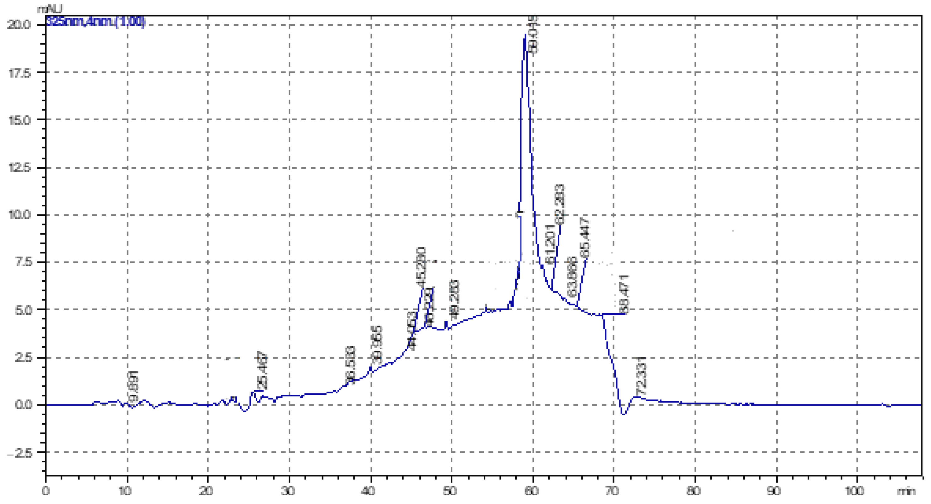
2.3.4. α-Tocopherol Analysis by HPLC

An HPLC method was used for the quantitative determination of α-tocopherol in the oily extract of HEAI based on Amin et al. [26], with slight modification. HPLC 10A VP (Shimadzu, Kyoto, Japan) equipped with two detectors, a diode-array detector ((DAD), 280, 325, and 365 nm) and a fluorescence-spectrophotometer detector at 295 nm excitation and 330 nm emission, temp: 40 °C, was used to analyze tocopherols. A volume of 250 μL of each of the oily preparations (HEAI) was diluted in 250 μL of chloroform (HPLC grade). A reverse-phase column C-18 ODS-M80 (L 100 mm, ID 25 mm) was used with degassed HPLC-grade methanol as a phase with two pumps (Table 3). The injection volume was 10 µL, the flow rate was 1 mL/min, and the oven temperature was maintained at 40 °C. α-tocopherol was identified by comparing it with the retention time of the authentic standard (α-tocopherol) and quantified by comparing the peak area of the standard.

2.3.5. Biological Analyses

Skin-Safety Assessment

In this assessment, two tests were performed: the eye-irritation test and the primary skin-irritation test.

Eye-Irritation Test

This method is used to assess the degree of eye irritation caused by substances following contact with the eye of rabbits (Official Journal of France (OJF) of 24/10/84 [27]). The eye is observed by direct examination, following a numerical quantitative scale from 0 to 4. The eye test in the current study was completed for the conjunctiva as follows: chemosis (A), assessed by swelling; tearing (B), assessed by humidification; and reddening (C), assessed by the condition and color of the blood vessels. The assessment of the conjunctiva was measured with a maximum of 20 according to the following equation:
(A + B + C) × 2
For the iris (D), the test showed congestion and swelling, as well as a reaction to light. The scores obtained (scaled from 0 to 4) and designated by (D) were multiplied by 5, with a maximum value of 10. For the cornea, the degree (E) and surface (F) of opacification were changed. The product of the scores E and F was multiplied by 5, with a maximum value of 80. The calculation of the individual eye-irritation index (IOI) was carried out as follows:
IOI index = [(A + B + C) × 2] + [D × 5] + [(E × F) × 5]
The mean eye-irritation index (IOM) represents the mean of the sum of the IOI (n = 6). The maximum eye-irritation index corresponds to the highest IOI value.

Primary Skin-Irritation Test

This method is applicable to products that do not permanently color the skin, and it assesses the skin reactions caused by a single application (OJF of 01/21/82 [27]).
The observations were carried out on two areas, scarified and non-scarified, on a numerical scale from 0 to 4. These following observations were made in the following order:
-
Erythema and pressure ulcer formation;
-
Edema formation.
The figures obtained on the six rabbits were added for erythema and edema after 24 h and 72 h on non-scarified and scarified areas. The figures were gathered (i.e., Ery + Oed), and the mean was calculated by dividing the total by 24. This mean represents the cutaneous primary index (CPI); its value provides information on the degree of irritation in the following order:
CPI ≤ 0.5: not irritating
0.5 ≤ CPI ≤ 2: slightly irritating
2 ≤ CPI ≤ 5: irritating
5 ≤ CPI ≤ 8: very irritating

Acute-Toxicity Test

The acute-toxicity test study was carried out on oily preparations administered via oral and intraperitoneal pathways. The aim was to assess the potential toxicity on mice as a function of the dose administered and expressed in mL/kg. The acute toxicity was calculated as the lethal dose (LD50), which corresponds to the dose that caused 50% mortality of treated mice.
The mice were divided into 15 groups of 10 individuals, distributed in plastic cages with free access to water and food (standard mixture of granules for mice). The animals were deprived of food for 24 h before the experiment. The mice were treated with a single dose of oily preparation by oral and intraperitoneal routes, with dosages of 10, 20, 30, 40, 50, 60, and 70 mL/kg (per OS) and 0.5, 1, 2, 3, 4, 5, and 6 mL/kg intraperitonial (IP), respectively, according to Zaoui et al. [28]. The control group was similar and did not receive any treatment.
Following the administration of oil, the mice were observed on the first day every 60 min for the first 8 h and afterward once a day for 14 days. During this period, the number of deaths and additional disorders (i.e., diarrhea, vomiting, and immobility) were noted. The monitoring of clinical signs of toxicity also included the observation of mass changes in mice from different experimental groups throughout the experiment [29].
The method of Kabber and Berhens [29] was adopted for the calculation of the LD50.
LD50 = LD 100 − ((Σa × b)/n)
where LD50 is the dose inducing 50% deaths, LD100 is the dose inducing 100% mortality, a is the mean number of deaths between two successive doses, b is the difference between two successive doses, and n is the mean number of animals used.

Assessment of Healing Activity

The healing activity was evaluated on excisional wounds on albino rabbits, as per Boulebda et al. [30]. This test evaluated the healing potential in vivo of the oily preparation (HEAI). The rabbits were randomly selected for this evaluation and were pre-shorn over the entire dorsal surface using an electric tensioner. At the beginning of the experiment, small hairs remaining on the animal’s back were shaved with a sterilized razor blade. The rabbits were anesthetized with ketamine hydrochloride at a dosage of 20 mg/kg combined with 2 mg/kg of acepromazine and injected intramuscularly (IM). Rectangular excision wounds of 2 cm × 1 cm dimension were performed at the level of the dorsolumbar spine.
Regarding wound treatment, the different groups, each comprising six animals, received the following treatments:
-
Cont (−): untreated operated animals;
-
CONT (+): operated animals receiving MADECASOL®;
-
HEAI: operated animals receiving the oily preparation.
Topical treatment was applied at a rate of 0.5 mL/24 h per wound until complete epithelialization.
The progress of wound regression was followed during the period until the end of the experiment (D = 1, 4, 8, 12, 16, and 20 days). This progress was photographed with a camera (Canon EOS 1000D) followed by the treatment of the surface with computer software (AUTOCAD®). The excision was determined at D1 (100% of the excised surface). The percent of wound regression on day x was determined with the following equation [31]:
% of wound contraction = ((surface day 1 − surface day x)/surface day 1) × 100
where day x: chosen day.
The body-mass gain of animals was measured weekly with an animal scale.

2.4. Statistical Analysis

The results were expressed as mean ± SD of various analyses in three replicates. One-way analysis of variance (ANOVA) was carried out, followed by Tukey’s HSD test for multiple comparisons, with a significance level set at p < 0.05.

3. Results and Discussion

3.1. Preliminary Organoleptic Characterization of the Herbal Drug

To better characterize the material, the organoleptic parameters of size, mass, and color were measured. The mean values (n = 100) are reported in Table 4 and Figure 1.
The measured organoleptic parameters, as well as the botanical descriptions of the three samples, were consistent with those reported by previous authors [32,33,34,35].

3.2. Determination of Protein, Ash, and Water Levels in DV

The plant material (PL and AS fruit, OFI seed) was tested for levels of water, protein, and ash. The values found are given in Table 5.
Several observations were made for the protein levels. Overall, the samples of the HEAI showed a substantial total protein value and were considered rich in protein matter [34,36].

3.3. Physicochemical Characterizations of Oily Preparations

3.3.1. Hexane-Extraction Yield

The hexane-extraction yield (combining lipophilic materials) of the studied sample was 21.34 ± 1.05% wt/dry wt of plant material. The HEAI yielded a highly extractable residue in fruits.

3.3.2. Organoleptic and Physicochemical Analyses of HEAI

The measurements of the organoleptic parameters (i.e., color, physical state, odor, and flavor) and overall physicochemical properties (i.e., density, acid index, and refractive index) are given in Table 6.
The density values of the HEAI were compared with those of other studies of unsaturated and saturated fatty oils. The density was highly dependent on the temperature and chemical composition of the oil and indicated the nature of the fatty-acid components—in particular, the length of the chains, the unsaturation, and the functionality of the carbon chains [3]. In addition, the fatty oils with a high proportion of unsaturated acids had high densities, above 0.900 [36]. However, oils with high proportions of saturated fatty acids had relatively lower densities, less than 0.9 [37,38,39]. The value obtained for the oily preparation of HEAI was 0.924 ± 0.1, showing that this product was dominated by unsaturated fatty oils. The refractive index depended, like the density, on the chemical composition of the oil and the temperature, as well as on the unsaturation and length of fatty chains of secondary functions [3]. The acid index indicated that the level of free fat was low for the HEAI, showing the importance of fruit preservation before oil extraction, as well as of the harvesting period in which the fruit ripened [3,40].

3.4. Characterizations by GC-MS of Fatty Acids and Phytosterols in the Oily Preparation

The oily preparation was analyzed by GC-MS for its content of fatty acids and phytosterols. The results are presented in the following section.

3.4.1. Chromatographic Characterization of Fatty Acids by GC-MS

The results of the analysis of the oily preparation of HEAI are reported in Table 7 and the chromatogram in Figure 2.
For the HEAI (see Table 7), the main fatty acids were oleic acid (46.05%), palmitic acid (23.94%), linoleic acid (11.39%), and stearic acid (7.87%). Other fatty acids were present in lower concentrations, and some were unidentified. The variable compositions of the fatty acids could explain fluctuations during the development of the plants [24].
At the current stage, the results showed that the unsaturated fatty acids (i.e., the monounsaturated and polyunsaturated fatty acids), oleic acid (46.05%), and linoleic acid (11.39%) were dominant in the composition of the oily HEAI (see Table 7). The ratio of polyunsaturated to saturated (PUFA/SFA) was 1.8, which indicated good nutritional quality. According to the recognized dietary recommendations, a ratio of PUFA/SFA higher than 1.5 indicates good nutritional value [41].

3.4.2. Chromatographic Characterization of Phytosterols by GC-MS

The results of the GC/MS analysis of the phytosterol fraction of the three samples are given in Table 8 and Figure 3.
The total phytosterol content was 128.56 ± 2.40 mg/100 g in the HEAI, reflecting a high concentration of dietary phytosterols that could efficiently reduce the cholesterol level in the blood [25,42]. The sterol fraction contained three major compounds; in decreasing order of importance, they were β-sitosterol, campesterol, and stigmasterol. Cholesterol was observed in trace amounts (i.e., 0.31 ± 0.02 mg/100 g). Beta-sitosterol was the most important sterol in the oil, and it is known to be involved in the reduction of cholesterol levels. This sterol also plays an important role in reducing swelling in the prostate, heart disease, rheumatoid arthritis, and male-pattern baldness [43].
The differences between phytosterol components can be explained by fluctuations in their quality or quantity, a phenomenon often occurring during the development of fruits [24]. It was reported that by the end of ripening, the enzymatic activity of phytosterol biosynthesis stops in fruits. During this period, the phytosterols are no longer synthesized but are converted into other compounds [44].

3.5. Determination of α-Tocopherols by HPLC in Oily Preparations

The chromatographic traces of α-tocopherol (Figure 4) were obtained by HPLC in the oily HEAI. At an absorbance of 460 nm, the concentration of α-tocopherol was calculated with the following equation:
Absorbance: Y = 0.223 X (mg) + 0.061 (R2: 0.981)
where the absorbance was A = 09.11% or 14.86 mg/100 g extract of HEAI.
The difference in the concentration of α-tocopherol (i.e., 14.86 mg/100 g extract) can be explained by the use of Soxhlet for extraction instead of a simple maceration in hexane. It is known that the heat generated during Soxhlet extraction can alter the status of tocopherols, which are thermo-fragile. This result suggests that the species A. indica could be a moderate source of vitamin E (α-tocopherol).

3.6. Biological Analyses

3.6.1. Toxicological Studies

The acute-toxicity tests were performed on adult mice. The percentages of mortality were observed according to the dose and route of administration (i.e., oral or intraperitoneal) for the oily preparations (HEAI, EHPL, and HAS) in mice during the two stages of the experiment (i.e., every hour on day 1 and once per day on days 2 to 14).
The results for the two observation stages D1 and D2–14 in all groups of treated and control mice were the following:
The groups of control mice that received physiological water orally or intraperitoneally showed no clinical signs or mortality during the 14 days of the experiment.
The groups of mice treated with HEAI (at different doses) and by the two routes of administration showed no mortality during the 14 days. However, the usual reversible clinical signs and nonserious symptoms (i.e., hypoactivity, asthenia, and anorexia) occurred during the first 3 days following the treatments. The two LD50s of HEAI oil (oral and peritoneal) were not calculated given the lack of mortality, indicating the relatively good tolerance of the HEAI treatments administered orally and intraperitoneally at maximum doses of 70 mL/kg and 6 mL/kg, respectively. The conclusion was that the HEAI oil showed weak toxicity [26].

3.6.2. Safety Studies

A safety study was carried out on the rabbits for eye and skin irritation, the cutaneous primary index, and rectal aggressiveness.

Eye Irritation

The eye-irritation index at the level of the iris and cornea was null regardless of the period. For the conjunctiva, the index of irritation was 4 during the first hour after instillation and then fell to zero within 24 h. Thus, it could be deduced that the application of the oil in the eye did not generate any macroscopically visible irritation, either for the iris or for the cornea. The value of the IOI was 4, measured at the level of the conjunctiva, and this was considered a low impact (Official Journal of France (OJF): 10/24/84, [27]).

Skin Irritation

The primary skin-irritation index evaluated by the action of the tested oil on scarified and non-scarified skin of rabbits was initially 0.079, but it dropped to zero within 48 h. In addition to the previous evaluation, the rectal-mucosa application of the HEAI in rabbits for 15 days, followed by macroscopic observation of the rectum, revealed the absence of any hemorrhage or ulceration, redness, or diarrhea. In conclusion, the three oily products tested on rabbits could be considered non-irritating at the ocular, cutaneous, and anal-mucous-membrane levels according to the norms specified by the OJF of 21/01/82 [27].

3.6.3. Assessment of Healing Activity

The results of the evaluation of the healing process (expressed as a percentage of contraction) of wounds surgically excised and treated with the oily preparation HEAI are reported in Table 9.
The treatment with HEAI showed significant healing progression compared with the negative control (CONT (−)) depending on the recovery time (see Table 9). After 4 days, all wounds of the HEAI group had comparable areas, as well as similar signs of inflammation and bleeding, but the healing was significantly accelerated (p < 0.01) compared with the CONT (−). On day 8, the oily HEAI led to a significantly accelerated healing process (p < 0.01) compared with the CONT (−); this pattern was observed consistently also on days 12 and 16 (p < 0.05). Overall, a gradual reduction in the wound area over time was observed in the different groups. The highest percentage of contraction was noticed for the wounds treated with HEAI oil, followed by the negative control group. The results observed in rabbits for the regression path of excised wounds treated with the oily preparation showed that at the end of the 20 days, treatment with HEAI at a rate of 0.5 mL oil/wound/day significantly accelerated the wound closure (p < 0.05) compared with the control group. Such a finding was discernibly supported by the comparable (p ≥ 0.05) healing potentials for HEAI oil and MADECASOL® (Table 9). A previous study on the evolution of the healing process of wounds of surgical excision in rats indicated that contraction of the wounds occurred following HEAI treatment [30]. The current study showed the potential for the hexane extract of neem fruits to provide healing at 0.5 mL oil/wound/day, with a significant positive effect (p < 0.01) compared with the control.
The current report on the cicatricial process induced by the oily HEAI extracted from Algerian neem almonds may be the first in this field of research. Previous studies showed that aqueous and alcoholic extracts of rackets extracted from the same species led to the contraction of treated wounds. The results of this work showed the following patterns:
The HEAI treatment significantly increased the healing process (contraction percentage) of surgical wounds in rabbits (p < 0.01 and p < 0.05) throughout the treatment period.
The presence of a potentiating effect that accelerated the healing significantly (p < 0.01 and p < 0.05) in rabbits treated with the oily preparation based on the seeds of A. indica was observable during the first half of the treatment period.

4. Conclusions

The Algerian and North African pharmacopeia has used semi-solid preparations for skin applications since antiquity. Some are based on oil extracts from fruits of the neem tree, A. indica. However, few previous scientific studies had focused on the evaluation of the healing properties of the extracts for wounds. Given the high nutritional value of these plant constituents, such as proteins, fatty acids, phytosterols, and tocopherols, as well as their overall beneficial effects on the skin, their physicochemical and phytochemical properties were characterized in the current study. High levels of tocopherols and phytosterols were also found in the hexane and fruits, respectively. From a toxicological perspective, the oily product HEAI tested on rabbits can be considered a nonirritant for the eyes, skin, and anal mucous membranes. The acute-toxicity study in mice also showed an absence of or weak toxicity of the oily HEAI when it was administered orally or intraperitoneally. These results indicated the relatively good tolerance of treatments with HEAI under experimental conditions. Because of the diversity of its active compounds, oily HEAI has important healing properties. This study’s results comprise the first scientific indication of the presence of healing properties of the hexane extract of A. indica seeds.

Author Contributions

A.B. and H.B.: conceptualization, methodology, acquisition of data, writing—original draft; M.K.: supervision; S.A.A., M.I.A. and F.B.: software, formal analysis, resources, investigation, writing—review and editing; G.P.: investigation, writing—review, and editing.; S.A.A. and O.H.A.-E.: funding acquisition, conceptualization, formal analysis. All authors have read and agreed to the published version of the manuscript.

Funding

The authors extend their appreciation to the Deanship of Scientific Research at Imam Mohammad Ibn Saud Islamic University for funding this work through Research Group No. RG-21-09-92.

Data Availability Statement

The data are not to be shared due to restrictions, e.g., privacy and regulation.

Acknowledgments

The authors extend their appreciation to the Deanship of Scientific Research at Imam Mohammad Ibn Saud Islamic University for funding this work through Research Group No. RG-21-09-92.

Conflicts of Interest

The authors declare no conflict of interest.

References

  1. López-Expósito, I.; Castillo, A.; Yang, N.; Liang, B.; Li, X.M. Chinese herbal extracts of Rubia cordifolia and Dianthus superbus suppress IgE production and prevent peanut-induced anaphylaxis. Chin. Med. 2011, 6, 35. [Google Scholar] [CrossRef] [PubMed]
  2. Harvey, A.L. Natural products in drug discovery. Drug Disco. Tod. 2008, 13, 894–901. [Google Scholar] [CrossRef] [PubMed]
  3. Bruneton, J. Pharmacognosie, Phytochimie, Plantes Médicinales, 3rd ed.; Editions TEC & DOC: Paris, France, 1999; pp. 319, 384, 680, 791–793. [Google Scholar]
  4. Baba Aïssa, F. Encyclopédie des Plantes Utiles, Flore Méditerranéenne (Maghreb et Europe Méridionale), Substances Végétales d’Afrique, d’Orient et d’Occident; Ed.El Maarifa: Algiers, Algeria, 2011. [Google Scholar]
  5. Sandanasamy, J.D.; Nour, A.H.; Tajuddin, S.N.B.; Hamid Nour, A. Fatty acid composition and antibacterial activity of neem (Azadirachta indica) seed oil. Open Conf. Proc. J. 2013, 4, 43–48. [Google Scholar] [CrossRef]
  6. Parmar, U.M.; Desai, B.S.; Chavda, J.R.; Tandel, M.B.; Jha, S.K. A. Juss. (Neem). J. Non-Timber Forest Prod. 2016, 23, 135–138. [Google Scholar] [CrossRef]
  7. Brototi, B.; Kaplay, R.D. Azadirachta indica (Neem): It’s economic utility and chances for commercial planned plantation in Nanded District. Int. J. Pharma. 2011, 1, 100–104. [Google Scholar]
  8. Biswas, K.; Chattopadhyay, I.; Banerjee, R.K.; Bandyopadhyay, U. Biological activities and medicinal properties of neem (Azadirachta indica). Curr. Sci. 2002, 82, 1336–1345. [Google Scholar]
  9. Hashmat, I.; Azad, H.; Ahmed, A. Neem (Azadirachta indica A. Juss)—A nature’s drugstore: An overview. Int. Res. J. Biol. Sci. 2012, 1, 76–79. [Google Scholar]
  10. Islas, J.F.; Acosta, E.; Zuca, G.; Delgado-Gallegos, J.L.; Moreno-Treviño, M.G.; Escalante, B.; Moreno-Cuevas, J.E. An overview of Neem (Azadirachta indica) and its potential impact on health. J. Funct. Foods 2020, 74, 104171. [Google Scholar] [CrossRef]
  11. Khanna, J.; Medvigy, D.; Fueglistaler, S.; Walko, R. Regional dry-season climate changes due to three decades of Amazonian deforestation. Nat. Clim. Chang. 2017, 7, 200–204. [Google Scholar] [CrossRef]
  12. Herbert, V. Vitamins in Medicine. JAMA 1983, 249, 281. [Google Scholar] [CrossRef]
  13. Barbosa, M.O.; Coutinho, D.J.G.; Santos, J.; Cordeiro, R.P.; Muniz, L.R.; Alves, R.C.; Bessa, C.M.A.S.; da Silva, M.V.; Oliveira, M.B.P.P.; de Oliveira, A.F.M. Composition of fatty acids, tocopherols, tocotrienols and β-carotene content in oils of seeds of Brazilian Sapindaceae and Meliaceae species. J. Food Sci. Technol. 2019, 56, 3164–3169. [Google Scholar] [CrossRef]
  14. Kaushik, N. Determination of azadirachtin and fatty acid methyl esters of Azadirachta indica seeds by HPLC and GLC. Anal. Bioanal. Chem. 2002, 374, 1199–1204. [Google Scholar] [CrossRef] [PubMed]
  15. Finglas, P.M.; Wigertz, K.; Vahteristo, L.; Witthöft, C.; Southon, S.; de Froidmont-Görtz, I. Standardisation of HPLC techniques for the determination of naturally-occurring folates in food. Food Chem. 1999, 64, 245–255. [Google Scholar] [CrossRef]
  16. AFNOR (Association française de normalization). Recueil des Normes Française, Graine oléagineuses–Détermination de la Teneur en Acides Gras par Chromatographie en Phase Gazeuse; T60-233 et T60-234; AFNOR: Paris, France, 1998. [Google Scholar]
  17. Lagarda, M.J.; García-Llatas, G.; Farré, R. Analysis of phytosterols in foods. J. Pharm. Biomed. Anal. 2006, 41, 1486–1496. [Google Scholar] [CrossRef] [PubMed]
  18. Sosulski, F.W.; Imafidon, G.I. Amino acid composition and nitrogen-to-protein conversion factors for animal and plant foods. J. Agric. Food Chem. 1990, 38, 1351–1356. [Google Scholar] [CrossRef]
  19. Sidibé-Anago, A.G.; Ouédraogo, G.A.; Ledin, I. Effect of partly replacing cottonseed cake with Mucuna spp. (var. Ghana) hay on feed intake and digestibility, milk yield and milk composition of zebu cows. Trop. Anim. Health Prod. 2006, 38, 563–570. [Google Scholar] [CrossRef]
  20. Boukeloua, A.; Belkhiri, A. Caractérisation Botanique et Chimique et Evaluation Pharmaco-Toxicologique D’une Préparation Topique à Base D’huile de Pistacia lentiscus L. (Anacardiaceae). Ph.D. Thesis, Université Frères Mentouri-Constantine 1, Constantine, Algeria, 2009. [Google Scholar]
  21. Boukeloua, A.; Belkhiri, A.; Djerrou, Z.; Bahri, L.; Boulebda, N.; Pacha, Y.H. Acute toxicity of Opuntia ficus indica and Pistacia lentiscus seed oils in mice. Afr. J. Tradit. Complement. Altern. Med. 2012, 9, 607–611. [Google Scholar] [CrossRef]
  22. Lecoq, R. Manuel D’analyses Alimentaires et D’expertises Usuelles. 1965. Available online: https://agris.fao.org/agris-search/search.do?recordID=US201300598471 (accessed on 16 May 2016).
  23. AOAC Association of Official Analytical Chemists. Official Methods of Analysis of the Association of Official Analytical Chemists, 16th ed.; AOAC International: Arlington, VA, USA, 1990. [Google Scholar]
  24. Trabelsi, H.; Cherif, O.A.; Sakouhi, F.; Villeneuve, P.; Renaud, J.; Barouh, N.; Sadok, B.; Mayer, P. Total lipid content, fatty acids and 4-desmethylsterols accumulation in developing fruit of Pistacia lentiscus L. growing wild in Tunisia. Food Chem. 2012, 131, 434–440. [Google Scholar] [CrossRef]
  25. Piironen, V.; Toivo, J.; Puupponen-Pimiä, R.; Lampi, A.M. Plant sterols in vegetables, fruits and berries. J. Sci. Food Agric. 2003, 83, 330–337. [Google Scholar] [CrossRef]
  26. Amin, M.Z.; Islam, T.; Mostofa, F.; Uddin, M.J.; Rahman, M.M.; Satter, M.A. Comparative assessment of the physicochemical and biochemical properties of native and hybrid varieties of pumpkin seed and seed oil (Cucurbita maxima Linn.). Heliyon 2019, 5, e02994. [Google Scholar] [CrossRef]
  27. Hamdi Pacha, Y. Effect Biologique d’un Nectar de Fleur. JAM 1993, 3, 104–114. [Google Scholar]
  28. Zaoui, A.; Cherrah, Y.; Mahassini, N.; Alaoui, K.; Amarouch, H.; Hassar, M. Acute and chronic toxicity of Nigella sativa fixed oil. Phytomedicine 2002, 9, 69–74. [Google Scholar] [CrossRef] [PubMed]
  29. Adjoungoua, A.L.; Koffi, A.; Traore, F.; Diafouka, F. Etude Phytochimique et Toxicologique de Ziziphus mauritiana, (Rhamnaceae), Une Plante Anti-hypertensive. Med. Pharm. Afr. 2008, 21, 82. [Google Scholar]
  30. Boulebda, N.; Belkhiri, A.; Belfadel, F.; Bensegueni, A.; Bahri, L. Dermal wound healing effect of Pistacia lentiscus fruit’s fatty oil. Pharmacog. Res. 2009, 1, 66–71. [Google Scholar]
  31. Srivastava, P.; Durgaprasad, S. Burn wound healing property of Cocos nucifera: An appraisal. Indian J. Pharmacol. 2008, 40, 144. [Google Scholar]
  32. Hichey, M.; King, C. 100 Families of Flowering Plants, 2nd ed.; Cambridge University (Botanic Garden): London, UK, 1988; 291p. [Google Scholar]
  33. Baba Aissa, F. Encyclopédie des Plantes Utiles, Flore d’Algérie et du Maghreb; Librairie Moderne: Rouiba, Algérie, 1999. [Google Scholar]
  34. Iserin, P.; Masson, M.; Restellini, J.; Ybert, E.; De Laage de Meux, A.; Moulard, F.; Vican, P. Larousse des Plantes Médicinales: Identification. Préparation, Soins, 2nd ed.; VUEF: Hong Kong, China, 2001; p. 335. [Google Scholar]
  35. El Monfalouti, H.; Guillaume, D.; Denhez, C.; Charrouf, Z. Therapeutic potential of argan oil: A review. J. Pharm. Pharmacol. 2010, 62, 1669–1675. [Google Scholar] [CrossRef]
  36. Karleskind, A. (Ed.) Manuel des Corps Gras; Technique et Documentation-Lavoisier: Paris, France, 1992. [Google Scholar]
  37. Farines, M.; Soulier, J.; Charrouf, M.; Soulier, R. Etude de l’huile de graines d’«Argania spinosa» (L.), Sapotaceae. 1. La fraction glycéridique. Rev. Fran. Corps. Gras. 1984, 31, 283–286. [Google Scholar]
  38. Maurin, R. Argan oil, Argania spinosa (L.) Skeels (Sapotaceae). Rev. Fr. Corps Gras 1992, 39, 139–146. [Google Scholar]
  39. Boudjira, M. Contribution à l’Etude Biochimique de l’Huile d’Arganier (Argania spinosa L.). Ph.D. Thesis, National Agronomic Institute (INA), Algiers, Algeria, 2002; 69p. [Google Scholar]
  40. Djerrou, Z. Anti-hypercholesterolemic effect of Pistacia lentiscus fatty oil in egg yolk-fed rabbits: A comparative study with simvastatin. Chin. J. Nat. Med. 2014, 12, 561–566. [Google Scholar] [CrossRef]
  41. Ribarova, F.; Zanev, R.; Shishkov, S.; Rizov, N. α-Tocopherol, fatty acids and their correlations in Bulgarian foodstuffs. J. Food Compo. Anal. 2003, 16, 659–667. [Google Scholar] [CrossRef]
  42. Ling, W.H.; Jones, P.J.H. Dietary phytosterols: A review of metabolism, benefits and side effects. Life Sci. 1995, 57, 195–206. [Google Scholar] [CrossRef] [PubMed]
  43. Saeidnia, S.; Manayi, A.; Gohari, A.R.; Abdollahi, M. The story of beta-sitosterol—A review. Eur. J. Med. Plant. 2014, 4, 590. [Google Scholar] [CrossRef]
  44. Menvielle-Bourg, F.J.; Joanny, F. L’extrait huileux au CO2 supercritique de baies et de graines d’argousier (Hippophaer hamnoides L.) et ses effets sur la peau et les muqueuses. Phytothérapie 2009, 1, 3–7. [Google Scholar] [CrossRef]
Figure 1. Plant material (seeds) of A. indica.
Processes 11 01692 g001
Figure 2. Chromatogram of the oily preparation (HEAI) related to composition in methyl esters of fatty acids.
Processes 11 01692 g002
Figure 3. GC-MS chromatogram of A. indica hexane extract related to composition in phytosterols: (1) cholesterol, (2) campesterol, (3) stigmasterol, (4) β-sitosterol.
Processes 11 01692 g003
Figure 4. Chromatogram of A. indica α-tocopherols determination by HPLC in oily preparations.
Processes 11 01692 g004
Table 1. The mass of the test portion and its precision according to the presumed acid value.
Presumed Acid ValueMass of Test Portion (g)Accuracy of Test Portion (g)
Below 1200.05
1 to 4100.02
4 to 152.50.01
15 to 750.50.001
Up to 750.10.0002
Table 2. Standard phytosterols (Sigma-Aldrich, Germany) used in the chromatographic analysis (GC-MS) by TMS derivatives.
RT minCommon NameSystematic Name
0.77CholesterolCholest-5-en-3β-ol
0.89CampesterolCampest-5-en-3β-ol
0.95Stigmasterol(22 E)-stigmasta-5,22-dien-3β-ol
1.04β-SitosterolStigmast-5-en-3β-ol
Table 3. Composition of the mobile phases of α-tocopherol analysis by HPLC.
Time (min)Mobile Phase B (%)Mobile Phase A (%)
0.010100
9.01496
30.01892
45.012278
50.012872
65.014555
66.010100
108.000100
Mobile phase A: solvent A = water, acetic acid, acetonitrile (94.5/0.5/5); mobile phase B: solvent B = acetonitrile (100%).
Table 4. Physicochemical properties of the plant material (seeds) of A. indica.
Parameters Sample
AI (Seed) (n = 100)
Dimension (diam./long, mm)2.16 ± 0.5/1.25 ± 0.3
Mass (g)4.95 ± 0.86
ColorYellowish or light brown
FormOval in shape, elongated to a rounded shape
Table 5. Protein, ash, and water levels of plant material.
Rate (%) of Physicochemical PropertiesHEAI (Hexane Extract of A. indica)
Water8.02 ± 0.35
Total proteins27.45 ± 0.84
Ash5.89 ± 0.13
Table 6. Organoleptic properties and physicochemical parameters of hexane extract of A. indica.
ParametersHexane Extract of A. indica
ColorGreen
Liquide stateViscous
FlavorAromatic
SmellOdorless
Density 20 °C (g/mL) *0.924 ± 0.1
Refraction index (20 °C) *1.471 ± 0.4
Acid index (mg KOH/g) *1.01 ± 0.13
Saponification index (mg KOH/g) *195.06 ± 0.1
Iodine value (I2 g/100 g) *90.78 ± 0.2
* Mean value ± SD (n = 3).
Table 7. Composition of the oily preparation (HEAI) in terms of fatty acids.
PeackRTPeack Report AreaHight Fatty AcidHexane Extract of A. indica (%)
117.6585,92023,912Myristic Acid (C14:0)0.77
219.8213,7346257Pentadecanoic A. (C15:0)0.32
321.958,442,9342,009,236Palmitic A. (C16:0)23.94
422.5653,68217,029Palmitoleic A. (C16:1)1.28
523.8539,31313,628Stearic A. (C18:0) 7.87
625.864,601,634957,939Elaidic A. (C18:1n9t)5.11
726.3633,537,2714,729,576Oleic A. (C18:1n9c) 46.05
828.4827,710,2694,631,303γ-linolenic A. (C18:3n6)2.27
927.3584,41627,068Linoleic A. (C18:2n6c)11.39
1028.48316,874101,678Linolenic A. (C18:3n6)0.30
1129.23301,16296,745Arachidic A. (C20:0)0.51
1229.6890,04627,936Cis-11-eicosenoic A. (C20:1)0.11
1334.10179,37149,466Cis-13,16-docosadienoic (C22:2)0.06
1435.4626,69810,159Lignoceric A. (C24:0)0.02
75,527,14612,716,469
Table 8. Composition of the A. indica hexane extract in phytosterols.
Peak SterolsRT (min)Hexane Extract of A. indica (mg/100 g)
1Cholesterol0.780.31 ± 0.02
2Campesterol0.8518.94 ± 0.63
3Stigmasterol0.9212.05 ± 0. 98
4β-sitosterol1.0397.26 ± 0.77
Total 128.56 ± 2.40
Mean value ± SD (n = 3).
Table 9. Wound-contraction rate % for 20 days following the neem oil treatment.
Wound Contraction (%) *
TreatmentDay 4Day 8Day 12Day 16Day 20
Cont (−)16.08 ± 1.71 29.87 ± 2.3636.24 ± 1.7569.17 ± 4.0591.66 ± 1.87Analysis of Variance
Cont (−)Processes 11 01692 i001Processes 11 01692 i002Processes 11 01692 i003Processes 11 01692 i004Processes 11 01692 i005df = 10
F(4,10) = 457.6
p < 0.0001
Tukey’s HSD test (time slots)p < 0.0001p < 0.0001p < 0.0001p < 0.0001p < 0.0001
HEAI23.93 ± 2.50 41.69 ± 1.25 49.08 ± 2.53 75.84 ± 3.00 96.78 ± 1.75 Analysis of Variance
HEAIProcesses 11 01692 i006Processes 11 01692 i007Processes 11 01692 i008Processes 11 01692 i009Processes 11 01692 i010df = 10
F(4,10) = 474.8
p < 0.0001
Tukey’s HSD test (time slots)p < 0.0001p < 0.0001p < 0.0001p < 0.0001p < 0.0001
CONT (+)31.16 ± 1.3647.92 ± 2.6964.82 ± 2.0981.12 ± 1.1498.03 ± 0.77Analysis of Variance
CONT (+)Processes 11 01692 i011Processes 11 01692 i012Processes 11 01692 i013Processes 11 01692 i014Processes 11 01692 i015df = 10
F(4,10) = 289.5
p < 0.0001
Tukey’s HSD test (time slots)p < 0.0001p < 0.0001p < 0.0001p < 0.0001p < 0.0001
Analysis of Variancedf = 6
F(2,6) = 61.65
p = 0.0209
df = 6
F(2,6) = 46.5
p = 0.0016
df = 6
F(2,6) = 202.4
p = 0.0010
df = 6
F(2,6) = 103.7
p = 0.0285
df = 6
F(2,6) = 73.8
p = 0.0461
Tukey’s HSD test [CONT (−) vs. HEAI]p = 0.0127p = 0.00425p = 0.0022p = 0.0409p = 0.0326
Tukey’s HSD test [CONT (+) vs. HEAI]p = 0.4819p = 0.4113p = 0.0727p = 0.3456p = 0.7501
CONT (−): negative control (operated rabbit without treatment). CONT (+): positive control (operated rabbit with MADECASOL®). HEAI: operated rabbit treated with hexane extract of A. indica (0.5 mL oil/wound/day). * Mean value ± SD (n = 3).
Disclaimer/Publisher’s Note: The statements, opinions and data contained in all publications are solely those of the individual author(s) and contributor(s) and not of MDPI and/or the editor(s). MDPI and/or the editor(s) disclaim responsibility for any injury to people or property resulting from any ideas, methods, instructions or products referred to in the content.