Abstract
Murals are a significant cultural heritage of humanity, and one of the conservation studies is to control the growth of microorganisms. General biocide agents can be used to preserve murals while also providing new organic carbon sources and increasing environmental pollution. In recent years, radiation technology has shown promising prospects for use in heritage protection. Five microorganisms often found in murals were irradiated with an electron beam in this study, and six mineral pigments were tested for color change, Raman spectra and pigment layer cohesion after irradiation. The result showed that irradiation at 20 kGy can basically eliminate Pseudomonas citronellolis, Bacillus sporothermodurans, Streptomyces vinaceus, and Streptomyces griseolus from the culture medium, but only inhibited the growth of Penicillium flavigenum. Lead white pigment showed a color difference of 5.56 (∆E*97) after irradiation, but lead tetroxide, azurite, malachite, ferrous oxide, and cinnabar showed no visible changes. The Raman spectra of the irradiated and unirradiated samples were basically the same. E-beam radiation did not affect the surface cohesion of the pigment layer. This preliminary work shows the potential of electron-beam technology in mural protection and provides basic research and relevant experience for the subsequent in situ mural protection work.
1. Introduction
Murals are an important part of human cultural heritage and have unique historical value. However, microbial contamination is damaging these artworks. This study showed that microorganism growth causes physical harm, and the generation of microbial pigments and organic acids weaken the aesthetic properties and degrade the mural base [1,2,3]; all these effects eventually cause incalculable damage to the murals. Today, physical and chemical methods that aim to inhibit microorganisms are being used to protect these precious artworks [4,5,6].
Some biocide agents are used to protect murals from biodegradation, which play an important role in conserving cultural heritage [7,8]. However, the use of biocide reagents might cause health and environmental problems [9,10]. Several studies have found that they make other communities become more prevalent, which are related to delivering new organic nitrogen and carbon [11,12]. Emerging biocide reagents, such as essential oils and nanomaterials, are becoming popular since they are less polluting and more environmentally friendly. Several essential oils have been shown to have antimicrobial properties [13,14], and some of them outperform conventional biocide agents [15]. However, research into concerns such as long-term performance, the interference with heritage materials, and other possible hazards is still lacking [16]. Franco-Castillo, et al. [17] provide a thorough examination of the use of antibacterial nanoparticles to protect cultural property items. Long-term evaluation, examining the impacts on specific microorganisms, historical materials, and developing multifunctional nanomaterials are all considered to be problems for this approach in heritage conservation study.
Ionizing radiation disinfection technology is a kind of physical technology with little influence on the environment and good disinfection effect. Radiation destroys the genetic material in the cell directly or through the radiolysis of the cell water, releasing oxidized free radicals, and finally leading to cell death [18]. Ionizing radiation mainly includes gamma radiation and electron-beam radiation; electron-beam radiation has better safety and predictability, and has broad prospects for development. Commercial high-energy electron accelerators can be used to sterilize movable artifacts [19]. Irradiation technology is widely used for sterilization in medical devices and, since the 1980s, also for disinfection of cultural objects. Decontaminating the Ramses II mummy and a specimen of a 42,000-year-old mammoth are two notable examples [20]. Research related to the use of radiation technology for heritage conservation is mainly concentrated in France, Croatia, Argentina, Italy, and other European regions.
As immovable cultural relics, murals need to be preserved in situ, so traditional electron-beam irradiation equipment cannot be used. The development of small, intelligent mobile low-energy electron-beam irradiation equipment has broadened the application range of electron-beam irradiation sterilization [21]. According to the present report, it is predicted that small electron-beam equipment can be used for in situ protection of tomb murals. However, the application of this technology needs to consider the radiation effect of the irradiated material.
Some powder pigments showed changes after irradiation. In the study of Negut, et al. [22], ultramarine, mars red, minium, raw sienna, and chrome yellow showed color shifts after gamma radiation at a dose of 36 kGy.
Electron-beam irradiation usually degrades or crosslinks organic materials and affects their properties. Effects of radiation on gelatin have also been reported. Low-dose (8 kGy) gamma ray irradiation did not significantly affect amino acid content in gelatin, but the increase in irradiation dose would make the adhesive viscosity continue to decline, which may be caused by the breaking of the protein peptide bond inside the adhesive [23]. However, at the same dose, there was no significant difference between gamma irradiation and electron-beam irradiation on gelatin [24].
Due to the limited electron-beam penetration ability of low-energy accelerators, electron-beam irradiation technology has been rarely reported in the field of color cultural relics protection. Most studies have only looked at the stain-removal effect of commercial high-energy electron accelerators on some microorganisms [23], or separately tested the effect of irradiation on pigment [24]. There are few studies on the irradiation effect of simulated wall blocks and no reports on the change of the cohesive force of the pigment layer of wall blocks after irradiation. Since the mobile electron-beam source belongs to the low-energy accelerator, in order to verify the feasibility of its application in non-mobile murals, the 200 keV electron accelerator was selected in this study. Under these conditions, the sterilizing effect on typical microorganisms was first verified, and the sterilizing dose was determined. Then the effect of sterilization dose on pigment composition and structure and the effect of pigment combination material on mural stability was determined. Thus, the feasibility of the low-energy electron-beam irradiation technology in mural sterilization protection was preliminarily verified.
Specifically in this study, we first conducted a series of experiments under laboratory conditions. The dose tolerance values of five typical microorganisms were determined by the irradiation test of gradient dose. Then, according to the disinfection dose, the irradiation experiment of a simulated mural block was carried out to study the irradiation effect on murals. A colorimeter was used to detect the change of color parameters of pigments, the Raman spectrometer was used to detect their chemical properties, and transparent tape was used to test the adhesion of the mural painting layer. The basic data obtained from the experiment can provide a basis for evaluating the feasibility of irradiation technology in mural protection and provide guidance for subsequent experiments of this technology on murals in the real tomb environment.
2. Materials and Methods
2.1. Microorganism
According to previous research, common bacterial communities in Dunhuang murals include Bacillus spp., Paenibacillus spp., Pseudomonas spp., Streptomyces spp., etc. Fungal communities are dominated by Penicillium spp., Cladosporium spp., Alternaria spp., Aspergillus spp., etc. [25,26]. Penicillium species are widely found in cultural heritage, including paper, paintings, textiles, etc. [27]. Streptomyces has also recently been found in the murals of the Southern Tang Dynasty, China. Pseudomonas and Bacillus also appear frequently in various cave paintings [28,29].
Considering the microorganisms in the tomb and cave paintings, we selected Penicillium flavigenum, Pseudomonas citronellolis, Bacillus sporothermodurans, Streptomyces vinaceus, and Streptomyces griseolus for irradiation experiments. The five microorganisms were abbreviated as PNC, Pse, Bac, Str.v, and Str.g. The strains were provided by researchers from the Environmental and Health Group of the Chinese Academy of Sciences [30].
Potato dextrose agar (PDA) was used to grow Penicillium, LB medium to grow Bacillus and Pseudomonas, and Gauze’s synthetic medium No. 1 to grow two species of Streptomyces. The culture medium was provided by Land Bridge Technology Co., Ltd., Beijing, China.
Culture plates of different microorganisms with varied incubation times (1,3,5,8 days) were made to explore the association between radiation resistance and incubation time. Microorganisms that were grown for different incubation times are shown in Figure 1.
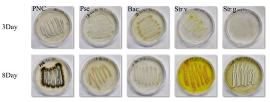
Figure 1.
Microorganisms that have grown for 3 and 8 days.
2.2. Mural Blocks and Pigments
Dunhuang Research Institute supplied 6 cm diameter mock mural blocks. The main body of the mural block was made of sand, clay, straw and hemp mixed with water, with a layer of lime on the surface. After the mural blocks were dried, a certain amount of mineral pigment powder was dispersed in 2% gelatin solution, mixed, and then applied to the mural block [31]. The pigments include lead tetroxide (Pb3O4), azurite (Cu3(CO3)2(OH)2), malachite (Cu2(OH)2CO3), ferrous oxide (Fe2O3), cinnabar (HgS), and lead white (2PbCO3Pb (OH)2), which are often used in the Dunhuang murals [31]. The pigments are purchased from Company Lhasa Zaxi Rainbow Tibetan Pigment Preparation Co., Ltd. (Lhasa, Tibet, China) and Shanghai Aladdin Biochemical Technology Co., Ltd. (Shanghai, China).
2.3. Electron-Beam Irradiation
The irradiation was performed with MEB-200 electron-beam equipment (Sichuan Zhiyan Technology Co., Ltd., Mianyang, China) at a dose rate of 4000 Gy/s and a source energy of 200 keV. To prevent the creation of ozone during the experiments, high purity nitrogen was pumped into the irradiation equipment. A B3 film dosimeter (GEX Corporation, Palm City, FL, USA) was used to determine radiation dose. The B3 film dosimeter transformed from colorless to purplish red after exposure to radiation. It was then placed in the oven for 15 min at 58.5 °C, and its absorbance at 552 nm was recorded. From the absorbance, we then calculated the absorbed dose for the experiment.
The microorganisms were irradiated at 5, 10, and 20 kGy, with one control and three experimental groups set up for each experiment. The mock mural blocks were irradiated with 20 kGy.
2.4. Characterization
The irradiated microorganisms were inoculated into a new medium and incubated at 25 ℃ for 48 h. The effect of irradiation was evaluated by checking whether microorganisms grew again.
We used a 3nh colorimeter (Shenzhen Threenh Technology) to measure the color change of pigments before and after irradiation; the test was repeated 5 times in the same area. In the CIELAB color space, the L* value represents lightness ranging from black (L* = 0) to white (L* = 100); the a* value represents red (+) and green (−), and the b* value represents yellow (+) and blue (−). We used color difference formula ΔE*97 (Equation (1)) to evaluate the color change. When E* < 3, the color change is practically imperceptible; when 3 < E* < 6, the color change is perceptible but acceptable; and when E* > 6, the color change is unsatisfactory [31,32,33].
[L1*, a1*, b1*] and [L2*, a2*, b2*] represent the color parameters of the control and experimental groups, respectively.
The Raman spectra of pigments were recorded using a 473 nm laser excitation line with a Horiba LabRAM HREvolution Raman spectrometer, and the spectral resolution was 0.6 cm−1. Excitation time in general was set at 20 s with a power of 7.5 mW; the excitation laser was focused with an objective lens 50 × (NA = 0.5). The wave number calibration was performed using a silicon wafer. For each test, a small amount of pigment was scraped from the mural block and applied to the slide.
We used the method described by Drdácký, et al. [34] to test the surface cohesion of the pigment layers. We applied a section of 19 mm × 28 mm Scotch transparent tape (3M, Maplewood, MN, USA) to the mural block and rubbed it with an eraser to ensure a solid bond. After 60 s, the free end was grasped and the tape quickly removed at an angle of 180° as far as possible. The test was repeated ten times in the same location, recognizing the difference in tape mass before and after the test (mg). The average peel amount (mg/cm2) was calculated by taking the sum of the 10 tests and dividing it by the area of each tape.
3. Results and Discussion
3.1. Irradiation Decontamination
Table 1 shows the survival of microorganisms after irradiation. In the table, × means no growth following reinoculation, and the numbers 1 and 3 mean which of the three experimental groups was not eliminated completely, with 1 or 3 groups still regrowing.

Table 1.
Survival results of microorganisms with different incubation time after irradiation.
Usually, the effectiveness of radiation decontamination is related to the type and the quantity of the microorganism, as well as temperature, humidity, oxygen, and other factors [18]. According to the results, a dose of 5 kGy already removes most microorganisms, which shows the excellent disinfection ability of irradiation technology. There were 4 bacterial species (Bac, Pse, Str.g, Str.v) that did not regrow after being exposed to 20 kGy of radiation in our experiment. Both Streptomyces species were eliminated at all doses, showing low resistance to irradiation. The fungus Penicillium flavigenum was not removed at a dose of 10 or 20 kGy and simply showed growth resistance (Figure 2, PNC, Red circular). After 30 days, eliminated bacteria did not regrow in the medium. These results showed that irradiation technology can efficiently reduce microbial contamination and give support for subsequent protective efforts.
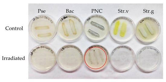
Figure 2.
Microorganisms with a cultivation time of 8 days are irradiated by 10 kGy.
The fungus was more radiation-resistant than the general bacteria [35]. Saleh, et al. [36] tested the relative resistance of ten types of fungi found in food, including Alternaria, Aspergillus, Cladosporium, Curvularia, Fusarium, and Penicillium, against γ radiation from a 137Cs source. Most fungi require less than 6.5 kGy to be inactivated, while some Curvularia and Alternaria species require 20 kGy to be destroyed. In the test performed by Maria and Wiendl [37], Penicillium was still resistant to γ irradiation at 17.5 kGy, while Aspergillus and Cladosporium could still survive at 20 kGy. Sakr, et al. [38] found that γ radiation at 20–25 kGy could clean most of Streptomyces spores, which is similar to our results.
In other decontamination studies, Li, et al. [6] tested six biocides against Aspergillus and Pseudomonas on limestone, and these biocides showed selectivity for microorganisms, with none being effective against both fungus and bacteria. Some microbes could gain increased radiation resistance through the synthesis of melanin, according to studies on the effects of radiation [39,40]. In Figure 1, Penicillium clearly darkened after 8 days of incubation, which could be due to its higher radiation resistance. Abdel-Haliem, et al. [41] found that a combination of γ radiation at 25 kGy and antibiotics was effective in removing several Streptomyces strains from the murals. The combination with biocide agents seems to be the future direction of irradiation technology.
The mural surface lacks nutrients and has far more microbial diversity in the real environment. Thus, irradiation experiments can have different results, which we will investigate in later tests. It is worth noting that following irradiation, the medium inevitably comes into contact with air, so after 48 h of incubation, some other microbe sometimes arises within the experimental group’s medium. Distinguishing between target strains and contaminants by their morphology, the presence of contaminants is not counted as not being disinfected by irradiation.
3.2. Effect on the Color of the Mock Mural Blocks
For comparison, before the experiment started, half of the mural blocks were covered (left) so as not to be affected by irradiation, and the other half were exposed to irradiation (right).
Figure 3 shows the color changes of cinnabar, malachite, azurite, ferrous oxide, lead tetroxide, and lead white mock mural blocks after electron-beam irradiation at a dose of 20 kGy. None of the five pigments except for lead white showed significant changes. With a color difference value of 5.56 (Table 2), lead white shows a large color change following irradiation. This is represented by a decrease in the L* value (−3.53), as well as an increase in the b* value (+4.27), suggesting a darkening and yellowing tendency. According to Cortella, et al. [42], ARC-Nucléart found similar results in a radiation study of pigments, with lead white being the only pigment that showed noticeable color change at the dose of 200 kGy with gamma radiation.
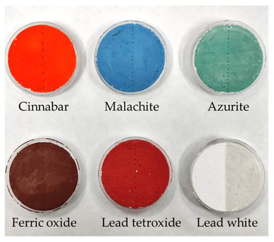
Figure 3.
The color changes of mural blocks. (The left half area of each one is the control group; the right half area is irradiated at 20 kGy).
Table 2.
Changes in color parameters of irradiated and unirradiated pigments.
Many studies have noted that the oxidizers, bases, salts, acids, hydrogen sulfide, and oxidizers, as well as other environmental conditions including temperature, humidity, and microorganisms, can cause lead white to discolor [43]. It is most commonly reported as blackening, formed mainly through producing both lead sulfide (PbS) and lead dioxide (β-PbO2) [44]. After exposure to radiation, lead white may have developed color centers, which could explain the discoloration [45]. The color difference of lead tetroxide after irradiation is 1.59, yet the human eye does not notice this effect.
3.3. Raman Spectra of Pigments
The Raman spectra of the six unirradiated and irradiated pigments are shown in Figure 4. “L” represents the unirradiated pigment on the left side of the mural block, and “R” represents the pigment irradiated at 20 kGy on the right side. The Raman shifts of the main peaks are labeled in the figure. The Raman spectrum of cinnabar exhibits three characteristic major bands at 256, 287, and 346 cm−1 [46]. The Raman bands of malachite show at 180, 219, 270, 355, 434, 538, 721, 1067, 1100, and 1493 cm−1 [47]. The Raman bands of azurite are 249, 401, 765, 838 and 1096 cm−1 [48]. The ferric oxide shows Raman bands at 223, 243, 292, 406, 495, and 609 cm−1 [47], and our results deviate from them by no more than 10 cm−1. Furthermore, the “L” curve (Figure 4D), after smoothing, shows a small peak on the far left side, which does not appear in the “R” curve, nor in the tests of others. We speculate that these variations are because the ferric oxide samples, out of the six pigments, had the worst signal-to-noise ratios at 473 nm excitation. And lead tetroxide with corresponding bands at 122, 153, 224, 314, 392, 480, and 550 cm−1 [49]. Lead white has characteristic Raman bands at 417 and 1049 cm−1 [50]. Our results are basically consistent with the data in these studies.
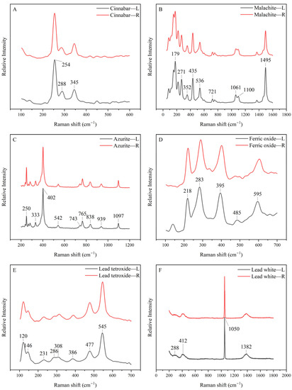
Figure 4.
Raman spectra of pigments, (A–F) represent cinnabar, malachite, azurite, ferric oxide, lead tetroxide and lead white, respectively. The black lines represent the unirradiated group and the red lines represent the irradiated group.
Previously, we detected a large color shift in the lead white in the test, but the Raman test (Figure 4F) results revealed no significant difference between the irradiated and unirradiated samples. In conclusion, Raman spectroscopy is able to confirm the chemical composition of these pigments; however, irradiation treatment does not change the chemical composition of the pigments. Changes in pigment color are more likely to be caused by changes in color centers, which needs some other tests to prove [51].
3.4. Effect on Surface Cohesion of Pigment Layer
Two identical mural blocks were used in each test. In the first test, using an unirradiated mural block, a Scotch Tape Test (STT) was performed simultaneously on the left and right portions (Table 3, unirradiated group, L, R). In the second test, the same test was performed on a mural block with only half of the area irradiated (Table 3, irradiated group, R), while the other part was masked with an absorbed dosage of 0 (Table 3, irradiated group, L). The differences in SST results between the two regions were used to assess the impact of irradiation. The standard deviation is not shown because the material released reduces each time a mural block is peeled [34]. Some pigments released almost the entire amount the first time, causing significant standard deviations that are not meaningful.
Table 3.
Scotch Tape Test results, ΔM = (L − R)/L.
The results of two tests performed on the unirradiated group showed that the maximum difference in released material (ΔM) between them was 9.13% (lead tetroxide), so that it can be considered that the Scotch Tape Test performed on handmade mural blocks had an unevenness of up to 9.13%. If the values in the irradiated group are also in the approximate range, it may indicate that the irradiation treatment does not affect this property. According to the results of the irradiated groups, the difference in released material of cinnabar pigments was higher than the other groups. The difference in released material was 0.89% in the unirradiated group and 11.08% in the irradiated group for the ferric oxide pigment, which also seems to show differences. To explain this, the variation in the amount of material released each time with the number of tests was plotted (Figure 5).
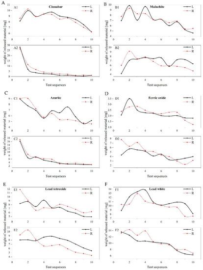
Figure 5.
The variation in the amount of material released each time with the number of tests, (A–F) represent cinnabar, malachite, azurite, ferric oxide, lead tetroxide and lead white, respectively. The letter suffix 1 represents the unirradiated group. Suffix 2 represents the irradiated group and only the “R” part is irradiated.
In Figure 5, the two sets of curves are intertwined in most cases and are not clearly separated, which supports the finding that the differences in released material in Table 3 are not large. Even though the ΔM for the cinnabar group was 25.24%, the two curves are still very similar, as can be seen in C2 of Figure 5, with the main difference originating from the first two releases. B2 in Figure 5 represents the lead tetroxide of the treatment group, and the “R” curve represents the irradiated part, which is consistently lower than the “L” curve representing the unirradiated part after the third test, which may be the reason for the difference in this group. However, the downward trend of the two curves is almost identical. To sum up, it can still be considered that irradiation treatment will not have a significant impact on the surface cohesion of the pigment layers of the mural blocks. (Detailed data can be obtained from the Supplementary Materials).
One limitation to this experiment is that it can only test dry mural blocks. It is important to use other methods to check the cohesiveness of the pigment layer if the mural is wet.
4. Conclusions
This study shows the feasibility of irradiation technology for mural decontamination under laboratory conditions, which provides a scientific basis for in situ mural conservation. Varied radiation doses were tested for their capacity to disinfect microorganisms with different incubation times. Irradiation at a minimum dose of 5 kGy showed good disinfection effects. All microorganisms but Penicillium flavigenum were disinfected following 20 kGy electron-beam irradiation, and the irradiation inhibited the growth of Penicillium flavigenum. Dunhuang mock mural blocks were used as samples to test changes in pigment properties such as color and surface cohesion following irradiation. The composition of the pigments and the changes after irradiation were confirmed using Raman spectroscopy. Lead white showed a significant color difference following irradiation at 20 kGy, but the other five pigments did not. Furthermore, at this dose, the surface cohesion of the pigment layer was not changed. The discoloration of lead white is more likely caused by a change in color center. More studies are required to find out the cause of the lead white pigment’s discoloration. Overall, the radiation technology disinfects the murals while causing less damage to them.
This study provides a disinfection technique using low-energy electron accelerator irradiation to control the growth of microorganisms on murals. The present research results show that the intelligent mobile electron accelerator has certain potential in the application of the in situ protection of murals. However, the storage environment of the real tomb or cave paintings is different from that of the laboratory, and the real mural pigments and painting layers will have some pollution and aging damage, and the micro-organisms attached to the surface of the murals will form a relatively stable microbial community. Therefore, it is necessary to conduct follow-up experiments on fresco samples that are closer to the actual situation. The feasibility of this technique is further verified and the corresponding irradiation process is optimized.
However, radiation techniques do not provide lasting protection and there is still a need to continuously monitor the condition of the frescoes and provide suitable storage conditions to prevent the recurrence of biometamorphism. The combination of various protection methods can provide new ideas for the protection of mural heritage.
Supplementary Materials
The following supporting information can be downloaded at: https://www.mdpi.com/article/10.3390/pr11061710/s1, Table S1: Release material of scotch tape test (mg).
Author Contributions
Article writing, Z.W. and M.L.; Experimental operation and data analysis, Z.W.; Irradiation experiment guidance, M.L.; Participant in some experiments, Y.S.; Writing—revision and editing, L.M. and M.W. All authors have read and agreed to the published version of the manuscript.
Funding
This research was funded by the National Key Research Program Projects of China (Grant/Award Number: 2019YFC1520700) and the Innovation Foundation of the Institute of High Energy Physics, CAS (Grant/Award Number: E15452JY10).
Institutional Review Board Statement
Not applicable.
Informed Consent Statement
Informed consent was obtained from all subjects involved in the study.
Data Availability Statement
The data presented in this study are available on request from the corresponding author.
Acknowledgments
We thank Dunhuang Academy and the University of the Chinese Academy of Sciences for its support.
Conflicts of Interest
The authors declare no conflict of interest.
References
- Gadd, G.M. Geomycology: Biogeochemical transformations of rocks, minerals, metals and radionuclides by fungi, bioweathering and bioremediation. Mycol. Res. 2007, 111, 3–49. [Google Scholar] [CrossRef] [PubMed]
- Garg, K.L.; Jain, K.K.; Mishra, A.K. Role of fungi in the deterioration of wall paintings. Sci. Total Environ. 1995, 167, 255–271. [Google Scholar] [CrossRef]
- Scheerer, S.; Ortega-Morales, O.; Gaylarde, C. Chapter 5 Microbial Deterioration of Stone Monuments—An Updated Overview. In Advances in Applied Microbiology; Academic Press: Cambridge, MA, USA, 2009; Volume 66, pp. 97–139. [Google Scholar] [CrossRef]
- Borderie, F.; Laurence, A.-S.; Naoufal, R.; Faisl, B.; Geneviève, O.; Dominique, R.; Badr, A.-S. UV–C irradiation as a tool to eradicate algae in caves. Int. Biodeterior. Biodegrad. 2011, 65, 579–584. [Google Scholar] [CrossRef]
- Gambino, M.; Ahmed, M.A.-A.A.; Villa, F.; Cappitelli, F. Zinc oxide nanoparticles hinder fungal biofilm development in an ancient Egyptian tomb. Int. Biodeterior. Biodegrad. 2017, 122, 92–99. [Google Scholar] [CrossRef]
- Li, T.; Hu, Y.; Zhang, B. Evaluation of efficiency of six biocides against microorganisms commonly found on Feilaifeng Limestone, China. J. Cult. Herit. 2020, 43, 45–50. [Google Scholar] [CrossRef]
- Bastian, F.; Alabouvette, C.; Jurado, V.; Saiz-Jimenez, C. Impact of biocide treatments on the bacterial communities of the Lascaux Cave. Naturwissenschaften 2009, 96, 863–868. [Google Scholar] [CrossRef]
- Kakakhel, M.A.; Wu, F.; Gu, J.-D.; Feng, H.; Shah, K.; Wang, W. Controlling biodeterioration of cultural heritage objects with biocides: A review. Int. Biodeterior. Biodegrad. 2019, 143, 104721. [Google Scholar] [CrossRef]
- Faimon, J.; Štelcl, J.; Kubešová, S.; Zimák, J. Environmentally acceptable effect of hydrogen peroxide on cave “lamp-flora”, calcite speleothems and limestones. Environ. Pollut. 2003, 122, 417–422. [Google Scholar] [CrossRef]
- Varnai, V.M.; Macan, J.; Ljubičić Ćalušić, A.; Prester, L.; Kanceljak Macan, B. Upper respiratory impairment in restorers of cultural heritage. Occup. Med. 2011, 61, 45–52. [Google Scholar] [CrossRef]
- Chelius, M.K.; Beresford, G.; Horton, H.; Quirk, M.; Selby, G.; Simpson, R.T.; Horrocks, R.; Moore, J.C. Impacts of alterations of organic inputs on the bacterial community within the sediments of Wind Cave, South Dakota, USA. Int. J. Speleol. 2009, 38, 1. [Google Scholar] [CrossRef]
- Martin-Sanchez, P.M.; Nováková, A.; Bastian, F.; Alabouvette, C.; Saiz-Jimenez, C. Use of Biocides for the Control of Fungal Outbreaks in Subterranean Environments: The Case of the Lascaux Cave in France. Environ. Sci. Technol. 2012, 46, 3762–3770. [Google Scholar] [CrossRef]
- Palla, F.; Bruno, M.; Mercurio, F.; Tantillo, A.; Rotolo, V. Essential Oils as Natural Biocides in Conservation of Cultural Heritage. Molecules 2020, 25, 730. [Google Scholar] [CrossRef]
- Veneranda, M.; Blanco-Zubiaguirre, L.; Roselli, G.; Di Girolami, G.; Castro, K.; Madariaga, J.M. Evaluating the exploitability of several essential oils constituents as a novel biological treatment against cultural heritage biocolonization. Microchem. J. 2018, 138, 1–6. [Google Scholar] [CrossRef]
- Marco, A.; Santos, S.; Caetano, J.; Pintado, M.; Vieira, E.; Moreira, P.R. Basil essential oil as an alternative to commercial biocides against fungi associated with black stains in mural painting. Build. Environ. 2020, 167, 106459. [Google Scholar] [CrossRef]
- Fidanza, M.R.; Caneva, G. Natural biocides for the conservation of stone cultural heritage: A review. J. Cult. Herit. 2019, 38, 271–286. [Google Scholar] [CrossRef]
- Franco-Castillo, I.; Hierro, L.; de la Fuente, J.M.; Seral-Ascaso, A.; Mitchell, S.G. Perspectives for antimicrobial nanomaterials in cultural heritage conservation. Chem 2021, 7, 629–669. [Google Scholar] [CrossRef]
- Calado, T.; Venâncio, A.; Abrunhosa, L. Irradiation for Mold and Mycotoxin Control: A Review. Compr. Rev. Food Sci. Food Saf. 2014, 13, 1049–1061. [Google Scholar] [CrossRef]
- Zhang, Y.; Moeller, R.; Tran, S.; Dubovcova, B.; Akepsimaidis, G.; Meneses, N.; Drissner, D.; Mathys, A. Geobacillus and Bacillus Spore Inactivation by Low Energy Electron Beam Technology: Resistance and Influencing Factors. Front. Microbiol. 2018, 9, 2720. Available online: https://www.frontiersin.org/articles/10.3389/fmicb.2018.02720 (accessed on 20 April 2023). [CrossRef]
- Głuszewski, W.; Zagórski, Z.P.; Tran, Q.K.; Cortella, L. Maria Skłodowska Curie—The precursor of radiation sterilization methods. Anal. Bioanal. Chem. 2011, 400, 1577–1582. [Google Scholar] [CrossRef]
- Xu, D.; Shi, R.; Ma, L.; Ruan, Y.; Luo, M.; Dai, M.; Shao, Y.; Zhuo, L.; Yang, X. Intelligently Controlled Mobile Electron Beam Irradiation Device. Chinese Patent ZL 202022634267.X, 11 February 2022. [Google Scholar]
- Negut, D.C.; Ponta, C.C.; Rodica, M.G.; Moise, I.V.; Gh, N.; Lupu, A.I.M. Effects of gamma irradiation on the colour of pigments. In Proceedings of the SPIE 6618, O3A: Optics for Arts, Architecture, and Archaeology, Munich, Germany, 19 July 2007; p. 66180R. [Google Scholar] [CrossRef]
- Fu, J.; Shen, W.; Bao, J.; Chen, Q. The decontamination effects of gamma irradiation on the edible gelatin. Radiat. Phys. Chem. 2000, 57, 345–348. [Google Scholar] [CrossRef]
- Vieira, F.F.; Del Mastro, N.L. Comparison of γ-radiation and electron beam irradiation effects on gelatin. Radiat. Phys. Chem. 2002, 63, 331–332. [Google Scholar] [CrossRef]
- Ma, W.; Wu, F.; He, D.; Li, J.; Zhang, Q.; Yang, X.; Gu, J.-D.; Wang, W.; Feng, H. The biodeterioration outbreak in Dunhuang Mogao Grottoes analyzed for the microbial communities and the occurrence time by C-14 dating. Int. Biodeterior. Biodegrad. 2023, 178, 105533. [Google Scholar] [CrossRef]
- Ma, Y.; Zhang, H.; Du, Y.; Tian, T.; Xiang, T.; Liu, X.; Wu, F.; An, L.; Wang, W.; Gu, J.-D.; et al. The community distribution of bacteria and fungi on ancient wall paintings of the Mogao Grottoes. Sci. Rep. 2015, 5, 7752. [Google Scholar] [CrossRef] [PubMed]
- Sterflinger, K. Fungi: Their role in deterioration of cultural heritage. Fungal Biol. Rev. 2010, 24, 47–55. [Google Scholar] [CrossRef]
- Gorbushina, A.A.; Heyrman, J.; Dornieden, T.; Gonzalez-Delvalle, M.; Krumbein, W.E.; Laiz, L.; Petersen, K.; Saiz-Jimenez, C.; Swings, J. Bacterial and fungal diversity and biodeterioration problems in mural painting environments of St. Martins church (Greene–Kreiensen, Germany). Int. Biodeterior. Biodegrad. 2004, 53, 13–24. [Google Scholar] [CrossRef]
- He, D.; Wu, F.; Ma, W.; Zhang, Y.; Gu, J.-D.; Duan, Y.; Xu, R.; Feng, H.; Wang, W.; Li, S.-W. Insights into the bacterial and fungal communities and microbiome that causes a microbe outbreak on ancient wall paintings in the Maijishan Grottoes. Int. Biodeterior. Biodegrad. 2021, 163, 105250. [Google Scholar] [CrossRef]
- Xing, W.; Qi, B.; Chen, R.; Ding, W.; Zhang, F. Metagenomic analysis reveals taxonomic and functional diversity of microbial communities on the deteriorated wall paintings of Qinling Tomb in the Southern Tang Dynasty, China. BMC Microbiol. 2023, 23, 140. [Google Scholar] [CrossRef]
- Han, P.; Zhang, H.; Zhang, R.; Tan, X.; Zhao, L.; Liang, Y.; Su, B. Evaluation of the effectiveness and compatibility of nanolime for the consolidation of earthen-based murals at Mogao Grottoes. J. Cult. Herit. 2022, 58, 266–273. [Google Scholar] [CrossRef]
- Coppola, F.; Fiorillo, F.; Modelli, A.; Montanari, M.; Vandini, M. Effects of γ-ray treatment on paper. Polym. Degrad. Stab. 2018, 150, 25–30. [Google Scholar] [CrossRef]
- Macera, L.; Daniele, V.; Duchetta, F.; Casciardi, S.; Taglieri, G. New nanolimes for eco-friendly and customized treatments to preserve the biocalcarenites of the “Valley of Temples” of Agrigento. Constr. Build. Mater. 2021, 306, 124811. [Google Scholar] [CrossRef]
- Drdácký, M.; Lesák, J.; Rescic, S.; Slížková, Z.; Tiano, P.; Valach, J. Standardization of peeling tests for assessing the cohesion and consolidation characteristics of historic stone surfaces. Mater. Struct. 2012, 45, 505–520. [Google Scholar] [CrossRef]
- Yang, X.; Feng, J.; Wang, F.; Hu, Y. Irradiation sterilization used for allogenetic tendon: A literature review of current concept. Cell Tissue Bank. 2019, 20, 129–139. [Google Scholar] [CrossRef]
- Saleh, Y.G.; Mayo, M.S.; Ahearn, D.G. Resistance of some common fungi to gamma irradiation. Appl. Environ. Microbiol. 1988, 54, 2134–2135. [Google Scholar] [CrossRef] [PubMed]
- Maria, G.C.T.; Wiendl, F.M. The Applicability of Gamma Radiation to the Control of Fungi in Naturally Contaminated Paper. Restaurator 1995, 16, 93–99. [Google Scholar] [CrossRef]
- Sakr, A.; Ghaly, M.; Ali, M. The Use of Gamma Irradiation in the Sterilization of Streptomyces Colonizing the Tempra Paintings in Ancient Egyptian Tombs. Int. J. Conserv. Sci. 2013, 4, 283–294. [Google Scholar]
- Dadachova, E.; Casadevall, A. Ionizing radiation: How fungi cope, adapt, and exploit with the help of melanin. Curr. Opin. Microbiol. 2008, 11, 525–531. [Google Scholar] [CrossRef] [PubMed]
- Pacelli, C.; Bryan, R.A.; Onofri, S.; Selbmann, L.; Shuryak, I.; Dadachova, E. Melanin is effective in protecting fast and slow growing fungi from various types of ionizing radiation. Environ. Microbiol. 2017, 19, 1612–1624. [Google Scholar] [CrossRef]
- Abdel-Haliem, M.E.F.; Ali, M.F.; Ghaly, M.F.; Sakr, A.A. Efficiency of antibiotics and gamma irradiation in eliminating Streptomyces strains isolated from paintings of ancient Egyptian tombs. J. Cult. Herit. 2013, 14, 45–50. [Google Scholar] [CrossRef]
- Cortella, L.; Albino, C.; Tran, Q.-K.; Froment, K. 50 years of French experience in using gamma rays as a tool for cultural heritage remedial conservation. Radiat. Phys. Chem. 2020, 171, 108726. [Google Scholar] [CrossRef]
- Gliozzo, E.; Ionescu, C. Pigments—Lead-based whites, reds, yellows and oranges and their alteration phases. Archaeol. Anthropol. Sci. 2021, 14, 17. [Google Scholar] [CrossRef]
- Vagnini, M.; Vivani, R.; Sgamellotti, A.; Miliani, C. Blackening of lead white: Study of model paintings. J. Raman Spectrosc. 2020, 51, 1118–1126. [Google Scholar] [CrossRef]
- Negut, C.-D.; Bercu, V.; Duliu, O.-G. Defects induced by gamma irradiation in historical pigments. J. Cult. Herit. 2012, 13, 397–403. [Google Scholar] [CrossRef]
- Philippidis, A.; Mikallou, A.; Anglos, D. Determining optimum irradiation conditions for the analysis of vermilion by Raman spectroscopy. Eur. Phys. J. Plus 2021, 136, 1194. [Google Scholar] [CrossRef]
- Yu, B.-S.; Fang, J.-N.; Huang, E.-P. Characteristics of the Raman spectra of archaeological Malachites. J. Raman Spectrosc. 2013, 44, 630–636. [Google Scholar] [CrossRef]
- Burgio, L.; Clark, R.J.H. Library of FT-Raman spectra of pigments, minerals, pigment media and varnishes, and supplement to existing library of Raman spectra of pigments with visible excitation. Spectrochim. Acta Part A Mol. Biomol. Spectrosc. 2001, 57, 1491–1521. [Google Scholar] [CrossRef] [PubMed]
- Gutman, M.; Lesar-Kikelj, M.; Mladenovič, A.; Čobal-Sedmak, V.; Križnar, A.; Kramar, S. Raman microspectroscopic analysis of pigments of the Gothic wall painting from the Dominican Monastery in Ptuj (Slovenia). J. Raman Spectrosc. 2014, 45, 1103–1109. [Google Scholar] [CrossRef]
- Petrova, O.; Pankin, D.; Povolotckaia, A.; Borisov, E.; Krivul’ko, T.; Kurganov, N.; Kurochkin, A. Pigment palette study of the XIX century plafond painting by Raman spectroscopy. J. Cult. Herit. 2019, 37, 233–237. [Google Scholar] [CrossRef]
- Luo, M.; Bo, P.; Shao, Y.; Liu, Z.; Xu, D.; Ma, L. Study on the Influence of Electron Beam Radiation Sterilization Method on Chinese Mural Pigment. Processes 2023, 11, 1403. [Google Scholar] [CrossRef]


Disclaimer/Publisher’s Note: The statements, opinions and data contained in all publications are solely those of the individual author(s) and contributor(s) and not of MDPI and/or the editor(s). MDPI and/or the editor(s) disclaim responsibility for any injury to people or property resulting from any ideas, methods, instructions or products referred to in the content. |
© 2023 by the authors. Licensee MDPI, Basel, Switzerland. This article is an open access article distributed under the terms and conditions of the Creative Commons Attribution (CC BY) license (https://creativecommons.org/licenses/by/4.0/).