Article

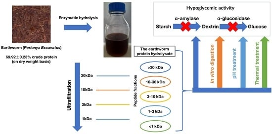
Earthworm (Perionyx excavatus) Protein Hydrolysate: Hypoglycemic Activity and Its Stability for the Hydrolysate and Its Peptide Fractions

1
Department of Food Technology, Faculty of Chemical Engineering, Ho Chi Minh City University of Technology (HCMUT), VNU-HCM, Ho Chi Minh City 700000, Vietnam
2
Department of Chemical and Biological Engineering, The University of Sheffield, Mappin Street, Sheffield S1 3JD, UK
*
Author to whom correspondence should be addressed.
Processes 2023, 11(8), 2490; https://doi.org/10.3390/pr11082490
Submission received: 3 July 2023 / Revised: 12 August 2023 / Accepted: 16 August 2023 / Published: 19 August 2023
(This article belongs to the Special Issue Current Trends in Food and Food Byproducts Processing)

Abstract

:
This study aims at exploring earthworm protein hydrolysate and its peptide fractions as a potential hypoglycemic agent by inhibiting α-amylase and α-glucosidase. Firstly, the best hydrolysis conditions to gain the hydrolysates with the highest α-amylase inhibitory activity (α-AIA) and α-glucosidase inhibitory activity (α-GIA) were figured out using a one-factor test. Next, the stability of the hypoglycemic activity of the hydrolysates and their 5 peptide fractions recovered using ultrafiltration membranes were assessed by employing the tests of in vitro digestion, thermal, and pH treatment. The results showed that at the best hydrolysis conditions, the hydrolysates exhibited α-AIA of 91.30 ± 2.51% and α-GIA of 44.69 ± 0.47%. Specifically, the <1 kDa peptide fraction from the hydrolysate expressed a greater α-AIA than that of acarbose, with nearly the same α-GIA as that of voglibose. The α-AIA and α-GIA of the hydrolysates and their fractions were enhanced after the in vitro digestion treatment, whereas they remained over 40% after the pH treatment in the range of 1 to 11 or heat treatment at 100 °C for 180 min. These data provide the preliminary evidence to develop the earthworm protein hydrolysate and its peptide fractions in functional food or nutraceutical products with hypoglycemic activity.

Graphical Abstract

1. Introduction

Earthworms are macro-invertebrates (oligochaeta), shouldering the responsibility to enhance soil fertility and productivity [1]. Perionyx excavatus is a popular species in Vietnam that has high protein content (55–70%, dry weight basis), thus it has been used as an alternative protein ingredient to feed common carp (Cyprinus carpio L.) [2], as well as a protein source for human in Asia, including India, Myanmar, China, Korea, and Vietnam for thousands of years [3]. The earthworm protein and its coelomic fluid were proven for their cytolytic, agglutinating, proteolytic, haemolytic, mitogenic, antipyretic, and tumorstatic activities [4]. Ding et al. [5] also documented that the earthworm was a potential source of pharmaceutical compounds expressing anti-hypertensive, anticoagulant, and anti-hyperlipidemic activities. Accordingly, it is interesting to utilize this high protein source and discover hidden bioactivities of this species.
The International Diabetes Federation (IDF) reported that in 2019, there were 463 million adults aged 20–79 years who had diabetes, and the number was predicted to reach over 700 million by 2045 [6]. Approximately 90% of diabetic patients had Type 2 diabetes, which is characterized by hyperglycemia (high blood glucose level) resulting from insulin-related disorders [7]. To control postprandial hyperglycemia and manage diabetes mellitus, the delay of glucose release into the bloodstream by inhibition of starch-digestive enzymes (including α-amylase and α-glucosidase) is one of the most effective strategies [8,9]. Iminosugar-derivatives (such as miglitol) and sugar-derivatives (including acarbose and voglibose) have been used as synthetic antidiabetic drugs [7,10]. However, their side effects, such as gastrointestinal disorders, bloating, stomach pain, diarrhea, and flatulence, need to be considered [11].
Earthworm-derived products, including dried earthworms, earthworm powder, and extract, have been used as medicinal ingredients to treat allergies in China [12]. In addition, in various countries such as Taiwan, Japan, South Africa, Brazil, and the Philippines, earthworms have been consumed in their meals [5]. Moreover, earthworm protein powder (BOCOM XUETONG) and smoked earthworms have been marketed in the USA and Venezuela [5]. In addition, commercial Lumbrokinase, a serine protease from earthworms, is globally available for anti-allergy [13]. Moreover, earthworm meals were made and evaluated safely in terms of microbiology, heavy metals, pesticide, mycotoxins, and antibiotic residues [14,15]. Recently, food wastes (fruit and vegetable residues and kitchen waste) have been utilized to raise earthworms for sustainable protein source recovery and meet global protein needs [16,17].
Regarding the nutritional value of earthworm protein hydrolysate, Alcalase hydrolysate was reported to be rich in essential amino acids (14.7 g/kg of hydrolysate) and expressed high protein digestibility in vitro (73%) [18]. Moreover, high α-AIA and α-GIA of earthworm extract have been found by Mir et al. [19]. Furthermore, Abdelaziz et al. [20] discovered that the therapeutic potential of earthworm extract against diabetic complications mainly depends on its amino acid composition when performing an in vivo test on diabetic rats. In addition, the peptide’s bioavailability was determined by its length, amino acid composition, charge, and hydrophobicity, and the peptide length might be the most important factor [21]. Patil et al. [22] found that for the small intestine, the absorbability of short-chain peptides (<6 amino acids) was more than 90% [9], which was about 45 times higher than that of acarbose [23]. Moreover, some animal-derived peptides were found to be hypoglycemic agents [11,24,25], due to their high affinity, specificity, bioactivity, and safety [26,27]. Our study is the very first to generate earthworm protein hydrolysate with hypoglycemic activity.
It was reported that smaller peptides may be more active than bigger peptides [28]. In addition, Félix-Medina et al. [29] and Li et al. [30] found that a better hypoglycemic activity of peptide fractions was observed as their molecular weights decreased. Moreover, Wang et al. [31] discovered that most peptides possessing antihyperglycemic activity have molecular weights below 1 kDa.
Félix-Medina et al. [29] published that α-AIA and α-GIA of the <3 kDa peptide fraction from maize protein hydrolysate were over 50% and 35%, respectively, after being treated at a temperature range from 30 to 90 °C or pH in the range of 2–8. In addition, the retention rate of α-GIA of hypoglycemic peptides in Moringa oleifera seed protein hydrolysates at pH = 2, 4, 6, and 8 was greater than 90%, however, it reduced to 60% at pH = 10 [31]. Meanwhile, their α-GIA could remain 100% at temperatures of 20, 40, and 60 °C, then quickly decreased to 18.73% at 100 °C [31]. On the other hand, the α-GIA of YPVEPF (a hypoglycemic peptide identified from whey hydrolysate) increased up to 4 times at pH = 2, and it reduced by approximately 9 times at pH = 10 [30]. Besides, this peptide was found to be stable at the temperature range of 20–80 °C, with its highest α-GIA retention rate of 94% at 60 °C [30].
Therefore, this study focused on investigating the α-AIA and α-GIA of the protein hydrolysate/peptide from earthworms. Firstly, the single factor test was employed to select the hydrolysis condition (enzyme type, earthworm: phosphate buffer ratio (w/v), temperature, pH, enzyme: substrate (E:S) ratio, and hydrolysis time) to obtain the hydrolysate that expresses the highest α-AIA and α-GIA. Subsequently, the hydrolysates were further fractionated using ultrafiltration membranes and 5 peptide fractions (>30 kDa, 10–30 kDa, 3–10 kDa, 1–3 kDa, and <1 kDa) were recovered and tested for their bioactivities. Then, the hydrolysates and 5 peptide fractions were assessed for their bioactivity stability using the tests of in vitro digestion, thermal, and pH treatment.

2. Materials and Methods

2.1. Materials

Earthworms (Perionyx excavatus) were purchased from the Biotechnology Center in Ho Chi Minh City, then they were crushed and stored in polyethylene bags at −20 °C before testing. The chemical compositions of the earthworms were determined using the methods of AOAC (1990) [32]. The earthworms contained 80.99 ± 0.53% moisture, 69.92 ± 0.23% crude protein, 6.97 ± 0.10% crude lipid, 12.78 ± 0.18% carbohydrate, and 10.25 ± 0.10% ash (on a dry weight basis).
In this study, α-glucosidase (from Saccharomyces cerevisiae, 10 units/mg protein), p-Nitrophenyl α-D-glucopyranoside (pNPG), α-amylase (from the porcine pancreas, 5 units/mg solid), and acarbose were purchased from Sigma Chemical Co. (St. Louis, MO, USA). Enzyme preparations, including Alcalase® 2.5 L (55 °C, pH = 8), Neutrase® 0.8 L (50 °C, pH = 7), Protamex® (55 °C, pH = 6.5), and Flavourzyme® 500 MG (50 °C, pH = 7), were bought from Novozymes Co. (Bagsvaerd, Denmark) and AB enzymes (Darmstadt, Germany). All reagents were of analytical grade.

2.2. Methods

2.2.1. Preparation of Earthworm Protein Hydrolysates

Earthworm hydrolysates were prepared based on the method described in Vo et al. [33] with slight modifications. The ground earthworm was mixed with 0.02 M phosphate buffer with an appropriate ratio so that the pH reached the required value for hydrolysis, and the mixture was heated at 90 °C for 10 min to deactivate endogenous enzymes. Then, the pH was readjusted using 1 M HCl or 1 M NaOH solution, and the temperature was controlled to the hydrolysis temperature before adding enzyme preparation with the appropriate E:S ratio. At a set time, the enzyme was deactivated by heating the hydrolysate for 10 min at 90 °C. The supernatant was then collected by centrifuging at 5000 rpm for 15 min, and the obtained supernatant was lyophilized and stored at −20 °C. The soluble protein content of the hydrolysate was determined by the Lowry method [34].

2.2.2. Effect of Hydrolysis Condition on α-AIA and α-GIA of the Earthworm Protein Hydrolysate

The effect of hydrolysis parameters, including enzyme type, earthworm: phosphate buffer ratio (w/v), temperature (°C), pH, E:S ratio (U/g protein), and hydrolysis time (h), on α-AIA and α-GIA of the earthworm protein hydrolysates was investigated using the single factor test method. More specifically, one factor varied at different levels while the others remained constant, as presented in Table 1.

2.2.3. Determination of α-AIA and α-GIA of the Earthworm Protein Hydrolysates and Their 5 Peptide Fractions

The method described in Liu et al. [35] was employed with minor modifications to determine α-AIA of the protein hydrolysates. In brief, 0.20 mL of the sample (2 mg protein/mL) was incubated with 0.04 mL of α-amylase (5 U/mL) and 0.36 mL of sodium phosphate buffer (0.02 M, pH = 6.9) containing 6 mM NaCl at 37 °C for 20 min. Then, 0.30 mL of starch solution (1%, substrate) in sodium phosphate buffer (0.02 M, pH = 6.9 with 6 mM NaCl) was added, and the mixture was further incubated at 37 °C for 20 min. Subsequently, it was mixed with 0.20 mL of dinitrosalicylic acid reagent before being kept in a boiling water pot for 5 min and cooled down to room temperature. The cooled mixture was then diluted with 10 mL of distilled water, and its absorbance was measured at 540 nm using a spectrophotometer (Shimadzu, Japan).
To test α-GIA, 0.1 mL of the sample (2 mg protein/mL) was incubated with 0.1 mL α-glucosidase (1 U/mL) in 0.01 M phosphate buffer (pH = 7.0) and 2.2 mL of 0.01 M phosphate buffer (pH = 7.0) at 37 °C for 5 min. Then, 0.1 mL of pNPG (3 mM, substrate) in 0.01 M phosphate buffer (pH = 7.0) was added to the mixture and was further incubated at 37 °C for 30 min. The reaction was stopped by adding 1.50 mL of 0.1 M Na2 CO3 solution before recording the absorbance at 405 nm by employing the spectrophotometer [36].
The α-AIA and α-GIA of the protein hydrolysate were calculated as follows:
α A I A   o r   α G I A % = A 1 A 2 A 3 A 4 A 1 A 2 × 100
where A1 is the absorbance of the control (the mixture containing enzyme, substrate, and no hydrolysate or peptide fraction); A2 is the absorbance of the control blank (the mixture containing substrate, no enzyme, and no hydrolysate or peptide fraction); A3 is the absorbance of the tested sample (the mixture containing enzyme, substrate, and hydrolysate or peptide fraction); A4 is the absorbance of the sample blank (the mixture containing hydrolysate or peptide fraction, substrate, and no enzyme).
Logarithmic regression analysis was employed to determine the half inhibitory concentration (IC50, the concentration of the inhibitor to inhibit 50% of enzyme activity) for α-AIA and α-GIA of the earthworm protein hydrolysates and their 5 peptide fractions.
Acarbose was used as the positive control with a concentration range of 50–250 (μg/mL) for the α-AIA test and 200–1000 (μg/mL) for the α-GIA test.
The effects of hydrolysis conditions on the α-AIA and α-GIA of the earthworm protein hydrolysate were investigated. The effects of 6 factors, including hydrolysis enzyme type, earthworm: phosphate buffer ratio, temperature, pH, enzyme: substrate (E:S) ratio, and hydrolysis time, were assessed to obtain the best condition that generates hydrolysate with the highest activity. After that, each collected hydrolysate was fractionated to gain its peptide fractions.

2.2.4. Fractionation of Earthworms Protein Hydrolysate

By using the ultrafiltration centrifugal devices of 30 kDa, 10 kDa, 3 kDa, and 1 kDa (Macrosep, Pall Laboratory, New Port Richey, FL, USA), the earthworm protein hydrolysate was further fractionated. Five peptide fractions (<1 kDa, 1–3 kDa, 3–10 kDa, 10–30 kDa, and >30 kDa) of each hydrolysate were obtained and their α-AIA and α-GIA were evaluated.

2.2.5. In Vitro Digestion

The in vitro digestion test of the earthworm protein hydrolysates and their peptide fractions was conducted following the method described in Kang et al. [37]. The sample, with a protein concentration of 5% (w/v), was adjusted to pH = 2 using 6 M HCl solution, and its temperature was increased to 37 °C. Pepsin was then added with an E:S ratio of 4% (w/w), and the mixture was further incubated at 37 °C for 1 h and shaken at 230 rpm. Subsequently, its pH was changed to 7.5 by utilizing 6 M NaOH solution before adding pancreatin with an E:S ratio of 4% (w/w). The mixture was continuously incubated at 37 °C and shaken at 230 rpm for 2 h. The enzymes were inactivated by heating the mixture at 90 °C for 10 min. After that, the sample was collected to determine its α-AIA and α-GIA.

2.2.6. Thermal and pH Stability

The method described in Sripokar et al. [38] was used to evaluate the thermal and pH stability of the protein hydrolysates and their peptide fractions.
To assess the thermal stability, 5 mL of the sample (40 mg protein/mL) was heated at 100 °C for 15, 30, 45, 60, 90, 120, 150, and 180 min. After that, the solution was cooled down to room temperature in iced water and diluted to the final volume of 10 mL with deionized water, and its α-AIA and α-GIA were measured.
In order to examine the pH stability, the pH of 5 mL of the sample (40 mg protein/mL) was adjusted to 1.0, 2.0, 3.0, 4.0, 5.0, 6.0, 7.0, 8.0, 9.0, 10.0, and 11.0 using 6 M HCl or 6 M NaOH solution and placed at room temperature for 30 min. The sample was then altered to pH = 7.0 by employing 1 M phosphate buffer and diluted to the final volume of 20 mL with deionized water, before performing the α-AIA and α-GIA assays.
Both the thermal stability and pH stability of the sample were expressed as the relative activity (%), which was defined as the percentage of bioactivity of the treated sample compared to that of the control (untreated sample).

2.2.7. Statistical Analysis

All experiments were performed in triplicate. The data presented as mean ± standard deviation were processed using the software Excel. The statistically significant differences were determined using the software SPSS (IBM SPSS Statistics 20).

3. Results and Discussion

3.1. Investigation Results of α-AIA and α-GIA of the Earthworm Hydrolysate under the Influence of Hydrolysis Condition

3.1.1. Effect of Hydrolysis Enzyme

Peptides’ bioactivity is significantly associated with their amino acid composition and sequence determined by the specificity of the enzymes employed in the hydrolysis process [39].
Regarding α-AIA, Protamex was the most suitable protease among the four enzyme preparations to acquire the earthworm protein hydrolysate, with the highest α-AIA of 73.91 ± 1.45% (Figure 1a). The enzyme was known to be a mixture of endoproteases and exopeptidases [40,41], which offered a broad specificity for aliphatic amino acids [42]. In addition, it produced peptides with a high frequency of Leu, Glu, Lys, Met, Val, and Arg at the C-terminal and Lys, Gly, Ala, Ile, His, Asp, and Phe at the N-terminal [43,44], fitting to the characteristics of α-amylase inhibitory peptides in the literature. For instance, the results of Baba et al. [7] showed that most α-amylase inhibitory peptides contained Leu and Met at their N- and C-ends. In addition, Balderas-León et al. [45] have emphasized the roles of hydrophobic (Ala, Leu, Val, and Gly) and hydrophilic amino acids (Met and His) in the α-AIA of the containing peptides [19]. Furthermore, the α-AIA of peptides was enhanced with the presence of Gly, although the exact mechanism was unknown [46]. Moreover, α-amylase has aromatic residues lying within the substrate-binding pocket and the aromatic-aromatic interactions seem to be predominantly involved in the α-AIA of peptides containing Phe [46]. In addition, Arg, Glu, and Asp were interpreted to own the highest α-amylase inhibitory effects with their binding energies of −5.2, −5.0, and −4.8 kcal/mol, respectively [11]. Additionally, the molecular docking results in Siahbalaei et al. [11] revealed that Glu and Ala could be bound to the allosteric binding site of α-amylase at its Arg398, Arg421, Asp402, Arg252, Glu23, Glu863, and Arg437, changing its conformation and thus diminishing its catalysis function. Protamex was used to obtain the Paeonia ostii cake protein hydrolysate with the highest α-AIA as well [46].
As for α-GIA, the hydrolysate of earthworm protein by Alcalase exhibited the highest activity of 37.66 ± 0.62% (Figure 2a). Alcalase (an endopeptidase) has a preferential cleavage of peptide bonds when the polypeptide chain contains amino acids Glu, Met, Leu, Tyr, Lys, Thr, Ile, Phe, Arg, and Gln [39,43]. In addition, it usually releases the peptides with N-terminals of Gly, Lys, Arg, Asp, Ala, Trp, and Ile [43]. These properties of the Alcalase-generated peptides were mostly similar to those of α-glucosidase inhibitory peptides published previously. Three α-glucosidase inhibitory peptides identified in the study of Feng et al. [47] had their N-terminals of Gly and C-terminals of Lys and Arg. Lys and Arg occupying the N-terminal of the peptide also appeared to greatly influence the α-glucosidase inhibition [8]. Similarly, Met located at any terminal ends was found to play a key role in peptides with α-GIA [7,8]. On the other hand, the contribution of hydrophobic amino acids, such as Phe, Leu, Ile, and Ala to α-GIA of the containing peptides, could not be belittled [6,48]. Additionally, Asp and Gln were demonstrated to attach to the α-glucosidase’s allosteric binding site through molecular docking analysis [10]. Alcalase was also the best choice to obtain the α-glucosidase inhibitory hydrolysates from various protein sources [25,47,49,50]. Therefore, Protamex and Alcalase were correspondingly chosen for further investigations on the α-AIA and α-GIA of the earthworm protein hydrolysates.

3.1.2. Effect of Earthworm: Phosphate Buffer Ratio

A large amount of buffer can reduce the probability of collisions between the substrate and enzyme molecules, obstructing the production of bioactive peptides from the intact protein and lowering the bioactivity of the protein hydrolysate [51,52]. In contrast, high viscosity is a major challenge for enzymatic hydrolysis at a small amount of buffer. It limits the diffusion rate of protein molecules, and thereby, protein aggregation easily happens, which inhibits the accessibility of the enzyme [51]. On the other hand, a sufficient amount of buffer not only provides high protein contents in the hydrolysate but also quickly scatters the products of hydrolysis, preventing the feedback effect and enhancing the bioactivity of the protein hydrolysate [52]. In this study, Figure 1b and Figure 2b indicate that α-AIA and α-GIA of the hydrolysates reached their peaks at the earthworm: phosphate buffer ratios of 1:8 (w/v) and 1:6 (w/v), respectively, which were the selected levels for further investigations.

3.1.3. Effect of Hydrolysis Temperature

As illustrated in Figure 1c and Figure 2c, the highest α-AIA of 80.30 ± 2.62% and α-GIA of 41.13 ± 0.50% were observed at 55 °C and 60 °C, respectively. At low temperatures, the protein and enzyme molecules own low kinetic energy, lessening the formation rate of the enzyme-substrate complex, and thus, a low content of bioactive peptides is generated in the hydrolysate [33,51]. However, irreversible denaturation of active catalyst molecules would occur at high temperatures, diminishing the generation of peptides with α-AIA and α-GIA [53]. Hence, 55 °C and 60 °C were set for further studies.

3.1.4. Effect of pH

The charge distribution and conformation of both substrate and enzyme molecules are remarkably affected by the environmental pH, influencing enzyme-substrate assemblies and the bioactivity of the protein hydrolysate [52,54]. In addition, pH could either enable or disable the solubility of protein molecules by changing their ionization ability and ameliorating or attenuating the hydrolysis process, altering the α-AIA and α-GIA of the hydrolysate [55]. Furthermore, under unfavorable pH, the specific spatial structures of the enzyme would be disrupted, and thus, the enzyme function will be negatively influenced [51,56]. In this study, the suitable pH for the earthworm protein hydrolysis to gain the hydrolysates with the highest α-AIA and α-GIA were 7.0 and 7.5, respectively.

3.1.5. Effect of E:S Ratio

Both α-AIA and α-GIA of the earthworm protein hydrolysates were in direct proportion to the E:S ratio within the range of 300–600 U/g protein, and then, their relation became an inverse proportion in an E:S ratio range from 600 to 800 U/g protein (Figure 1e and Figure 2e). It is a common observation that could be found in many studies on the relationship between the E:S ratio and bioactivity of the obtained protein hydrolysates [33,54,55]. A popular explanation for this case is that with the increase in enzyme amount, protein hydrolysis proceeds more swiftly to create bioactive peptides, although they could be further degraded if a very high enzyme dosage was used [51]. Therefore, 600 U/g protein was applied in the following experiments.

3.1.6. Effect of Hydrolysis Time

Figure 1f and Figure 2f indicate that α-AIA and α-GIA of the earthworm protein hydrolysates increased up to a threshold of hydrolysis time of 4 h and declined afterward. This could be due to the fact that in the first period of hydrolysis, the enzyme disrupts the quaternary, tertiary, and secondary structure of the intact earthworm proteins to release peptides with high α-AIA and α-GIA into the hydrolysate [9,55]. An extensive hydrolysis time, however, may trigger the degradation of these bioactive peptides, decreasing the bioactivities [53]. A similar pattern of α-AIA and α-GIA in relation to the hydrolysis time was reported by Mojica and Mejía [49] and Kang et al. [37]. Therefore, the hydrolysis time of 4 h was chosen for further analysis.

3.2. α-AIA and α-GIA of Peptide Fractions

As seen in Figure 3, the lower the molecular weight of the peptide fraction was, the greater its α-AIA and α-GIA were. The finding in this study was in agreement with those of several studies [47,57,58]. Probably, the low-molecular-weight peptides can straightforwardly attach to the active site of the α-amylase or α-glucosidase, changing the enzyme’s configurations and blocking their substrate-binding sites [57,58]. In addition, Zhu et al. [9] reported that small peptides are more flexible than large peptides, and they can bind to the enzyme-substrate complex, causing an uncompetitive inhibition phenomenon and reducing the enzyme’s activity. The high flexibility of small peptides also promotes the hydrophobic, hydrogen bonding, and electrostatic interactions with starches (substrate of α-amylase), hindering the formation of enzyme-substrate complex [9]. In contrast, the sterical effect is the main problem that disrupts the attachment of large peptides to the enzymes’ catalytic sites, lessening their α-AIA and α-GIA [47,59]. In our study, the <1 kDa peptide fraction expressed the highest α-AIA and α-GIA (Figure 3). Nevertheless, the highest α-AIA was found at the >10 kDa fraction from the lima bean hydrolysate [57] and at the 3–5 kDa fraction from the yellow field pea hydrolysate [60]. Meanwhile, the 5–10 kDa fraction of the lima bean hydrolysate and the 1–3 kDa fraction of the yellow field pea hydrolysate displayed the greatest α-GIA [57,60]. The difference in the peptide composition in various protein sources and used hydrolysis enzyme preparations may cause the dissimilarity in the findings of these different publications [57,60].
In terms of the inhibition potential of the earthworm protein hydrolysates/peptide fractions, the α-AIA of the <1 kDa fraction was 335.42, 3.06, and 5.47 times greater than those of the >10 kDa peptide fraction from the lima bean hydrolysate [57], Scutellaria baicalensis shoots extract [61], and Persicaria hydropiper L. leaves essential oils [62], respectively. Meanwhile, its α-GIA was 390.18 and 1.82 folds higher than those of the 5–10 kDa peptide fraction from the lima bean hydrolysate [57] and Scutellaria baicalensis shoots extract [61], respectively. In addition, the <1 kDa peptide fraction in our study showed higher α-AIA (its IC50 being 4.05 times lower) and lower α-GIA (its IC50 being 1.86 folds higher) than that of Acarbose, a common antidiabetic drug (Figure 3). However, its α-GIA was approximately the same as that of voglibose, a reversible α-glucosidase inhibitor (IC50 of 310 µg/mL) [63].
For the action mechanism of the protein hydrolysates/peptide fractions, peptides were known to inhibit these enzymes by binding to either enzyme’s catalytic sites to block the access of substrate molecules or to other sites on the enzymes to alter their conformation [27]. The interactions between the peptides and enzymes could comprise aromatic-aromatic interactions, hydrogen bonds, electrostatic, and Van der Waals interactions [46]. Similarly, acarbose (a common antidiabetic drug) inhibits α-amylase or α-glucosidase by competing with the substrates for the enzymes’ active sites [64]. In the same way, phytochemicals in plant-based extracts or essential oils attach to α-amylase or α-glucosidase through aromatic-aromatic interactions, hydrogen bonds, and Van der Waals interactions that are similar to those between peptides and the enzymes, except for the electrostatic interactions [65,66].
The specificity of the protein hydrolysates/peptide fractions for α-amylase and α-glucosidase was mainly affected by their conformations [67]. Their high specificity and affinity to the enzymes were also reported in Ibrahim et al. [68] and Chelliah et al. [26]. In a similar way, Acarbose (a pseudo-tetrasaccharide consisting of maltose bridged to acarvosine) displays an excellent specificity for these starch-digestive enzymes [23] by mimicking the substrate structure [68].
Therefore, the <1 kDa peptide fraction from the earthworm protein hydrolysate could be considered a potential antidiabetic agent.

3.3. Investigation Results of In Vitro Digestion Stability of α-AIA and α-GIA of the Hydrolysates and Their Peptide Fractions

For exploitation as an α-amylase or α-glucosidase inhibitor, the bioactive peptides must reach their targets (α-amylase and α-glucosidase), which mostly act in the small intestine [8,69]. This could be achieved if the peptides tolerate harsh acidic conditions and with digestive enzymes in the gastrointestinal tract. The bioactivity of the protein hydrolysates/peptides after in vitro digestion seems to be a good indicator of their bioavailability in the human body [70]. In this study, after the in vitro digestion test, the α-AIA of the earthworm hydrolysate and its 5 peptide fractions enhanced from 1.15 to 1.56 folds, whereas the α-GIA of the tested samples increased from 1.15 to 1.29 times. It could be explained by several hypotheses. Firstly, the active sequence of the peptides would not be degraded by digestive enzymes [71]. Secondly, some peptides could resist gastrointestinal peptidases, especially for short-chained peptides and Pro-containing peptides [71,72]. Thirdly, Qiao et al. [46] and Zhu et al. [9] proposed that the hydrolysed proteins from pepsin-pancreatin could enhance the amount of V-type resistant starch, restraining the activity of α-amylase. In addition, the newer short peptides generated by pepsin and pancreatin were reported to express higher α-AIA and α-GIA than the initial peptides [72,73]. Similar observations could be found in the studies of Mojica and Mejía [49], Ninomiya et al. [74], and Wang et al. [75]. The result indicated that the earthworm protein hydrolysates and their 5 derived-peptide fractions could be potent α-amylase or α-glucosidase inhibitors.

3.4. Investigation Results of Thermal and pH Stability of α-AIA and α-GIA of the Hydrolysates and Their Peptide Fractions

The stability of the hydrolysate and peptide fractions towards thermal and pH treatments (two popular food treatments) should be taken into account as a prerequisite for incorporating them into food or nutraceutical products [76].
As seen in Figure 4, the stability of α-AIA and α-GIA of the hydrolysate and 5 peptide fractions reached the peaks at pH = 7 and 8. It is acknowledged that most proteins are stable at around neutral pH because of the low net electrostatic repulsive energy, which minimizes the swelling and unfolding of protein molecules, and thereby, remaining their bioactivity [77]. Nevertheless, the α-AIA and α-GIA stability of these samples gradually decreased as the pH shifted to very low or very high values (Figure 4). This could be due to the changes in the charges of peptides, particularly at the N- and C-terminus at high acidic and alkaline conditions [78], altering the electrostatic interactions and hydrogen bonds and transforming their secondary structures and solubility, resulting in the loss of bioactivity [79,80]. The change in the stability of these samples could be due to the impact of pH on one particular or more amino acids. For instance, Gln and Arg were vulnerable under acid treatment while alkaline conditions could destroy Cys, Ser, and Thr [71]. In addition, deamination and racemization reactions under alkaline conditions could take charge of the decrease in the α-AIA and α-GIA stability of the samples at high pH values [72,81].
On the other hand, temperature is considered the most common factor impacting protein stability. High temperatures not only break down the noncovalent interactions (hydrophobic interactions, electrostatic interactions, and hydrogen bonds) but also enhance the conformational entropy of the protein molecules, causing protein destabilization and losing their bioactivity [77]. In this study, in the first 15 min of heat treatment, the α-AIA and α-GIA of all samples remained unchanged (Figure 5). This could be due to the presence of high contents of peptides containing specific amino acids that enhanced their thermal stability. For example, Ile assists in greatly bundling up the interior core of the protein, reducing its void spaces. Consequently, the mobility of the polypeptide chain at high temperatures decreased, minimizing its conformational entropy and contributing to stabilizing protein/peptide at high temperatures [77]. Val, Leu, and Phe were also found to have the same role as Ile in protein stability [82]. However, the bioactivity stability of these samples progressively diminished as heating time was continuously prolonged to 180 min (Figure 5). The observation was similar to the previous publication of Wali et al. [81] that with different heating times, the bioactivity of peptides declined after the threshold value. The reason behind this could be attributed to protein denaturation, aggregation, and amino acid degradation. High temperatures break down the intermolecular and intramolecular interactions, exposing hydrophobic domains and causing protein aggregation [79]. It also resulted from the random reformation of intramolecular disulphide bonds [74]. Simultaneously, the deamination of Asn and Gln, as well as oxidation of Cys, Met, and Trp at high temperatures, lowered the α-AIA and α-GIA stability of the protein hydrolysate and peptide fractions [77].
Regarding the peptide molecular weight, an inverse proportion between the peptide molecular weight and thermal stability was observed (Figure 5). This agreed with the findings of Klomklao and Benjakul [79] that peptides with smaller sizes are more stable to aggregation at high temperatures than the larger peptides. López-Sánchez et al. [71] also stated that large peptides easily form clusters which may prevent them from binding to the active site of the enzymes, reducing their α-AIA and α-GIA.
In general, the earthworm protein hydrolysates and their 5 peptide fractions remained over 40% of α-AIA and α-GIA after wide-range pH treatment or heat treatment at 100 °C for 180 min. It comes to a suggestion that these samples can be applied to a broad range of food or nutraceutical products.

4. Conclusions

This study has discovered a new bioactivity (antidiabetic activity) of the protein hydrolysates/peptide fractions derived from the earthworm. Especially, the <1 kDa peptide fraction of the hydrolysate showed a higher α-AIA than that of Acarbose, with approximately the same α-GIA as that of voglibose. Thus, the protein hydrolysates/peptide fractions could act as potential α-amylase or α-glucosidase inhibitors, being fortified to a broad range of functional foods or nutraceutical products, supporting diabetes treatment and reducing the utilization of synthetic drugs and their side effects. However, further characterization of the peptides, identification of their active components and potential mechanisms of the action of the peptides within a living organism, in vivo tests, allergy assays, or clinical trials, should be done to strengthen their application probability in reality.

Author Contributions

Conceptualization, P.T.B. and T.D.L.V.; Data curation, P.T.B. and T.D.L.V.; Formal analysis, P.T.B.; Investigation, P.T.B.; Methodology, P.T.B. and T.D.L.V.; Project administration, P.T.B. and T.D.L.V.; Resources, P.T.B.; Supervision, T.D.L.V.; Validation, P.T.B., K.T.P. and T.D.L.V.; Visualization, P.T.B., K.T.P. and T.D.L.V.; Writing—original draft, T.D.L.V.; Writing—review & editing, T.D.L.V. All authors have read and agreed to the published version of the manuscript.

Funding

This research received no external funding.

Institutional Review Board Statement

Not applicable.

Informed Consent Statement

Not applicable.

Data Availability Statement

The data presented in this study are available on request from the corresponding author.

Acknowledgments

We acknowledge Ho Chi Minh City University of Technology (HCMUT), VNU-HCM for supporting this study.

Conflicts of Interest

The authors declare no conflict of interest.

References

  1. Bansal, N.; Gupta, R.K.; Singh, D.; Shashank, S. Comparative study of antibacterial activity of two different earthworm species, Perionyx excavatus and Pheretima posthuma against pathogenic bacteria. J. Appl. Nat. Sci. 2015, 7, 666–671. [Google Scholar] [CrossRef]
  2. Ngoc, T.N.; Pucher, J.; Becker, K.; Focken, U. Earthworm powder as an alternative protein source in diets for common carp (Cyprinus Carpio L.). Aquac. Res. 2016, 47, 2917–2927. [Google Scholar] [CrossRef]
  3. Williamson, E.M.; Lorenc, A.; Booker, A.; Robinson, N. The rise of traditional Chinese medicine and its materia medica: A comparison of the frequency and safety of materials and species used in Europe and China. J. Ethnopharmacol. 2013, 149, 453–462. [Google Scholar] [CrossRef]
  4. Prakash, M.; Gunasekaran, G.; Elumalai, K. Effect of earthworm powder on antioxidant enzymes in alcohol induced hepatotoxic rats. Eur. Rev. Med. Pharmacol. Sci. 2008, 12, 237–243. [Google Scholar] [PubMed]
  5. Ding, S.; Lin, X.; He, S. Earthworms: A Source of Protein. J. Food Sci. Eng. 2019, 9, 159–170. [Google Scholar] [CrossRef]
  6. Zhao, Q.; Wei, G.; Li, K.; Duan, S.; Ye, R.; Huang, A. Identification and molecular docking of novel α-glucosidase inhibitory peptides from hydrolysates of Binglangjiang buffalo casein. LWT—Food Sci. Technol. 2022, 156, 113062. [Google Scholar] [CrossRef]
  7. Baba, W.N.; Mudgil, P.; Kamal, H.; Kilari, B.P.; Gan, C.-Y.; Maqsood, S. Identification and characterization of novel α-amylase and α-glucosidase inhibitory peptides from camel whey proteins. J. Dairy Sci. 2021, 104, 1364–1377. [Google Scholar] [CrossRef] [PubMed]
  8. Ibrahim, M.A.; Bester, M.J.; Neitz, A.W.; Gaspar, A.R. Structural properties of bioactive peptides with α-glucosidase inhibitory activity. Chem. Biol. Drug Des. 2018, 91, 370–379. [Google Scholar] [CrossRef]
  9. Zhu, J.; Chen, X.; Luo, J.; Liu, Y.; Wang, B.; Liang, Z.; Li, L. Insight into the binding modes and mechanisms of inhibition between soybean-peptides and α-amylase based on spectrofluorimetry and kinetic analysis. LWT—Food Sci. Technol. 2021, 142, 110977. [Google Scholar] [CrossRef]
  10. Tseng, P.-S.; Ande, C.; Moremen, K.W.; Crich, D. Influence of Side Chain Conformation on the Activity of Glycosidase Inhibitors. Angew. Chem. Int. Ed. 2023, 62, e202217809. [Google Scholar] [CrossRef]
  11. Siahbalaei, R.; Kavoosi, G.; Noroozi, M. Protein nutritional quality, amino acid profile, anti-amylase and anti-glucosidase properties of microalgae: Inhibition and mechanisms of action through in vitro and in silico studies. LWT—Food Sci. Technol. 2021, 150, 112023. [Google Scholar] [CrossRef]
  12. Edwards, C.A.; Arancon, N.Q. Biology and Ecology of Earthworms, 4th ed.; Springer Nature: New York, NY, USA, 2022; p. 578. [Google Scholar] [CrossRef]
  13. Verma, M.K.; Pulicherla, K.K. Enzyme promiscuity in earthworm serine protease: Substrate versatility and therapeutic potential. Amino Acids 2016, 48, 941–948. [Google Scholar] [CrossRef] [PubMed]
  14. Conti, C.; Castrica, M.; Balzaretti, C.M.; Tedesco, D.E.A. Edible earthworms in a food safety perspective: Preliminary data. Ital. J. Food Saf. 2019, 8, 7695. [Google Scholar] [CrossRef] [PubMed]
  15. Tedesco, D.E.A.; Castrica, M.; Tava, A.; Panseri, S.; Balzaretti, C.M. From a Food Safety Prospective: The Role of Earthworms as Food and Feed in Assuring Food Security and in Valuing Food Waste. Insects 2020, 11, 293. [Google Scholar] [CrossRef]
  16. Tedesco, D.E.A.; Conti, C.; Lovarelli, D.; Biazzi, E.; Bacenetti, J. Bioconversion of fruit and vegetable waste into earthworms as a new protein source: The environmental impact of earthworm meal production. Sci. Total Environ. 2019, 683, 690–698. [Google Scholar] [CrossRef]
  17. Kostecka, J.; Garczyńska, M.; Pączka, G.; Mazur-Pączka, A. Chemical Composition of Earthworm (Eisenia Fetida Sav.) Biomass and Selected Determ for its Production. J. Ecol. Eng. 2022, 23, 169–179. [Google Scholar] [CrossRef]
  18. Rodrigues, M.; Carlesso, W.M.; Kuhn, D.; Altmayer, T.; Martini, M.C.; Tamiosso, C.D.; Mallmann, C.A.; Souza, C.F.V.D.; Ethur, E.M.; Hoehne, L. Enzymatic hydrolysis of the Eisenia andrei earthworm: Characterization and evaluation of its properties. Biocatal. Biotransformation 2017, 35, 110–119. [Google Scholar] [CrossRef]
  19. Mir, M.A.; Upadhyay, S.; Mir, B.A. Inhibition of Alpha Amylase and Alpha Glycosidase Enzymes by Various Earth Worm Extracts. Biomed. Pharmacol. J. 2018, 11, 1261–1268. [Google Scholar] [CrossRef]
  20. Abdelaziz, M.H.; Abdelfattah, M.A.; Bahaaeldine, M.A.; Rashed, A.R.; Mohamed, A.S.; Ali, M.F.; Elbatran, M.M.; Saad, D.Y. Earthworm Extract Enhanced Organ Functions in Diabetic Rats by Ameliorating Physiological and Structural Changes. Biointerface Res. Appl. Chem. 2023, 13, 445. [Google Scholar] [CrossRef]
  21. Liu, D.; Guo, Y.; Ma, H. Production, bioactivities and bioavailability of bioactive peptides derived from walnut origin byproducts: A review. Crit. Rev. Food Sci. Nutr. 2022. [Google Scholar] [CrossRef]
  22. Patil, P.J.; Usman, M.; Zhang, C.; Mehmood, A.; Zhou, M.; Teng, C.; Li, X. An updated review on food-derived bioactive peptides: Focus on the regulatory requirements, safety, and bioavailability. Compr. Rev. Food Sci. Food Saf. 2022, 21, 1732–1776. [Google Scholar] [CrossRef] [PubMed]
  23. Dash, R.P.; Babu, R.J.; Srinivas, N.R. Reappraisal and perspectives of clinical drug-drug interaction potential of α-glucosidase inhibitors such as acarbose, voglibose and miglitol in the treatment of type 2 diabetes mellitus. Xenobiotica 2018, 48, 89–108. [Google Scholar] [CrossRef] [PubMed]
  24. Gong, P.-X.; Wang, B.-K.; Wu, Y.-C.; Li, Q.-Y.; Qin, B.-W.; Li, H.-J. Release of antidiabetic peptides from Stichopus japonicas by simulated gastrointestinal digestion. Food Chem. 2020, 315, 126273. [Google Scholar] [CrossRef] [PubMed]
  25. Wang, J.; Wu, T.; Fang, L.; Liu, C.; Liu, X.; Li, H.; Shi, J.; Li, M.; Min, W. Anti-diabetic effect by walnut (Juglans mandshurica Maxim.)-derived peptide LPLLR through inhibiting α-glucosidase and α-amylase, and alleviating insulin resistance of hepatic HepG2 cells. J. Funct. Foods 2020, 69, 103944. [Google Scholar] [CrossRef]
  26. Chelliah, R.; Wei, S.; Daliri, E.B.-M.; Elahi, F.; Yeon, S.-J.; Tyagi, A.; Liu, S.; Madar, I.H.; Sultan, G.; Oh, D.-H. The Role of Bioactive Peptides in Diabetes and Obesity. Foods 2021, 10, 2220. [Google Scholar] [CrossRef] [PubMed]
  27. Yan, J.; Zhao, J.; Yang, R.; Zhao, W. Bioactive peptides with antidiabetic properties: A review. Int. J. Food Sci. Technol. 2019, 54, 1909–1919. [Google Scholar] [CrossRef]
  28. Karami, Z.; Akbari-adergani, B. Bioactive food derived peptides: A review on correlation between structure of bioactive peptides and their functional properties. J. Food Sci. Technol. 2019, 56, 535–547. [Google Scholar] [CrossRef] [PubMed]
  29. Félix-Medina, J.V.; Sepúlveda-Haro, A.G.; Quintero-Soto, M.F. Stability of antioxidant and hypoglycemic activities of peptide fractions of Maize (Zea mays L.) under different processes. J. Food Meas. Charact. 2023, 17, 362–370. [Google Scholar] [CrossRef]
  30. Li, Y.; Fan, Y.; Liu, J.; Meng, Z.; Huang, A.; Xu, F.; Wang, X. Identification, characterization and in vitro activity of hypoglycemic peptides in whey hydrolysates from rubing cheese by-product. Food Res. Int. 2023, 164, 112382. [Google Scholar] [CrossRef]
  31. Wang, X.; Fan, Y.; Xu, F.; Xie, J.; Gao, X.; Li, L.; Tian, Y.; Sheng, J. Characterization of the structure, stability, and activity of hypoglycemic peptides from Moringa oleifera seed protein hydrolysates. Food Funct. 2022, 13, 3481–3494. [Google Scholar] [CrossRef]
  32. Helrich, K. Official Methods of Analysis of the Association of Official Analytical Chemists, 15th ed.; The Association of Official Analytical Chemists: Washington, DC, USA, 1990; pp. 69–79. [Google Scholar]
  33. Vo, T.D.L.; Pham, K.T.; Le, V.M.V.; Lam, H.H.; Huynh, O.N.; Vo, B.C. Evaluation of iron-binding capacity, amino acid composition, functional properties of Acetes japonicus proteolysate and identification of iron-binding peptides. Process Biochem. 2020, 91, 374–386. [Google Scholar] [CrossRef]
  34. Lowry, O.H.; Rosebrough, N.J.; Farr, A.L.; Randall, R.J. Protein measurement with the Folin phenol reagent. J. Biol. Chem. 1951, 193, 265–275. [Google Scholar] [CrossRef] [PubMed]
  35. Liu, S.; Li, D.; Huang, B.; Chen, Y.; Lu, X.; Wang, Y. Inhibition of pancreatic lipase, α-glucosidase, α-amylase, and hypolipidemic effects of the total flavonoids from Nelumbo nucifera leaves. J. Ethnopharmacol. 2013, 149, 263–269. [Google Scholar] [CrossRef] [PubMed]
  36. Kim, K.Y.; Nguyen, T.H.; Kurihara, H.; Kim, S.M. α-Glucosidase inhibitory activity of bromophenol purified from the red alga Polyopes lancifolia. J. Food Sci. 2010, 75, H145–H150. [Google Scholar] [CrossRef]
  37. Kang, B.; Skonberg, D.I.; Myracle, A.D. Anti-Hyperglycemic Effects of Green Crab Hydrolysates Derived by Commercially Available Enzymes. Foods 2020, 9, 258. [Google Scholar] [CrossRef]
  38. Sripokar, P.; Benjakul, S.; Klomklao, S. Antioxidant and functional properties of protein hydrolysates obtained from starry triggerfish muscle using trypsin from albacore tuna liver. Biocatal. Agric. Biotechnol. 2019, 17, 447–454. [Google Scholar] [CrossRef]
  39. Tacias-Pascacio, V.G.; Morellon-Sterling, R.; Siar, E.-H.; Tavanoe, O.; Berenguer-Murcia, Á.; Fernandez-Lafuente, R. Use of Alcalase in the production of bioactive peptides: A review. Int. J. Biol. Macromol. 2020, 165, 2143–2196. [Google Scholar] [CrossRef]
  40. Marson, G.V.; Castro, R.J.S.d.; Machado, M.T.d.C.; Zandonadi, F.d.S.; Barros, H.D.d.F.Q.; Júnior, M.R.M.; Sussulini, A.; Hubinger, M.D. Proteolytic enzymes positively modulated the physicochemical and antioxidant properties of spent yeast protein hydrolysates. Process Biochem. 2020, 91, 34–45. [Google Scholar] [CrossRef]
  41. Zheng, L.; Zhao, Y.; Xiao, C.; Sun-Waterhouse, D.; Zhao, M.; Su, G. Mechanism of the discrepancy in the enzymatic hydrolysis efficiency between defatted peanut flour and peanut protein isolate by Flavorzyme. Food Chem. 2015, 168, 100–106. [Google Scholar] [CrossRef]
  42. Fernández, A.; Kelly, P. pH-stat vs. free-fall pH techniques in the enzymatic hydrolysis of whey proteins. Food Chem. 2016, 199, 409–415. [Google Scholar] [CrossRef]
  43. Fu, Y.; Liu, J.; Hansen, E.T.; Bredie, W.L.P.; Lametsch, R. Structural characteristics of low bitter and high umami protein hydrolysates prepared from bovine muscle and porcine plasma. Food Chem. 2018, 257, 163–171. [Google Scholar] [CrossRef] [PubMed]
  44. Esfandi, R.; Willmore, W.G.; Tsopmo, A. Peptidomic analysis of hydrolyzed oat bran proteins, and their in vitro antioxidant and metal chelating properties. Food Chem. 2019, 279, 49–57. [Google Scholar] [CrossRef]
  45. Balderas-León, I.; Baigts-Allende, D.; Cardador-Martínez, A. Antioxidant, angiotensin-converting enzyme, and α-amylase inhibitory activities of protein hydrolysates of Leucaena leucocephala seeds. CYTA—J. Food 2021, 19, 349–359. [Google Scholar] [CrossRef]
  46. Qiao, H.; Bi, X.; Zhang, Y.; Liu, M.; Zu, S.; Jia, N.; Jiang, S.; Lu, Q.; Zu, Y.; Bao, Y. Enzymic polypeptide antioxidant activity and inhibitory activity on α-glucosidase and α-amylase from Paeonia ostii cake. Ind. Crops Prod. 2020, 146, 112158. [Google Scholar] [CrossRef]
  47. Feng, J.; Ma, Y.-L.; Sun, P.; Thakur, K.; Wang, S.; Zhang, J.-G.; Wei, Z.-J. Purification and characterization of α-glucosidase inhibitory peptides from defatted camellia seed cake. Int. J. Food Sci. Technol. 2021, 56, 138–147. [Google Scholar] [CrossRef]
  48. Ujiroghene, O.J.; Liu, L.; Zhang, S.; Jing, L.; Pang, X.; Lv, J. α-glucosidase and ACE Dual Inhibitory Protein Hydrolysates and Peptide Fractions of Sprouted Quinoa Yoghurt Beverages Inoculated with Lactobacillus casei. Food Chem. 2019, 299, 124985. [Google Scholar] [CrossRef]
  49. Mojica, L.; de Mejía, E.G. Optimization of enzymatic production of anti-diabetic peptides from black bean (Phaseolus vulgaris L.) proteins, their characterization and biological potential. Food Funct. 2016, 7, 713–727. [Google Scholar] [CrossRef] [PubMed]
  50. Arise, R.O.; Idi, J.J.; Mic-Braimoh, I.M.; Korode, E.; Ahmed, R.N.; Osemwegie, O. In vitro Angiotesin-1-converting enzyme, α-amylase and α-glucosidase inhibitory and antioxidant activities of Luffa cylindrical (L.) M. Roem seed protein hydrolysate. Heliyon 2019, 5, e01634. [Google Scholar] [CrossRef]
  51. Zhang, H.; Yu, L.; Yang, Q.; Sun, J.; Bi, J.; Liu, S.; Zhang, C.; Tang, L. Optimization of a Microwave-Coupled Enzymatic Digestion Process to Prepare Peanut Peptides. Molecules 2012, 17, 5661–5674. [Google Scholar] [CrossRef]
  52. Shu, G.; Zhang, B.; Zhang, Q.; Wan, H.; Li, H. Effect of Temperature, pH, Enzyme to Substrate Ratio, Substrate Concentration and Time on the Antioxidative Activity of Hydrolysates from Goat Milk Casein by Alcalase. Acta Univ. Cibiniensis. Ser. E Food Technol. 2017, 20, 29–38. [Google Scholar] [CrossRef]
  53. Liu, L.; Wang, Y.; Peng, C.; Wang, J. Optimization of the Preparation of Fish Protein Anti-Obesity Hydrolysates Using Response Surface Methodology. Int. J. Mol. Sci. 2013, 14, 3124–3139. [Google Scholar] [CrossRef]
  54. Vo, T.D.L.; Pham, K.T.; Doan, K.T. Identification of Copper-Binding Peptides and Investigation of Functional Properties of Acetes japonicus Proteolysate. Waste Biomass Valorization 2021, 12, 1565–1579. [Google Scholar] [CrossRef]
  55. Ngoh, Y.-Y.; Gan, C.-Y. Enzyme-assisted extraction and identification of antioxidative and α-amylase inhibitory peptides from Pinto beans (Phaseolus vulgaris cv. Pinto). Food Chem. 2016, 190, 331–337. [Google Scholar] [CrossRef] [PubMed]
  56. Thiansilakul, Y.; Benjakul, S.; Shahidi, F. Compositions, functional properties and antioxidative activity of protein hydrolysates prepared from round scad (Decapterus maruadsi). Food Chem. 2007, 103, 1385–1394. [Google Scholar] [CrossRef]
  57. Castañeda-Pérez, E.; Jiménez-Morales, K.; Quintal-Novelo, C.; Moo-Puc, R.; Chel-Guerrero, L.; Betancur-Ancona, D. Enzymatic protein hydrolysates and ultrafiltered peptide fractions from Cowpea Vigna unguiculata L. bean with in vitro antidiabetic potential. J. Iran. Chem. Soc. 2019, 16, 1773–1781. [Google Scholar] [CrossRef]
  58. Suwanangul, S.; Alashi, M.A.; Aluko, R.E.; Tochampa, W.; Ruttarattanamongkol, K. Inhibition of α-amylase, α-glucosidase and pancreatic lipase activities in vitro by sacha inchi (Plukenetia volubilis L.) protein hydrolysates and their fractionated peptides. Maejo Int. J. Sci. Technol. 2021, 15, 13–26. [Google Scholar]
  59. Jiang, M.; Yan, H.; He, R.; Ma, Y. Purification and a molecular docking study of α-glucosidase-inhibitory peptides from a soybean protein hydrolysate with ultrasonic pretreatment. Eur. Food Res. Technol. 2018, 244, 1995–2005. [Google Scholar] [CrossRef]
  60. Awosika, T.O.; Aluko, R.E. Inhibition of the in vitro activities of a-amylase, a-glucosidase and pancreatic lipase by yellow field pea (Pisum sativum L.) protein hydrolysates. Int. J. Food Sci. Technol. 2019, 54, 2021–2034. [Google Scholar] [CrossRef]
  61. Li, K.; Yao, F.; Xue, Q.; Fan, H.; Yang, L.; Li, X.; Sun, L.; Liu, Y. Inhibitory effects against α-glucosidase and α-amylase of the flavonoids-rich extract from Scutellaria baicalensis shoots and interpretation of structure–activity relationship of its eight flavonoids by a refined assign-score method. Chem. Cent. J. 2018, 12, 82. [Google Scholar] [CrossRef]
  62. Mahnashi, M.H.; Alqahtani, Y.S.; Alyami, B.A.; Alqarni, A.O.; Ayaz, M.; Ghufran, M.; Ullah, F.; Sadiq, A.; Ullah, I.; Haq, I.U.; et al. Phytochemical Analysis, α-Glucosidase and Amylase Inhibitory, and Molecular Docking Studies on Persicaria hydropiper L. Leaves Essential Oils. Evid.-Based Complement. Altern. Med. 2022, 2022, 7924171. [Google Scholar] [CrossRef]
  63. Hasan, T.; Islam, A.; Riva, R.a.k.; Rahman, N.; Ahmed, S.; Islam, A.; Daula, A.F.M.S.U. Phytochemicals from Zingiber capitatum rhizome as potential a-glucosidase, a-amylase, and glycogen phosphorylase inhibitors for the management of Type-II diabetes mellitus: Inferences from in-vitro, in-vivo and in-silico investigations. Arab. J. Chem. 2023, 16, 105128. [Google Scholar] [CrossRef]
  64. Han, L.; Wang, H.; Cao, J.; Li, Y.; Jin, X.; He, C.; Wang, M. Inhibition mechanism of α-glucosidase inhibitors screened from Tartary buckwheat and synergistic effect with acarbose. Food Chem. 2023, 420, 136102. [Google Scholar] [CrossRef]
  65. Mudgil, P.; Alblooshi, M.; Singh, B.P.; Devarajan, A.R.; Maqsood, S. Pearl millet protein hydrolysates exhibiting effective in-vitro antioxidant, antidiabetic and anti-lipidemic properties as potential functional food ingredient. Int. J. Food Sci. Technol. 2023, 58, 3264–3272. [Google Scholar] [CrossRef]
  66. Sun, L.; Wang, Y.; Miao, M. Inhibition of α-amylase by polyphenolic compounds: Substrate digestion, binding interactions and nutritional intervention. Trends Food Sci. Technol. 2020, 104, 190–207. [Google Scholar] [CrossRef]
  67. Rane, A.S.; Joshi, R.S.; Giri, A.P. Molecular determinant for specificity: Differential interaction of α-amylases with their proteinaceous inhibitors. Biochim. Biophys. Acta BBA—Gen. Subj. 2020, 1864, 129703. [Google Scholar] [CrossRef]
  68. Ibrahim, M.A.; Bester, M.J.; Neitz, A.W.; Gaspar, A.R.M. Rational in silico design of novel α-glucosidase inhibitory peptides and in vitro evaluation of promising candidates. Biomed. Pharmacother. 2018, 107, 234–242. [Google Scholar] [CrossRef]
  69. Brownlee, I.A.; Gill, S.; Wilcox, M.D.; Pearson, J.P.; Chater, P.I. Starch Digestion in the Upper Gastrointestinal Tract of Humans. Starch—Stärke 2018, 70, 1700111. [Google Scholar] [CrossRef]
  70. Harnedy, P.A.; Parthsarathy, V.; McLaughlin, C.M.; O’Keeffe, M.B.; Allsopp, P.J.; McSorley, E.M.; O’Harte, F.P.M.; FitzGerald, R.J. Blue whiting (Micromesistius poutassou) muscle protein hydrolysate with in vitro and in vivo antidiabetic properties. J. Funct. Foods 2018, 40, 137–145. [Google Scholar] [CrossRef]
  71. López-Sánchez, J.; Ponce-Alquicira, E.; Pedroza-Islas, R.; de la Pena-Díaz, A.; Soriano-Santos, J. Effects of heat and pH treatments and in vitro digestion on the biological activity of protein hydrolysates of Amaranthus hypochondriacus L. grain. J. Food Sci. Technol. 2016, 53, 4298–4307. [Google Scholar] [CrossRef]
  72. Zhang, X.; Dai, Z.; Zhang, Y.; Dong, Y.; Hu, X. Structural characteristics and stability of salmon skin protein hydrolysates obtained with different proteases. LWT—Food Sci. Technol. 2022, 153, 112460. [Google Scholar] [CrossRef]
  73. Mudgil, P.; Kamal, H.; Kilari, B.P.; Salim, M.A.S.M.; Gan, C.-Y.; Maqsood, S. Simulated gastrointestinal digestion of camel and bovine casein hydrolysates: Identification and characterization of novel anti-diabetic bioactive peptides. Food Chem. 2021, 353, 129374. [Google Scholar] [CrossRef] [PubMed]
  74. Ninomiya, K.; Ina, S.; Hamada, A.; Yamaguchi, Y.; Akao, M.; Shinmachi, F.; Kumagai, H.; Kumagai, H. Suppressive Effect of the α-Amylase Inhibitor Albumin from Buckwheat (Fagopyrum esculentum Moench) on Postprandial Hyperglycaemia. Nutrients 2018, 10, 1503. [Google Scholar] [CrossRef] [PubMed]
  75. Wang, R.; Zhao, H.; Pan, X.; Orfila, C.; Lu, W.; Ma, Y. Preparation of bioactive peptides with antidiabetic, antihypertensive, and antioxidant activities and identification of α-glucosidase inhibitory peptides from soy protein. Food Sci. Nutr. 2019, 7, 1848–1856. [Google Scholar] [CrossRef] [PubMed]
  76. Wang, K.; Luo, Q.; Hong, H.; Liu, H.; Luo, Y. Novel antioxidant and ACE inhibitory peptide identified from Arthrospira platensis protein and stability against thermal/pH treatments and simulated gastrointestinal digestion. Food Res. Int. 2021, 139, 109908. [Google Scholar] [CrossRef] [PubMed]
  77. Damodaran, S.; Parkin, K.L. Fennema’s Food Chemistry; CRC Press: New York, NY, USA, 2017; pp. 284–297. [Google Scholar] [CrossRef]
  78. Ketnawa, S.; Benjakul, S.; Martinez-Alvarez, O.; Rawdkuen, S. Fish skin gelatin hydrolysates produced by visceral peptidase and bovine trypsin: Bioactivity and stability. Food Chem. 2017, 215, 383–390. [Google Scholar] [CrossRef] [PubMed]
  79. Klomklao, S.; Benjakul, S. Protein Hydrolysates Prepared from the Viscera of Skipjack Tuna (Katsuwonus pelmamis): Antioxidative Activity and Functional Properties. Turk. J. Fish. Aquat. Sci. 2018, 18, 69–79. [Google Scholar] [CrossRef]
  80. Jang, H.L.; Liceaga, A.M.; Yoon, K.Y. Purification, characterisation and stability of an antioxidant peptide derived from sandfish (Arctoscopus japonicus) protein hydrolysates. J. Funct. Foods 2016, 20, 433–442. [Google Scholar] [CrossRef]
  81. Wali, A.; Ma, H.; Shahnawaz, M.; Hayat, K.; Xiaong, J.; Jing, L. Impact of Power Ultrasound on Antihypertensive Activity, Functional Properties, and Thermal Stability of Rapeseed Protein Hydrolysates. J. Chem. 2017, 2017, 4373859. [Google Scholar] [CrossRef]
  82. Nourmohammadi, E.; SadeghiMahoonak, A.; Alami, M.; Ghorbani, M. Amino acid composition and antioxidative properties of hydrolysed pumpkin (Cucurbita Pepo L.) oil cake protein. Int. J. Food Prop. 2017, 20, 3244–3255. [Google Scholar] [CrossRef]
Figure 1. Effect of hydrolysis enzyme type (a), earthworm: phosphate buffer ratio (b), temperature (c), pH (d), enzyme: substrate (E:S) ratio (e), and hydrolysis time (f) on the α-amylase inhibitory activity (α-AIA) of the earthworm protein hydrolysate. The bars with different letters indicate the significant differences (p < 0.05).
Processes 11 02490 g001
Figure 2. Effect of hydrolysis enzyme type (a), earthworm: phosphate buffer ratio (b), temperature (c), pH (d), E:S ratio (e), and hydrolysis time (f) on the α-glucosidase inhibitory activity (α-GIA) of the earthworm protein hydrolysate. The bars with different letters indicate the significant differences (p < 0.05).
Processes 11 02490 g002
Figure 3. α-AIA (a) and α-GIA (b) expressed by the half inhibitory concentration (IC50) (µg/mL) of the earthworm protein hydrolysate, their 5 peptide fractions, and the positive control (Acarbose) before and after the simulated digestion.
Processes 11 02490 g003
Figure 4. pH stability of α-AIA (a) and α-GIA (b) of the earthworm protein hydrolysate and their 5 peptide fractions. The same color bars with different letters indicate the significant differences (p < 0.05).
Processes 11 02490 g004
Figure 5. Thermal stability of α-AIA (a) and α-GIA (b) of the earthworm protein hydrolysate and their 5 peptide fractions. The same color bars with different letters indicate the significant differences (p < 0.05).
Processes 11 02490 g005
Table 1. Hydrolysis condition of each investigation.
Tested ParameterX1 *X2 *X3 *X4 *X5 *X6 *
Investigation 14 proteases **1:6Optimal **Optimal **5004
Investigation 2X ***Tested rangeOptimal **Optimal **5004
Investigation 3X ***X ***Tested rangeOptimal **5004
Investigation 4X ***X ***X ***Tested range5004
Investigation 5X ***X ***X ***X ***Tested range4
Investigation 6X ***X ***X ***X ***X ***Tested range
* X1: enzyme type; X2: earthworm:phosphate buffer ratio (w/v); X3: temperature (°C); X4: pH; X5: E:S ratio (U/g protein); and X6: hydrolysis time (h). ** Value is shown in Table 2. *** The chosen level of the hydrolysis parameter after each investigation.
Table 2. Optimal pH and temperature of each enzyme preparation.
Enzyme PreparationOptimal pHOptimal Temperature
Alcalase® 2.5 L8.055 °C
Neutrase® 0.8 L7.050 °C
Protamex®6.555 °C
Flavourzyme® 500 MG7.050 °C
Disclaimer/Publisher’s Note: The statements, opinions and data contained in all publications are solely those of the individual author(s) and contributor(s) and not of MDPI and/or the editor(s). MDPI and/or the editor(s) disclaim responsibility for any injury to people or property resulting from any ideas, methods, instructions or products referred to in the content.

