
Anti-Inflammatory and Antinociceptive Activity of Herbal Lipospheres of Pentaclethra macrophylla (Fabaceae) Stem Bark Extract

 , , , , and
Abstract
:
1. Introduction
2. Materials and Methods
2.1. Collection and Identification of Pentaclethra macrophylla Stem Bark
2.2. Maceration and Extraction of Plant Material
2.3. Extraction of Goat Fat from Capra hircus
2.4. Preparation of Lipospheres
2.5. Characterization of Lipospheres
2.5.1. Determination of Particle Size and Morphology of Lipospheres
2.5.2. pH—Time Dependent Analysis
2.5.3. Encapsulation Efficiency (EE %)
2.5.4. HPLC Method
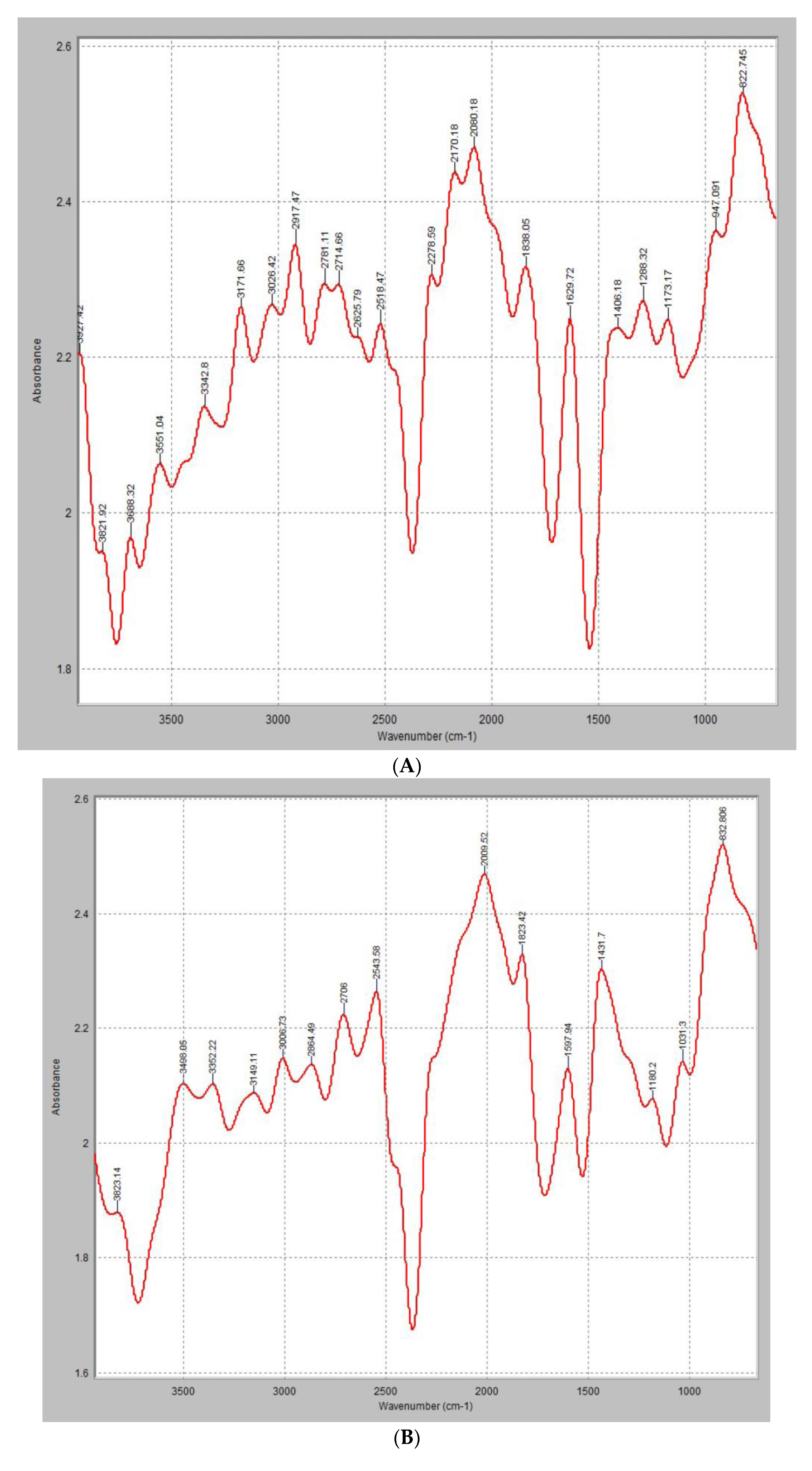
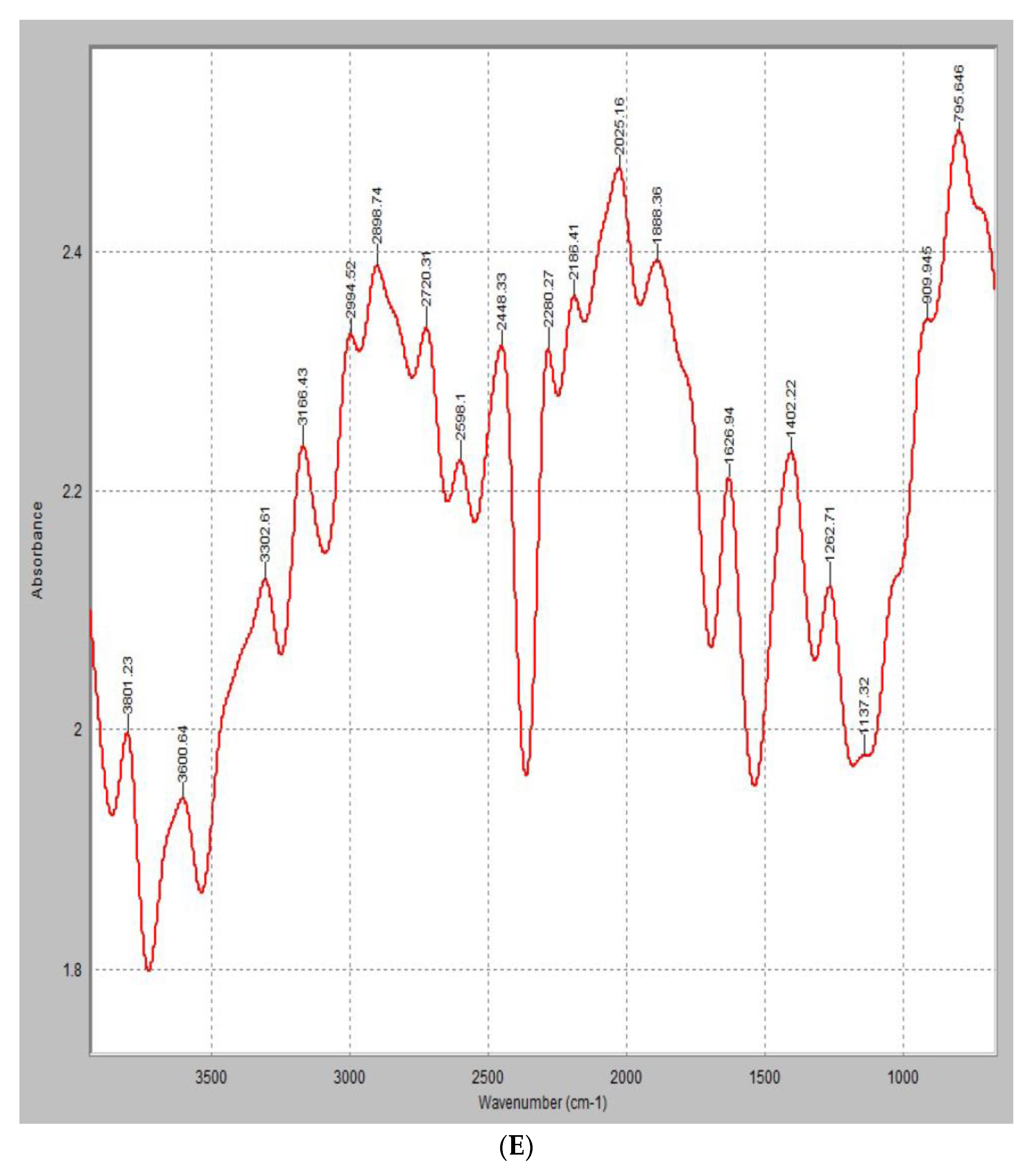
2.6. FTIR Analysis
2.7. In Vitro Anti-Nociceptive and Anti-Inflammatory Study
2.7.1. Assay of Membrane Stabilization by Hypotonicity Induced Hemolysis
2.7.2. Determination of Anti-Platelet Aggregatory Activity
2.8. In Vivo Antinociceptive and Anti-Inflammatory Study
2.8.1. Egg Albumin Induced Rat Paw Edema Inflammatory Model
2.8.2. 2% Formaldehyde Induced Arthritis Model
Total White Blood Cell Count
Differential Blood Count
3. Results and Discussion
3.1. pH Time Dependent Analysis
3.2. Encapsulation Efficiency (EE) and Loading Capacity (LC)
3.3. Particle Size and Morphology of Lipospheres
3.4. FTIR Analysis
3.5. In Vitro Anti-Inflammatory Studies
3.5.1. Membrane Stabilization
3.5.2. Anti-Platelet Aggregation Activity
3.6. In Vivo Anti-Inflammatory Studies
3.6.1. 2% Formaldehyde-Induced Arthritis Model
3.6.2. Egg Albumin-Induced Rat Paw Edema Inflammatory Model
3.6.3. Blood Cell Counts of Infected Rats
4. Conclusions
Author Contributions
Funding
Data Availability Statement
Conflicts of Interest
References
- Viana, F.A.; Pouliquen, Y.B.M.; Andrade-Neto, M.; Santiago, G.M.P.; Pessoa, O.D.L.; Rodrigues-Filho, E.; Braz-Filho, R. Complete 1H and 13C NMR assignments for two new monodesmosidesaponins from Pentaclethra macroloba (Willd.) Kuntze. MagnReson Chem. 2004, 42, 695–699. [Google Scholar] [CrossRef]
- Pertuit, D.; Kapundu, M.; Mitaine-Offer, A.C.; Miyamoto, T.; Tanaka, C.; Delaude, C.; Lacaille-Dubois, M.-A. Triterpenoid SaponinsFrom the Stem Bark of Pentaclethraeet veldeana. Nat. Prod. Commun. 2019, 14. [Google Scholar] [CrossRef]
- Babady-Byla, H.W. Triterpenes and 1-(ω-hydroxyceratyl) glycerols from Pentaclethraeet veldeana root bark. Phytochemistry 1996, 42, 501–504. [Google Scholar] [CrossRef]
- Viana, F.A.; Braz-Filho, R.; Pouliquen, Y.B.M.; Andrade Neto, M.; Santiago, G.M.P.; Rodrigues-Filho, E. Triterpenoid saponins from stem bark of Pentaclethra macroloba. J. Braz. Chem. Soc. 2004, 15, 595–602. [Google Scholar] [CrossRef]
- Chinaka, C.N.; Garuba, A.S.; Ahmadu, A.A. Chemical Constituents from the Stem Bark of Pentaclethra macrophylla Benth (Fabaceae). Nig. J. Pharm. Res. 2017, 13, 37–44. [Google Scholar]
- Folefoc, G.N.; Bisseck, J.P.; Fomum, Z.T.; Bodo, B. A new Secokauranediterpenoid and O-glucoside from seeds of Pentacletheramacrophylla. J. Cameroon Acad. Sci. 2004, 4, 227–231. [Google Scholar]
- Akindahunsi, A.A. Physicochemical studies on African oil bean (Pentaclethra macrophylla Benth.) seed. J. Food Agric. Environ. 2004, 2, 14–17. [Google Scholar]
- Asoegwu, S.; Ohanyere, S.; Kanu, O.; Iwueke, C. Physical Properties of African Oil Bean Seed (Pentaclethra macrophylla). Agric. Eng. Int. CIGR J. 2006, VIII. [Google Scholar]
- Nnamani, P.O.; Kenechukwu, F.C.; Asogwa, F.O.; Momoh, M.A.; Lehr, C.-M.; Attama, A.A. Novel Anti-Ulcer Phytosomal Formulation of Ethanol Extract of Pentaclethra macrophylla Stem-Bark. Trop. J. Nat. Prod. Res. 2020, 4, 385–391. [Google Scholar]
- Igbe, I.; Osigwe, C. Hypoglycaemic activity of aqueous extract of Pentaclethra macrophylla Benth. (Fabaceae) stem bark in streptozotocin-induced diabetic rats. J. Pharm. Bioresour. 2012, 9, 39–44. [Google Scholar]
- Ukoro, B.; Chike, C.P.R.; Olorunfemi, O.J. Effects of Aqueous Extract of Pentaclethra macrophylla Leaves on Blood Pressure and Oxidative Stress in Albino Wistar Rats. Int. J. Sci. Res. Eng. Dev. 2020, 3, 1095–1108. [Google Scholar]
- Iwu, I.C.; Uchegbu, R. Isolation and characterization of 4, 15- Cholestene-3-benzoate from the stem bark of Pentaclethra macrophylla (P. Benth). Int. J. Emerg. Knowl. 2014, 1, 185–192. [Google Scholar]
- Tico Etnobotanical Dictionary. Pentaclethra macrophylla (Benth). 2005. Available online: https://www.uni-hamburg.de/search.html?q=Pentaclethra+macrophylla (accessed on 25 August 2023).
- Okunrobo, L.O.; Ching, F.P.; Ifijeh, F. Antinociceptive activity of methanol extract and aqueous fraction of the stem bark of Pentaclethra macrophylla Benth (Mimosaceae). J. Med. Plants Res. 2009, 3, 101–104. [Google Scholar]
- Chidozie, J.I. African oil bean seed as an essential food supplement in the control of cancer and tobacco related diseases. In Proceedings of the U1CC World Cancer Congress, Washington, DC, USA, 8–12 July 2006. [Google Scholar]
- Balogun, B.I. Evaluation of the Nutritional Potentials of Fermented Oil Beans Seed Pentaclethra macrophyllah Benth. PAT 2013, 9, 73–87. [Google Scholar]
- Akah, P.A.; Aguwa, C.N.; Agu, R.U. Studies on the antidiarrhoeal properties of Pentaclethra macrophylla leaf extracts. Phytother. Res. 1999, 13, 292–295. [Google Scholar] [CrossRef]
- Alinnor, I.J.; Oze, R. Chemical Evaluation of the Nutritive Value of Pentaclethra macrophylla (African Oil Bean) seed. Pak. J. Nutr. 2011, 10, 355–359. [Google Scholar] [CrossRef]
- Osabor, V.N.; Pkpnkwo, P.C.; Ikeuba, A.I. Chemical profile of leaves and seeds of Pentaclethra macrophylla Benth. J. Med. Plant Herbal Ther. Res. 2017, 5, 11–17. [Google Scholar]
- Githens, T.S. African Handbooks—Drug Plants of Africa; University of Pennsylvania Press: Philadelphia, PA, USA, 1948; p. 64. [Google Scholar]
- Yesmin, S.; Paul, A.; Naz, T.; Rahman AB, M.A.; Akhter, S.F.; Wahed MI, I.; Emran, T.B.; Siddiqui, S.A. Membrane stabilization as a mechanism of the antiinflammatory activity of ethanolic root extract of Choi (Piper chaba). Clin. Phytosci. 2020, 6, 59. [Google Scholar] [CrossRef]
- Nnamani, P.O.; Ibezim, E.C.; Attama, A.A.; Adikwu, M.U. Surface modified solid lipid microparticles based on homolipids and softisan® 142: Preliminary characterization. Asian Pac. J. Trop. Med. 2010, 3, 205–210. [Google Scholar] [CrossRef]
- Sur, T.K.; Biswas, T.K.; Ali, L.; Mukherjee, B. Anti-inflammatory and anti-platelet aggregation activity of human placental extract. Acta Pharmacol. Sin. 2003, 24, 187–192. [Google Scholar]
- Zheng, Y.-Y.; Wu, T.-T.; Yang, Y.; Xian-Geng, H.; Gao, Y.; Chen, Y.; Yang, Y.-N.; Li, X.-M.; Ma, X.; Ma, Y.-T.; et al. Personalized antiplatelet therapy guided by a novel detection of platelet aggregation function in stable coronary artery disease patients undergoing percutaneous coronary intervention: A randomized controlled clinical trial. Eur. Heart J. Cardiovasc. Pharmacother. 2020, 6, 211–221. [Google Scholar] [CrossRef]
- Nnamani, P.O.; Attama, A.A.; Kenechukwu, F.C.; Ibezim, E.C.; Adikwu, M.U. Pharmacodynamics of Piroxicam from Novel Solid Lipid Microparticles Formulated with Homolipids from Bos indicus. Curr. Drug Deliv. 2013, 10, 645–655. [Google Scholar] [CrossRef]
- Ochei, J.; Kolhatkar, A. Medical Laboratory Science Theory and Practice; McGraw Hill Education: New York, NY, USA, 2000. [Google Scholar]
- Halliwell, B.; Whiteman, M. Measuring reactive species and oxidative damage in vivo and in cell culture: How should you do it and what do the results mean? Br. J. Pharmacol. 2004, 142, 231–255. [Google Scholar] [CrossRef]
- Chaitanya, R.; Sandhya, S.; Banji, D.; Ravindran, V.; Murali, S. HRBC Membrane Stabilizing Property of Root, Stem and Leaf of Glochidionelutinum. Int. J. Res. Pharm. Biomed. Sci. 2011, 2, 256–259. [Google Scholar]
- Shinde, U.A.; Phadke, A.S.; Nair, A.M.; Mungantiwar, A.A.; Dikshit, V.J.; Saraf, M.N. Membrane stabilizing activity—A possible mechanism of action for the anti-inflammatory activity of Cedrusdeodara wood oil. Fitoterapia 2008, 70, 251–257. [Google Scholar] [CrossRef]
- Serafini, M.; Peluso, I.; Raguzzini, A. Flavonoids as anti-inflammatory agents. Proc. Nutr. Soc. 2010, 69, 273–278. [Google Scholar] [CrossRef] [PubMed]
- Choi, J.H.; Kim, D.W.; Park, S.E.; Lee, H.J.; Kim, K.M.; Kim, K.J.; Kim, M.K.; Kim, S.J.; Kim, S. Anti-thrombotic effect of rutin isolated from Dendropanax morbifera Leveille. J. Biosci. Bioeng. 2015, 120, 181–186. [Google Scholar] [CrossRef]
- Ashorobi, D.; Ameer, M.A.; Fernandez, R. Thrombosis. In StatPearls; StatPearls Publishing: Treasure Island, FL, USA, 2022. [Google Scholar]
- Zhou, Y.J.; Xiang, J.Z.; Yuan, H.; Liu, H.; Tang, Q.; Hao, H.Z.; Yin, Z.; Wang, J.; Ming, Z.Y. Neferine exerts its antithrombotic effect by inhibiting platelet aggregation and promoting dissociation of platelet aggregates. Thromb. Res. 2013, 132, 202–210. [Google Scholar] [CrossRef] [PubMed]
- Nair, V.; Singh, S.; Gupta, Y. Evaluation of disease modifying activity of Coriandrum sativum in experimental models. Indian J. Med. Res. 2012, 135, 240–245. [Google Scholar]
- Bischoff, S.C. Quercetin: Potentials in the prevention and therapy of disease. Curr. Opin. Clin. Nutr. Metab. Care 2008, 11, 733–740. [Google Scholar] [CrossRef]
- Desai Nilesh, V.; Patkar Atul, A.; Shinde Shilpa, S.; Arwade Aboli, S. Protective effect of aqueous extract of Aegle marmelos against formaldehyde induced arthritis in rats. Int. Res. J. Pharm. Appl. Sci. 2012, 2, 66–72. [Google Scholar]
- Uttra, A.M.; Hasan, U.H. Anti-arthritic activity of aqueous-methanolic extract and various fractions of Berberis orthobotrys Bien ex Aitch. BMC Complement. Altern. Med. 2017, 17, 371. [Google Scholar]
- Farrukh, M.; Saleem, U.; Qasim, M.; Manan, M.; Shah, M.A. Sarcococcasaligna extract attenuates formaldehyde-induced arthritis in Wistar rats via modulation of pro-inflammatory and inflammatory biomarkers. Inflammopharmacology 2022, 30, 579–597. [Google Scholar] [CrossRef] [PubMed]
- Adeyemi, O.O.; Okpo, S.O.; Okpaka, O. The analgesic effect of the methanolic extract of Acanthus montanus. J. Ethnopharmacol. 2004, 90, 45–48. [Google Scholar] [CrossRef] [PubMed]


| PM Batches (g) | Goat Fat (g) | Phospholipon 90H (g) | Sorbitol (g) | Sorbic Acid (g) | Tween 80 (g) | Distilled Water (qs) w/w |
|---|---|---|---|---|---|---|
| 0 | 0.5 | 0.75 | 4 | 0.05 | 1.5 | 100 |
| 2.5 | 0.5 | 0.75 | 4 | 0.05 | 1.5 | 100 |
| 5.0 | 0.5 | 0.75 | 4 | 0.05 | 1.5 | 100 |
| 7.5 | 0.5 | 0.75 | 4 | 0.05 | 1.5 | 100 |
| 10.0 | 0.5 | 0.75 | 4 | 0.05 | 1.5 | 100 |
| Liposphere Batches (g) | Particle Size (µm) | Encapsulation Efficiency (EE %) | Loading Capacity (LC) | pH Time Dependence (Days) | ||
|---|---|---|---|---|---|---|
| 1 | 7 | 30 | ||||
| 0 | 12.56 ± 2.18 | - | - | 3.5 | 3.5 | 3.5 |
| 2.5 | 26.87 ± 4.21 | 35.2 | 4.4 | 3.8 | 3.8 | 3.8 |
| 5.0 | 42.42 ± 3.64 | 75.6 | 1.89 | 3.8 | 3.9 | 3.9 |
| 7.5 | 82.27 ± 6.24 | 89.4 | 3.4 | 3.7 | 3.9 | 3.9 |
| 10.0 | 98.67 ± 10.23 | 94.1 | 4.7 | 3.7 | 3.8 | 3.8 |
| Sample (µg/mL) | % Protection of Membrane |
|---|---|
| 50 | 24.60 |
| 100 | 51.70 |
| 200 | 100.0 |
| 400 | 300.0 |
| 800 | 200.0 |
| Aspirin (µg/mL) | |
| 50 | 89.00 |
| 100 | 5.90 |
| 200 | 100.00 |
| 400 | 100.00 |
| 800 | 106.25 |
| Sample (µg/mL) | Change 1 | Change 2 | Change 3 | Change 4 |
|---|---|---|---|---|
| Mean ± SEM | Mean ± SEM | Mean ± SEM | Mean ± SEM | |
| 50 | 0.007 ± 0.0025 cd | 0.0043 ± 0.0013 bc | 0.0017 ± 0.0007 bc | 0.001 ± 0.00 b |
| 100 | 0.004 ± 0.001 abcd | 0.0047 ± 0.0017 bc | 0.005 ± 0.0021 ab | 0.0037 ± 0.002 a |
| 200 | 0.0013 ± 0.009 ab | 0.002 ± 0.0006 ab | 0.007 ± 0.0026 a | 0.0023 ± 0.009 b |
| 400 | 0.0017 ± 0.002 ab | 0.0033 ± 0.0007 bc | 0.0007 ± 0.0003 c | 0.0057 ± 0.002 a |
| 800 | 0.000 ± 0.00 a | 0.000 ± 0.00 a | 0.00 ± 0.00 c | 0.001 ± 0.001 b |
| Aspirin (µg/mL) | ||||
| 50 | 0.005 ± 0.001 abcd | 0.0063 ± 0.0018 c | 0.0023 ± 0.009 bc | 0.002 ± 0.006 b |
| 100 | 0.0033 ± 0.0007 abc | 0.002 ± 0.0006 ab | 0.0010 ± 0.00 c | 0.0010 ± 0.006 b |
| 200 | 0.004 ± 0.0021 abcd | 0.003 ± 0.001 abc | 0.003 ± 0.00 bc | 0.0013 ± 0.0007 b |
| 400 | 0.0057 ± 0.002 bcd | 0.0043 ± 0.0015 abc | 0.0027 ± 0.0007 bc | 0.0013 ± 0.0003 b |
| 800 | 0.009 ± 0.0012 d | 0.0067 ± 0.0015 c | 0.0027 ± 0.0007 bc | 0.0023 ± 0.0009 b |
| Treatments (mg/kg) | Edema (mL) ± SEM over Time (0–48 h) | ||||||
|---|---|---|---|---|---|---|---|
| 0 | 1 | 2 | 3 | 4 | 5 | 48 | |
| T50 | 0.375 ± 0.025 | 0.750 ± 0.029 (0.00) | 0.650 ± 0.029 a (7.14) | 0.525 ± 0.025 a (0.00) | 0.500 ± 0.00 b (16.7) | 0.500 ± 0.00 b (0.00) | 0.425 ± 0.048 (−6.2) |
| T100 | 0.300 ± 0.00 | 0.700 ± 0.00 (6.7) | 0.600 ± 0.00 b (14.3) | 0.525 ± 0.025 a (0.00) | 0.525 ± 0.025 b (12.5) | 0.475 ± 0.025 c (0.00) | 0.400 ± 0.00 (0.00) |
| T150 | 0.325 ± 0.025 | 0.675 ± 0.025 (10.0) | 0.575 ± 0.025 b (17.8) | 0.525 ± 0.025 a (0.00) | 0.525 ± 0.025 b (12.5) | 0.500 ± 0.00 b (0.00) | 0.425 ± 0.025 (−6.25) |
| T200 | 0.325 ± 0.025 | 0.725 ± 0.025 (3.33) | 0.600 ± 0.00 b (14.29) | 0.500 ± 0.00 b (4.8) | 0.525 ± 0.025 b (12.5) | 0.550± 0.029 b (10.0) | 0.425 ± 0.025 (−6.25) |
| PC | 0.325 ± 0.025 | 0.70 ± 0.04 (6.7) | 0.60 ± 0.04 b (6.7) | 0.50 ± 0.041 b (4.77) | 0.475 ± 0.00 b (21.0) | 0.40 ± 0.00 a (20.0) | 0.40 ± 0.00 (0.00) |
| NC | 0.30 ± 0.00 | 0.75 ± 0.041 | 0.70 ± 0.029 a | 0.525 ± 0.025 a | 0.60 ± 0.00 a | 0.50 ± 0.00 b | 0.40 ± 0.00 |
| Normal | 0.35 ± 0.05 | 0.75 ± 0.05 | 0.65 ± 0.00 a | 0.60 ± 0.00 a | 0.5 ± 0.025 b | 0.475 ± 0.025 c | 0.40 ± 0.00 |
| Treatments (mg/kg) | Edema (mL) ± SEM over Time (h) | |||||
|---|---|---|---|---|---|---|
| 0 | 1 | 2 | 3 | 4 | 5 | |
| T50 | 0.350 ± 0.029 | 0.625 ± 0.025 (−4.17) | 0.550 ± 0.029 abc (−4.7) | 0.500 ± 0.058 a (−17.65) | 0.475 ± 0.048 a (−15.79) | 0.650 ± 0.029 a (−8.33) |
| T100 | 0.275 ± 0.025 | 0.625 ± 0.025 (−4.17) | 0.600 ± 0.00 ab (−14.29) | 0.525 ± 0.025 a (−23.5) | 0.525 ± 0.025 b (−31.25) | 0.650 ± 0.029 a (−4.17) |
| T150 | 0.300 ± 0.00 | 0.500 ± 0.00 (16.6) | 0.500 ± 0.058 c (4.76) | 0.433 ± 0.033 b (−1.88) | 0.433 ± 0.33 a (−8.25) | 0.633 ± 0.033 a (−5.5) |
| T200 | 0.267 ± 0.033 | 0.600 ± 0.058 (0.00) | 0.533 ± 0.033 ac (−1.5) | 0.500 ± 0.00 a (−17.64) | 0.533 ± 0.033 b (−33.25) | 0.700 ± 0.00 a (−6.0) |
| PC | 0.30 ± 0.00 | 0.625 ± 0.025 (4.17) | 0.500 ± 0.00 c (4.76) | 0.425 ± 0.025 b (0.00) | 0.425 ± 0.025 a (−6.25) | 0.600 ± 0.00 b (0.00) |
| NC | 0.30 ± 0.00 | 0.6 ± 0.00 | 0.525 ± 0.025 b | 0.425 ± 0.029 a | 0.400 ± 0.00 a | 0.60 ± 0.00 a |
| Normal | 0.275 ± 0.025 | 0.55 ± 0.029 | 0.625 ± 0.025 ac | 0.575 ± 0.025 b | 0.50 ± 0.041 a | 0.65 ± 0.029 b |
| Treatments (mg/kg) | Percentage Compositions of Different Blood Cells Count ± SEM | |||||
|---|---|---|---|---|---|---|
| WBC (%) | Neutrophil (%) | Lymphocytes (%) | Eosinophil (%) | Monocyte (%) | Basophil (%) | |
| T50 | 77.50 ± 3.59 | 65.50 ± 1.50 bc | 34.00 ± 1.414 abc | 0.50 ± 0.5 | 0.00 | 0.00 |
| T100 | 70.00 ± 1.83 | 58.50 ± 1.71 a | 40.00 ± 1.63 c | 1.50 ± 0.5 | 0.00 | 0.00 |
| T150 | 70.50 ± 1.71 | 61.50 ± 1.71 abc | 37.50 ± 2.22 abc | 1.00 ± 0.57 | 0.00 | 0.00 |
| T200 | 67.50 ± 3.50 | 61.00 ± 1.29 b | 38.50 ± 0.96 bc | 0.50 ± 0.5 | 0.00 | 0.00 |
| PC | 71.00 ± 3.512 | 63.50 ± 3.403 abc | 36.00 ± 3.747 abc | 0.50 ± 0.50 | 0.00 | 0.00 |
| NC | 67.00 ± 2.646 | 67.25 ± 0.479 c | 31.00 ± 0.577 a | 1.75 ± 0.629 | 0.00 | 0.00 |
| Normal 1 | 72.00 ± 4.320 | 61.50 ± 1.50 abc | 36.50 ± 0.751 abc | 2.0 ± 0.816 | 0.00 | 0.00 |
| Normal 2 | 68.80 ± 2.062 | 65.00 ± 1.915 bc | 32.00 ± 1.414 ab | 2.50 ± 0.500 | 0.00 | 0.00 |
Disclaimer/Publisher’s Note: The statements, opinions and data contained in all publications are solely those of the individual author(s) and contributor(s) and not of MDPI and/or the editor(s). MDPI and/or the editor(s) disclaim responsibility for any injury to people or property resulting from any ideas, methods, instructions or products referred to in the content. |
© 2023 by the authors. Licensee MDPI, Basel, Switzerland. This article is an open access article distributed under the terms and conditions of the Creative Commons Attribution (CC BY) license (https://creativecommons.org/licenses/by/4.0/).
Share and Cite
Nnamani, P.O.; Diovu, E.O.; Peter, I.E.; Onwuka, A.M.; Ogbuanwu, C.V.; Abonyi, O.E.; Loretz, B.; Lehr, C.-M. Anti-Inflammatory and Antinociceptive Activity of Herbal Lipospheres of Pentaclethra macrophylla (Fabaceae) Stem Bark Extract. Processes 2023, 11, 2557. https://doi.org/10.3390/pr11092557
Nnamani PO, Diovu EO, Peter IE, Onwuka AM, Ogbuanwu CV, Abonyi OE, Loretz B, Lehr C-M. Anti-Inflammatory and Antinociceptive Activity of Herbal Lipospheres of Pentaclethra macrophylla (Fabaceae) Stem Bark Extract. Processes. 2023; 11(9):2557. https://doi.org/10.3390/pr11092557
Chicago/Turabian StyleNnamani, Petra Obioma, Edith Obioma Diovu, Ikechukwu Emmanuel Peter, Akachukwu Marytheresa Onwuka, Chiamaka Victory Ogbuanwu, Obiora Emmanuel Abonyi, Brigitta Loretz, and Claus-Michael Lehr. 2023. "Anti-Inflammatory and Antinociceptive Activity of Herbal Lipospheres of Pentaclethra macrophylla (Fabaceae) Stem Bark Extract" Processes 11, no. 9: 2557. https://doi.org/10.3390/pr11092557
APA StyleNnamani, P. O., Diovu, E. O., Peter, I. E., Onwuka, A. M., Ogbuanwu, C. V., Abonyi, O. E., Loretz, B., & Lehr, C.-M. (2023). Anti-Inflammatory and Antinociceptive Activity of Herbal Lipospheres of Pentaclethra macrophylla (Fabaceae) Stem Bark Extract. Processes, 11(9), 2557. https://doi.org/10.3390/pr11092557