

Exploring the Antimelanoma Potential of Betulinic Acid Esters and Their Liposomal Nanoformulations

 , ,
, , .jpg) , ,
, , 
Abstract
1. Introduction
2. Results
2.1. Chemistry
2.1.1. Synthesis and Characterization of Fatty Acid Esters of BA
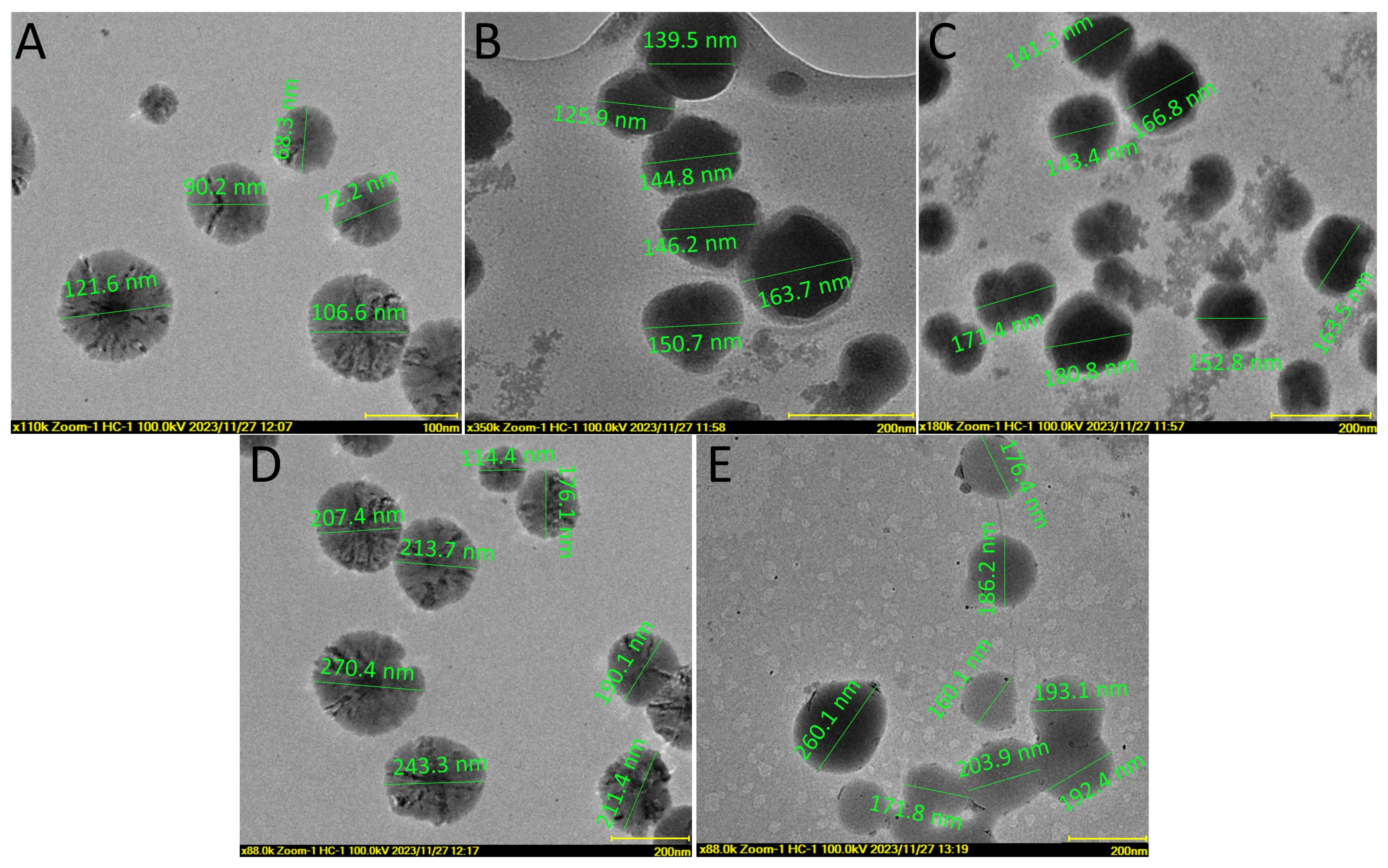
2.1.2. Synthesis and Characterization of BA-Fatty Acid Ester Liposomal Formulation
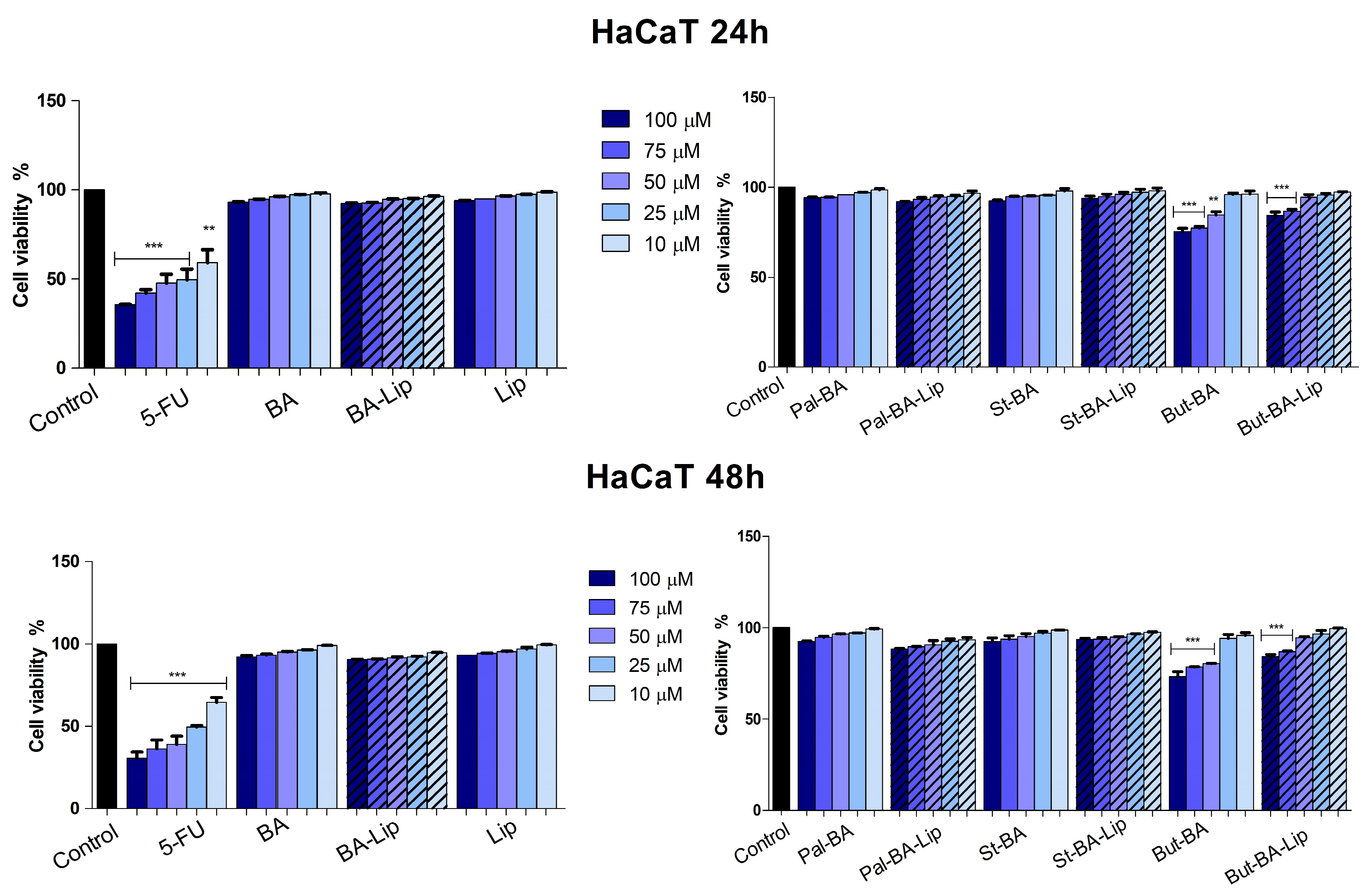
2.2. Evaluation of Betulinic Acid Fatty Esters and Liposomes Cytotoxic Effect
2.3. Fatty Ester Derivatives Effects on Cell Morphology
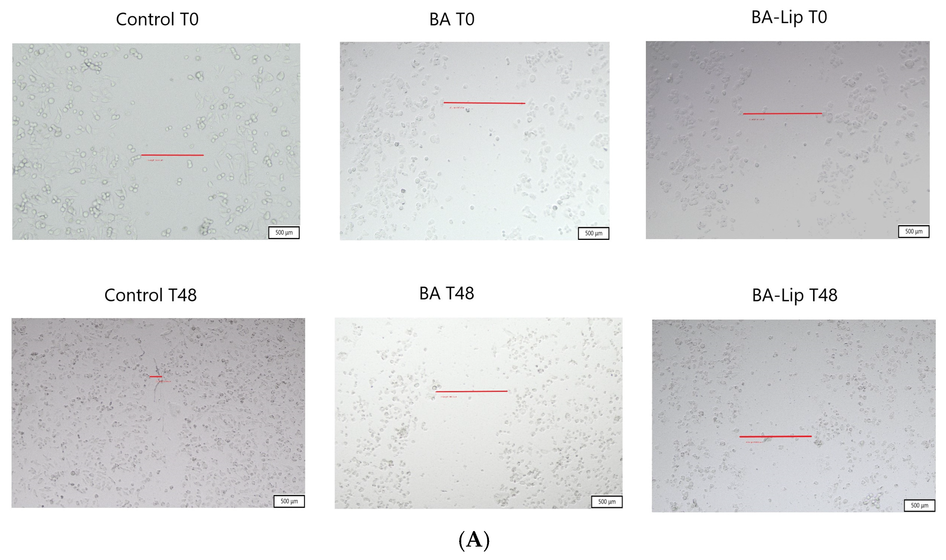
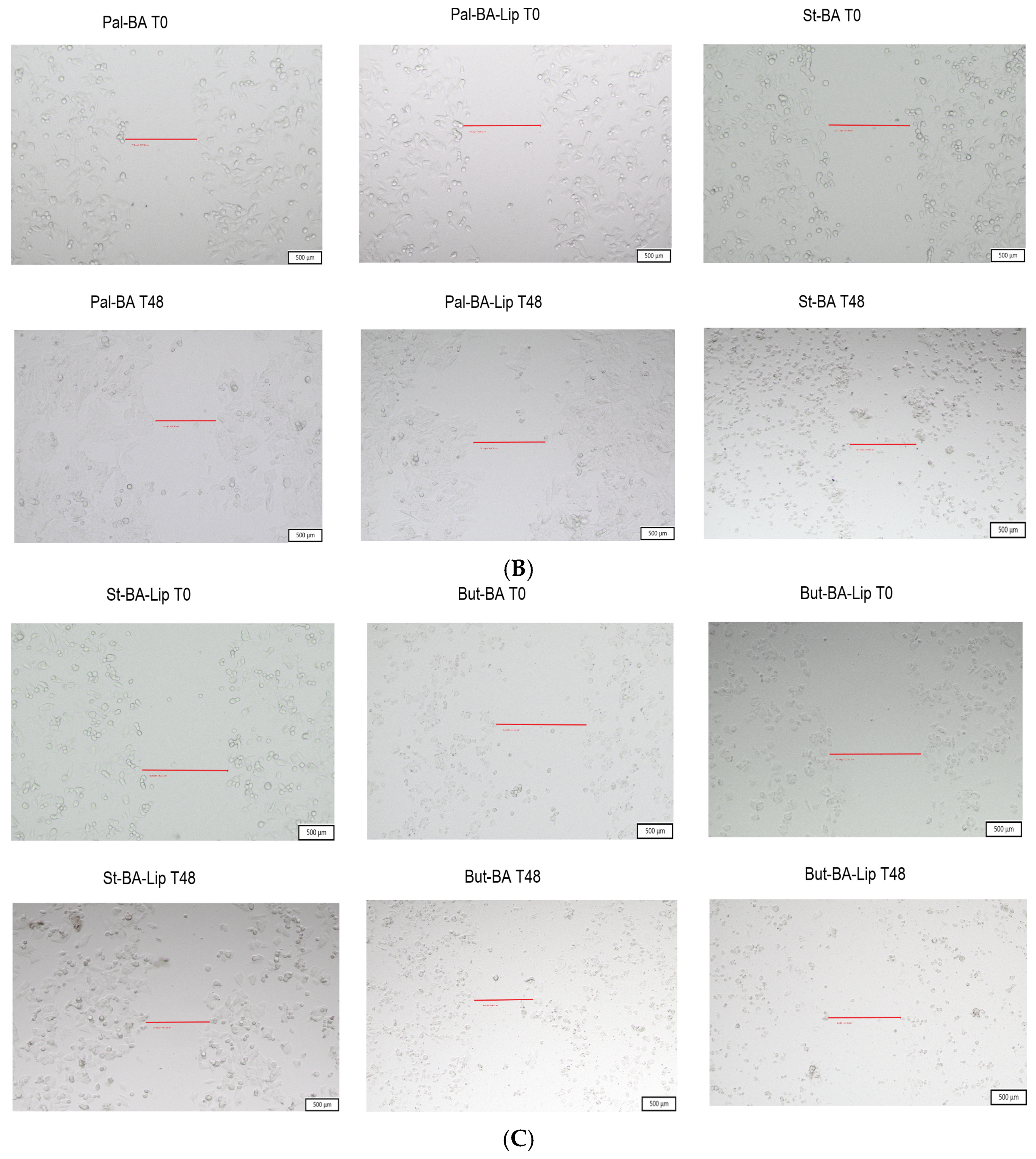
2.4. Scratch Assay
2.5. Betulinic Acid Esters Effect on BAX and Bcl-2 Protein Levels
3. Discussion
4. Materials and Methods
4.1. Chemistry
4.1.1. Instruments and Reagents
4.1.2. Synthesis Procedure for BA Fatty Acid Esters
4.1.3. Synthesis Procedure for BA Fatty Acid Esters Liposomal Formulations
4.2. In Vitro Assessment
4.2.1. Cell Culture
4.2.2. Cell Viability Assessment
4.2.3. Fatty Ester Derivatives Effects on Cell Morphology
4.2.4. Immunofluorescence Assay—Morphological Assessment of Apoptotic Cells
4.2.5. Real-Time PCR Quantification of Apoptotic Markers
4.2.6. Scratch Assay
4.3. BAX and Bcl-2 Detection
4.4. Statistical Analysis
5. Conclusions
Supplementary Materials
Author Contributions
Funding
Data Availability Statement
Conflicts of Interest
References
- Majolo, F.; de Oliveira Becker Delwing, L.K.; Marmitt, D.J.; Bustamante-Filho, I.C.; Goettert, M.I. Medicinal Plants and Bioactive Natural Compounds for Cancer Treatment: Important Advances for Drug Discovery. Phytochem. Lett. 2019, 31, 196–207. [Google Scholar] [CrossRef]
- Harvey, A. Natural Products in Drug Discovery. Drug Discov. Today 2008, 13, 894–901. [Google Scholar] [CrossRef] [PubMed]
- Ríos, J.; Máñez, S. New Pharmacological Opportunities for Betulinic Acid. Planta Med. 2018, 84, 8–19. [Google Scholar] [CrossRef] [PubMed]
- Mioc, M.; Milan, A.; Malița, D.; Mioc, A.; Prodea, A.; Racoviceanu, R.; Ghiulai, R.; Cristea, A.; Căruntu, F.; Șoica, C. Recent Advances Regarding the Molecular Mechanisms of Triterpenic Acids: A Review (Part I). Int. J. Mol. Sci. 2022, 23, 7740. [Google Scholar] [CrossRef] [PubMed]
- Seca, A.; Pinto, D. Plant Secondary Metabolites as Anticancer Agents: Successes in Clinical Trials and Therapeutic Application. Int. J. Mol. Sci. 2018, 19, 263. [Google Scholar] [CrossRef] [PubMed]
- Ghiulai, R.; Roşca, O.J.; Antal, D.S.; Mioc, M.; Mioc, A.; Racoviceanu, R.; Macaşoi, I.; Olariu, T.; Dehelean, C.; Creţu, O.M.; et al. Tetracyclic and Pentacyclic Triterpenes with High Therapeutic Efficiency in Wound Healing Approaches. Molecules 2020, 25, 5557. [Google Scholar] [CrossRef] [PubMed]
- Paduch, R.; Kandefer-Szerszen, M. Antitumor and Antiviral Activity of Pentacyclic Triterpenes. Mini. Rev. Org. Chem. 2014, 11, 262–268. [Google Scholar] [CrossRef]
- Mioc, M.; Prodea, A.; Racoviceanu, R.; Mioc, A.; Ghiulai, R.; Milan, A.; Voicu, M.; Mardale, G.; Șoica, C. Recent Advances Regarding the Molecular Mechanisms of Triterpenic Acids: A Review (Part II). Int. J. Mol. Sci. 2022, 23, 8896. [Google Scholar] [CrossRef]
- Furtado, N.A.J.C.; Pirson, L.; Edelberg, H.; Miranda, M.L.; Loira-Pastoriza, C.; Preat, V.; Larondelle, Y.; André, C. Pentacyclic Triterpene Bioavailability: An Overview of In Vitro and In Vivo Studies. Molecules 2017, 22, 400. [Google Scholar] [CrossRef]
- Prodea, A.; Mioc, A.; Banciu, C.; Trandafirescu, C.; Milan, A.; Racoviceanu, R.; Ghiulai, R.; Mioc, M.; Soica, C. The Role of Cyclodextrins in the Design and Development of Triterpene-Based Therapeutic Agents. Int. J. Mol. Sci. 2022, 23, 736. [Google Scholar] [CrossRef]
- Valdés, K.; Morales, J.; Rodríguez, L.; Günther, G. Potential Use of Nanocarriers with Pentacyclic Triterpenes in Cancer Treatments. Nanomedicine 2016, 11, 3139–3156. [Google Scholar] [CrossRef]
- Liu, Y.; Gao, D.; Zhang, X.; Liu, Z.; Dai, K.; Ji, B.; Wang, Q.; Luo, L. Antitumor Drug Effect of Betulinic Acid Mediated by Polyethylene Glycol Modified Liposomes. Mater. Sci. Eng. C 2016, 64, 124–132. [Google Scholar] [CrossRef] [PubMed]
- Nistor, G.; Trandafirescu, C.; Prodea, A.; Milan, A.; Cristea, A.; Ghiulai, R.; Racoviceanu, R.; Mioc, A.; Mioc, M.; Ivan, V.; et al. Semisynthetic Derivatives of Pentacyclic Triterpenes Bearing Heterocyclic Moieties with Therapeutic Potential. Molecules 2022, 27, 6552. [Google Scholar] [CrossRef] [PubMed]
- Wang, X.; Zhang, C.; Bao, N. Molecular Mechanism of Palmitic Acid and Its Derivatives in Tumor Progression. Front. Oncol. 2023, 13, 1224125. [Google Scholar] [CrossRef] [PubMed]
- Al-Hwaiti, M.S.; Alsbou, E.M.; Abu Sheikha, G.; Bakchiche, B.; Pham, T.H.; Thomas, R.H.; Bardaweel, S.K. Evaluation of the Anticancer Activity and Fatty Acids Composition of “Handal” (Citrullus Colocynthis L.) Seed Oil, a Desert Plant from South Jordan. Food Sci. Nutr. 2021, 9, 282–289. [Google Scholar] [CrossRef] [PubMed]
- Abe, A.; Sugiyama, K. Growth Inhibition and Apoptosis Induction of Human Melanoma Cells by ω-Hydroxy Fatty Acids. Anticancer. Drugs 2005, 16, 543–549. [Google Scholar] [CrossRef] [PubMed]
- Chodurek, E.; Orchel, A.; Gawlik, N.; Kulczycka, A.; Gruchlik, A.; Dzierzewicz, Z. Proliferation and Cellular Death of A375 Cell Line in the Presence of HDACs Inhibitors. Acta Pol. Pharm.—Drug Res. 2010, 67, 686–689. [Google Scholar]
- Pinzaru, I.; Trandafirescu, C.; Szabadai, Z.; Mioc, M.; Ledeti, I.; Coricovac, D.; Ciurlea, S.; Ghiulai, R.M.; Crainiceanu, Z.; Simu, G. Synthesis and Biological Evaluation of Some Pentacyclic Lupane Triterpenoid Esters. Rev. Chim. 2014, 65, 848–851. [Google Scholar]
- Mallavadhani, U.V.; Mahapatra, A.; Jamil, K.; Reddy, P.S. Antimicrobial Activity of Some Pentacyclic Triterpenes and Their Synthesized 3-O-Lipophilic Chains. Biol. Pharm. Bull. 2004, 27, 1576–1579. [Google Scholar] [CrossRef]
- Hodges, L.D.; Kweifio-Okai, G.; Macrides, T.A. Antiprotease Effect of Anti-Inflammatory Lupeol Esters. Mol. Cell. Biochem. 2003, 252, 97–101. [Google Scholar] [CrossRef]
- Fotie, J.; Bohle, D.S.; Leimanis, M.L.; Georges, E.; Rukunga, G.; Nkengfack, A.E. Lupeol Long-Chain Fatty Acid Esters with Antimalarial Activity from Holarrhena f Loribunda. J. Nat. Prod. 2006, 69, 62–67. [Google Scholar] [CrossRef] [PubMed]
- Gregory, G. Engineering Liposomes for Drug Delivery: Progress and Problems. Trends Biotechnol. 1995, 13, 527–537. [Google Scholar]
- Sofou, S. Surface-Active Liposomes for Targeted Cancer Therapy. Nanomedicine 2007, 2, 711–724. [Google Scholar] [CrossRef] [PubMed]
- Mineart, K.P.; Venkataraman, S.; Yang, Y.Y.; Hedrick, J.L.; Prabhu, V.M. Fabrication and Characterization of Hybrid Stealth Liposomes. Macromolecules 2018, 51, 3184–3192. [Google Scholar] [CrossRef] [PubMed]
- Habib, N.; Wood, C.; Apostolov, K.; Barker, W.; Hershman, M.; Aslam, M.; Heinemann, D.; Fermor, B.; Williamson, R.; Jenkins, W. Stearic Acid and Carcinogenesis. Br. J. Cancer 1987, 56, 455–458. [Google Scholar] [CrossRef] [PubMed]
- Zhu, S.; Jiao, W.; Xu, Y.; Hou, L.; Li, H.; Shao, J.; Zhang, X.; Wang, R.; Kong, D. Palmitic Acid Inhibits Prostate Cancer Cell Proliferation and Metastasis by Suppressing the PI3K/Akt Pathway. Life Sci. 2021, 286, 120046. [Google Scholar] [CrossRef] [PubMed]
- Zafaryab, M.; Fakhri, K.K.; Hajela, K.; Moshahid, M.; Rizvi, A.; Moshahid, A.; Rizvi, M. In Vitro Assessment of Cytotoxic and Apoptotic Potential of Palmitic Acid for Breast Cancer Treatment. J. Life Sci. Res. 2019, 7, 166–174. [Google Scholar] [CrossRef]
- Farcas, C.G.; Dehelean, C.; Pinzaru, I.A.; Mioc, M.; Socoliuc, V.; Moaca, E.-A.; Avram, S.; Ghiulai, R.; Coricovac, D.; Pavel, I.; et al. Thermosensitive Betulinic Acid-Loaded Magnetoliposomes: A Promising Antitumor Potential for Highly Aggressive Human Breast Adenocarcinoma Cells Under Hyperthermic Conditions. Int. J. Nanomed. 2020, 15, 8175–8200. [Google Scholar] [CrossRef]
- Naeem, A.; Hu, P.; Yang, M.; Zhang, J.; Liu, Y.; Zhu, W.; Zheng, Q. Natural Products as Anticancer Agents: Current Status and Future Perspectives. Molecules 2022, 27, 8367. [Google Scholar] [CrossRef]
- Hordyjewska, A.; Ostapiuk, A.; Horecka, A.; Kurzepa, J. Betulin and Betulinic Acid: Triterpenoids Derivatives with a Powerful Biological Potential. Phytochem. Rev. 2019, 18, 929–951. [Google Scholar] [CrossRef]
- Zhang, X.; Hu, J.; Chen, Y. Betulinic Acid and the Pharmacological Effects of Tumor Suppression. Mol. Med. Rep. 2016, 14, 4489–4495. [Google Scholar] [CrossRef] [PubMed]
- Ali-Seyed, M.; Jantan, I.; Vijayaraghavan, K.; Bukhari, S.N.A. Betulinic Acid: Recent Advances in Chemical Modifications, Effective Delivery, and Molecular Mechanisms of a Promising Anticancer Therapy. Chem. Biol. Drug Des. 2016, 87, 517–536. [Google Scholar] [CrossRef] [PubMed]
- Fulda, S. Betulinic Acid for Cancer Treatment and Prevention. Int. J. Mol. Sci. 2008, 9, 1096–1107. [Google Scholar] [CrossRef] [PubMed]
- Wang, J.; Shi, Y.-m. Recent Updates on Anticancer Activity of Betulin and Betulinic Acid Hybrids (A Review). Russ. J. Gen. Chem. 2023, 93, 610–627. [Google Scholar] [CrossRef]
- Wang, K.-W. A New Fatty Acid Ester of Triterpenoid from Celastrus rosthornianus with Anti-Tumor Activitives. Nat. Prod. Res. 2007, 21, 669–674. [Google Scholar] [CrossRef]
- Legrand, P.; Rioux, V. The Complex and Important Cellular and Metabolic Functions of Saturated Fatty Acids. Lipids 2010, 45, 941–946. [Google Scholar] [CrossRef] [PubMed]
- Jóźwiak, M.; Filipowska, A.; Fiorino, F.; Struga, M. Anticancer Activities of Fatty Acids and Their Heterocyclic Derivatives. Eur. J. Pharmacol. 2020, 871, 172937. [Google Scholar] [CrossRef]
- Jóźwiak, M.; Struga, M.; Roszkowski, P.; Filipek, A.; Nowicka, G.; Olejarz, W. Anticancer Effects of Alloxanthoxyletin and Fatty Acids Esters—In Vitro Study on Cancer HTB-140 and A549 Cells. Biomed. Pharmacother. 2019, 110, 618–630. [Google Scholar] [CrossRef]
- Ahmad, F.B.H.; Ghafari, M.; Basri, M.; Abdul, M.B. Anticancer Activity of 3- O -Acylated Betulinic Acid Derivatives Obtained by Enzymatic Synthesis. Biosci. Biotechnol. Biochem. 2010, 74, 1025–1029. [Google Scholar] [CrossRef]
- Abbasi, H.; Kouchak, M.; Mirveis, Z.; Hajipour, F.; Khodarahmi, M.; Rahbar, N.; Handali, S. What We Need to Know about Liposomes as Drug Nanocarriers: An Updated Review. Adv. Pharm. Bull. 2022, 13, 7. [Google Scholar] [CrossRef]
- Mullauer, F.B.; Van Bloois, L.; Daalhuisen, J.B.; Ten Brink, M.S.; Storm, G.; Medema, J.P.; Schiffelers, R.M.; Kessler, J.H. Betulinic Acid Delivered in Liposomes Reduces Growth of Human Lung and Colon Cancers in Mice without Causing Systemic Toxicity. Anticancer. Drugs 2011, 22, 223–233. [Google Scholar] [CrossRef] [PubMed]
- Gao, D.; Tang, S.; Tong, Q. Acid Liposomes with Polyethylene Glycol Modification: Promising Antitumor Drug Delivery. Int. J. Nanomed. 2012, 7, 3517–3526. [Google Scholar] [CrossRef] [PubMed]
- Németh, Z.; Csóka, I.; Semnani Jazani, R.; Sipos, B.; Haspel, H.; Kozma, G.; Kónya, Z.; Dobó, D.G. Quality by Design-Driven Zeta Potential Optimisation Study of Liposomes with Charge Imparting Membrane Additives. Pharmaceutics 2022, 14, 1798. [Google Scholar] [CrossRef] [PubMed]
- Berbel Manaia, E.; Paiva Abuçafy, M.; Chiari-Andréo, B.G.; Lallo Silva, B.; Oshiro-Júnior, J.A.; Chiavacci, L. Physicochemical Characterization of Drug Nanocarriers. Int. J. Nanomed. 2017, 12, 4991–5011. [Google Scholar] [CrossRef] [PubMed]
- Jin, X.; Yang, Q.; Cai, N.; Zhang, Z. A Cocktail of Betulinic Acid, Parthenolide, Honokiol and Ginsenoside Rh2 in Liposome Systems for Lung Cancer Treatment. Nanomedicine 2020, 15, 41–54. [Google Scholar] [CrossRef] [PubMed]
- Guo, B.; Xu, D.; Liu, X.; Yi, J. Enzymatic Synthesis and in Vitro Evaluation of Folate-Functionalized Liposomes. Drug Des. Devel. Ther. 2017, 11, 1839–1847. [Google Scholar] [CrossRef]
- Colombo, I.; Sangiovanni, E.; Maggio, R.; Mattozzi, C.; Zava, S.; Corbett, Y.; Fumagalli, M.; Carlino, C.; Corsetto, P.A.; Scaccabarozzi, D.; et al. HaCaT Cells as a Reliable In Vitro Differentiation Model to Dissect the Inflammatory/Repair Response of Human Keratinocytes. Mediators Inflamm. 2017, 2017, 7435621. [Google Scholar] [CrossRef]
- Keshari, S.; Balasubramaniam, A.; Myagmardoloonjin, B.; Herr, D.R.; Negari, I.P.; Huang, C.-M. Butyric Acid from Probiotic Staphylococcus Epidermidis in the Skin Microbiome Down-Regulates the Ultraviolet-Induced Pro-Inflammatory IL-6 Cytokine via Short-Chain Fatty Acid Receptor. Int. J. Mol. Sci. 2019, 20, 4477. [Google Scholar] [CrossRef]
- Suresh, C.; Zhao, H.; Gumbs, A.; Chetty, C.S.; Bose, H.S. New Ionic Derivatives of Betulinic Acid as Highly Potent Anti-Cancer Agents. Bioorg. Med. Chem. Lett. 2012, 22, 1734–1738. [Google Scholar] [CrossRef]
- Coricovac, D.; Dehelean, C.A.; Pinzaru, I.; Mioc, A.; Aburel, O.-M.; Macasoi, I.; Draghici, G.A.; Petean, C.; Soica, C.; Boruga, M.; et al. Assessment of Betulinic Acid Cytotoxicity and Mitochondrial Metabolism Impairment in a Human Melanoma Cell Line. Int. J. Mol. Sci. 2021, 22, 4870. [Google Scholar] [CrossRef]
- Jiang, W.; Li, X.; Dong, S.; Zhou, W. Betulinic Acid in the Treatment of Tumour Diseases: Application and Research Progress. Biomed. Pharmacother. 2021, 142, 111990. [Google Scholar] [CrossRef] [PubMed]
- Jan, R.; Chaudhry, G.-E.-S. Understanding Apoptosis and Apoptotic Pathways Targeted Cancer Therapeutics. Adv. Pharm. Bull. 2019, 9, 205–218. [Google Scholar] [CrossRef] [PubMed]
- Shen, M.; Hu, Y.; Yang, Y.; Wang, L.; Yang, X.; Wang, B.; Huang, M. Betulinic Acid Induces ROS-Dependent Apoptosis and S-Phase Arrest by Inhibiting the NF- κ B Pathway in Human Multiple Myeloma. Oxid. Med. Cell. Longev. 2019, 2019, 5083158. [Google Scholar] [CrossRef] [PubMed]
- Khan, A.A.; Alanazi, A.M.; Jabeen, M.; Chauhan, A.; Abdelhameed, A.S. Design, Synthesis and in Vitro Anticancer Evaluation of a Stearic Acid-Based Ester Conjugate. Anticancer Res. 2013, 33, 2517–2524. [Google Scholar] [PubMed]
- Nakkarach, A.; Foo, H.L.; Song, A.A.-L.; Mutalib, N.E.A.; Nitisinprasert, S.; Withayagiat, U. Anti-Cancer and Anti-Inflammatory Effects Elicited by Short Chain Fatty Acids Produced by Escherichia Coli Isolated from Healthy Human Gut Microbiota. Microb. Cell Fact. 2021, 20, 36. [Google Scholar] [CrossRef] [PubMed]
- Salomone, B.; Ponti, R.; Gasco, M.R.; Ugazio, E.; Quaglino, P.; Osella-Abate, S.; Bernengo, M.G. In Vitro Effects of Cholesteryl Butyrate Solid Lipid Nanospheres as a Butyric Acid Pro-Drug on Melanoma Cells: Evaluation of Antiproliferative Activity and Apoptosis Induction. Clin. Exp. Metastasis 2000, 18, 663–673. [Google Scholar] [CrossRef] [PubMed]
- Quagliariello, V.; Masarone, M.; Armenia, E.; Giudice, A.; Barbarisi, M.; Caraglia, M.; Barbarisi, A.; Persico, M. Chitosan-Coated Liposomes Loaded with Butyric Acid Demonstrate Anticancer and Anti-Inflammatory Activity in Human Hepatoma HepG2 Cells. Oncol. Rep. 2019, 41, 1476–1486. [Google Scholar] [CrossRef] [PubMed]
- Pisha, E.; Chai, H.; Lee, I.-S.; Chagwedera, T.E.; Farnsworth, N.R.; Cordell, G.A.; Beecher, C.W.W.; Fong, H.H.S.; Kinghorn, A.D.; Brown, D.M.; et al. Discovery of Betulinic Acid as a Selective Inhibitor of Human Melanoma That Functions by Induction of Apoptosis. Nat. Med. 1995, 1, 1046–1051. [Google Scholar] [CrossRef]
- Guo, Y.; Zhu, H.; Weng, M.; Wang, C.; Sun, L. Chemopreventive Effect of Betulinic Acid via MTOR -Caspases/Bcl2/Bax Apoptotic Signaling in Pancreatic Cancer. BMC Complement. Med. Ther. 2020, 20, 178. [Google Scholar] [CrossRef]
- Rzeski, W.; Stepulak, A.; Szymański, M.; Sifringer, M.; Kaczor, J.; Wejksza, K.; Zdzisińska, B.; Kandefer-Szerszeń, M. Betulinic Acid Decreases Expression of Bcl-2 and Cyclin D1, Inhibits Proliferation, Migration and Induces Apoptosis in Cancer Cells. Naunyn. Schmiedebergs. Arch. Pharmacol. 2006, 374, 11–20. [Google Scholar] [CrossRef]
- Khan, I.; Guru, S.K.; Rath, S.K.; Chinthakindi, P.K.; Singh, B.; Koul, S.; Bhushan, S.; Sangwan, P.L. A Novel Triazole Derivative of Betulinic Acid Induces Extrinsic and Intrinsic Apoptosis in Human Leukemia HL-60 Cells. Eur. J. Med. Chem. 2016, 108, 104–116. [Google Scholar] [CrossRef] [PubMed]
- Mioc, M.; Mioc, A.; Racoviceanu, R.; Ghiulai, R.; Prodea, A.; Milan, A.; Barbu Tudoran, L.; Oprean, C.; Ivan, V.; Șoica, C. The Antimelanoma Biological Assessment of Triterpenic Acid Functionalized Gold Nanoparticles. Molecules 2023, 28, 421. [Google Scholar] [CrossRef] [PubMed]
- Nistor, G.; Mioc, A.; Mioc, M.; Balan-Porcarasu, M.; Ghiulai, R.; Racoviceanu, R.; Avram, Ș.; Prodea, A.; Semenescu, A.; Milan, A.; et al. Novel Semisynthetic Betulinic Acid−Triazole Hybrids with In Vitro Antiproliferative Potential. Processes 2022, 11, 101. [Google Scholar] [CrossRef]
- Kazakova, O.; Șoica, C.; Babaev, M.; Petrova, A.; Khusnutdinova, E.; Poptsov, A.; Macașoi, I.; Drăghici, G.; Avram, Ș.; Vlaia, L. 3-Pyridinylidene Derivatives of Chemically Modified Lupane and Ursane Triterpenes as Promising Anticancer Agents by Targeting Apoptosis. Int. J. Mol. Sci. 2021, 22, 10695. [Google Scholar] [CrossRef] [PubMed]
- Saha, S.; Ghosh, M.; Dutta, S.K. A Potent Tumoricidal Co-Drug ‘Bet-CA’—an Ester Derivative of Betulinic Acid and Dichloroacetate Selectively and Synergistically Kills Cancer Cells. Sci. Rep. 2015, 5, 7762. [Google Scholar] [CrossRef] [PubMed]
- Drag-Zalesinska, M.; Kulbacka, J.; Saczko, J.; Wysocka, T.; Zabel, M.; Surowiak, P.; Drag, M. Esters of Betulin and Betulinic Acid with Amino Acids Have Improved Water Solubility and Are Selectively Cytotoxic toward Cancer Cells. Bioorg. Med. Chem. Lett. 2009, 19, 4814–4817. [Google Scholar] [CrossRef] [PubMed]
- Bobadilla, A.V.P.; Arévalo, J.; Sarró, E.; Byrne, H.M.; Maini, P.K.; Carraro, T.; Balocco, S.; Meseguer, A.; Alarcón, T. In Vitro Cell Migration Quantification Method for Scratch Assays. J. R. Soc. Interface 2019, 16, 20180709. [Google Scholar] [CrossRef]
- Ghiƫu, A.; Pavel, I.Z.; Avram, S.; Kis, B.; Minda, D.; Dehelean, C.A.; Buda, V.; Folescu, R.; Danciu, C. An In Vitro-In Vivo Evaluation of the Antiproliferative and Antiangiogenic Effect of Flavone Apigenin against SK-MEL-24 Human Melanoma Cell Line. Anal. Cell. Pathol. 2021, 2021, 5552664. [Google Scholar] [CrossRef]
- Polacheck, W.J.; Zervantonakis, I.K.; Kamm, R.D. Tumor Cell Migration in Complex Microenvironments. Cell. Mol. Life Sci. 2013, 70, 1335–1356. [Google Scholar] [CrossRef]
- Bache, M.; Zschornak, M.P.; Passin, S.; Keßler, J.; Wichmann, H.; Kappler, M.; Paschke, R.; Kaluđerović, G.N.; Kommera, H.; Taubert, H.; et al. Increased Betulinic Acid Induced Cytotoxicity and Radiosensitivity in Glioma Cells under Hypoxic Conditions. Radiat. Oncol. 2011, 6, 111. [Google Scholar] [CrossRef]
- Wang, H.; Wang, H.; Ge, L.; Zhao, Y.; Zhu, K.; Chen, Z.; Wu, Q.; Xin, Y.; Guo, J. Betulinic Acid Targets Drug-Resistant Human Gastric Cancer Cells by Inducing Autophagic Cell Death, Suppresses Cell Migration and Invasion, and Modulates the ERK/MEK Signaling Pathway. Acta Biochim. Pol. 2021, 69, 25–30. [Google Scholar] [CrossRef]
- El-Desouky, S.K. A Cytotoxic Lupeol Fatty Acid Ester and Other Pentacyclic Triterpenes from Salvadora Persica Seeds. Nat. Prod. Sci. 2023, 29, 121–126. [Google Scholar] [CrossRef]
- Nunez, C.V.; de Vasconcellos, M.C.; Alaniz, L. Editorial: Are Natural Products, Used as Antitumoral/Antiangiogenic Agents, Less Toxic than Synthetic Conventional Chemotherapy? Front. Pharmacol. 2022, 13, 1055516. [Google Scholar] [CrossRef]
- Zhou, M.; Zhang, R.-H.; Wang, M.; Xu, G.-B.; Liao, S.-G. Prodrugs of Triterpenoids and Their Derivatives. Eur. J. Med. Chem. 2017, 131, 222–236. [Google Scholar] [CrossRef]
- Moacă, E.A.; Pavel, I.Z.; Danciu, C.; Crăiniceanu, Z.; Minda, D.; Ardelean, F.; Antal, D.S.; Ghiulai, R.; Cioca, A.; Derban, M.; et al. Romanian Wormwood (Artemisia Absinthium L.): Physicochemical and Nutraceutical Screening. Molecules 2019, 24, 3087. [Google Scholar] [CrossRef] [PubMed]
- Assay Procedure. Available online: https://www.abcam.com/en-tr/products/elisa-kits/human-bcl-2-elisa-kit-ab119506 (accessed on 30 January 2024).


| Melting Point | Yield | Appearance | m/z [M-H+]− | |
|---|---|---|---|---|
| Compound | ||||
| But-BA | 265–280 °C | 74% | white powder | 525 |
| Pal-BA | 160–170 °C | 70% | translucent crystals | 693 |
| St-BA | 150–162 °C | 65% | translucent crystals | 721 |
| Drug Loading Efficiency (DLE %) | ||||||||
|---|---|---|---|---|---|---|---|---|
| BA-Lip | But-BA-Lip | Pal-BA-Lip | St-BA-Lip | |||||
| 4 °C | 25 °C | 4 °C | 25 °C | 4 °C | 25 °C | 4 °C | 25 °C | |
| Day 1 | 78.12 | 78.12 | 85.22 | 85.22 | 82.06 | 82.06 | 80.18 | 80.18 |
| Day 3 | 78.08 | 77.66 | 84.91 | 84.51 | 81.65 | 81.42 | 79.72 | 79.71 |
| Day 5 | 77.65 | 77.13 | 84.58 | 83.72 | 81.17 | 80.68 | 79.16 | 78.88 |
| Day 7 | 76.87 | 76.35 | 83.96 | 82.82 | 80.58 | 79.81 | 78.44 | 77.85 |
| Day 9 | 76.11 | 75.38 | 83.15 | 80.91 | 79.83 | 78.87 | 77.53 | 76.71 |
| Day 11 | 75.76 | 74.4 | 82.21 | 78.82 | 78.95 | 77.72 | 76.49 | 75.46 |
| Day 13 | 74.58 | 73.17 | 81.03 | 76.51 | 78.01 | 76.25 | 75.35 | 73.89 |
| Day 15 | 73.89 | 71.11 | 78.84 | 73.64 | 76.63 | 74.56 | 74.07 | 72.14 |
| Day 1–15 difference | 4.23 | 7.01 | 6.38 | 11.58 | 5.43 | 7.5 | 6.11 | 8.04 |
| Compounds | HaCaT | A375 |
|---|---|---|
| 5-FU | 40.14 ± 1.2 | 26.61 ± 0.82 |
| BA | >100 | 65.9 ± 1.07 |
| BA-LIP | >100 | 59.01 ± 0.45 |
| PAL-BA | >100 | 85.58 ± 1.32 |
| PAL-BA-LIP | >100 | 67.59 ± 0.33 |
| ST-BA | >100 | 75.75 ± 0.75 |
| ST-BA-LIP | >100 | 60.11 ±1.56 |
| BUT-BA | >100 | 60.77 ± 0.29 |
| BUT-BA-LIP | >100 | 50.71 ± 0.67 |
Disclaimer/Publisher’s Note: The statements, opinions and data contained in all publications are solely those of the individual author(s) and contributor(s) and not of MDPI and/or the editor(s). MDPI and/or the editor(s) disclaim responsibility for any injury to people or property resulting from any ideas, methods, instructions or products referred to in the content. |
© 2024 by the authors. Licensee MDPI, Basel, Switzerland. This article is an open access article distributed under the terms and conditions of the Creative Commons Attribution (CC BY) license (https://creativecommons.org/licenses/by/4.0/).
Share and Cite
Milan, A.; Mioc, M.; Mioc, A.; Marangoci, N.; Racoviceanu, R.; Mardale, G.; Bălan-Porcărașu, M.; Rotunjanu, S.; Şoica, I.; Șoica, C. Exploring the Antimelanoma Potential of Betulinic Acid Esters and Their Liposomal Nanoformulations. Processes 2024, 12, 416. https://doi.org/10.3390/pr12020416
Milan A, Mioc M, Mioc A, Marangoci N, Racoviceanu R, Mardale G, Bălan-Porcărașu M, Rotunjanu S, Şoica I, Șoica C. Exploring the Antimelanoma Potential of Betulinic Acid Esters and Their Liposomal Nanoformulations. Processes. 2024; 12(2):416. https://doi.org/10.3390/pr12020416
Chicago/Turabian StyleMilan, Andreea, Marius Mioc, Alexandra Mioc, Narcisa Marangoci, Roxana Racoviceanu, Gabriel Mardale, Mihaela Bălan-Porcărașu, Slavița Rotunjanu, Irina Şoica, and Codruța Șoica. 2024. "Exploring the Antimelanoma Potential of Betulinic Acid Esters and Their Liposomal Nanoformulations" Processes 12, no. 2: 416. https://doi.org/10.3390/pr12020416
APA StyleMilan, A., Mioc, M., Mioc, A., Marangoci, N., Racoviceanu, R., Mardale, G., Bălan-Porcărașu, M., Rotunjanu, S., Şoica, I., & Șoica, C. (2024). Exploring the Antimelanoma Potential of Betulinic Acid Esters and Their Liposomal Nanoformulations. Processes, 12(2), 416. https://doi.org/10.3390/pr12020416