Article

Identification and Absorption–Distribution–Metabolism–Excretion–Toxicity Prediction of Potential MTHFD2 Enzyme Inhibitors from Urtica dioica Ethanolic Leaf Extract

by
Shifaa O. Alshammari
Department of Biology, College of Science, University of Hafr Al Batin, P.O. Box 1803, Hafr Al Batin 31991, Saudi Arabia
Processes 2024, 12(6), 1177; https://doi.org/10.3390/pr12061177
Submission received: 3 April 2024 / Revised: 1 June 2024 / Accepted: 4 June 2024 / Published: 7 June 2024

Abstract

:
This study aimed to explore the potential of Urtica dioica (U. dioica) ethanolic leaf extract for cancer treatment by identifying its components, evaluating its effects on cancer cell lines, and analyzing its molecular docking. The objective of this study was to investigate the anticancer properties of U. dioica ethanolic leaf extract and assess its potential as a therapeutic strategy for cancer treatment. This study utilized high-performance liquid chromatography (HPLC) to analyze the chemical composition of U. dioica ethanolic leaf extract. The anticancer effects of the extract were evaluated by assessing cell viability, determining IC50 values, and conducting ADMET analysis after oral administration. U. dioica ethanolic leaf extract was found to contain methyl hexadecanoate as its primary component, along with flavonoids and polyphenols. It effectively reduced cell viability in various tested cancer cell lines, with IC50 values varying for each cell line. The duration of treatment significantly influenced cell viability, with the most significant reduction observed after 48 h. Molecular docking studies suggested that catechin, kaempferol, and quercetin-3-O-rutinoside may have potential as inhibitors of the MTHFD2 enzyme. This study revealed the potential of U. dioica and its compounds in cancer treatment. Ethanolic leaf extract has been shown to have anticancer effects on various cancer cell lines, with catechin and kaempferol showing promise as inhibitors of the MTHFD2 enzyme. Further research is warranted to explore the therapeutic implications of U. dioica in cancer treatment.

1. Introduction

Cancer is a complex disease characterized by multifactorial origins, including genetic mutations, epigenetic alterations, and metabolic dysregulation [1,2]. This disease often manifests from the interplay of these factors, leading to irreversible impairment of cellular homeostasis [1,2]. This impairment can arise from internal sources such as mutations, apoptosis, oxidative stress, hypoxia, and loss of or reduction in cellular functions, or from external sources such as prolonged exposure to radiation, ultraviolet rays, pollution, smoking, and stress [3,4]. The progression of cancer is characterized by six main hallmarks, including uncontrolled cell growth and differentiation, replicative immortality, promotion of angiogenesis, increased proliferative signaling, resistance to cell death, and metastatic invasion [3,4]. To control and treat cancer, it is necessary to target these molecular pathways and attempt to repair the disturbed balance [5].
The treatment of cancer is a challenging problem despite the availability of numerous anticancer drugs due to the side effects of traditional chemotherapy and the emergence of resistance associated with molecularly targeted antitumor drugs [6]. To develop more effective and reliable antitumor agents, researchers have turned to natural sources, particularly plants used in traditional folk medicine [7,8]. Plants and their constituents have long been recognized for their potential in the search for new anticancer drugs [9]. Urtica dioica L. (U. dioica), also known as stinging nettle, is one such plant that has gained interest in cancer treatment [10]. Its leaves are edible and have nutritional and medicinal properties [11]. Studies identify bioactive compounds such as flavonoids and alkaloids in U. dioica that are being investigated for their ability to induce apoptosis—a critical therapeutic target in cancer treatment. This process, typically suppressed in cancer cells due to their enhanced survival mechanisms, is crucial for the anticancer potential of U. dioica, underscoring its importance in the development of new anticancer drugs [12,13,14,15].
The potential therapeutic effects of U. dioica in cancer treatment have been extensively studied. For instance, Fattahi et al. observed a significant cytotoxic effect of U. dioica on the BT-474 and HeLa cell lines [16]. Similarly, Karakol et al. reported that U. dioica exhibits anti-proliferative and anti-apoptotic properties against various human cancers, potentially linked to its bioactive compounds, such as polyphenols known for their antioxidant, anti-mutagenic, and anti-proliferative characteristics [11]. Additionally, Rashidbaghan et al. reported that U. dioica extract could inhibit cancer cell migration by regulating certain genes and proteins involved in cell migration and metastasis, such as miR-21, MMP1, MMP9, MMP13, vimentin, CXCR4, and E1. U. dioica root extracts have also been evaluated for their anticancer effects, particularly in human prostate cancer [17]. In another study, U. dioica extract inhibited cell proliferation in NSCLC cell models with low sensitivity to cisplatin, a cytotoxic agent largely employed to cure NSCLCs [18]. Furthermore, U. dioica has shown promise in cancer treatment due to its ability to block the epidermal growth factor receptor pathway, which is commonly dysregulated in cancer [19]. These investigations suggest that U. dioica may possess antitumor characteristics, making it a potential candidate for the management of various cancer types, specifically colon [20,21], gastric [13,20], lung [13,18], and breast cancer [11,14]. The ongoing exploration of this plant’s potential in cancer research holds great promise for future investigations in this field.
Methylenetetrahydrofolate dehydrogenase 2 (MTHFD2) is an enzyme that has recently come to the forefront due to its high expression in various cancer types, including non-small cell lung cancer, breast cancer, prostate cancer, and blood cancer [22,23,24,25,26,27]. This enzyme plays a pivotal role in mitochondrial folate one-carbon metabolism and is also referred to as NMDMC (NAD-dependent mitochondrial methylenetetrahydrofolate dehydrogenase-cyclohydrolase) [28]. Studies have consistently demonstrated that MTHFD2 is a critical factor in driving cancer cell proliferation, making it a significant contributor to tumor growth and advancement [25,29,30,31]. Targeting MTHFD2, which is primarily expressed in tumor cells and notably absent in most normal tissues, has emerged as a promising strategy for anticancer therapies [23,27,32]. The use of drugs designed to selectively target MTHFD2 has the potential to effectively eliminate cancer cells dependent on this enzyme while minimizing adverse effects on healthy cells with low levels of the MTHFD2 protein [33,34].
The primary objectives of this study are twofold: First, to conduct an extensive phytochemical analysis and establish the metabolic profile of an ethanolic leaf extract derived from U. dioica. Second, to investigate the in vitro anti-proliferative effects of the ethanolic leaf extract on four distinct cancer cell lines: NB4 (representing acute promyelocytic leukemia [APL]) [35], Hs578T (breast cancer) [36], H460 (characteristic of non-small cell lung cancer) [37], and PC-3 (representing prostate cancer) [38]. The evaluation of anti-proliferative effects will be accomplished through the use of the MTT assay. Following this, we will employ computational tools to facilitate the docking of the identified compounds with the MTHFD2 enzyme, which is present in the cancer cell lines used in our in vitro examinations. This approach will be supplemented by an ADMET (absorption–distribution–metabolism–excretion–toxicity) assessment to identify potential candidate inhibitors against MTHFD2. These selected candidates will be further explored in subsequent studies.

2. Materials and Methods

2.1. Collection and Extraction

Sigma Aldrich (St. Louis, MO, USA, CAS No. 9048-46-8) supplied DMSO, methanol, ethanol, and bovine serum. Sigma Aldrich also provided the DMEM medium (D6429-500ML C-L-Number 890890P), and Pharma Egypt (Cairo, Egypt) supplied the penicillin (DEPO-PEN 1.2 MIU VIALMPU). Streptomycin 1 gm vial was procured from Nile Pharma Company, Cairo, Egypt, whereas phosphate-buffered saline (CAS Number: 7730-18-5) was obtained from Fisher Scientific (Waltham, MA, USA). Tetrazolium blue chloride (ET11162) was acquired from Biosynth Company in Qism Kafr El-Shaikh, Egypt. The cancer cell lines used in the current research, including NB4-acute promyelocytic leukemia-APL (573EGCBC), Hs578T breast cancer (573EGCBC), H460 lung cancer (573EGCBC), PC-3 prostate cancer (573EGCBC), and HepG2 liver cancer (hepatocellular carcinoma, 573EGCBC), were obtained from the Cell Banking Facility of Cell Biology located in Cairo, Egypt, where the experimentation with these cell lines took place in June 2022. Leaves of U. dioica were obtained from a garden in Hafr Al Batin, Saudi Arabia. The plant was authenticated and compared to a reference specimen (Eg-N. S5722) at Cairo University’s Herbarium in Egypt for verification purposes. To obtain a coarse powder, the dried U. dioica leaf material was ground and subsequently sieved through a 40-mesh sieve. The hot ethanol extraction method was employed using 100 g of powdered leaves with the assistance of a Soxhlet apparatus. The resulting extract was then filtered and left to cool, followed by vacuum evaporation of the filtrate, which resulted in residue formation [39,40].

2.2. GC–MS Method

The phytochemicals were extracted from the plant material by mixing it with absolute ethanol in a 100 mL conical flask at a solid-to-liquid ratio of 1:2 (w/v). The extraction procedure involved refluxing the mixture in a water bath at 34.4 °C. After extraction, excess ethanol was removed from the liquid extract through evaporation using a vacuum rotating evaporator set to 50 °C. The resulting extract was dried completely and stored in a desiccator for further use.
In this study, a GC system consisting of an Agilent Technology type 6890 GC and a mass spectrometric detector was used. The analysis utilized an HP-5MS capillary column with dimensions of 30 m × 0.32 mm and a film thickness of 0.25 m coated with a 5% phenylmethyl-polysiloxane film. To ensure complete elution, the temperature in the column oven was increased from 60 °C to 300 °C at an average rate of 15 °C/min and held at 300 °C for seven minutes. The injector clearance time lasted for fifteen seconds before opening the split valves for three minutes. Each injection was carried out using a syringe with a volume of 10 μL while maintaining helium gas as the carrier gas at a flow rate of 1.0 mL/min. Additionally, fluorescence detection was used during the analysis to capture the emissions observed at wavelengths of approximately 440 nm and 335 nm. The retention times were then analyzed to identify the different amino acids present within the sample.

2.3. HPLC Method

Plant leaf powder was obtained by immersing one gram of powdered seeds in hexane and soaking for an hour. The soaked leaf was then extracted using 5 mL of extraction solution containing ethanol and 0.2% metaphoric acid. The resulting mixture was centrifuged at 10,000 rpm for five minutes, after which the supernatant was collected. The dried samples were transferred to an HPLC glass vial and analyzed using an HPLC system. Liquid chromatography analysis was performed using a 250 × 4.6 mm OmniSpher C18 column with a particle diameter of 5 μm. The mobile phases were water mixed with phosphoric acid (0.1% concentration) for Phase A and HPLC-grade methanol for Phase B. The flow rate was maintained at 0.8 mL/minute, and both the samples and standards were injected in volumes of 20 µL each at room temperature. A re-equilibration period of 10 min was implemented between runs to ensure accuracy. UV–visible spectroscopy was performed at a wavelength of 280 nm. The column temperature was maintained at 25 °C, and the injection volume remained consistent at 20 µL. By comparing the relative retention times of each compound with those in the standard chromatogram mixture, all substances were successfully identified.

2.4. Cell Cultures

The carcinoma cells used in this study were obtained from the Cell Banking Facility of Cell Biology, Cairo, Egypt. Four different cell cultures, namely, NB4, Hs578T, H460, and PC-3, were used in the experiments. These cells were cultured in DMEM supplemented with 10% heat-inactivated fetal bovine serum, 100 units/mL penicillin, and 100 mg/mL streptomycin. The cultures were maintained at 37 °C in a humid environment with an atmospheric concentration of 5% (v/v) CO2 [39,40].

2.4.1. Cell Viability

The potential of U. dioica extract to inhibit the proliferation of cancer cells was evaluated using the MTT assay. Exponentially growing MDA-MB231 and MCF-7 cells were plated in 96-well plates at a density of 1 × 10⁴ cells/mL each, along with 100 µL of media [41], and left to adhere for 24 h. Subsequently, the cells were treated with varying concentrations of ethanolic leaf extract from U. dioica and incubated for an additional 48 h. The control group received media containing a maximum concentration of 0.1% DMSO only. The media used was discarded, and the cells were washed with 200 mL of phosphate-buffered saline. Then, a solution of tetrazolium dye in PBS at a concentration of 5 mg/mL was added. The mixture was then incubated at 37 °C for 4 h. After that, the medium was removed and replaced with 100 µL of DMSO. The absorbance measurements were taken using a LAB-TECH-FLUOstar Omega microplate reader from Ortenberg, Germany, to calculate the cell viability [39].

2.4.2. Cell Viability (%)

Cell viability refers to the assessment of the proportion of live, functional cells within a given population. The calculation for determining the percentage of viable cells in a particular sample was as follows:
N u m b e r   o f   l i v e   a s   w e l l   a s   h e a l t h y   c e l l s × 100 % T o t a l   c e l l s
This is the formula for calculating the cell viability percentage. When we talk about cell proliferation, we are talking about a cell’s capacity to divide or undergo cytokinesis. Only healthy cells divide and multiply, whereas damaged, dying, or dead cells do not influence cell proliferation [42,43]. The following formula was used to determine the percentage of viable cells:
A o A t × 100 % A o
where At denotes the computed absorbance of the cells treated with the U. dioica extract, and Ao denotes the absorbance of the cells treated with 0.1% DMSO. For comparison, a negative control containing 0.1% (v/v) DMSO in the medium was used. Each therapy was applied three times, with doxorubicin serving as the control. The IC50 values were calculated using GraphPad Prism v8 dose–response inhibition curves [39].

2.5. Molecular Docking and ADMET Prediction

2.5.1. Protein Structure Preparation

This study focused on the enzyme target MTHFD2 in complex with a potent isoenzyme inhibitor (DS44960156) (PDB ID: 6JIB) [44], which was chosen due to its presence in various cancer cell lines and potential impact on cell proliferation and viability [22,24,25,27]. The crystal structure of the enzyme–inhibitor complex (PDB ID: 6JIB) was obtained from the Protein Data Bank in PDB [45] format on 5 October 2023. To establish a comparison, the co-crystalized ligand (DS44960156) within this complex served as a reference drug. In order to facilitate a molecular docking study, the enzyme target’s 3D structures underwent preprocessing procedures. Biovia Discovery Studio Visualizer [46] was used to remove heteroatoms and non-essential water molecules [47,48,49,50,51]. The resulting structure was saved in PDB format. Any missing amino acids in the target structure were integrated using the YASARA web server tool [52,53,54]. The missing residues integrated were MET16, GLY17, SER18, SER19, HIS20, HIS21, HIS22, HIS23, HIS24, HIS25, SER26, SER27, GLY28, GLU29, ASN30, LEU31, TYR32, PHE33, GLN34, GLY35, ASP281, PRO282, VAL283, THR284, ALA285, LEU334, GLU335, GLU336, ARG337, and GLU338 [44]. To determine titratable amino acid groups’ ionization states at pH 7.4, the H++ web server tool was utilized [53,54,55]. Finally, AutoDock Tools v1.5.6 [56] added polar hydrogen atoms and Kollman charges to transform the output into PDBQT format.

2.5.2. Ligand Preparation

The identified compounds (ligands) found in the GC and HPLC analyses were retrieved from the PubChem database [57] and stored in a file format representing their structural data. To ensure the stability of each ligand conformation, energy minimization was carried out using the universal force field method with a conjugate gradient optimization algorithm involving 1000 steps. Open-source Babel software v3.0.1 was employed for this optimization process, resulting in structures being saved as PDB files. Subsequently, AutoDock Tools v1.5.6 incorporated Gasteiger charges into the ligands, enabling them to be converted into PDBQT files suitable for molecular docking simulations.

2.5.3. Molecular Docking Preparation

Molecular docking analyses were performed to examine the binding energy, binding pattern, and interactions between the ligand with the highest binding energy and the active site in MTHFD2. The AutoDock 4.2 Release 4.2.6 program was utilized for these docking calculations. To optimize compound poses within the binding site, a Lamarckian genetic algorithm was implemented while maintaining flexibility in the ligands and rigidity in the macromolecules. The grid box size used for the enzyme spanned dimensions of 40 × 40 × 40 along the X, Y, and Z axes.

2.6. ADMET Prediction

This study utilized the ADMETlab 2.0 online web server [58] to predict the anticancer properties of various compounds. The criteria for selecting specific properties were determined based on their significance in terms of oral bioavailability, absorption rate, and ability to penetrate the blood–brain barrier. These selections were made after extensive molecular docking investigations, focusing on binding energy and pattern similarity with a reference compound. The team uploaded the desired compounds in the SMILES format to the web server, selected specific ADMET properties for prediction, and retrieved the predicted values along with their corresponding confidence levels. They also conducted computational analysis on toxicity and drug-like characteristics using the comprehensive toolset available in ADMETlab 2.0. This research provides valuable insights into the anticancer potential of U. dioica ethanolic leaf extract. Future directions may involve the isolation and individual testing of promising compounds identified within the extract, enabling a more detailed exploration of their specific mechanisms of action and therapeutic potential. Further studies could include in vivo experiments to confirm the safety and efficacy of the extract, paving the way for potential clinical trials.

2.7. Statistical Analysis

The IC50 values are reported as the mean ± standard error of the mean (SEM). For statistical analysis, significance was determined based on a p-value of ≤ 0.05.

3. Results

3.1. GC–MS Analysis of the Extract

U. dioica, a medicinal plant, contains compounds with unique chemical properties, such as α-pinene and limonene, which have anti-inflammatory and antimicrobial properties. The GC–MS analysis identified these compounds, paving the way for their pharmacological exploration [59,60,61,62]. The GC–MS analysis of U. dioica leaf samples yielded interesting results. The results revealed the presence of 19 distinct phytochemical substances, each represented by separate peaks in the GC–MS chromatogram (refer to Figure 1 and Table 1). Although Peak 1 was detected, its identity remains unknown. Among the identified compounds, some of the most abundant included methyl hexadecanoate, cis-vaccenic acid, 3,7,11,15-tetramethyl-2-hexadecen-1-ol, β-cis-caryophyllene, and β-sitosterol stearate. Furthermore, ethyl hexadecanoate, octadecanal, γ-tocopherol, geraniol, isoledene, hexahydrofarnesyl, and acetone were also identified. The presence of these compounds in U. dioica leaf extract suggests that they may have potential antioxidant, phytotoxic, and antibacterial activities [63]. For example, a study investigated the antioxidant, phytotoxic, and antibacterial activities of the essential oil of U. dioica in vitro [64]. The GC–MS analysis data assisted in identifying the compounds responsible for these activities. Another study analyzed the polyphenolic profile and mass spectrometric data of nettle leaves and stalks [65]. The study revealed that the polyphenolic profile of nettle leaves and stalks varied depending on the season and habitat.

3.2. HPLC Profiles of the Extracts

High-performance liquid chromatography (HPLC) is a valuable analytical method for separating and analyzing complex mixtures [66], particularly in the study of U. dioica. It has been used to identify and study compounds in U. dioica extracts, such as isorhamnetin-3-O-rutinoside, rutin, quercetin-3-O-glucoside, and kaempferol-3-O-glucoside. HPLC has also been used to examine phenolic compounds in various parts of U. dioica, including the leaves, stalks, and textile fibers [67]. These compounds have potential applications in inhibiting disaccharidase activity, facilitating glucose transport within Caco-2 cells, and potentially in cancer treatment research [68,69]. Figure 2 shows the chromatogram of the crude U. dioica extract. UV–vis spectra were measured at a detection wavelength of 280 nm, where the major flavonoid peaks exhibited the highest absorption levels, making them the chosen wavelengths for analysis. The eluted compounds were observed between 9 and 15 min after elution, indicating the presence of compounds with varying polarities. The primary components identified were as follows: kaempferol, with a retention time of 13.11 min and an area percentage of 38.22; quercetin-3-O-rutinoside, with a retention time of 15.24 min and an area percentage of 35.33; and catechin, with a retention time of 9.44 min and an area percentage of 26.45. Furthermore, in Figure 2, the chromatogram of the crude U. dioica extract is depicted, with the detection wavelength set at 280 nm. This choice of wavelength was based on the highest levels of absorption exhibited by the major polyphenol peaks. The eluted compounds were observed between 6 and 19 min after elution, indicating the presence of compounds with varying polarities. Notably, four primary components were identified at retention times (RTs) of 6.35 (caffeic acid), 10.30 (gallic acid), 11.26 (ferulic acid), and 18.31 (quinic acid) minutes.

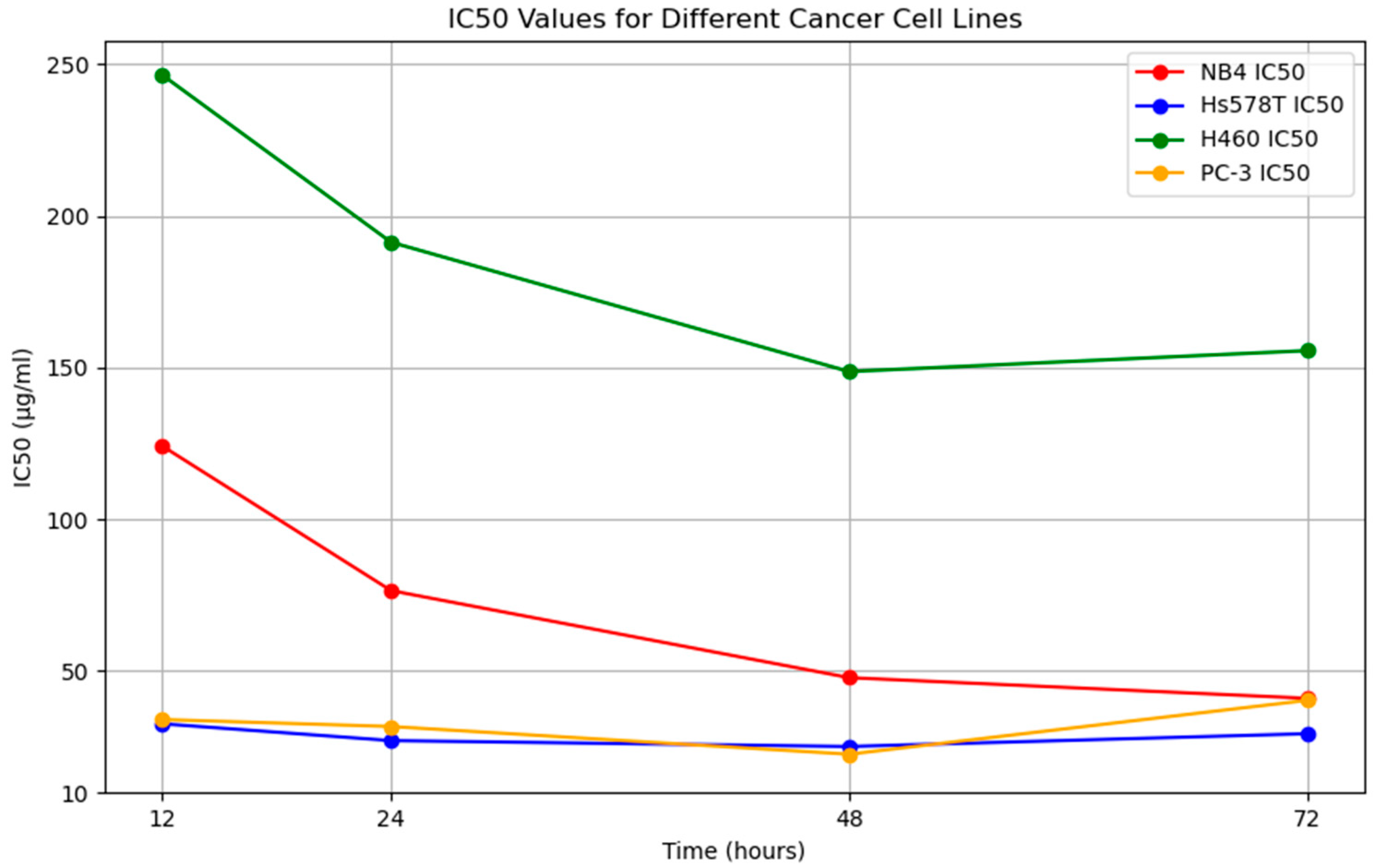
3.3. Anticancer Activity

This study evaluated the potential anticancer effects of U. dioica extract on various cancer cell lines at various time points. The IC50 values, which indicate the concentration of the extract required to impede cell growth by 50%, were determined at various time points. The results showed significant fluctuations across time points and cell lines. The U. dioica extract significantly influenced the NB4 and H460 cell lines, with the highest IC50 values recorded at 48 h for NB4 and 41.00 µg/mL for H460 (Table 2 and Figure 3). The extract not only hindered cancer cell proliferation but also sustained the viability of normal cells to a notable degree. These findings highlight the potential of U. dioica ethanolic leaf extracts as promising contenders for cancer therapy, especially in the NB4 and H460 cell lines (Figure 4).
This study examined the morphological changes in various cancer cell lines, both in the control cells without treatment and after treatment with U. dioica. The results, depicted in Figure 3, clearly demonstrate the morphological alterations observed in all cancer cell types, indicating the impact on the targeted cells. These morphological changes were found to be dependent on the concentration of the U. dioica extracts. The viability of the cells treated with U. dioica for different exposure times was reduced compared to the control cells, which exhibited a normal morphology. Specifically, leukemia cancer cells (NB4) incubated with U. dioica for 48 h experienced a significant decrease in viability, with a 64.60% reduction compared to the control cells’ viability of 98.44%. Moreover, cell adhesion capacity and viability were decreased by 33.84% compared to the control cells (Table 2). Overall, most cells exposed to U. dioica exhibited atypical morphology and appeared smaller in size. The Urtica dioica extracts demonstrated promising IC50 values, indicating their potential to inhibit the proliferation of these specific cancer cell lines (Table 2, Figure 4).

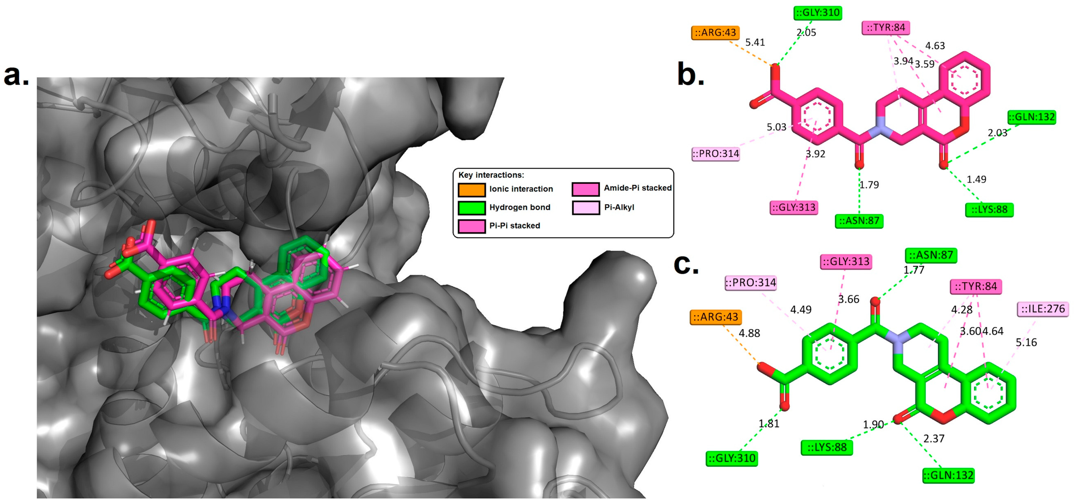
3.4. Molecular Docking Analysis

In this study, molecular docking simulations were conducted for the compounds identified through GC and HPLC analysis, along with the co-crystallized ligand (DS44960156), against the MTHFD2 enzyme. The results are detailed in Table 3 and Figure 5 and Figure 6.

3.5. Drug-Likeness and ADMET Properties

Table 4 presents the predicted drug-likeness and ADMET properties of catechin, kaempferol, and quercetin-3-O-rutinoside, as determined by ADMETlab 2.0.

4. Discussion

4.1. GC–MS and HPLC Analyses of the Extract

An ethanolic extract made from U. dioica roots was shown to have cytotoxic effects on human colon (HT29) and stomach (MKN45) cancer cells in an additional investigation by Ghasemi et al. (2016). The results support the hypothesis put forth by numerous epidemiological studies that consuming urine may help to lower cancer-related fatalities [20]. According to the GC–MS results (Table 1), methyl hexadecanoate, the main component, was crucial to the antitumor effects of the U. dioica extract. The impact of U. dioica leaf extract on the cytotoxic activity of cancerous cell lines was investigated. It was found to significantly induce apoptosis in HepG-2 cells as well as in the cancer cell line MCF-7. In line with an earlier investigation by Ahmed et al. (2020), U. dioica extract significantly increased the rate of apoptosis in a bladder cancer cell line [70].
Methyl hexadecanoate was examined as a potential breast cancer treatment by Karakol et al. (2020) using the same plant species [11]. Comparatively, MCF-7 human mammary adenocarcinoma cells are directly inhibited from becoming carcinogenic by cis-vaccenic acid, according to Lim et al. (2014) [71,72]. Similarly, oleyl alcohol has been demonstrated to have an anti-inflammatory effect as well as an inhibitory effect on cell lysis in human leukemia HL-60 cells. Silva et al. (2022) described the effects of geranial, which has been shown to have a variety of pharmacological impacts, including antimicrobial, antioxidant, anti-inflammatory, and anticancer effects [73].
According to the HPLC results (Figure 2), the U. dioica leaf alcoholic extract contains a high concentration of natural flavonoid compounds such as kaempferol, quercetin-3-O-rutinoside, and catechin. These compounds may have medical advantages, such as anticancer, anti-inflammatory, and antioxidant properties. Nevertheless, additional investigations are necessary to fully elucidate the mechanisms of action of these chemicals as well as their potential uses in medicine [74,75,76]. Additionally, the results demonstrate that the extract contains significant amounts of compounds, including gallic acid, quinic acid, ferulic acid, and caffeine acid (Figure 2). These polyphenols may all have anti-inflammatory, antioxidant, and cancer-preventing properties, according to prior research [64,65,66,67,68].
A histological examination of cancer cells treated with U. dioica leaf extract revealed a substantial decrease in the viability (61.80%) of acute promyelocytic leukemia (NB4) cells after 72 h compared to that of the control cells (98.44%), as indicated by Table 2 and Figure 3, along with Figure 4. The IC50 values showed that the extract effectively reduced the growth of acute promyelocytic leukemia cells at the concentrations tested (5–305 g/mL) during this incubation period, with an IC50 value of approximately 41 ± 0.05 µg/mL, indicating that the extract significantly decreased cell development. Notably, a mild reduction in viability was observed after 48 h of treatment with the plant extract compared to that of the untreated cells (IC50 = 47.80 ± 0.05 µg/mL). However, only minor-to-moderate changes were noted in viability after treatment for 12 and 24 h.
Moreover, based on the results depicted in Table 2 and Figure 3 as well as Figure 4, the results of administering U. dioica leaf extract to breast cancer cells (Hs578T) indicated a highly significant decrease in cell viability (65.88%) after 48 h of treatment compared to that of the control cells (98.00%). The IC50 value was determined to be 25.11 ± 0.05 µg/mL. However, after 24 h, the percentage of cells treated with the plant extract decreased somewhat significantly (76.90%, IC50 = 27.06 ± 0.05 µg/mL) compared to that of the control group. Furthermore, mild-to-moderately notable changes in viability were also observed after 12 and 72 h post-treatment.
The results revealed that the viability of non-small cell lung cancer (H460) cells was significantly reduced after a 48 h treatment with U. dioica leaf extract. Compared to that of the untreated cells at 98% viability, the IC50 of U. dioica extract against H460 cells was determined to be 148.70 ± 0.05 µg/mL. Moreover, a mild but significant decrease in lung cell viability (78.51%, IC50 = 191.33 ± 0.05 µg/mL) was observed after a 24 h treatment with the plant extract compared to that of the control group. These trends were further supported by the moderate changes in cell viability observed after treatments lasting for both 12 and 72 h.
After 48 h of treatment, the viability of prostate cancer cells treated with an extract from U. dioica was highly significantly lower (68.90%) than that of the control cells (98.55%). The IC50 value for the U. dioica extracts against PC-3 cells was 22.57 ± 0.05 µg/mL, indicating that U. dioica is effective as an anticancer agent in this model system. In addition, after 24 h, there was a mildly significant decrease in cell viability compared to that of the control group (76.50%, IC50 = 22.57 ± 0/05 µg/mL), and similar mild-to-moderate effects were observed at later time points, such as 72 and 12 h of treatment, as shown in Figure 3 and Figure 4, respectively.
The ethanolic leaf extract of U. dioica has been found to have potential antitumor effects, as it significantly reduced cell viability and decreased IC50 values. This finding suggested that U. dioica could be beneficial for treating various cancer cell lines, including leukemia, breast cancer, non-small cell lung cancer, and prostate cancer cell lines. Further investigation into the molecular mechanisms underlying these effects is needed, particularly using molecular docking techniques to probe interactions between the compounds and the MTHFD2 enzyme. The MTHFD2 enzyme is crucial in cancer cell metabolism and could provide insights into the underlying mechanisms of these anticancer effects, allowing for a more comprehensive understanding of its therapeutic potential [22,23,24,25,26,27].

4.2. Molecular Docking

Molecular docking is a computational technique used to study the interaction between a ligand and its protein target [77,78]. It helps predict the binding mode and affinity of small molecules for specific protein receptors, which is crucial in drug discovery [77,78]. AutoDock 4.2 [79], a widely used docking software tool, has been used to identify potential hit compounds. In this study, compounds from GC and HPLC analyses were analyzed using AutoDock 4.2 with the MTHFD2 enzyme. The results showed that the docking method successfully positioned the control ligand in a conformation closely resembling its original conformation, with a root mean square deviation (RMSD) value of ~0.84 Å and a binding affinity of approximately −9.89 kcal/mol. The obtained RMSD was within an acceptable range, under 2 Å, as noted in refs. [47,48,49,50,51,54,80,81,82]. Such validation lends credibility to the docking parameters used for predicting the binding orientations of the identified compounds [47,48,49,50,51,54,80,81,82].
Remarkably, three of the identified compounds demonstrated binding affinities remarkably close to those of the control ligand, which had a binding free energy of −9.89 kcal/mol. These three compounds, namely, catechin, kaempferol, and quercetin-3-O-rutinoside, exhibited binding free energies of −9.31 kcal/mol, −9.44 kcal/mol, and −9.82 kcal/mol, respectively. This indicates that these compounds established notably robust interactions with the active site of the MTHFD2 enzyme. These findings underscore the potential of catechin, kaempferol, and quercetin-3-O-rutinoside as promising candidates for further investigation in the context of their anticancer properties. These compounds have a substantial affinity for MTHFD2, which is a significant finding in the pursuit of anticancer drug development.
To further elucidate the molecular interactions between these compounds and the MTHFD2 enzyme, we conducted a comprehensive analysis, as demonstrated in Figure 6. This figure provides a visual representation of the 3D and 2D molecular interactions within the active binding site of the MTHFD2 enzyme. Conducting an in-depth analysis of the specific amino acid residues involved, hydrogen-bonding patterns, and the stability of the binding complexes may provide valuable insights into their potential as inhibitor agents. Molecular docking analysis revealed intricate interactions between the identified compounds, catechin, kaempferol, quercetin-3-O-rutinoside, and the MTHFD2 enzyme. In this investigation, we performed a comprehensive analysis of the specific amino acid residues involved and the nature of these interactions, which provides critical insights into their potential as inhibitors of MTHFD2.
Catechin (−9.31 kcal/mol) displayed remarkable interactions with several key residues within the enzyme’s active site. Notably, it established hydrogen-bond interactions with SER83 (2.18 Å), SER83 (2.43 Å), ASN87 (1.64 Å), GLY310 (1.87 Å), and GLY311 (2.07 Å). No ionic or pi-sigma interactions were detected, but a pi-cation interaction was observed with ARG43. Furthermore, hydrophobic interactions with TYR84, LEU289, and PRO314 suggest a stable binding orientation. Kaempferol (−9.44 kcal/mol) exhibited a binding affinity close to that of the control, indicating its strong potential as an inhibitor. This compound formed hydrogen bonds with ARG43 (2.45 Å), ARG43 (3.06 Å), TYR84 (2.47 Å), ASN87 (1.81 Å), LYS88 (2.96 Å), and THR176 (1.92 Å). Notably, no ionic or pi-cation interactions were detected, and it displayed a pi-sigma interaction with THR316. Hydrophobic interactions were evident with ARG43, GLY313, PRO314, and VAL317, further supporting its binding stability. Quercetin-3-O-rutinoside (−9.82 kcal/mol) displayed a strong binding affinity, indicating that it may be a potential MTHFD2 inhibitor. It established hydrogen-bond interactions with THR176 (2.06 Å), VAL274 (1.88 Å), VAL274 (1.93 Å), and GLY310 (1.78 Å). Although no ionic interactions were detected, they exhibited pi-cation and pi-sigma interactions with ARG43 and TYR84, respectively. Hydrophobic interactions with ALA80, TYR84, VAL205, LEU289, and PRO309 contribute to its robust binding. The interactions observed suggest that quercetin-3-O-rutinoside is a promising candidate for MTHFD2 inhibition. Comparatively, the co-crystallized ligand (DS44960156) (−9.89 kcal/mol) interacted with ASN87 (1.79 Å), LYS88 (1.49 Å), GLN132 (2.03 Å), and GLY310 (2.05 Å) through hydrogen bonding. Additionally, it exhibited ionic interactions with ARG43. The absence of pi-cation or pi-sigma interactions differentiates it from the test compounds. Hydrophobic interactions with TYR84, GLY313, and PRO314 contributed to stable binding.
Catechin, kaempferol, and quercetin-3-O-rutinoside are polyphenolic antioxidants that have been reported to have chemotherapeutic potential in various forms of cancer [83]. For example, quercetin, in combination with catechin, inhibited breast cancer cell proliferation and cell cycle progression [84]. A high intake of quercetin and kaempferol has also been associated with a reduced risk of stomach cancer [85]. Research has shown that kaempferol can overcome resistance to the chemical drug 5-fluorouracil in human colon cancer cells [86]. Quercetin has been shown to induce cell cycle arrest and apoptosis in human cervical cancer cells and to suppress the mobility of breast cancer cells by suppressing glycolysis [87]. Catechin can inhibit the growth of human breast cancer cells by inducing apoptosis and can inhibit the proliferation of human colon cancer cells by inducing cell cycle arrest [88]. These studies suggest that catechin, kaempferol, and quercetin-3-O-rutinoside have potential as chemotherapeutic agents for various forms of cancer. However, more research is needed to fully understand their mechanisms of action and to determine their efficacy and safety in clinical trials.
This study suggested that catechin, kaempferol, and quercetin-3-O-rutinoside may have significant potential as inhibitors of the MTHFD2 enzyme, with binding energies close to those of the control. These compounds can modulate MTHFD2 function through hydrogen bonding, pi-sigma or pi-cation interactions, and hydrophobic contacts with residues. Further investigation into their ADMET profiles is needed to determine their suitability for drug development.

4.3. Drug-Likeness and ADMET Properties

The assessment of drug-likeness and ADMET (absorption–distribution–metabolism–excretion–toxicity) properties is crucial in the selection process of potential drugs during discovery and development [89]. In addition to efficacy against the therapeutic target, a promising drug candidate must exhibit favorable ADMET characteristics at an appropriate dosage [90]. This research focuses on evaluating the drug-likeness and ADMET properties of three chemical entities: catechin, kaempferol, and quercetin-3-O-rutinoside. To obtain valuable insights into their potential for drug development, we utilize the reliable computational tool ADMETlab 2.0, known for its accurate predictions of key pharmacokinetic parameters [58]. The systematic assessment aims to provide a comprehensive understanding of these compounds’ suitability for further in silico exploration.
Table 4 shows the predicted drug-likeness and ADMET properties for catechin, kaempferol, and quercetin-3-O-rutinoside using ADMETlab 2.0. The results revealed valuable insights into the pharmacokinetic and safety profiles of these compounds. Notably, catechin and kaempferol demonstrated a more favorable pharmacokinetic profile, as evidenced by their adherence to the Lipinski Rule, Pfizer Rule, Golden Triangle, and GSK Rule, indicating their potential for better ADMET outcomes. The analysis also shed light on their absorption capabilities, where they displayed higher Papp (Caco-2 permeability) values and MDCK permeability, along with HIA (Human Intestinal Absorption) values that suggested improved bioavailability. In contrast, quercetin-3-O-rutinoside, while still within acceptable parameters, showed variations in some properties, potentially indicating a somewhat less favorable ADMET profile. In addition, the analysis delved into their distribution within the body, plasma protein binding (PPB), and their ability to cross the blood–brain barrier (BBB), with all demonstrating strong PPB percentages and an inability to penetrate the BBB, which can be seen as a safety advantage. Furthermore, the metabolism assessment indicated that both compounds are neither substrates nor inhibitors of various cytochrome P450 (CYP) enzymes, suggesting a lower likelihood of drug interactions. The excretion parameters, including clearance rate (CL) and half-life (T ½), also favored catechin and kaempferol, signifying efficient clearance and manageable half-life values. In terms of toxicity, all three compounds exhibited a lack of human hepatotoxicity (H-HT), which is a favorable sign. Quercetin-3-O-rutinoside, however, showed a potential mutagenic effect in the Ames test. The favorable pharmacokinetic and safety profiles of catechin and kaempferol present promising opportunities for inhibiting the MTHFD2 enzyme. Subsequent in vitro and in vivo investigations are necessary to validate these findings and explore the specific mechanisms by which they interact with MTHFD2. This research paves the way for a deeper understanding of the therapeutic potential of catechin and kaempferol in relation to MTHFD2 inhibition, as well as their contributions to innovative cancer treatment approaches.

5. Conclusions

This study involved a thorough examination of the chemical composition of U. dioica leaf samples using the GC–MS and HPLC techniques. The results from the GC–MS analysis identified nineteen phytochemicals, with six major compounds being prominently detected. Additionally, the HPLC analysis revealed three significant flavonoids and four significant polyphenols in the crude extract. Following this comprehensive chemical profiling, attention turned towards evaluating the effects of the extract on cancer cells. Four different human tumor cell lines were subjected to U. dioica extract, and the outcomes were quite remarkable. The extract significantly inhibited the rate of cellular proliferation, accompanied by notable alterations in the visual appearance of cancer cells. Across various types of cancer cell lines, there was a substantial decrease in cell viability upon treatment with the extract, which was particularly evident after 48 h; however, more subtle-to-moderate changes were observed at 24 and 72 h. These results underscore the potential therapeutic applications of U. dioica extract as a natural remedy for multiple forms of cancer, including breast cancer. Furthermore, our research went beyond this stage to investigate the molecular mechanisms that underlie these anticancer effects. We conducted molecular docking studies on all the identified compounds against the MTHFD2 enzyme, which is known to be abundantly expressed in various cancer cell lines, including those utilized in our study. The calculations of free binding energy and the examination of molecular interactions and binding modes revealed that catechin, kaempferol, and quercetin-3-O-rutinoside may serve as potential inhibitors, similar to the co-crystallized ligand used as a control in this study. Furthermore, we gained valuable insights from our predictions regarding ADMET (absorption–distribution–metabolism–excretion–toxicity) properties and drug-likeness. Importantly, both catechin and kaempferol have emerged as promising candidates with favorable pharmacokinetic profiles, indicating their potential for oral administration. The diverse capabilities of U. dioica and its component compounds in cancer treatment are highlighted by these combined discoveries. Although our study provides a solid starting point for future investigations, it also highlights the importance of conducting additional laboratory experiments and animal studies to confirm the effectiveness and therapeutic potential of these encouraging compounds, particularly catechin and kaempferol, when considering MTHFD2 inhibition and cancer therapy.

Funding

This research received no external funding.

Data Availability Statement

Data are contained within the article.

Acknowledgments

The author wishes to express her gratitude to the Deanship of Scientific Research at the University of Hafr Al Batin in Saudi Arabia for their unending support of this research.

Conflicts of Interest

The author declares no conflicts of interest.

References

  1. Seyfried, T.N.; Shelton, L.M. Cancer as a metabolic disease. Nutr. Metab. 2010, 7, 7. [Google Scholar] [CrossRef]
  2. Seyfried, T.N.; Flores, R.E.; Poff, A.M.; D’Agostino, D.P. Cancer as a metabolic disease: Implications for novel therapeutics. Carcinogenesis 2014, 35, 515–527. [Google Scholar] [CrossRef]
  3. Abotaleb, M.; Liskova, A.; Kubatka, P.; Büsselberg, D. Therapeutic potential of plant phenolic acids in the treatment of cancer. Biomolecules 2020, 10, 221. [Google Scholar] [CrossRef]
  4. Hanahan, D.; Weinberg, R.A. Hallmarks of cancer: The next generation. Cell 2011, 144, 646–674. [Google Scholar] [CrossRef]
  5. Weis, S.M.; Cheresh, D.A. Tumor angiogenesis: Molecular pathways and therapeutic targets. Nat. Med. 2011, 17, 1359–1370. [Google Scholar] [CrossRef]
  6. Tamburrino, A.; Piro, G.; Carbone, C.; Tortora, G.; Melisi, D. Mechanisms of resistance to chemotherapeutic and anti-angiogenic drugs as novel targets for pancreatic cancer therapy. Front. Pharmacol. 2013, 4, 56. [Google Scholar] [CrossRef]
  7. Du, G.-H. Natural Small Molecule Drugs from Plants; Springer: Singapore, 2018. [Google Scholar]
  8. Wangchuk, P. Therapeutic applications of natural products in herbal medicines, biodiscovery programs, and biomedicine. J. Biol. Act. Prod. Nat. 2018, 8, 1–20. [Google Scholar] [CrossRef]
  9. Cragg, G.M.; Newman, D.J. Discovery and development of antineoplastic agents from natural sources. Cancer Investig. 1999, 17, 153–163. [Google Scholar] [CrossRef]
  10. Devkota, H.P.; Paudel, K.R.; Khanal, S.; Baral, A.; Panth, N.; Adhikari-Devkota, A.; Jha, N.K.; Das, N.; Singh, S.K.; Chellappan, D.K. Stinging nettle (Urtica dioica L.): Nutritional composition, bioactive compounds, and food functional properties. Molecules 2022, 27, 5219. [Google Scholar] [CrossRef]
  11. Karakol, P.; Saraydin, S.U.; Bozkurt, M.; Hepokur, C.; Inan, Z.D.S.; Turan, M. Anticancer Effects of Urtica Dioica in Breast Cancer. Asian Pac. J. Cancer Prev. 2022, 23, 673–681. [Google Scholar] [CrossRef]
  12. Mohammadi, A.; Mansoori, B.; Aghapour, M.; Baradaran, B. Urtica dioica dichloromethane extract induce apoptosis from intrinsic pathway on human prostate cancer cells (PC3). Cell. Mol. Biol. 2016, 62, 78–83. [Google Scholar]
  13. Esposito, S.; Bianco, A.; Russo, R.; Di Maro, A.; Isernia, C.; Pedone, P.V. Therapeutic Perspectives of Molecules from Urtica dioica Extracts for Cancer Treatment. Molecules 2019, 24, 2753. [Google Scholar] [CrossRef]
  14. Mohammadi, A.; Mansoori, B.; Baradaran, P.C.; Khaze, V.; Aghapour, M.; Farhadi, M.; Baradaran, B. Urtica dioica extract inhibits proliferation and induces apoptosis and related gene expression of breast cancer cells in vitro and in vivo. Clin. Breast Cancer 2017, 17, 463–470. [Google Scholar] [CrossRef]
  15. Semwal, P.; Rauf, A.; Olatunde, A.; Singh, P.; Zaky, M.Y.; Islam, M.M.; Khalil, A.A.; Aljohani, A.S.; Al Abdulmonem, W.; Ribaudo, G. The medicinal chemistry of Urtica dioica L.: From preliminary evidence to clinical studies supporting its neuroprotective activity. Nat. Prod. Bioprospecting 2023, 13, 16. [Google Scholar] [CrossRef]
  16. Fattahi, S.; Ghadami, E.; Asouri, M.; Ardekanid, A.M.; Akhavan-Niaki, H. Urtica dioica inhibits cell growth and induces apoptosis by targeting Ornithine decarboxylase and Adenosine deaminase as key regulatory enzymes in adenosine and polyamines homeostasis in human breast cancer cell lines. Cell. Mol. Biol. 2018, 64, 97–102. [Google Scholar] [CrossRef]
  17. Rashidbaghan, A.; Mostafaie, A.; Yazdani, Y.; Mansouri, K. Urtica dioica agglutinin (a plant lectin) has a caspase-dependent apoptosis induction effect on the acute lymphoblastic leukemia cell line. Cell. Mol. Biol. 2020, 66, 121–126. [Google Scholar] [CrossRef]
  18. D’Abrosca, B.; Ciaramella, V.; Graziani, V.; Papaccio, F.; Della Corte, C.M.; Potenza, N.; Fiorentino, A.; Ciardiello, F.; Morgillo, F. Urtica dioica L. inhibits proliferation and enhances cisplatin cytotoxicity in NSCLC cells via Endoplasmic Reticulum-stress mediated apoptosis. Sci. Rep. 2019, 9, 4986. [Google Scholar] [CrossRef]
  19. Wagner, H.; Geiger, W.; Boos, G.; Samtleben, R. Studies on the binding of Urtica dioica agglutinin (UDA) and other lectins in an in vitro epidermal growth factor receptor test. Phytomedicine 1995, 1, 287–290. [Google Scholar] [CrossRef]
  20. Ghasemi, S.; Moradzadeh, M.; Mousavi, S.; Sadeghnia, H.R. Cytotoxic effects of Urtica dioica radix on human colon (HT29) and gastric (MKN45) cancer cells mediated through oxidative and apoptotic mechanisms. Cell. Mol. Biol. 2016, 62, 90–96. [Google Scholar]
  21. Uyar, A.; Doğan, A.; Yaman, T.; Keleş, Ö.F.; Yener, Z.; Çelik, İ.; Alkan, E.E. The protective role of Urtica dioica seed extract against azoxymethane-induced colon carcinogenesis in rats. Nutr. Cancer 2022, 74, 306–319. [Google Scholar] [CrossRef]
  22. Gao, Y.; Feng, L.; Zhang, L.; Geng, J.; Zhang, E. ATF4/MYC Regulates MTHFD2 to Promote NSCLC Progression by Mediating Redox Homeostasis. Dis. Markers 2022, 2022, 7527996. [Google Scholar] [CrossRef]
  23. Zhu, Z.; Leung, G.K.K. More than a metabolic enzyme: MTHFD2 as a novel target for anticancer therapy? Front. Oncol. 2020, 10, 658. [Google Scholar] [CrossRef]
  24. Zhou, F.; Yuan, Z.; Gong, Y.; Li, L.; Wang, Y.; Wang, X.; Ma, C.; Yang, L.; Liu, Z.; Wang, L. Pharmacological targeting of MTHFD2 suppresses NSCLC via the regulation of ILK signaling pathway. Biomed. Pharmacother. 2023, 161, 114412. [Google Scholar] [CrossRef]
  25. Huang, J.; Qin, Y.; Lin, C.; Huang, X.; Zhang, F. MTHFD2 facilitates breast cancer cell proliferation via the AKT signaling pathway. Exp. Ther. Med. 2021, 22, 703. [Google Scholar] [CrossRef]
  26. Zhao, R.; Feng, T.; Gao, L.; Sun, F.; Zhou, Q.; Wang, X.; Liu, J.; Zhang, W.; Wang, M.; Xiong, X. PPFIA4 promotes castration-resistant prostate cancer by enhancing mitochondrial metabolism through MTHFD2. J. Exp. Clin. Cancer Res. 2022, 41, 125. [Google Scholar] [CrossRef]
  27. Pikman, Y.; Puissant, A.; Alexe, G.; Furman, A.; Chen, L.M.; Frumm, S.M.; Ross, L.; Fenouille, N.; Bassil, C.F.; Lewis, C.A. Targeting MTHFD2 in acute myeloid leukemia. J. Exp. Med. 2016, 213, 1285–1306. [Google Scholar] [CrossRef]
  28. Hertweck, K.L.; Vikramdeo, K.S.; Galeas, J.N.; Marbut, S.M.; Pramanik, P.; Yunus, F.; Singh, S.; Singh, A.P.; Dasgupta, S. Clinicopathological significance of unraveling mitochondrial pathway alterations in non-small-cell lung cancer. FASEB J. 2023, 37, e23018. [Google Scholar] [CrossRef]
  29. Wei, Y.; Liu, P.; Li, Q.; Du, J.; Chen, Y.; Wang, Y.; Shi, H.; Wang, Y.; Zhang, H.; Xue, W. The effect of MTHFD2 on the proliferation and migration of colorectal cancer cell lines. OncoTargets Ther. 2019, 12, 6361–6370. [Google Scholar] [CrossRef]
  30. Gustafsson Sheppard, N.; Jarl, L.; Mahadessian, D.; Strittmatter, L.; Schmidt, A.; Madhusudan, N.; Tegnér, J.; Lundberg, E.K.; Asplund, A.; Jain, M. The folate-coupled enzyme MTHFD2 is a nuclear protein and promotes cell proliferation. Sci. Rep. 2015, 5, 15029. [Google Scholar]
  31. Li, G.; Wu, J.; Li, L.; Jiang, P. p53 deficiency induces MTHFD2 transcription to promote cell proliferation and restrain DNA damage. Proc. Natl. Acad. Sci. USA 2021, 118, e2019822118. [Google Scholar] [CrossRef]
  32. Bonagas, N.; Gustafsson, N.M.; Henriksson, M.; Marttila, P.; Gustafsson, R.; Wiita, E.; Borhade, S.; Green, A.C.; Vallin, K.S.; Sarno, A. Pharmacological targeting of MTHFD2 suppresses acute myeloid leukemia by inducing thymidine depletion and replication stress. Nat. Cancer 2022, 3, 156–172. [Google Scholar] [CrossRef]
  33. Yang, C.; Zhang, J.; Liao, M.; Yang, Y.; Wang, Y.; Yuan, Y.; Ouyang, L. Folate-mediated one-carbon metabolism: A targeting strategy in cancer therapy. Drug Discov. Today 2021, 26, 817–825. [Google Scholar] [CrossRef]
  34. Zhao, L.N.; Björklund, M.; Caldez, M.J.; Zheng, J.; Kaldis, P. Therapeutic targeting of the mitochondrial one-carbon pathway: Perspectives, pitfalls, and potential. Oncogene 2021, 40, 2339–2354. [Google Scholar] [CrossRef]
  35. Albanesi, J.; Noguera, N.I.; Banella, C.; Colangelo, T.; De Marinis, E.; Leone, S.; Palumbo, O.; Voso, M.T.; Ascenzi, P.; Nervi, C. Transcriptional and metabolic dissection of ATRA-induced granulocytic differentiation in NB4 acute promyelocytic leukemia cells. Cells 2020, 9, 2423. [Google Scholar] [CrossRef]
  36. Ugur, D.; Gungul, T.B.; Yucel, S.; Ozcivici, E.; Yalcin-Ozuysal, O.; Mese, G. Connexin 32 overexpression increases proliferation, reduces gap junctional intercellular communication, motility and epithelial-to-mesenchymal transition in Hs578T breast cancer cells. J. Cell Commun. Signal. 2022, 16, 361–376. [Google Scholar] [CrossRef]
  37. Lei, Z.-N.; Teng, Q.-X.; Zhang, W.; Fan, Y.-F.; Wang, J.-Q.; Cai, C.-Y.; Lu, K.W.; Yang, D.-H.; Wurpel, J.N.; Chen, Z.-S. Establishment and characterization of a topotecan resistant non-small cell lung cancer NCI-H460/TPT10 cell line. Front. Cell Dev. Biol. 2020, 8, 607275. [Google Scholar] [CrossRef]
  38. Rapuano, R.; Riccio, A.; Mercuri, A.; Madera, J.R.; Dallavalle, S.; Moricca, S.; Lupo, A. Proliferation and migration of PC-3 prostate cancer cells is counteracted by PPARγ-cladosporol binding-mediated apoptosis and a decreased lipid biosynthesis and accumulation. Biochem. Pharmacol. 2024, 222, 116097. [Google Scholar] [CrossRef]
  39. Sodde, V.K.; Lobo, R.; Kumar, N.; Maheshwari, R.; Shreedhara, C.S. Cytotoxic activity of Macrosolen parasiticus (L.) Danser on the growth of breast cancer cell line (MCF-7). Pharmacogn. Mag. 2015, 11, S156–S160. [Google Scholar] [CrossRef]
  40. Savić, M.; Nikolic, D.V.; Savic-Gajic, M.I.; Kundakovic, D.T.; Stanojkovic, P.T.; Najman, J.S. Chemical composition and biological activity of the plum seed extract. Adv. Technol. 2016, 5, 38–45. [Google Scholar] [CrossRef]
  41. Popolin, C.P.; Reis, J.P.; Becceneri, A.B.; Graminha, A.E.; Almeida, M.A.; Correa, R.S.; Colina-Vegas, L.A.; Ellena, J.; Batista, A.A.; Cominetti, M.R. Cytotoxicity and anti-tumor effects of new ruthenium complexes on triple negative breast cancer cells. PLoS ONE 2017, 12, e0183275. [Google Scholar] [CrossRef]
  42. Zhang, R.; Wei, M.; Chen, S.; Li, G.; Zhang, F.; Yang, N.; Huang, L. A cell viability assessment method based on area-normalized impedance spectrum (ANIS). Biosens. Bioelectron. 2018, 110, 193–200. [Google Scholar] [CrossRef]
  43. Gudipaty, S.A.; Conner, C.M.; Rosenblatt, J.; Montell, D.J. Unconventional ways to live and die: Cell death and survival in development, homeostasis, and disease. Annu. Rev. Cell Dev. Biol. 2018, 34, 311–332. [Google Scholar] [CrossRef]
  44. Kawai, J.; Ota, M.; Ohki, H.; Toki, T.; Suzuki, M.; Shimada, T.; Matsui, S.; Inoue, H.; Sugihara, C.; Matsuhashi, N. Structure-based design and synthesis of an isozyme-selective MTHFD2 inhibitor with a tricyclic coumarin scaffold. ACS Med. Chem. Lett. 2019, 10, 893–898. [Google Scholar] [CrossRef]
  45. Westbrook, J.; Feng, Z.; Chen, L.; Yang, H.; Berman, H.M. The protein data bank and structural genomics. Nucleic Acids Res. 2003, 31, 489–491. [Google Scholar] [CrossRef]
  46. Dassault Systèmes (BIOVIA). Discovery Studio Visualizer; Release 2019; Dassault Systèmes: San Diego, CA, USA, 2020. [Google Scholar]
  47. Alhawarri, M.B.; Al-Thiabat, M.G.; Dubey, A.; Tufail, A.; Fouad, D.; Alrimawi, B.H.; Dayoob, M. ADME profiling, molecular docking, DFT, and MEP analysis reveal cissamaline, cissamanine, and cissamdine from Cissampelos capensis Lf as potential anti-Alzheimer’s agents. RSC Adv. 2024, 14, 9878–9891. [Google Scholar] [CrossRef]
  48. Alhawarri, M.B.; Olimat, S. Potential Serotonin 5-HT2A Receptor Agonist of Psychoactive Components of Silene undulata Aiton: LC-MS/MS, ADMET, and Molecular Docking Studies. Curr. Pharm. Biotechnol. 2024, 25, 1–15. [Google Scholar] [CrossRef]
  49. Yunos, N.M.; Al-Thiabat, M.G.; Sallehudin, N.J. Quassinoids from Eurycoma longifolia as Potential Dihydrofolate Reductase Inhibitors: A Computational Study. Curr. Pharm. Biotechnol. 2024, 25, 1–12. [Google Scholar] [CrossRef]
  50. Alhawarri, M.; Dianita, R.; Rawa, M.; Nogawa, T.; Wahab, H. Potential Anti-Cholinesterase Activity of Bioactive Compounds Extracted from Cassia grandis Lf and Cassia timoriensis DC. Plants 2023, 12, 344. [Google Scholar] [CrossRef]
  51. Amir Rawa, M.S.; Al-Thiabat, M.G.; Nogawa, T.; Futamura, Y.; Okano, A.; Wahab, H.A. Naturally Occurring 8ß, 13ß-kaur-15-en-17-al and Anti-Malarial Activity from Podocarpus polystachyus Leaves. Pharmaceuticals 2022, 15, 902. [Google Scholar] [CrossRef]
  52. Land, H.; Humble, M.S. YASARA: A tool to obtain structural guidance in biocatalytic investigations. In Protein Engineering: Methods and Protocols; Humana Press: New York, NY, USA, 2018; pp. 43–67. [Google Scholar]
  53. Shalayel, M.H.F.; Al-Mazaideh, G.M.; Alanezi, A.A.; Almuqati, A.F.; Alotaibi, M. Diosgenin and Monohydroxy Spirostanol from Prunus amygdalus var amara Seeds as Potential Suppressors of EGFR and HER2 Tyrosine Kinases: A Computational Approach. Pharmaceuticals 2023, 16, 704. [Google Scholar] [CrossRef]
  54. Abdelbagi, M.E.; Al-Mazaideh, G.M.; Ahmed, A.E.; Al-Rimawi, F.; Ayyal Salman, H.; Almutairi, A.; Abuilaiwi, F.A.; Wedian, F. Exploring Securigera securidaca Seeds as a Source of Potential CDK1 Inhibitors: Identification of Hippeastrine and Naringenin as Promising Hit Candidates. Processes 2023, 11, 1478. [Google Scholar] [CrossRef]
  55. Gordon, J.C.; Myers, J.B.; Folta, T.; Shoja, V.; Heath, L.S.; Onufriev, A. H++: A server for estimating p K as and adding missing hydrogens to macromolecules. Nucleic Acids Res. 2005, 33, W368–W371. [Google Scholar] [CrossRef]
  56. Norgan, A.P.; Coffman, P.K.; Kocher, J.-P.A.; Katzmann, D.J.; Sosa, C.P. Multilevel parallelization of AutoDock 4.2. J. Cheminformatics 2011, 3, 12. [Google Scholar] [CrossRef]
  57. Wang, Y.; Xiao, J.; Suzek, T.O.; Zhang, J.; Wang, J.; Bryant, S.H. PubChem: A public information system for analyzing bioactivities of small molecules. Nucleic Acids Res. 2009, 37, W623–W633. [Google Scholar] [CrossRef]
  58. Xiong, G.; Wu, Z.; Yi, J.; Fu, L.; Yang, Z.; Hsieh, C.; Yin, M.; Zeng, X.; Wu, C.; Lu, A. ADMETlab 2.0: An integrated online platform for accurate and comprehensive predictions of ADMET properties. Nucleic Acids Res. 2021, 49, W5–W14. [Google Scholar] [CrossRef]
  59. Dar, S.A.; Yousuf, A.; Ganai, F.A.; Sharma, P.; Kumar, N.; Singh, R. Bioassay guided isolation and identification of anti-inflammatory and anti-microbial compounds from Urtica dioica L.(Urticaceae) leaves. Afr. J. Biotechnol. 2012, 11, 12910–12920. [Google Scholar]
  60. Asgarpanah, J.; Mohajerani, R. Phytochemistry and pharmacologic properties of Urtica dioica L. J. Med. Plants Res. 2012, 6, 5714–5719. [Google Scholar]
  61. Gülçin, I.; Küfrevioǧlu, Ö.İ.; Oktay, M.; Büyükokuroǧlu, M.E. Antioxidant, antimicrobial, antiulcer and analgesic activities of nettle (Urtica dioica L.). J. Ethnopharmacol. 2004, 90, 205–215. [Google Scholar] [CrossRef]
  62. Joshi, B.C.; Mukhija, M.; Kalia, A.N. Pharmacognostical review of Urtica dioica L. Int. J. Green Pharm. 2014, 8, 201. [Google Scholar]
  63. Krajewska, A.; Mietlińska, K. Determining the parameters of the Stinging Nettle (Urtica dioica L.) hydrolate distillation process. Molecules 2022, 27, 3912. [Google Scholar] [CrossRef]
  64. Al-Tameme, H.J.; Hadi, M.Y.; Hameed, I.H. Phytochemical analysis of Urtica dioica leaves by fourier-transform infrared spectroscopy and gas chromatography-mass spectrometry. J. Pharmacogn. Phytother. 2015, 7, 238–252. [Google Scholar]
  65. Otles, S.; Yalcin, B. Phenolic compounds analysis of root, stalk, and leaves of nettle. Sci. World J. 2012, 2012, 564367. [Google Scholar] [CrossRef]
  66. Masłowski, M.; Aleksieiev, A.; Miedzianowska, J.; Strzelec, K. Common nettle (Urtica dioica L.) as an active filler of natural rubber biocomposites. Materials 2021, 14, 1616. [Google Scholar] [CrossRef]
  67. Silva, J.d.O.; Dias, M.I.; Barros, L.; Tanamati, A.A.; Ferreira, I.C.; Amaral, J.S. Bioactive and nutritional characterization of stinging nettle (Urtica dioica L.) harvested in Portugal. In Proceedings of the XX EuroFoodChem Conference, Porto, Portugal, 17–19 June 2019. [Google Scholar]
  68. Pinelli, P.; Ieri, F.; Vignolini, P.; Bacci, L.; Baronti, S.; Romani, A. Extraction and HPLC analysis of phenolic compounds in leaves, stalks, and textile fibers of Urtica dioica L. J. Agric. Food Chem. 2008, 56, 9127–9132. [Google Scholar] [CrossRef]
  69. Altamimi, M.A.; Abu-Reidah, I.M.; Altamimi, A.; Jaradat, N. Hydroethanolic extract of Urtica dioica L.(stinging nettle) leaves as disaccharidase inhibitor and glucose transport in Caco-2 hinderer. Molecules 2022, 27, 8872. [Google Scholar] [CrossRef]
  70. Levy, A.; Sivanesan, D.; Murugan, R.K.; Jornadal, J.; Quiñónez, Y.B.; Jaffe, M.; Rathinavelu, A. Urtica dioica Induces Cytotoxicity in Human Prostate Carcinoma LNCaP Cells: Involvement of Oxidative Stress, Mitochondrial Depolarization and Apoptosis. Trop. J. Pharm. Res. 2014, 13, 711–717. [Google Scholar] [CrossRef]
  71. Lim, J.N.; Oh, J.J.; Wang, T.; Lee, J.S.; Kim, S.H.; Kim, Y.J.; Lee, H.G. trans-11 18:1 vaccenic acid (TVA) has a direct anti-carcinogenic effect on MCF-7 human mammary adenocarcinoma cells. Nutrients 2014, 6, 627–636. [Google Scholar] [CrossRef]
  72. Mahnashi, M.H.; Alqahtani, Y.S.; Alyami, B.A.; Alqarni, A.O.; Ahmed Alshrahili, M.; Abou-Salim, M.A.; Alqahtani, M.N.; Mushtaq, S.; Sadiq, A.; Jan, M.S. GC-MS Analysis and Various In Vitro and In Vivo Pharmacological Potential of Habenaria plantaginea Lindl. Evid.-Based Complement. Altern. Med. 2022, 2022, 7921408. [Google Scholar] [CrossRef]
  73. Silva, G.; Marques, J.N.J.; Linhares, E.P.M.; Bonora, C.M.; Costa, É.T.; Saraiva, M.F. Review of anticancer activity of monoterpenoids: Geraniol, nerol, geranial and neral. Chem. Biol. Interact. 2022, 362, 109994. [Google Scholar] [CrossRef]
  74. Yao, S.; Wang, X.; Li, C.; Zhao, T.; Jin, H.; Fang, W. Kaempferol inhibits cell proliferation and glycolysis in esophagus squamous cell carcinoma via targeting EGFR signaling pathway. Tumor Biol. 2016, 37, 10247–10256. [Google Scholar] [CrossRef]
  75. Ziaee, A.; Zamansoltani, F.; Nassiri-Asl, M.; Abbasi, E. Effects of rutin on lipid profile in hypercholesterolaemic rats. Basic Clin. Pharmacol. Toxicol. 2009, 104, 253–258. [Google Scholar] [CrossRef]
  76. Khan, N.; Mukhtar, H. Tea polyphenols for health promotion. Life Sci. 2007, 81, 519–533. [Google Scholar] [CrossRef]
  77. Naqvi, A.A.; Mohammad, T.; Hasan, G.M.; Hassan, M.I. Advancements in docking and molecular dynamics simulations towards ligand-receptor interactions and structure-function relationships. Curr. Top. Med. Chem. 2018, 18, 1755–1768. [Google Scholar] [CrossRef]
  78. Ferreira, L.G.; Dos Santos, R.N.; Oliva, G.; Andricopulo, A.D. Molecular docking and structure-based drug design strategies. Molecules 2015, 20, 13384–13421. [Google Scholar] [CrossRef]
  79. Hou, X.; Du, J.; Zhang, J.; Du, L.; Fang, H.; Li, M. How to improve docking accuracy of AutoDock4. 2: A case study using different electrostatic potentials. J. Chem. Inf. Model. 2013, 53, 188–200. [Google Scholar] [CrossRef]
  80. Yunos, N.M.; Wahab, H.A.; Al-Thiabat, M.G.; Sallehudin, N.J.; Jauri, M.H. In Vitro and In Silico Analysis of the Anticancer Effects of Eurycomanone and Eurycomalactone from Eurycoma longifolia. Plants 2023, 12, 2827. [Google Scholar] [CrossRef]
  81. Larue, L.; Kenzhebayeva, B.; Al-Thiabat, M.G.; Jouan–Hureaux, V.; Mohd–Gazzali, A.; Wahab, H.A.; Boura, C.; Yeligbayeva, G.; Nakan, U.; Frochot, C. tLyp–1: A peptide suitable to target NRP–1 receptor. Bioorg. Chem. 2023, 130, 106200. [Google Scholar] [CrossRef]
  82. Alidmat, M.M.; Khairuddean, M.; Kamal, N.N.S.N.M.; Muhammad, M.; Wahab, H.A.; Althiabat, M.G.; Alhawarri, M.B. Synthesis, Characterization, Molecular Docking and Cytotoxicity Evaluation of New Thienyl Chalcone Derivatives against Breast Cancer Cells. Syst. Rev. Pharm. 2022, 13, 1–11. [Google Scholar]
  83. Sharma, A.; Kaur, M.; Katnoria, J.K.; Nagpal, A.K. Polyphenols in food: Cancer prevention and apoptosis induction. Curr. Med. Chem. 2018, 25, 4740–4757. [Google Scholar] [CrossRef]
  84. Imran, M.; Insaf, A.; Hasan, N.; Sugandhi, V.V.; Shrestha, D.; Paudel, K.R.; Jha, S.K.; Hansbro, P.M.; Dua, K.; Devkota, H.P. Exploring the Remarkable Chemotherapeutic Potential of Polyphenolic Antioxidants in Battling Various Forms of Cancer. Molecules 2023, 28, 3475. [Google Scholar] [CrossRef]
  85. Batra, P.; Sharma, A.K. Anti-cancer potential of flavonoids: Recent trends and future perspectives. 3 Biotech 2013, 3, 439–459. [Google Scholar] [CrossRef]
  86. Brglez Mojzer, E.; Knez Hrnčič, M.; Škerget, M.; Knez, Ž.; Bren, U. Polyphenols: Extraction methods, antioxidative action, bioavailability and anticarcinogenic effects. Molecules 2016, 21, 901. [Google Scholar] [CrossRef]
  87. Aghababaei, F.; Hadidi, M. Recent advances in potential health benefits of quercetin. Pharmaceuticals 2023, 16, 1020. [Google Scholar] [CrossRef]
  88. Sun, J.; Liu, R.H. Cranberry phytochemical extracts induce cell cycle arrest and apoptosis in human MCF-7 breast cancer cells. Cancer Lett. 2006, 241, 124–134. [Google Scholar] [CrossRef]
  89. Kar, S.; Roy, K.; Leszczynski, J. In silico tools and software to predict ADMET of new drug candidates. In In Silico Methods for Predicting Drug Toxicity; Humana: New York, NY, USA, 2022; pp. 85–115. [Google Scholar]
  90. Kar, S.; Leszczynski, J. Open access in silico tools to predict the ADMET profiling of drug candidates. Expert Opin. Drug Discov. 2020, 15, 1473–1487. [Google Scholar] [CrossRef]
Figure 1. U. dioica GC–MS chromatogram.
Processes 12 01177 g001
Figure 2. HPLC-chromatogram of U. dioica leaf extract.
Processes 12 01177 g002
Figure 3. Microscopy analysis of a stained cell line (Hematoxylin, Eosin, Giemsa, and Sox-1) using alcoholic leaf extracts of U. dioica treatments. For the Hs578T and H460 cell lines, the scale bar is set at 50 µm, while for the Pc-3 and NB4 cell lines, it is set at 100 µm.
Processes 12 01177 g003
Figure 4. The anticancer effects of U. dioica on four different cancer cell lines over time (in hours), presenting data on IC50 (µg/mL).
Processes 12 01177 g004
Figure 5. (a) Superimposition and (b) 2D interaction analysis of the co-crystallized ligand (DS44960156) (highlighted in pink for carbon (C), red for oxygen (O), and navy blue for nitrogen (N)). (c) Redocked ligand (highlighted in green for carbon (C), red for oxygen (O), and navy blue for nitrogen (N)) within the crystal structure of the human MTHFD2 enzyme (PDB ID: 6JIB).
Processes 12 01177 g005
Figure 6. Three- and two-dimensional interactions of catechin (a,b), kaempferol (c,d), and quercetin-3-O-rutinoside (e,f) within the active binding site of the human MTHFD2 enzyme. These models were generated using the Discovery Studio Visualizer (Biovia, 2020).
Processes 12 01177 g006
Table 1. GC–MS outcomes of U. dioica phytoconstituents.
Peak No.Component NameRT (min)Area %
1Hexadecane4.7080.832
2Geraniol5.2363.101
3Geranyl Acetate6.4830.724
4β-cis-Caryophyllene6.6128.309
5Isoledene7.2374.574
6Hexyl 2-[(4-butylbenzoyl) amino] propanoate7.4835.72
7Isocadinene7.8233.387
8Hexahydrofarnesyl acetone8.0334.089
93,7,11,15-Tetramethyl-2-Hexadecen-1-ol8.2329.043
10Methyl hexadecanoate8.73414.16
11Ethyl hexadecanoate9.1735.144
12Pentadecanoic Acid9.4680.866
13Methyl (Z)-octadec-13-enoate10.720.527
14Phytol12.321.332
15Cis-Vaccenic Acid12.7412.04
16Octadecanal15.474.871
17Oleyl acetate16.38.321
18γ-Tocopherol17.54.651
19β-Sitosteryl stearate20.518.301
Table 2. Anticancer activity of U. dioica ethanolic leaf extracts.
Anticancer ImpactsTime (Hours)Cancer Cell Line
NB4Hs578TH460PC-3
IC50 (µg/mL)12124.2032.60246.5034.00
2476.5027.06191.3331.70
4847.8025.11148.7022.57
7241.0029.33155.6040.33
Cell Viability %1282.0083.5082.0880.20
2470.0776.9078.5176.50
4864.6065.8871.0068.90
7261.8081.0274.3078.90
Cell Viability % Untreated (Control)1298.4498.009898.78
2498.4498.009898.66
4898.4498.009898.55
7298.4498.009898.9
Table 3. Docking scores (*ΔGbind in kcal/mol) of the identified compounds obtained from the GC and HPLC analyses, along with the co-crystallized ligand (DS44960156), within the active binding site of the MTHFD2 enzyme (PDB ID: 6JIB), assessed using AutoDock 4.2.
Identified Compound*ΔGbind (kcal/mol)
3,7,11,15-Tetramethyl-2-hexadecen-1-ol−4.93
β-cis-Caryophyllene−6.84
β-Sitosteryl stearate−6.84
γ-Tocopherol−7.06
Caffeic acid−6.76
Catechin−9.31
Cis-Vaccenic Acid−3.49
Ethyl hexadecanoate−6.06
Ferulic acid−6.55
Gallic acid−6.72
Geraniol−4.93
Geranyl Acetate−6.85
Hexahydrofarnesyl acetone−5.91
Hexyl 2-[(4-butylbenzoyl) amino] propanoate−7.12
Isocadinene−6.42
Isoledene−7.06
Kaempferol−9.44
Methyl (Z)-octadec-13-enoate−4.87
Methyl hexadecanoate−6.11
Octadecanal−2.68
Oleyl acetate−2.53
Pentadecanoic Acid−5.99
Phytol−5.25
Quercetin-3-O-rutinoside−9.82
Quinic acid−6.73
Co-crystalized ligand (control)−9.89
Table 4. Predicted drug-likeness and ADMET properties of catechin, kaempferol, and quercetin-3-O-rutinoside using ADMETlab 2.0.
PropertyModel NamePredicted Value
CatechinKaempferolQuercetin-3-O-rutinoside
Drug-Likeness*Lipinski RuleAcceptedAcceptedRejected
*Pfizer RuleAcceptedAcceptedAccepted
*Golden TriangleAcceptedAcceptedRejected
*GSK RuleAcceptedAcceptedRejected
AbsorptionPapp (Caco-2 Permeability) cm/s−4.65−4.97−6.33
MDCK Permeability (cm/s)5 × 10−69 × 10−630 × 10−6
HIA (Human Intestinal Absorption)0.0350.0040.920
DistributionPPB (Plasma Protein Binding) %89.2397.8683.81
BBB (Blood–Brain Barrier)Cannot crossCannot crossCan penetrate
VD (Volume Distribution) L/kg0.660.520.75
Fu (Fraction unbound in plasms) %12.914.4120.86
MetabolismCYP1A2 substrateNoNoNo
CYP2C19 substrateNoNoNo
CYP2C9 substrateNoNoYes
CYP2D6 substrateNoNoNo
CYP1A2 inhibitorNoNoYes
CYP2C19 inhibitorNoNoYes
CYP2C9 inhibitorNoNoNo
CYP3A4 inhibitorNoNoNo
Excretion*CL (Clearance Rate) mL/min/kg17.306.861.34
T ½ (Half-Lifetime) hr0.890.910.52
ToxicityH-HT (Human Hepatotoxicity)---
AMES (Ames Mutagenicity)--+
Carcinogenicity---
M.W: molecular weight (g/mol), *logP: partition coefficient (lipophilicity), *Hacc: hydrogen-bond acceptor, *Hdon: hydrogen-bond donor, *logD: distribution coefficient. *Lipinski rule: *M. At W ≤ 500, logP ≤ 5, Hacc ≤ 10, and *Hdon ≤ 5; if two properties are out of range, poor absorption or permeability is possible, and one is acceptable. *Pfizer rule: 200 ≤ MW ≤ 50, −2 ≤ logD ≤ 5. *Golden Triangle: 200 ≤ MW ≤ 50, −2 ≤ *logD ≤ 5. *GSK Rule: *M.W ≤ 400, logP ≤ 4. Compounds satisfying the Golden Triangle and GSK rules may have more favorable ADMET profiles. Caco-2 permeability: high: >−5.15 cm/s; low: <−5.15 cm/s. MDCK permeability (cm/s): low: < 2 × 10⁻⁶; medium: 2–20 × 10−6; high: > 20 × 10−6. HIA: high: <0.030; low: >0.030. Optimal PPB: <90%. Drugs with a protein binding lower than 90% may have a high therapeutic index. BBB crossing: ≥0.1; BBB crossing: <0.1. Optimal VD: 0.04–20 L/kg. Fu: low: 5%; moderate: 5~20%; high: >20%. CL: high: >15 mL/min/kg; moderate: CL 5–15 mL/min/kg; low: CL <5 mL/min/kg. T ½: long half-life: > 3 h; short half-life: < 3 h. - Negative values: low affinity to be toxic. Positive values: high affinity for toxicity.
Disclaimer/Publisher’s Note: The statements, opinions and data contained in all publications are solely those of the individual author(s) and contributor(s) and not of MDPI and/or the editor(s). MDPI and/or the editor(s) disclaim responsibility for any injury to people or property resulting from any ideas, methods, instructions or products referred to in the content.

