Article

Exopolysaccharides Synthesized by Lacticaseibacillus rhamnosus ŁOCK 0943: Structural Characteristics and Evaluation of Biological and Technological Properties

by
Magdalena Oleksy-Sobczak
1,
Sabina Górska
2,
Lidia Piekarska-Radzik
1,*,
Sylwia Ścieszka
1,* and
Elżbieta Klewicka
1
1
Institute of Fermentation Technology and Microbiology, Faculty of Biotechnology and Food Science, Lodz University of Technology, Wólczańska 171/173, 90-530 Łódź, Poland
2
Laboratory of Microbiome Immunobiology, Hirszfeld Institute of Immunology and Experimental Therapy, Polish Academy of Sciences, Rudolfa Weigla 12, 53-114 Wrocław, Poland
*
Authors to whom correspondence should be addressed.
Processes 2024, 12(6), 1192; https://doi.org/10.3390/pr12061192
Submission received: 10 May 2024 / Revised: 2 June 2024 / Accepted: 6 June 2024 / Published: 11 June 2024
(This article belongs to the Section Food Process Engineering)

Abstract

:
Lactic acid bacteria can synthesize extracellular exopolysaccharides (EPSs) that have versatile physicochemical and biological properties. In this paper, the EPSs synthesized by Lacticaseibacillus rhamnosus ŁOCK 0943 were characterized. Their structure, biological, and technological activity, as well as application potential, were analyzed. Chemical analysis showed that this strain produces mannan and β-1,6-glucan. Their emulsifying, antagonistic, and antioxidant properties, along with their prebiotic potential, were assessed. The analysis of the tested polymers’ ability to create a stable emulsion showed that their emulsifying activity depends mainly on the type of oily substance used. The analysis of the antagonistic activity revealed that these EPSs can inhibit the growth of yeasts (e.g., Candida albicans ATCC 10231) and potentially pathogenic bacteria (e.g., Clostridium acetobutylicum ŁOCK 0831, Enterococcus faecalis ATCC 29212). Moreover, EPSs positively influenced the growth of all tested probiotic bacteria. Furthermore, EPSs can be successfully used as a preservative in cosmetic products. The most effective results were obtained with the use of a 0.05% solution of a chemical preservative (bronopol) and 0.25 mg/mL of the EPSs.

1. Introduction

In recent years, the interest in exopolysaccharides (EPSs) synthesized by lactic acid bacteria (LAB) has increased significantly. Due to their favorable physicochemical properties, they are excellent candidates for many commercial applications in various industrial sectors. They are used not only to improve the texture and taste of dairy products, but also in the textile, cosmetics, medical, and pharmaceutical industries and environmental protection (reclamation, flocculation, heavy metal absorbing agents, etc.) [1,2]. EPSs have several biological properties: they show antioxidant, prebiotic, and antimicrobial activity and have the ability to bind proteins and heavy metals. In vivo studies have shown that they can lower the concentration of cholesterol and glucose in the blood. However, these biological properties are influenced by several factors, such as the structure of the EPSs, the presence of branches in the molecule, the molecular weight, the number of EPSs, and many others [3,4].
Although many beneficial features of EPSs produced by LAB are known, the efficiency of their synthesis is still too low. Research shows that LAB synthesizes EPSs mainly in the phase of logarithmic growth and that they can then be used as an alternative source of carbon. The homopolysaccharide yield can be several grams per liter of the growth medium, in contrast to the heteropolysaccharide yield, which is only 0.15–0.6 g/L under optimal breeding conditions. Compared to homopolysaccharide synthesis, heteropolysaccharide synthesis is more complex and challenging to understand (Figure 1).
In Stage I, saccharide transport into the cell cytoplasm, regulated by specialized proteins, occurs via three main routes: ATP-coupled primary transport, ion-coupled secondary transport, and the crucial bacterial PEP–PTS (phosphoenolpyruvate–phosphotransferase) system [5,6,7]. In Stage II, the synthesis of sugar 1-phosphates takes place in the cytoplasm. The course of synthesis is determined by the site of phosphorylation, i.e., 1-phosphates may take part in the synthesis of exopolysaccharides, while 6-phosphates undergo catabolic processes and become food for bacteria. The catabolic and metabolic pathways are coupled by phosphoglucomutase [8]. In Stage III, sugar nucleotide and EPS subunit synthesis involves two gene sets: sugar nucleotide synthesis genes and EPS-specific genes, which are not adjacent in the bacterial genome. Key components include UDP-glucose, UDP-galactose, and dTDP-rhamnose. These nucleotides, derived from glucose-1-phosphate, polymerize into EPS polymers, catalyzed by EPS-specific enzymes [9,10]. Although the exact mechanism of EPS polymerization and transport out of the cell is not fully understood, in Stage IV, it is known that EPSs form through the polymerization of hundreds to thousands of subunits. The initial sugar residue links to a phosphorylated lipid carrier (undecaprenyl phosphate—C55-P) by a β-glycosidic bond, and this carrier, anchored in the bacterial membrane, transports the EPS subunit across the membrane. Polymerization and cleavage from the carrier likely occur outside the cell [9].
Among LAB, Lacticaseibacillus rhamnosus is known as the highest producer [7,11]. The literature reports that the overall efficiency of EPS production by LAB depends on environmental conditions. The environmental conditions affect not only the efficiency of EPS synthesis but also their chemical structure and thus their biological and technological properties. Therefore, in our previous study [12,13], stable breeding conditions were developed for effective EPS synthesis, thanks to which the repeatability of the analysis can be maintained at a significantly lower production cost. While the biological activities of EPSs are well-documented, there remains a need for a detailed understanding of their structure, bioactivity, and technological applications, which our study endeavors to fulfill. The work we conducted was aimed at extensively characterizing the EPSs produced by high-yield EPS synthesis with Lacticaseibacillus rhamnosus ŁOCK 0943.

2. Materials and Methods

2.1. Bacterial Strain and Isolation of Exopolysaccharides

Lacticaseibacillus rhamnosus ŁOCK 0943 bacteria obtained from the Pure Culture Collection of Industrial Microorganisms (ŁOCK 105) of the Institute of Fermentation Technology and Microbiology at Lodz University of Technology (Łódź, Poland) were used in the study. The nucleotide sequence was deposited in the GenBank database (the National Center for Biotechnology Information) under the accession number KY576901.
The analysis was conducted on bacterial cultures cultivated under culture conditions developed in earlier studies [12,13]. A modified MRS medium (De Man, Rogosa, and Sharpe, Merck, Darmstadt, Germany) with an initial pH of 5.7 was inoculated with 5% (v/v) bacteria at a density of approx. 1 × 10⁸ CFU/mL, and cultivated at a room temperature of 25 °C for 30 h. The cultures were heated (100 °C, 15 min) and bacterial cells were removed by centrifugation (15 min, 14,534× g, 4 °C). Then, one volume of cold 96% ethanol was added to the supernatant in order to precipitate the EPSs. The mixture was left for 24 h at 4 °C. The EPSs were collected by centrifugation (11,772× g, 20 min, 4 °C) and dissolved in 10 mL of distilled water. The EPSs were purified by dissolution in 15% (w/v) trichloroacetic acid and the precipitates were removed by centrifugation (20,000× g, 10 min, 4 °C), suspended in water, dialyzed for 48 h against water, and freeze-dried.

2.2. Purification of Exopolysaccharides for Structural Analysis

The lyophilized EPSs were dissolved in 1 mL of buffer (50 mM Tris-HCl, pH 7.5, 10 mM MgCl2) and treated with DNase (210 μg; Sigma-Aldrich, St. Louis, MO, USA) and RNase (210 μg; Sigma-Aldrich, St. Louis, MO, USA) at 37 °C for 6 h. The samples were then treated with protease obtained from Streptomyces griseus (447 μg, 37 °C, overnight incubation, Sigma-Aldrich, St. Louis, MO, USA). After the interfering macromolecules were removed, the polysaccharides were separated from low-molecular-weight contaminants by dialysis (MWCO: 1200–1400 Da, 4 °C, 24 h) in the presence of distilled water. The samples were then lyophilized and dissolved in 1 mL of 20 mM Tris-HCl (pH 8.2) and purified by ion exchange chromatography on a DEAE-Sephadex A-25 packed column (1.6 cm × 20 cm; Amersham Pharmacia Biotech, Uppsala, Sweden). Neutral fractions were eluted with 20 mM Tris-HCl buffer (pH 8.2), while charged fractions were obtained via elution with a NaCl gradient (0–2 M) in 20 mM Tris-HCl buffer (pH 8.2, flow rate: 1.2 mL/min). The process was monitored at 220 nm with a UV-VIS absorbance detector and a differential refractometer (Knauer, Berlin, Germany). The carbohydrate content of the eluates was analyzed with a modified Dubois et al. method [14]. The polysaccharide-containing fractions were pooled, desalted by dialysis against water at 4 °C for 24 h, lyophilized, and further purified by gel permeation chromatography on a TSK HW-55S column (1.6 cm × 100 cm; Amersham Pharmacia Biotech, Uppsala, Sweden) calibrated with dextran standards (10, 70, 200, and 500 kDa), with 0.1 M ammonium acetate buffer as the eluent. The eluates were monitored with an absorbance detector at 220, 260, and 280 nm and with a differential refractometer (Knauer, Berlin, Germany). The collected eluates were reanalyzed for the carbohydrate content and the polysaccharide-containing fractions were pooled and lyophilized 3 times in order to remove ammonium acetate from the environment.

2.3. Determination of Monosaccharide Composition

The purified, freeze-dried EPSs (0.5 mg) were subjected to acid hydrolysis by adding 300 μL of 10 M HCl (80 °C, 25 min). The hydrochloric acid was then evaporated under a stream of N2; next, 100 μL of 1 M ammonia and 0.5 mL of NaBH4 in DMSO (10 mg/mL) were added and the samples were incubated for 2 h at 37 °C. After the 2 h incubation at 37 °C, 10 µL of 80% acetic acid, 100 µL of methyl-imidazole, and 1 mL of acetic anhydride were added to the samples. Then, 3 mL of MiliQ water and 3 mL of dichloromethane were added to the samples and shaken 3 times. After each shaking, the upper aqueous layer was discarded and 3 mL of MiliQ water was added. The samples were then dried in the presence of N2. The alditol derivatives thus formed were analyzed by gas chromatography coupled to mass spectrometry (GC-MS) on a Hewlett-Packard 5971A system equipped with an HP-1 capillary column (temperature gradient: 150–270 °C at 8 °C/min, Agilent, Santa Clara, CA, USA). The saccharides were identified by comparing the retention times with the standard compounds and by analyzing the mass spectra. The standards used were glucose, galactose, mannose, fructose, arabinose, ribose, rhamnose, D-fucose, 2-deoxyribose, xylose, glucosamine, galactosamine, and mannosamine. Spectra were obtained for individual saccharide standards as well as for mixtures thereof.

2.4. Methylation Analysis

Next, 2 mg samples were suspended in 2 mL of DMSO and sonicated in a Branson ultrasonic bath (model 2210) for 30 min. Then, 4 “pinches” of NaOH were added and the mixture was sonicated and vortexed twice (5 min) per shift. These samples were subjected to low temperature (−20 °C, 10–15 min) before 1 mL of methyl iodide was added with a glass pipette and the whole was mixed 3 times (3 min). An amount of 1 M acetic acid that would neutralize the solutions was added to the samples. These samples were purified by water-chloroform extraction. The samples were then dried in the presence of N2 before the monosaccharide composition analysis was performed (as above), with some exceptions: the hydrolysis step was performed with 0.5 mL of 10 M HCl, and 10 mg/mL of NaBD4 in H2O was used instead of NaBH4 in the reduction step. The resulting samples were analyzed by GC-MS using the same conditions as for the monosaccharide analysis described above.

2.5. NMR Spectroscopy

Nuclear magnetic resonance (NMR) spectra were obtained on a Bruker 600-MHz Avance III spectrometer with a 5-mm QCI 1H/13C/15N/31P probe equipped with a z-gradient. The NMR spectra were obtained for 2H2O solutions of the polysaccharides at 25 °C by using acetone (δH H 2.225, δC C 31.05 ppm) as an internal reference. The polysaccharide (10 mg) was repeatedly exchanged with 2H2O with intermediate lyophilization. The data were acquired and processed using Bruker Topspin (version 3.1) and SPARKY (version 3.106) software. The signals were assigned using 1- and 2-dimensional experiments, correlation spectroscopy, total correlation spectroscopy (TOCSY), nuclear Overhauser effect spectroscopy (NOESY), 1H-detected heteronuclear single-quantum coherence spectroscopy (HSQC) with and without carbon decoupling, and 1H-detected heteronuclear multiple-bond correlation spectroscopy (HMBC). The TOCSY experiments were carried out with mixing times of 30, 60, and 100 ms; NOESY was performed with mixing times of 100 ms and 300 ms; and HMBC was performed with a 60 ms mixing time.

2.6. Emulsifying Properties

The rheological properties of the EPSs were analyzed with the use of sunflower, castor, and linseed oils. For this purpose, 4 concentrations of EPSs were prepared in distilled water: 1 mg/mL, 0.5 mg/mL, 0.1 mg/mL, and 0.05 mg/mL. Then, 2 mL of the oily substance was added to 2 mL of the prepared EPS solutions. The samples were vigorously mixed and left for 24 h at 4 °C. After this time, the first measurement of the volume of the emulsion layer was taken and the emulsification coefficient (Wem) was calculated according to the following Formula (1).
W e m % = v o l u m e   o f   t h e   e m u l s i o n   l a y e r   [ m L ]   t o t a l   v o l u m e   [ m L ]   × 100
Measurements were also made 10 and 20 days after incubation in order to determine the stability of the resulting emulsion. The samples were analyzed in 3 independent replications.

2.7. Antagonistic Activity

The antimicrobial activity of the EPSs was assessed against Bacillus subtilis ATCC 6633, Clostridium acetobutylicum ŁOCK 0831, Enterococcus faecalis ATCC 29212, Escherichia coli ATCC 10536, Pseudomonas fluorescens PCM 2123, Salmonella Enteritidis ATCC 1307, Staphylococcus aureus ATCC 27734, Candida albicans ATCC 10231, Aspergillus niger ŁOCK 0433, Rhizopus nigricans ŁOCK 0548, and Geotrichum candidum ŁOCK 0511. Two EPS concentrations were used: 0.05 and 0.5 mg/mL. The antagonistic activity of EPSs was assessed using the turbidimetric method for bacteria and yeast and the diffusion-well method for filamentous fungi. The growth dynamics of bacteria and yeast in the presence of EPSs were examined using a 96-well plate using Nutrient Broth (Merck, Darmstadt, Germany). Depending on the tested microorganisms, the plates were incubated at either 37 °C (Enterococcus faecalis, Escherichia coli, Salmonella Enteritidis, Staphylococcus aureus), 30 °C (Bacillus subtilis, Pseudomonas fluorescens, Candida albicans), or 30 °C for Clostridium acetobutylicum in an incubator (S@fegrow 188 PRO, EUROCLONE S.p.A, Pero, Italy) using a CO2 concentration of 5% v/v. The absorbance was measured with a TriStar2 S LB 942 microplate reader (Berthold Technologies, Bad Wildbad, Germany) at specified time intervals (0, 3, 5, 7, 9, 19, 24, 30, 48, and 72 h) at a wavelength of 540 nm. In the case of filamentous fungi, antagonistic activity was analyzed using the diffusion-well method. Aqueous solutions of EPSs were used in this study. After 72 h of incubation, the growth of fungi around wells with EPSs was evaluated.

2.8. Prebiotic Potential

The prebiotic potential of the tested EPSs was investigated with the use of strains with documented probiotic properties: Lacticaseibacillus rhamnosus ŁOCK 0908, Lacticaseibacillus casei ŁOCK 0919, and Levilactobacillus brevis ŁOCK 0944 from Pure Culture Collection of Industrial Microorganisms of the Institute of Fermentation Technology and Microbiology ŁOCK 105, at Lodz University of Technology (Lodz, Poland). LAB were cultured in MRS broth (De Man, Rogosa, and Sharpe, Merck, Darmstadt, Germany) at 30 °C (Levilactobacillus brevis) or 37 °C (Lacticaseibacillus rhamnosus and Lacticaseibacillus casei) for 24 h. Two concentrations of EPSs, namely 0.05 mg/mL and 0.5 mg/mL, were employed. The study was conducted using the turbidimetric method with a microplate reader TriStar2S LB 942 (Berthold Technologies, Bad Wildbad, Germany), and absorbance measurements were taken at 0, 3, 5, 7, 9, 19, 24, 30, 48, and 72 h of incubation at λ = 540 nm. The experiment was conducted in three independent replicates.

2.9. Antioxidant Activity

The antioxidant activity of the tested EPSs was determined using a solution of the radical DPPH (2,2-diphenyl-1-picrylhydrazyl); 1.95 mL of DPPH reagent was added to 50 μL of the 0.05 mg/mL and 0.5 mg/mL EPS aqueous solutions. After the samples were mixed, they were incubated at 23 ± 2 °C for 30 min, protected from light. The absorbance of each sample was measured at a wavelength of 517 nm. The control sample was distilled water instead of an EPS solution. Low absorbance results indicate high antioxidant activity, which was calculated from Formula (2).
t h e   d e g r e e   o f   r a d i c a l   i n h i b i t i o n   D P P H % = A 0 A E P S s A 0 × 100 %
AEPSs—the absorbance of the specific sample; A0—the absorbance of the sample with water.

2.10. Evaluation of the Effectiveness of EPSs as a Preservative in Cosmetic Products

The technological suitability of EPSs in the cosmetics industry was investigated using EPSs at a concentration of 0.5 mg/mL as an ingredient in hand cream. This concentration of EPSs was selected on the basis of studies of the biological properties. The cream was made by hand from individual, unrefined ingredients: shea butter (30 g), coconut oil (30 g), and jojoba oil (20 g).
The hand cream preservative test was performed according to The Polish Pharmacopoeia (X, 2014) with the use of 4 test strains: Pseudomonas aeruginosa ATCC 9027, Staphylococcus aureus ATCC 6538, Candida albicans ATCC 10231, and Aspergillus brasilensis ATCC 16404. As a positive control, the commonly used cosmetics preservative, bronopol (2-Bromo-2-nitro-1,3-propanediol; Supelco® Analytical Products, Bellefonte, PA, USA), was used at a concentration of 0.1%. Another test was performed, where the EPSs and bronopol were used in one system at a concentration twice as low (0.25 mg/mL and 0.05%). The antimicrobial activity of the preservative system in the hand cream was assessed in accordance with the criteria adopted by The Polish Pharmacopoeia (X, 2014) (Table 1).
Samples of 5 g of hand cream were transferred to sterile containers and a suspension of one strain was added to each container so as to obtain from 105 to 106 cells per 1 g. The analysis of microbial activity was carried out at intervals depending on the strain tested (Table 1). The samples were stored at room temperature (23 ± 2 °C) and were protected from light throughout the duration of the study. In order to determine the number of microorganisms in the contaminated samples of the product, the samples were incubated at 44 °C until the cream was dissolved. Then, the number of microorganisms was determined using the plate method. After incubation, colonies grown on specific media (Nutrient agar (Merck, Darmstadt, Germany) for P. aeruginosa and S. aureus, and Sabouraud Dextrose Agar (Merck, Darmstadt, Germany) for C. albicans and A. brasilensis) were counted and the results were reported in log colony-forming units per mL of the sample (log (CFU/mL)).

3. Results

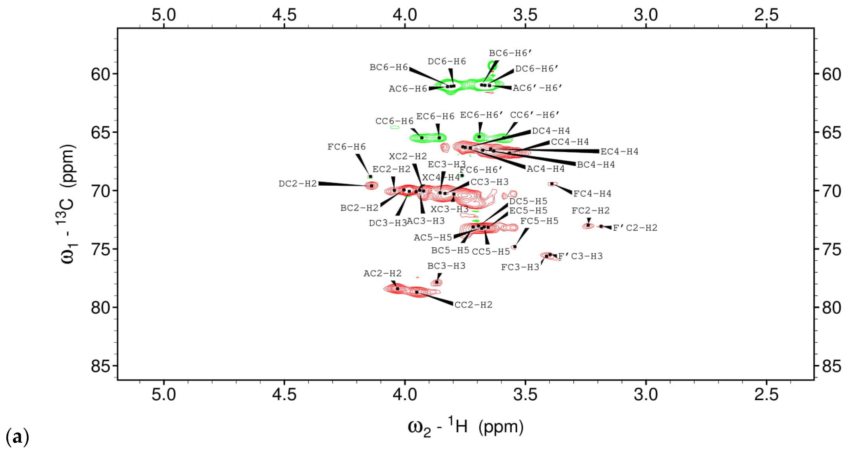
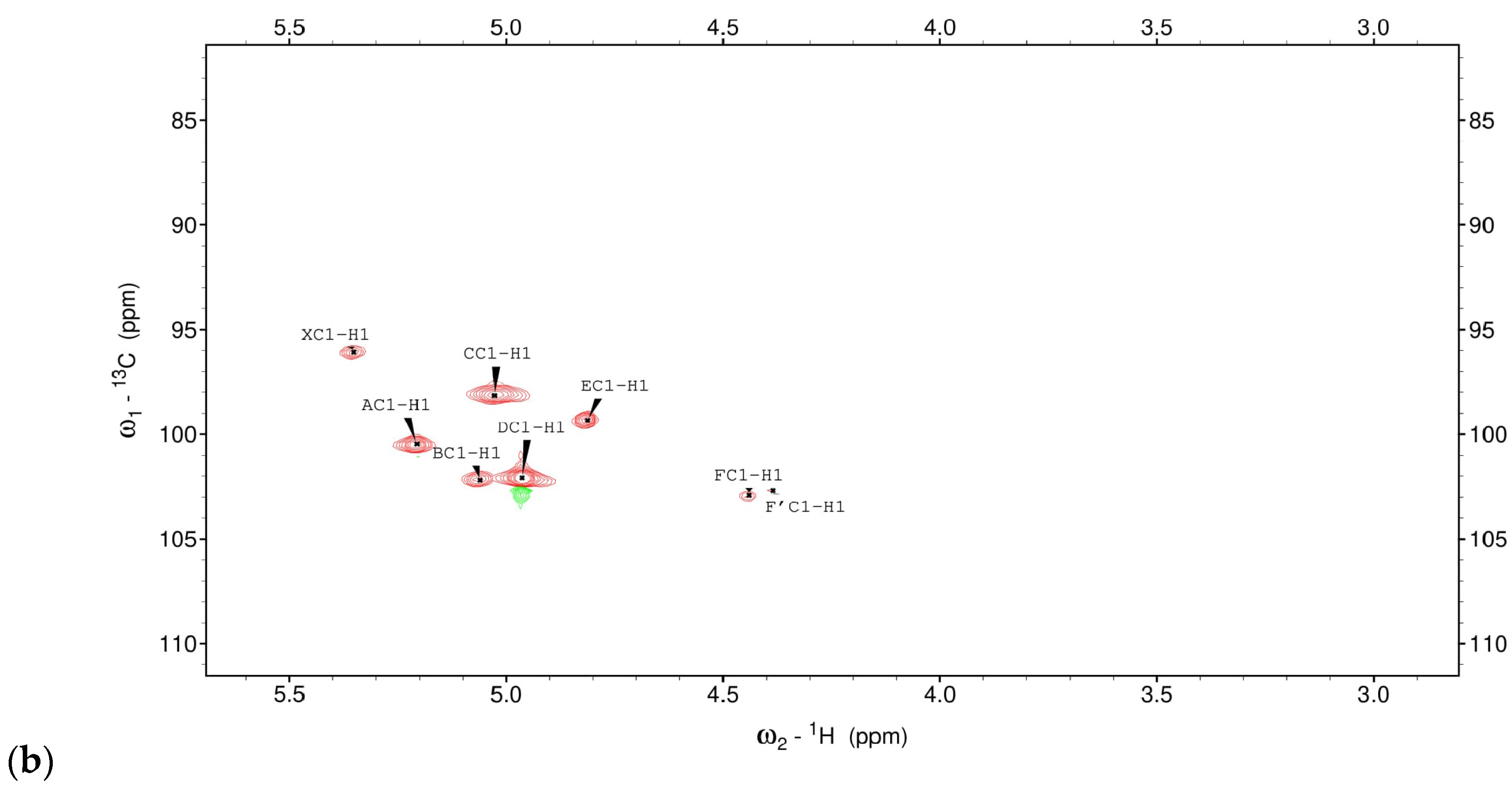
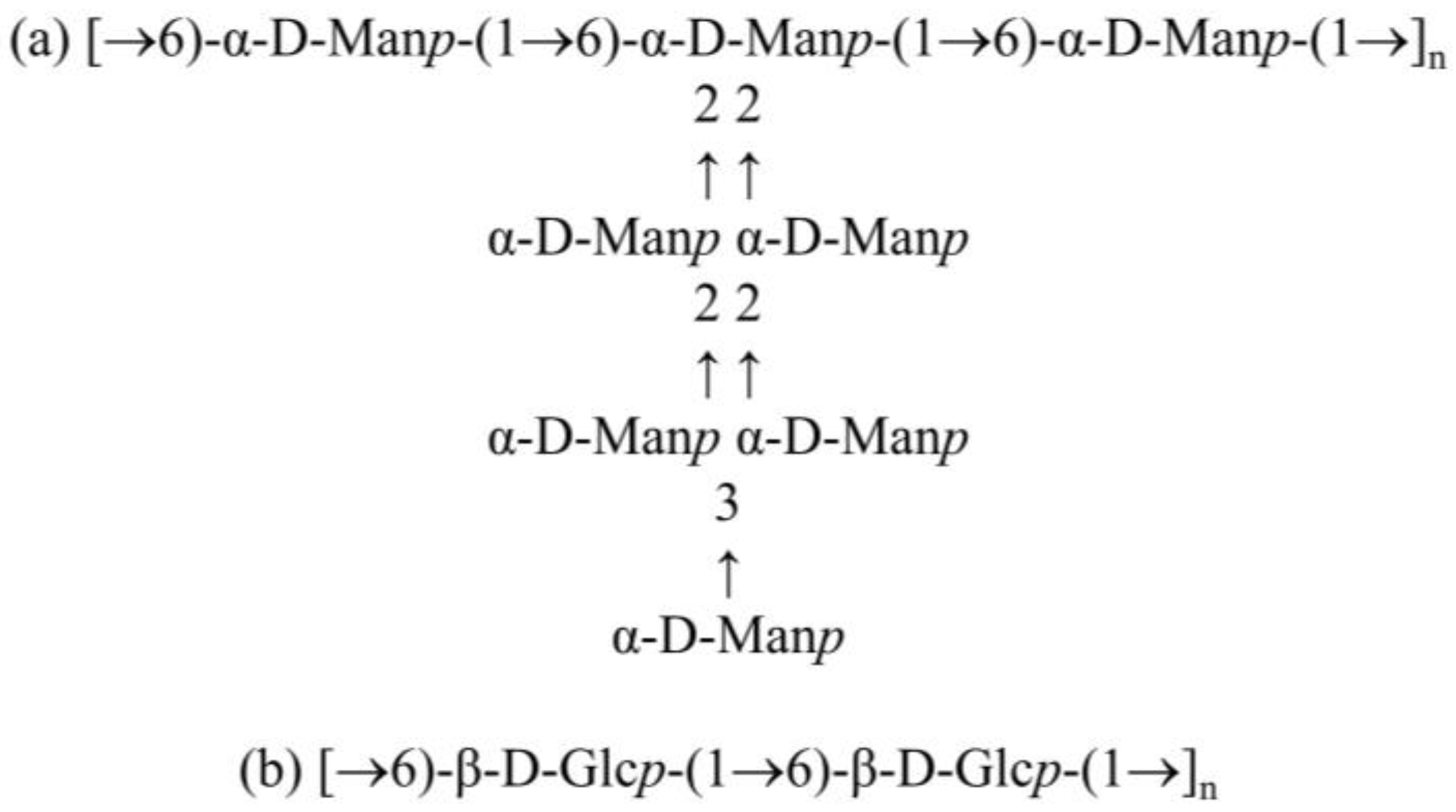
3.1. Structural Studies of EPSs Isolated from L. rhamnosus ŁOCK 0943

Monosaccharides and substitution sites were identified based on the retention times of the resulting volatile derivatives—alditol acetates. Analysis of the monosaccharide composition and EPS methylation revealed the presence of various mannose derivatives, such as terminal Manp, 2-Manp, 3-Manp, 6-Manp, and 2,6-Manp.
The 1-H NMR spectrum of EPSs in L. rhamnosus ŁOCK 0943 showed a complex pattern of signals in the anomeric region (4.4–5.5 ppm) and broad signals in the backbone sugar proton region (3.0–4.4 ppm). The sugar spin systems were marked with capital letters as X and from A to F/F’ according to the increasing δH value. The assignment of H and C resonances was performed on the basis of several homo- (1H-1H) and hetero- (1H-13C) correlated two-dimensional NMR experiments (Table 2, Figure 2) as well as by comparison with previously published NMR data [15,16,17]. Residue X (δHC 5.35/96.07 ppm) was proposed to represent α-D-Galp-(1→, according to previously presented terminal galactose in the literature [18]; however, the signals for H5/C5 and H6/C6 of X were not identified and the very low intensity of the signals precluded any consecutive cross peaks in the NMR spectra. Thus, the interpretation of the linkage types was ambiguous. Five spin systems—A, B, C, D, E—with chemical shifts of the anomeric proton/carbon at δHC 5.21/100.9, 5.06/102.2, 5.03/98.3, 4.96/102.1, and 4.82/99.4, respectively, were recognized as different mannose residues. The downfield shifts of C-2 (78.5 ppm) of residue A indicated that it was →2)-α-D-Manp-(1→ residue [19]. Position C-3 (77.8 ppm) of residue B was glycosidically substituted since significant downfield shifts for C-3 were observed. Furthermore, residue C possessed C-2 (78.7 ppm) and C-6 (65.5 ppm) chemical shifts at low fields due to glycosylation, and was therefore identified as →2,6)-α-D-Manp-(1→ residue. Likewise, at 4.96 ppm (residue D), it was possible to identify non-substituted mannose residue, as inferred by its 1H and 13C resonances. Finally, the last residue, E, which possessed an α-manno configuration, was identified as →6)-α-D-Manp-(1→ according to downfield shifts of C-6 (65.5 ppm). The other anomeric signals (F/F’) were identified as β-D-Glc based on the TOCSY and HSQC spectra analysis [20]. This residue had substituted hydroxyl groups at position C-6. The sequence of the monosaccharide residues within the repeating unit of the polysaccharides was obtained by assigning the inter-residue interactions observed in the 2D NOESY and HMBC spectra. Connectivities were found between C-1 of C and C-6 of C or E, between C-1 of A and C-2 of C, between C-1 of B and C-2 of A and C-1 of D and C-3 of B and C-2 of A, and between C-1 of E and C-6 of C or E. Based on the previously published mannan polysaccharide [21,22], it was clear that the structure could be presented as in Figure 3a.
No correlation was observed between the H-1 of residues A, B, C, D, or E and the C-6 of residue F, indicating that residues A, B, C, D, or E cannot substitute the F residue. However, we observed connections between the C-1 of F/F’ and the C-6 of F. Thus, we indicated that this is a separate polymer—β-1,6-glucan—as in Figure 3b.

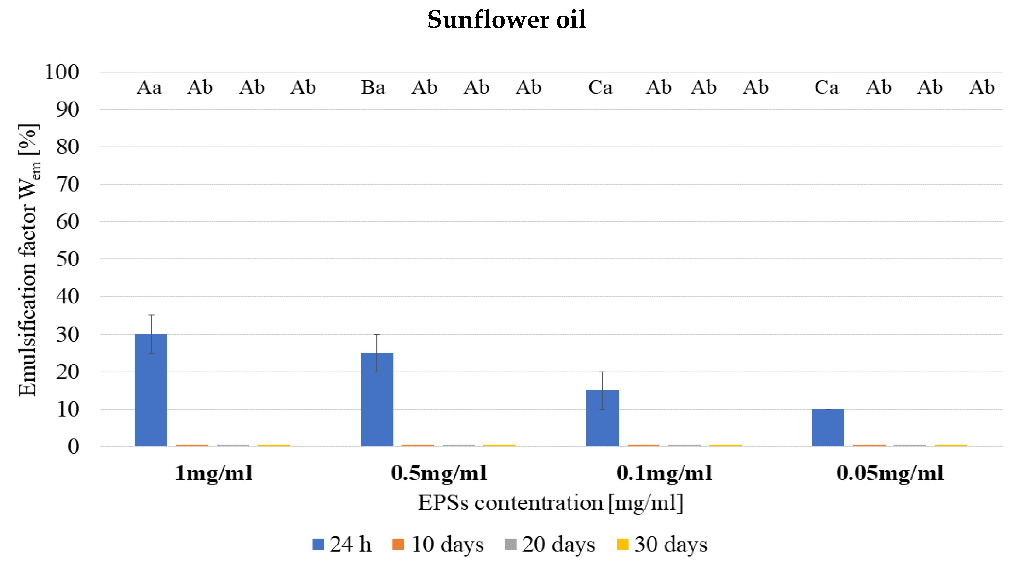
3.2. Emulsifying Properties

The emulsifying properties of the examined EPSs of L. rhamnosus bacteria were investigated with the use of three different oily substances: linseed oil, sunflower oil, and castor oil. The analysis revealed that the tested EPSs’ ability to emulsify and stabilize the oily substance depends on the type of oil used.
The highest emulsifying properties were demonstrated for castor oil. In this case, the most effective was the use of an EPS at a concentration of 0.1 mg/mL, obtaining an emulsion with a highly stable Wem of 90% (Wem = 80% after 20 days) after 24 h. The use of a concentration ten times lower still yielded high rheological effects with much lower consumption of EPSs. The least favorable emulsifying properties were found for the tested EPSs and sunflower oil. The highest emulsification coefficient was obtained here at the level of 30% (test with 1.0 mg/mL of EPSs) (Figure 4). Unfortunately, all the resulting emulsions turned out to be unstable and after 10 days of incubation, only the emulsions formed at the border of the water and oily phases were observed.

3.3. Antimicrobial Activity of EPSs

The EPSs under study did not demonstrate any antagonistic activity against the tested filamentous fungi in the experiments based on the diffusion-well method. No zones of growth inhibition were observed for the selected strains. Moreover, in the samples containing the strains Rhizopus nigricans ŁOCK 0548 and Geotrichum candidum ŁOCK 0511, growth of fungi was also observed in wells filled with EPS aqueous solutions, regardless of the concentration. Therefore, the examined EPSs did not exhibit antifungal properties.
The study of EPSs’ antagonistic activity confirmed that these biopolymers may affect the growth of bacteria and yeasts (Figure 5). The use of EPSs at concentrations of 0.5 and 0.05 mg/mL significantly inhibited the growth of Clostridium acetobutylicum ŁOCK 0831 (Figure 5a). Higher antagonistic activity of EPSs was noted in the cultures with 0.5 mg/mL of EPSs, where a more than 2-fold reduction in the number of bacteria was observed at 48 h compared to the control. Moreover, the EPSs inhibited the growth of Bacillus subtilis ATCC 6633 during a 24-h incubation, after which bacterial growth was observed with the use of a higher concentration of EPSs (Figure 5b). A significant inhibition of the growth of Candida albicans ATCC 10231 was also observed in samples with the addition of EPSs from 48 h of incubation (Figure 5c), as well as Enterococcus faecalis ATCC 29212 for up to 30 h of incubation (Figure 5d). The antibacterial activity of EPSs at a concentration of 0.5 mg/mL against Staphylococcus aureus ATCC 27734 was observed for up to 30 h of incubation, after which bacterial growth was noted at 48 h, followed by a significant inhibition of growth at 72 h of incubation (Figure 5e). The analysis also showed that Escherichia coli ATCC 10536 (Figure 5f), Pseudomonas fluorescens PCM 2123 (Figure 5g), and Salmonella Enteritidis ATCC 1307 (Figure 5h) were insensitive to the selected EPSs and their addition to the culture medium did not affect the growth curve of these bacteria.

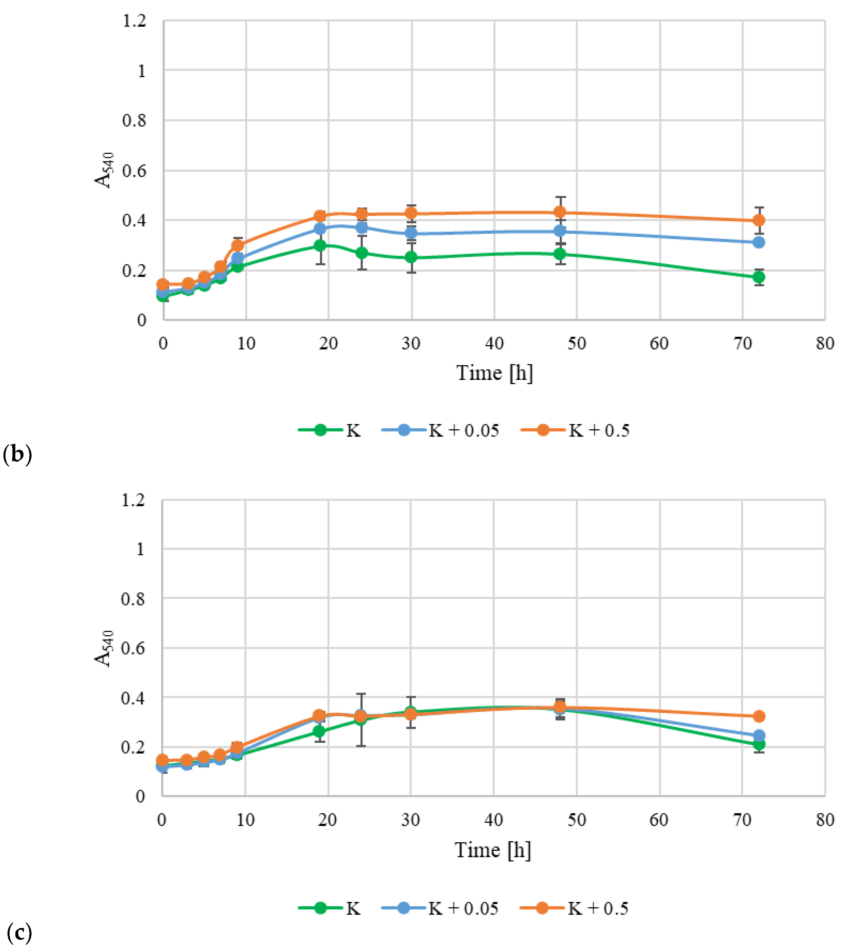
3.4. Prebiotic Potential of Exopolysaccharides

The experiments showed that EPSs synthesized by L. rhamnosus ŁOCK 0943 have a high prebiotic potential compared to those synthesized by Levilactobacillus brevis ŁOCK 0944 and L. rhamnosus ŁOCK 0908 (Figure 6a,b). The addition of EPSs to the culture medium stimulated the growth of the probiotic bacteria tested throughout the culture period. A much better prebiotic effect was obtained using EPSs at a 0.5 mg/mL concentration. In the culture of L. brevis ŁOCK 0944 with EPSs at a concentration of 0.5 mg/mL, a greater number of bacteria (approx. 40%) was recorded 24 h after cultivation; 48 h after cultivation, it was approx. 90% higher than in the control sample. However, in the test with 0.05 mg/mL of EPSs, there was 15% more bacterial growth at 24 h, and 35% at 48 h. On the other hand, for L. rhamnosus ŁOCK 0908, after the addition of 0.5 mg/mL of EPSs, bacterial growth increased by approx. 60%; in the sample with 0.05 mg/mL it was approx. 35% greater than in the control bacterial culture after 10 h of incubation. In the case of Lacticaseibacillus casei ŁOCK 0919, the addition of EPSs did not significantly affect their growth during 48 h of cultivation (Figure 6c). In cultures with EPSs, a shortening of the logarithmic growth phase was observed, while the maximum cell proliferation was at the same level regardless of the sample being tested (absorbance: approx. 0.36). A positive effect was obtained with the use of EPSs at a concentration of 0.5 mg/mL, where the viability of L. casei ŁOCK 0919 cells was extended by approx. 24 h—the stationary phase lasted up to 72 h.

3.5. Antioxidant Activity

Analysis of the antioxidant properties of the biopolymers under study showed that they have a modest potential to extinguish free radicals. The inhibition of the radical DPPH by EPSs at a concentration of 0.05 mg/mL was 8.09 ± 0.12%, while at concentrations of 0.5 mg/mL, this value slightly (but statistically significantly) increased to 8.71 ± 0.03% (Table 3). A higher degree of DPPH inhibition was observed with the use of EPSs from Leuconostoc suionicum LSBM1 at a concentration of 8 mg/mL [23]. However, it is noteworthy to mention that the EPSs from Lactiplantibacillus plantarum SP8 exhibited lower DPPH radical scavenging activity at a concentration of 0.05 mg/mL [24] compared to the EPSs derived from Lacticaseibacillus rhamnosus ŁOCK 0943.

3.6. Assessment of the Technological Suitability of EPSs as a Preservative in the Cosmetics Industry

The numbers of microorganisms measured in the tested samples (Figure 7) were compared with the criteria for assessing the antimicrobial activity of a preservative system according to The Polish Pharmacopoeia (X, 2014) (Table 1).
The analysis revealed that the synthetic preservative used in the tests (bronopol) can be used as a preservative in hand cream (Figure 7). Less satisfactory effects were obtained with the use of EPSs obtained from the cultures of bacteria. For both P. aeruginosa ATCC 9027 and S. aureus ATCC 6538 strains, a 1-log order of reduction was observed after 2 days of incubation, and 2-log orders of reduction were observed after 14 days of incubation. On the other hand, interesting results were obtained in systems where both the tested EPSs and bronopol were used at concentrations reduced by half (respectively, 0.25 mg/mL and 0.05%). In the case of S. aureus ATCC 6538, the same antagonistic activity was observed in the tests with bronopol and with bronopol and EPSs (after 2 days of incubation). In turn, for P. aeruginosa ATCC 9027, after the use of a synthetic preservative and EPSs, a reduction in the number of bacterial cells by 3 logarithmic units was observed after 2 days. Moreover, in this sample, the lowest number of bacteria was observed throughout the entire incubation period (statistically significant differences). In the case of fungi, the preservative system reduced their numbers by 3 log units after 14 days of incubation. Thus, the use of bronopol and EPSs at lower concentrations in hand cream allows a high microbiological purity of cosmetic products to be maintained with the limited use of a synthetic preservative. It can be assumed that the resulting product is safer to use on the skin.

4. Discussion

The impulse for research on bacterial EPSs was due to their enormous application potential in many industries. They have a number of biological and technological properties, thanks to which they can revolutionize not only the market of fermented milk products, but also the cosmetics, medical, and pharmaceutical industries. The amount of research into the use of EPSs from LAB in the food industry is steadily increasing and it is believed that this increase will lead to the production of higher quality food and cosmetic products.
The analysis was carried out in previously determined stable environmental conditions, which were selected to maintain the economic and technical parameters of the process while maintaining the highest possible efficiency of EPS synthesis. Due to the various structures of the synthesized EPSs, only after establishing the exact culture conditions is it possible to analyze them chemically and evaluate their biological/technological properties. Bearing in mind the possibility of using EPSs in industry, the safety of use should be maintained, which is why the selected strains had not been subjected to genetic mutations [12,13]. However, despite the widely documented benefits of EPSs, there is a need to improve the efficiency of their synthesis and to better understand how environmental conditions affect their chemical structures and properties.
Advanced chemical analysis revealed that the strain L. rhamnosus ŁOCK 0943 synthesized mannan and β-1,6-glucan. In the GC-MS spectrum, a high peak was observed at 11.89, which is indicative of a high amount of mannose in the sample. There were also low signals at 11.92 and 11.98, which were identified as glucose and galactose by NMR analysis. Similar results were obtained by Sandal et al. [22] and Donnarumma et al. [26]. The Lactobacillus crispatus L1 and Histophilus somni 2336 bacteria studied synthesized mannan that was identical in structure to that from L. rhamnosus ŁOCK 0943.
The assessment of biological and technological properties of EPSs included emulsifying, antagonistic, prebiotic, and antioxidant properties. In the first stage of the research, the emulsifying properties—i.e., the ability of the tested EPSs to form a stable emulsion—were analyzed. The emulsifying properties of EPSs are important in food and cosmetics production. These properties were tested using four different concentrations of EPSs dissolved in distilled water (0.05, 0.1, 0.5, and 1.0 mg/mL) along with three oily substances: linseed oil, sunflower oil, and castor oil. It was found that the emulsifying activity of EPSs depends mainly on the type of oily substance used. The most durable emulsion and the one with the largest volume was obtained with castor oil. On the other hand, the least satisfactory results were recorded in the trials with sunflower oil, where the emulsions turned out to be unstable: after 10 days, the emulsion was observed only at the interface. Denser oil leads to more emulsification due to the increase in viscosity of the water-in-oil emulsion, which reduces the mobility of heavy oil [27]. Polysaccharides are a group of carbohydrate emulsifiers. Their ability to reduce oil-water interfacial tension is influenced by the presence of nonpolar groups and their interactions with surfactants and emulsifiers [28]. Emulsions are mixtures of two immiscible liquids, typically with small droplets of oil dispersed in water [29]. Emulsifiers, which are amphiphilic molecules containing both hydrophilic and hydrophobic groups, are essential for stabilizing these emulsions. Natural food-grade emulsifiers, such as proteins and polysaccharides, are commonly used in the food industry. Bacterial exopolysaccharides (EPSs) are increasingly important in the food, pharmaceutical, and cosmetic industries due to their rheological properties, viscosity, and emulsifying capabilities [30]. EPSs act as thickeners, gelling agents, adhesives, emulsifiers, and stabilizers [31]. They can form and stabilize both water-in-oil and oil-in-water emulsions, enhancing the texture and sensory qualities of food products [30]. Han et al. [32] obtained different results when analyzing the emulsifying properties of EPSs (at a concentration of 1 mg/mL) synthesized by Bacillus amyloliquefaciens LPL061. These EPSs showed similar emulsifying activity with sunflower oil, olive oil, nut oil, and rice oil (emulsifying coefficient: approx. 60%). In turn, Rajoka et al. [3] demonstrated the high emulsifying activity of EPSs synthesized by Lacticaseibacillus rhamnosus in sunflower oil. These researchers observed that the EPSs of all six of the tested strains showed an emulsification coefficient ranging from 38.1% to 65.4%. However, after 15 days, no significant (p ≤ 0.05) decrease in the Wem value was observed, which proves the high stability of the resulting emulsions.
The study of EPSs’ antagonistic activity confirmed that the tested biopolymers may affect the growth of microorganisms (bacteria and yeast), while the filamentous fungi turned out to be insensitive. EPSs from the bacterial strain L. rhamnosus ŁOCK 0943 showed an inhibitory effect on the growth of the bacteria and yeasts. In many cases, increased growth of the tested microorganisms was observed with a higher EPS concentration of 0.5 mg/mL (e.g., Clostridium acetobutylicum ŁOCK 0831). Zhou et al. [33] reported that the molecular mass (Mw) of EPSs plays a very important role in their antagonistic activity (higher antimicrobial properties of EPSs were observed at low molecular weights). Similar results were obtained by Ayyash et al. [34], who found high antagonistic activity for EPSs synthesized by Lactiplantibacillus plantarum C70, which was isolated from camel milk. In turn, both L. monocytogenes ATCC 51776 and S. Enteritidis 108 were completely inhibited in a nutrient broth supplemented with 10 and 25 mg/mL of EPSs obtained from Lactobacillus kefiranofaciens DN1 cultures [35]. It is important to note that in the results of all the other studies, a high concentration of EPSs was used (at least 10 times higher than in this study). However, given the possibility of EPSs from LAB being used for industrial purposes, concentrations of polymers in excess of 1 mg/mL become uneconomical on an industrial scale.
Many studies have indicated that some EPSs are resistant to biodegradation when passing through the human digestive system [36]. Therefore, in the next stage of the research, the influence of EPSs on the growth of bacteria with probiotic features (L. brevis ŁOCK 0944, L. casei ŁOCK 0919, and L. rhamnosus ŁOCK 0908) was examined. The addition of EPSs at concentrations of 0.05 and 0.5 mg/mL resulted in a higher multiplication of bacterial cells of L. brevis ŁOCK 0919 and L. rhamnosus ŁOCK 0908 and increased the viability of all the tested probiotic bacteria. Thus, these bacteria have the ability to hydrolyze EPSs into easily digestible compounds, thanks to the ability to synthesize extracellular enzymes in the family of glycosidic hydrolases (2, 13, 36, and 42), such as α-galactosidases, β-galactosidases, and enzymes that are active against gluco-oligosaccharides [37]. The prebiotic potential of EPSs has been confirmed in many scientific reports. For example, the EPSs synthesized by L. plantarum DM5 stimulated the growth of bacteria with probiotic features—Bifidobacterium infantis NRRL B-41661 and L. acidophilus NRRL B-4495—without affecting the growth of potential pathogenic bacteria, Enterobacter aerogenes MTCC 7016 [38]. In turn, Sarikaya et al. [39] demonstrated the bifidogenic properties (relative to Bifidobacterium breve BASO-1) of EPSs synthesized by strains of LAB isolated from human feces and dairy products. Their analyses indicated that EPSs synthesized by LAB can potentially have a positive effect on the host microflora, though this has not yet been directly confirmed in intervention trials in humans. This area, therefore, requires further research.
In this study, it was shown that the EPSs under study had limited potential to extinguish free radicals. Moreover, slightly higher results were obtained with a higher concentration of EPSs (0.5 mg/mL). The literature reports that there is a significant relationship between the monosaccharide composition of EPSs and their ability to extinguish free radicals. An important aspect is the amounts of arabinose, glucose, and mannose, as well as the presence of hydroxyl groups that can act as electron donors to bind radicals [40]. Rani et al. [41] observed an increase in antioxidant activity with an increase in the concentration of EPSs synthesized by Lactobacillus gasseri FR4: at a concentration of 4 mg/mL of EPSs, the inhibition rate of DPPH was 75.95%, while for an EPSs concentration of 0.5 mg/mL, it was only about 23%. On the other hand, Yan et al. [25] did not observe significant differences in the antioxidant activity of tested EPSs from Lactiplantibacillus plantarum LPC-1 at concentrations of 1–9 mg/mL. It is noteworthy that in the majority of studies, the concentration of EPS is substantially higher compared to our research. The inhibition of the radical DPPH EPSs obtained from Lactobacillus helveticus SMN2-1 bacteria was about 15% when using EPSs at a concentration of 0.5 mg/mL [42]. The antioxidant activity of EPSs from Lactiplantibacillus plantarum SP8 at a concentration of 0.2 mg/L was 8.98%, while at a concentration of 0.05 mg/mL, it was less than 2% [24]. Therefore, considering the low concentration of EPSs used in our study (0.05 mg/mL), the obtained results of antioxidant activity are not low at all (8.09%).
The analyses carried out so far on the chemical and biological properties of EPSs from L. rhamnosus ŁOCK 0943 have shown that they have favorable features, thanks to which they can be used for industrial applications. Therefore, we decided to use the tested EPSs as a preservative in the production of natural hand cream, which is one of the innovative aspects of this work. Based on the analysis of antibacterial and prebiotic properties for investigating the use of EPSs as a preservative in cosmetics, an EPS concentration of 0.5 mg/mL was selected. It was shown that biopolymers can be successfully used as a preservative in cosmetic products. The best results were obtained after using both the synthetic preservative and EPSs for the production of the cream, in halved concentrations: 0.05% bronopol and 0.25 mg/mL EPSs. This is very beneficial from the point of view of product safety, as synthetic compounds may cause side effects (allergic reactions), particularly on sensitive skin. The use of such a preservative system is in line with the current trend of developing natural products while maintaining microbiological safety. In the face of global discussions on the side effects of preservatives, it is important to study newly acquired compounds and structures that could serve as alternatives to the chemical substances used in industries. Polysaccharides are now commonly being used to improve the properties of cosmetic products; however, they are mainly of plant origin. Polysaccharides of bacterial origin, due to the large variety in their composition and physicochemical properties, are gradually becoming competitive with polymers obtained from plants. Levan synthesized by Zymomonas mobillis shows good stability in ethanol and has strong moisturizing properties; it, therefore, can be used as an active ingredient in cosmetics [43]. In turn, EPSs produced by Pseudoalteromonas spp. can be successfully used in dermopharmaceutical products for the treatment or care of the skin, mucous membranes, hair, and nails [44].

5. Conclusions

An innovative and comprehensive approach to the subject of EPSs synthesized by Lacticaseibacillus rhamnosus ŁOCK 0943 confirmed the research hypothesis that they have favorable biological and technological properties. It has been shown that these polymers can be successfully used in a preservative system in cosmetics. Despite the fact that these bacteria have obtained the Generally Regarded as Safe status and the polymers synthesized using them may have high potential applications, their use in industry is still limited. The analysis carried out in this research constitutes the basis for further research on EPSs, including in pharmaceutical or cosmetic products and the production of “new-generation” foods.

Author Contributions

Conceptualization, M.O.-S., S.G., L.P.-R. and E.K.; methodology, M.O.-S., S.G., L.P.-R. and E.K.; formal analysis, M.O.-S., S.G., L.P.-R. and S.Ś.; investigation, M.O.-S., S.G. and L.P.-R.; resources, S.G. and E.K.; data curation, M.O.-S.; writing—original draft preparation, M.O.-S., S.G., L.P.-R., S.Ś. and E.K.; writing—review and editing, S.G., L.P.-R., S.Ś. and E.K.; visualization, M.O.-S., S.G., L.P.-R., S.Ś. and E.K.; supervision, E.K.; project administration, E.K. All authors have read and agreed to the published version of the manuscript.

Funding

This research received no external funding.

Data Availability Statement

The raw data supporting the conclusions of this article will be made available by the authors on request.

Conflicts of Interest

The authors declare no conflicts of interest.

References

  1. Saadat, Y.R.; Khosroushahi, A.Y.; Gargari, B.P. A comprehensive review of anticancer, immunomodulatory and health beneficial effects of the lactic acid bacteria exopolysaccharides. Carbohydr. Polym. 2019, 217, 79–89. [Google Scholar] [CrossRef]
  2. Trabelsi, I.; Slima, B.S.; Chaabane, H.; Riadh, S.B. Purification and characterization of a novel exopolysaccharides produced by Lactobacillus sp. Ca6. Int. J. Biol. Macromol. 2015, 74, 541–546. [Google Scholar] [CrossRef]
  3. Rajoka, M.S.R.; Jin, M.; Haobin, Z.; Li, Q.; Shao, D.; Jiang, C.; Huang, Q.; Yang, H.; Shi, J.; Hussain, N. Functional characterization and biotechnological potential of exopolysaccharide produced by Lactobacillus rhamnosus strains isolated from human breast milk. LWT—Food Sci. Technol. 2018, 89, 638–647. [Google Scholar] [CrossRef]
  4. Yilmaz, M.T.; Dertli, E.; Toker, O.S.; Tatlisu, N.B.; Sagdic, O.; Arici, M. Effect of in situ exopolysaccharide production on physicochemical, rheological, sensory, and microstructural properties of the yogurt drink Aryan: An optimization study based on fermentation kinetics. J. Diary Sci. 2015, 98, 1604–1624. [Google Scholar] [CrossRef]
  5. De Vuyst, L.; De Vin, F.; Vaningelgem, F.; Degeest, B. Recent developments in the biosynthesis and applications of heteropolysaccharides from lactic acid bacteria. Int. Dairy J. 2001, 11, 687–707. [Google Scholar] [CrossRef]
  6. Madhuri, K.V.; Prabhakar, K.V. Microbial exopolysaccharides: Biosynthesis and potential applications. Orient. J. Chem. 2014, 30, 1401–1410. [Google Scholar] [CrossRef]
  7. Badel, S.; Bernardi, T.; Michaud, P. New perspectives for Lactobacilli exopolysaccharides. Biotechnol. Adv. 2011, 29, 54–66. [Google Scholar] [CrossRef]
  8. Grobben, G.J.; Smith, M.R.; Sikkema, J.; Bont, J.A.M. Influence of fructose and glucose on the production of exopolysaccharides and the activities of enzymes involved in the sugar metabolism and the synthesis of sugar nucleotides in Lactobacillus delbrueckii subsp. bulgaricus NCFB 2772. Appl. Microbiol. Biotechnol. 1996, 46, 279–284. [Google Scholar]
  9. Górska, S.; Grycko, P.; Rybka, J.; Gamian, A. Egzopolisacharydy bakterii kwasu mlekowego–biosynteza I struktura. Postępy Hig. I Med. Dośw. 2007, 61, 805–818. [Google Scholar]
  10. Kim, D.; Robyt, J.F. Production and selection of mutants of Leuconostoc mesenteroides constitutive for glucansucrases. Enzym. Microb. Technol. 1994, 16, 659–664. [Google Scholar] [CrossRef]
  11. Bajpai, V.K.; Rather, I.A.; Majumder, R.; Shukla, S.; Aeron, A.; Kim, K.; Park, Y.H. Exopolysaccharide and lactic acid bacteria: Perception, functionality and prospects. Bangladesh J. Pharmacol. 2016, 11, 1–23. [Google Scholar] [CrossRef]
  12. Oleksy-Sobczak, M.; Klewicka, E.; Piekarska-Radzik, L. Exopolysaccharides production by Lactobacillus rhamnosus strains–Optimization of synthesis and extraction conditions. LWT—Food Sci. Technol. 2020, 122, 109055. [Google Scholar] [CrossRef]
  13. Oleksy-Sobczak, M.; Klewicka, E. Optimization of media composition to maximize the yield of exopolysaccharides production by Lactobacillus rhamnosus strains. Probiotics Antimicrob. Proteins 2020, 12, 774–783. [Google Scholar] [CrossRef]
  14. Dubois, M.; Giller, K.A.; Rebers, P.A.; Smith, F.A. Colorimetric method for determination of sugars and related substances. Anal. Chem. 1956, 28, 350–356. [Google Scholar] [CrossRef]
  15. Bock, K.; Pedersen, C. Carbon-13 nuclear magnetic resonance spectroscopy of monosaccharides. Adv. Carbohydr. Chem. Biochem. 1983, 41, 27–66. [Google Scholar]
  16. Gerwig, G.J.; Kamerling, J.P.; Vliegenthart, J.F.G. Determination of the absolute configuration of monosaccharides in complex carbohydrates by capillary G.L.C. Carbohydr. Res. 1979, 77, 10–17. [Google Scholar] [CrossRef]
  17. Gorin, P.A.J.; Mazurek, M. Further studies on the assignment of signals in 13C magnetic resonance spectra of aldoses and derived methyl glycosides. Can. J. Chem. 1975, 53, 1212–1223. [Google Scholar] [CrossRef]
  18. Sauvageau, J.; Ryan, J.; Lagutin, K.; Sims, I.M.; Stocker, B.L.; Timmer, M.S. Isolation and structural characterisation of the major glycolipids from Lactobacillus plantarum. Carbohydr. Res. 2012, 357, 151–156. [Google Scholar] [CrossRef]
  19. Katzenellenbogen, E.; Kocharova, N.A.; Toukach, P.V.; Górska, S.; Korzeniowska-Kowal, A.; Bogulska, M.; Gamian, A.; Knirel, Y.A. Structure of an abequose-containing O-polysaccharide from Citrobacter freundii O22 strain PCM 1555. Carbohydr. Res. 2009, 344, 1724–1728. [Google Scholar] [CrossRef]
  20. Górska-Frączek, S.; Sandström, C.; Kenne, L.; Rybka, J.; Strus, M.; Heczko, P.; Gamian, A. Structural studies of the exopolysaccharide consisting of a nonasaccharide repeating unit isolated from Lactobacillus rhamnosus KL37B. Carbohydr. Res. 2011, 346, 2926–2932. [Google Scholar] [CrossRef]
  21. Molinaro, A.; Piscopo, V.; Lanzetta, R.; Parrilli, M. Structural determination of the complex exopolysaccharide from the virulent strain of Cryphonectria parasitica. Carbohydr. Res. 2002, 337, 1707–1713. [Google Scholar] [CrossRef]
  22. Sandal, I.; Inzana, T.J.; Molinaro, A.; De Castro, C.; Shao, J.Q.; Apicella, M.A.; Cox, A.D.; St Michael, F.; Berg, G. Identification, structure, and characterization of an exopolysaccharide produced by Histophilus somni during biofilm formation. BMC Microbiol. 2011, 11, 186. [Google Scholar] [CrossRef]
  23. Long, X.; Hou, X.; Li, S.; Chen, A.; Zhang, Z.; Shen, G. Fermentation Optimization and In-Vitro Antioxidant Activity of Exopolysaccharides Produced by Leuconostoc suionicum LSBM1 Using Sugar Beet Molasses. Sugar Tech 2024, 1–10. [Google Scholar] [CrossRef]
  24. Zhang, L.; Zhao, B.; Liu, C.-J.; Yang, E. Optimization of biosynthesis conditions for the production of exopolysaccharides by Lactobacillus plantarum SP8 and the exopolysaccharides antioxidant activity test. Indian J. Microbiol. 2020, 60, 334–345. [Google Scholar] [CrossRef]
  25. Yan, Z.X.; Li, M.; Wei, H.Y.; Peng, S.Y.; Xu, D.J.; Zhang, B.; Cheng, X. Characterization and antioxidant activity of the polysaccharide hydrolysate from Lactobacillus plantarum LPC-1 and their effect on spinach (Spinach oleracea L.) growth. Appl. Biochem. Biotechnol. 2024, 1–23. [Google Scholar] [CrossRef]
  26. Donnarumma, G.; Molinaro, A.; Cimini, D.; De Castro, C.; Valli, V.; De Gregorio, V.; De Rosa, M.; Schiraldi, C. Lactobacillus crispatus L1: High cell density cultivation and exopolysaccharide structure characterization to highlight potentially beneficial effects against vaginal pathogens. BMC Microbiol. 2014, 14, 137. [Google Scholar] [CrossRef]
  27. Liu, J.; Zhong, L.; Yuan, X.; Liu, Y.; Zou, J.; Wang, Q.; Zhang, H. Study on the re-emulsification process of water in heavy oil emulsion with addition of water-soluble viscosity reducer solution. Energy Fuels 2019, 33, 10852–10860. [Google Scholar] [CrossRef]
  28. Chen, S.; AlSofi, A.M.; Wang, J.; Alotaibi, M.B. A Polycyclic–Aromatic Hydrocarbon-Based Water-Soluble Formulation for Heavy Oil Viscosity Reduction and Oil Displacement. Energy Fuels 2023, 37, 11864–11880. [Google Scholar] [CrossRef]
  29. McClements, D.J.; Jafari, S.M. Improving emulsion formation, stability and performance using mixed emulsifiers: A review. Adv. Colloid Interface Sci. 2018, 251, 55–79. [Google Scholar] [CrossRef]
  30. Kavitake, D.; Balyan, S.; Devi, P.B.; Shetty, P.H. Evaluation of oil-in-water (O/W) emulsifying properties of galactan exopolysaccharide from Weissella confusa KR780676. J. Food Sci. Technol. 2020, 57, 1579–1585. [Google Scholar] [CrossRef]
  31. Kavitake, D.; Marchawala, F.Z.; Delattre, C.; Shetty, P.H.; Pathak, H.; Andhare, P. Biotechnological potential of exopolysaccharide as a bioemulsifier produced by Rhizobium radiobacter CAS isolated from curd. Bioact. Carbohydr. Diet. Fibre 2019, 20, 100202. [Google Scholar] [CrossRef]
  32. Han, Y.; Liu, E.; Liu, L.; Zhang, B.; Wang, Y.; Gui, M.; Wu, R.; Li, P. Rheological, emulsifying and thermostability properties of two exopolysaccharides produced by Bacillus amyloliquefaciens LPL061. Carbohydr. Polym. 2015, 115, 230–237. [Google Scholar] [CrossRef] [PubMed]
  33. Zhou, Y.; Cui, Y.; Qu, X. Exopolysaccharides of lactic acid bacteria: Structure, bioactivity and associations: A review. Carbohydr. Polym. 2019, 207, 317–332. [Google Scholar] [CrossRef]
  34. Ayyash, M.; Abu-Jdayil, B.; Itsaranuwat, P.; Galiwango, E.; Tamiello-Rosa, C.; Abdullah, H.; Esposito, G.; Hunashald, Y.; Obaide, R.S.; Hamed, F. Characterization, bioactivities, and rheological properties of exopolysaccharide produced by novel probiotic Lactobacillus plantarum C70 isolated from camel milk. Int. J. Biol. Macromol. 2020, 144, 938–946. [Google Scholar] [CrossRef]
  35. Jeong, D.; Kim, D.H.; Kang, I.B.; Kim, H.; Song, K.Y.; Kim, H.S.; Seo, K.H. Characterization and antibacterial activity of a novel exopolysaccharide produced by Lactobacillus kefiranofaciens DN1 isolated from kefir. Food Control 2017, 78, 436–442. [Google Scholar] [CrossRef]
  36. Ryan, P.M.; Ross, R.P.; Fitzgerald, G.F.; Caplice, N.M.; Stanson, C. Sugarcoated: Exopolysaccharides producing lactic acid bacteria for food and human health applications. Food Funct. 2015, 6, 679–693. [Google Scholar] [CrossRef] [PubMed]
  37. Pokusaeva, K.; Fitzgerald, G.F.; van Sinderen, D. Carbohydrate metabolism in Bifidobacteria. Genes Nutr. 2011, 6, 285–306. [Google Scholar] [CrossRef]
  38. Das, S.; Mishra, B.K.; Hati, S. Techno-functional characterization of indigenous Lactobacillus isolates from the traditional fermented foods of Meghalaya, India. Curr. Res. Food Sci. 2020, 3, 9–18. [Google Scholar] [CrossRef]
  39. Sarikaya, H.; Aslim, B.; Yuksekdag, Z. Assessment of anti-biofilm activity and bifidogenic growth stimulator (BGS) effect of lyophilized exopolysaccharides (l-EPSs) from Lactobacilli strains. Int. J. Food Prop. 2017, 20, 362–371. [Google Scholar] [CrossRef]
  40. Trabelsi, I.; Ktari, N.; Slima, S.B.; Triki, M.; Bardaa, S.; Mnif, H.; Salah, R.B. Evaluation of dermal wound healing activity and in vitro antibacterial and antioxidant activities of a new exopolysaccharide produced by Lactobacillus sp. Ca6. Int. J. Biol. Macromol. 2017, 103, 194–201. [Google Scholar] [CrossRef]
  41. Rani, R.P.; Anandharaj, M.; Ravindran, A.D. Characterization of a novel exopolysaccharide produced by Lactobacillus gasseri FR4 and demonstration of its in vitro biological properties. Int. J. Biol. Macromol. 2018, 109, 772–783. [Google Scholar] [CrossRef]
  42. Bai, L.J.; Wang, L.; Ji, S.J. Structural Elucidation and Antioxidant Activities of Exopolysaccharide from L. helveticus SMN2-1. Chem. Eng. Trans. 2016, 55, 61–66. [Google Scholar]
  43. Srikanth, R.; Reddy, C.H.S.; Siddartha, G.; Ramaiah, M.J.; Uppuluri, K.B. Review on production, characterization and applications of microbial levan. Carbohydr. Polym. 2015, 120, 102–114. [Google Scholar] [CrossRef] [PubMed]
  44. Courtois, A.; Thollas, B.; Gonzalez, R.D.; Puche, J.C.; Astals, A.S. Exopolysaccharide for Treatment or Care of Skin, Mucous Membranes, Hair or Nails. U.S. Patent No. 9,770,400, 26 September 2017. [Google Scholar]
Figure 1. Scheme of the synthesis of extracellular heteropolysaccharides.
Processes 12 01192 g001
Figure 2. Fragments of the 1H, 13C HSQC spectra obtained for EPSs synthesized by L. rhamnosus ŁOCK 0943 (a) region of ring carbons (green colors—negative signals for the CH2, red color—positive signals for the CH carbons); (b) region of anomeric carbons in the ring. Signals are shown in capital letters (Table 2).
Processes 12 01192 g002aProcesses 12 01192 g002b
Figure 3. The structure of (a) mannan and (b) β-glucan synthesized by the strain Lacticaseibacillus rhamnosus ŁOCK 0943.
Processes 12 01192 g003
Figure 4. Emulsifying properties of exopolysaccharides synthesized by the tested Lacticaseibacillus rhamnosus ŁOCK 0943 bacteria. A, B, C—statistically significant differences within the same time (ANOVA, Tukey’s post hoc test (p ≤ 0.05)); a, b, c—statistically significant differences within the same concentration of exopolysaccharides (ANOVA, Tukey’s post hoc test (p ≤ 0.05)).
Processes 12 01192 g004aProcesses 12 01192 g004b
Figure 5. The influence of EPSs on the growth of selected microorganisms: (a) culture of Clostridium acetobutylicum ŁOCK 0831; (b) culture of Bacillus subtilis ATCC 6633; (c) culture of Candida albicans ATCC 10231; (d) culture of Enterococcus faecalis ATCC 29212; (e) culture of Staphylococcus aureus ATCC 27734; (f) culture of Escherichia coli ATCC 10536; (g) culture of Pseudomonas fluorescens PCM 2123; (h) culture of Salmonella Enteritidis ATCC 1307. K—control culture—culture without the addition of EPSs; K + 0.05—culture with EPSs at a concentration of 0.05 mg/mL; K + 0.5—culture with EPSs at a concentration of 0.5 mg/mL.
Processes 12 01192 g005
Figure 6. The influence of EPSs on the growth of (a) culture of Levilactobacillus brevis ŁOCK 0944; (b) culture of Lacticaseibacillus rhamnosus ŁOCK 0908; (c) culture of Lacticaseibacillus casei ŁOCK 0919; K—control culture—culture without additional EPSs; K + 0.05—culture with EPSs at a concentration of 0.05 mg/mL; K + 0.5—culture with EPSs at a concentration of 0.5 mg/mL.
Processes 12 01192 g006aProcesses 12 01192 g006b
Figure 7. Number of (a) Pseudomonas aeruginosa ATCC 9027; (b) Staphylococcus aureus ATCC 6538; (c) Candida albicans ATCC 10231; and (d) Aspergillus brasilensis ATCC 16404 tested in hand cream after application of different preservative system properties. a, b, c, d, e, f, g—statistically significant differences between samples (ANOVA, Tukey’s post hoc test (p ≤ 0.05)).
Processes 12 01192 g007
Table 1. Assessment of the antimicrobial activity of the preservative system in cosmetic products according to the Polish Pharmacopoeia (X, 2014).
The Value of Microbial Reduction *
2 Days7 Days14 Days28 Days
Bacteria23-NI
Fungi--2NI
* Log reduction value for the microbial count; NI—the microbial count does not increase from the previous test.
Table 2. 1H and 13C NMR chemical shifts in sample B.
Chemical Shifts 1H, 13C (ppm)
Sugar ResidueH1H2H3H4H5H6H6’
C1C2C3C4C5C6
X
α-D-Galp-(1→
5.353.903.803.95n.dn.d
96.0770.270.370.4n.dn.d
A
→2)-α-D-Manp-(1→
5.214.033.943.763.683.82/3.65
100.978.570.066.373.361.1
B
→3)-α-D-Manp-(1→
5.064.013.873.733.723.81/3.68
102.270.277.866.573.261.1
C
→2,6)-α-D-Manp-(1→
5.033.953.833.573.673.93/3.59
98.378.770.366.873.265.5
D
α-D-Manp-(1→
4.964.143.983.753.703.79/3.66
102.169.670.166.373.161.1
E
→6)-α-D-Manp-(1→
4.823.913.853.643.663.86/3.70
99.469.970.366.473.265.6
F/F’
→6)-β-D-Glcp-(1→
4.44/4.393.24/3.193.41/3.393.403.544.14/3.76
102.9/102.772.9/73.175.6/75.569.574.868.72
Spectra were obtained for 2H2O solutions at 25 °C and acetone (δH 2.225, δC 31.05 ppm) was used as an internal reference; n.d.—not detected.
Table 3. Inhibition of the radical DPPH by EPSs.
LABEPS Concentration (mg/mL)DPPH Inhibition (%)References
Lacticaseibacillus
rhamnosus
ŁOCK 0943
0.058.09 ± 0.12 aOur data
0.58.71 ± 0.03 b
Lactiplantibacillus plantarum SP80.05<2.00Zhang et al., 2020 [24]
0.28.98
Lactiplantibacillus plantarum LPC-11.0–9.0<20Yan et al., 2024 [25]
Leuconostoc suionicum LSBM18.067.41 ± 0.33Long et al., 2024 [23]
a, b—statistically significant differences (ANOVA, Tukey’s post hoc test (p ≤ 0.05)).
Disclaimer/Publisher’s Note: The statements, opinions and data contained in all publications are solely those of the individual author(s) and contributor(s) and not of MDPI and/or the editor(s). MDPI and/or the editor(s) disclaim responsibility for any injury to people or property resulting from any ideas, methods, instructions or products referred to in the content.

