Article

Synthesis of Transparent Bacterial Cellulose Films as a Platform for Targeted Drug Delivery in Wound Care

by
Julia Didier Pedrosa de Amorim
1,2,
Yasmim de Farias Cavalcanti
2,3,
Alexandre D’Lamare Maia de Medeiros
1,2,
Cláudio José Galdino da Silva Junior
1,2,
Italo José Batista Durval
2,
Andréa Fernanda de Santana Costa
2,4 and
Leonie Asfora Sarubbo
2,3,*
1
Rede Nordeste de Biotecnologia (RENORBIO), Universidade Federal Rural de Pernambuco, Rua Dom Manuel de Medeiros, s/n—Dois Irmãos, Recife 52171-900, PE, Brazil
2
Instituto Avançado de Tecnologia e Inovação (IATI), Rua Potyra, n. 31, Prado, Recife 50751-310, PE, Brazil
3
Escola Icam Tech, Universidade Católica de Pernambuco (UNICAP), Rua do Príncipe, n. 526, Boa Vista, Recife 50050-900, PE, Brazil
4
Centro de Design Comunicação, Centro Acadêmico da Região Agreste, Universidade Federal de Pernambuco (UFPE), Av Marielle Franco, s/n—Nova Caruaru, Caruaru 50670-900, PE, Brazil
*
Author to whom correspondence should be addressed.
Processes 2024, 12(7), 1282; https://doi.org/10.3390/pr12071282
Submission received: 22 May 2024 / Revised: 13 June 2024 / Accepted: 17 June 2024 / Published: 21 June 2024
(This article belongs to the Section Pharmaceutical Processes)

Abstract

:
Bacterial cellulose (BC) can be chemically modified and combined with other materials to create composites with enhanced properties. In the medical field, biomaterials offer advantages, such as biocompatibility and sustainability, enabling improved therapeutic strategies and patient outcomes. Incorporating lidocaine into wound dressings offers significant potential benefits. In this study, transparent BC films were produced in situ with an undefined minimal culture medium with a yeast and bacteria co-culture system on black tea (Camellia sinensis) and white sugar medium for three days. Lidocaine was incorporated ex situ into the BC matrix, and the composite film was sterilized using gamma radiation. Drug-release studies showed a two-stage release profile, with an initial fast release (24.6%) followed by a slower secondary release (27.2% cumulative release). The results confirmed the incorporation of lidocaine into the BC, producing highly transparent films with excellent thermal stability, essential for the storage and transportation of wound dressings. This study highlighted BC properties and drug incorporation and release behavior. The findings contribute towards optimizing wound dressings with controlled drug release, showcasing the potential of transparent BC films as an effective platform for wound care and drug-delivery applications.

Graphical Abstract

1. Introduction

Chronic wounds, such as burns, infected surgical incisions, and diabetic ulcers, present a significant challenge in modern healthcare. Effective wound-care strategies that promote pain relief, enhance patient comfort, and facilitate optimal healing are of utmost importance to prevent infections [1,2]. Depending on the severity and depth of the wound, different types of dressings may be used, including non-adherent dressings, hydrogels, hydrocolloids, or biosynthetic dressings. These help to create a moist environment, protect the wound, and facilitate healing [3].
Various polymers have been investigated and utilized for wound-dressing applications, including both synthetic and natural options. Synthetic polymers, such as polyurethane [4] and polyethylene glycol [5], are commonly utilized, while natural polymers, like alginate [2,6], chitosan [7,8], collagen [9], hyaluronic acid [10,11], and bacterial cellulose (BC) [12,13], are also prominent choices. Natural polymers offer a range of appealing properties, including biocompatibility, biodegradability, and often, excellent moisture retention capabilities.
Biofilms are complex communities of multiple species of microorganisms enveloped in a self-produced extracellular polymeric substance (EPS). The EPS matrix, composed of polysaccharides, proteins, lipids, and nucleic acids, provides structural support, facilitates adhesion to surfaces, and offers protection to the microbial cells from environmental stresses and antimicrobial agents. These structures exhibit a high level of self-organization and cell-to-cell communication [14]. The formation of biofilms is a common occurrence in bacteria, algae, fungi, and protozoa in diverse environmental settings [15]. In recent years, there has been growing interest in the use of EPSs in wound management. Biocompatible and sustainable, BC has emerged as a promising candidate due to its unique properties and versatility [12].
BC is a biopolymer synthesized by certain bacterial species, often present in co-cultures such as the symbiotic culture of bacteria and yeast (SCOBY). The biosynthesis occurs through the fermentation of a liquid medium, such as black tea and sugar, containing carbon and nitrogen as nutrients [16]. Specific metabolic processes can be activated or deactivated based on changes in culture conditions, leading to different cellular responses and the characteristics of the obtained cellulosic byproduct [17,18]. BC possesses remarkable physicochemical properties, including high purity, biocompatibility, high water-holding capacity, and a three-dimensional nanostructured matrix [19]. These characteristics make BC an attractive material for various biomedical applications, particularly in wound healing.
BC possesses a highly porous and interconnected structure, facilitating efficient gas exchange and fluid absorption. When used as a wound-dressing material, this structure aids in maintaining a moist wound environment, which is crucial for optimal wound healing. By promoting moisture balance, inhibiting bacterial colonization, and supporting tissue regeneration, BC contributes significantly to the wound-healing process. Moreover, BC is biocompatible, meaning it is well-tolerated by the human body and typically does not cause adverse reactions or allergies during use [20].
BC’s versatility allows for easy tailoring into different formats, such as films, particles, hydrogels, and fibers, making it highly suitable for integration into various wound dressings [21,22,23]. Its properties can also be tailored to address specific needs for various types of wounds. This material can be combined with other active agents, such as antioxidants [24], antimicrobials [25] or growth factors [22], to enhance their therapeutic properties and address specific wound-healing challenges. Additionally, modifications to the polymeric properties can be made to improve flexibility [26], transparency [27], or other desired characteristics.
From a clinical perspective, the optimal dressing enables in situ monitoring of wound healing without necessitating its removal. Hence, the transparency of wound dressings offers numerous benefits, including enhanced wound monitoring and patient comfort, contributing to improved outcomes in wound-care management. A common approach to achieve transparency in BC involves incorporating composite materials such as poly(2-hydroxyethyl methacrylate) [28] or poly(ethylene glycol) [29]. While effective, these methods increase production costs and time. Undefined minimal media (UMM), also referred to as complex or non-synthetic media, provide only the essential nutrients [30]. This approach has been relatively underexplored in the context of BC production and its property modifications, particularly when employing co-cultures in black tea media. Employing an UMM can notably influence the characteristics of the resultant polymer, exhibiting distinct structural and physical features, including improved optical transparency [31]. In this study, we investigate the synthesis of transparent BC using an UMM comprising black tea, sugar, and a kombucha co-culture with reduced oxygen supply during fermentation. This novel approach has not been previously reported. By employing this technique, we simplify the production process and reduce costs by eliminating the need for complex additives. This method offers a cost-effective and straightforward alternative for transparent BC synthesis, highlighting its potential for large-scale production and various applications.
Functionalizing materials for wound dressings is a critical advancement in biomedical engineering, aimed at enhancing the therapeutic efficacy and adaptability of dressings to various wound types. To further enhance the functionality of BC-based wound dressings, the incorporation of lidocaine has gained attention [31,32]. Lidocaine is a local anesthetic of the amide type. It primarily acts by blocking sodium channels, inhibiting the generation and propagation of action potentials along neural membranes, thereby interrupting the transmission of nerve signals [33]. This inhibition occurs by binding reversibly to voltage-gated sodium channels in their open or inactivated state, impeding the influx of sodium ions and preventing the depolarization required for nerve impulse conduction [34]. This action is particularly significant in pain management, as it effectively attenuates the transmission of pain signals from peripheral nerves to the central nervous system [35,36]. Additionally, lidocaine’s reversible binding nature allows for the restoration of normal nerve function once the drug dissociates from the sodium channels, being widely used in surgical procedures [37]. Lidocaine dressings function as topical analgesics, effectively interrupting pain signals within peripheral nociceptors. Their action is primarily localized, reducing systemic adsorption, and thereby minimizing adverse effects [36,37,38,39].
The amount of an adsorbate absorbed onto a porous adsorbent typically depends on the adsorbent’s surface area, provided there are no specific interactions involved other than van der Waals (VDW) forces [40]. These forces are not the only factors that can influence the adsorption process in various drug-loading mechanisms [41,42]. BC has a highly porous structure, extensive surface area, and numerous sites for VDW interactions [43]. Lidocaine, a small molecule with polar functional groups, can engage in these weak interactions with the cellulose fibers. The extensive surface area of BC facilitates the adsorption of lidocaine, enhancing the overall adsorption capacity of the material [33].
The incorporation of drugs into BC dressings offers the potential for targeted pain management directly at the wound site, minimizing the need for additional anesthetic procedures during dressing changes or wound care [44]. Understanding the correlation between membrane permeability, BC properties, and drug incorporation is essential for optimizing the design of wound dressings with tailored drug-release profiles and therapeutic efficacy [45]. The combination of transparent BC films and lidocaine in wound dressings offers a multifaceted approach to wound management. The inherent properties of BC, coupled with the analgesic effects of lidocaine, can alleviate pain, enhance patient comfort, and support the wound-healing process [33]. Additionally, the transparency of BC reduces the need for frequent dressing changes. Lidocaine hydrochloride, chosen as a representative hydrophilic drug, was utilized to investigate its loading into BC’s polymeric matrix.
While our study focuses on the in situ production of transparent BC films and the incorporation of lidocaine for wound-care applications, it is important to highlight how it differs from previous works. Various processing methods involving additional steps, such as the isolation of bacteria and yeast from SCOBY or the use of BC as a slurry for biomedical applications, have been reported [28,46]. These methods would extend production time in an industrial setting and increase costs. In contrast, our straightforward method employs black tea media for both pre-inoculum and production, requiring only three days to achieve transparent films. In addition, unlike studies where BC is used as a filler in different polymer matrices [28,47], our approach uses the intact BC film as the primary matrix for incorporating other materials.
Current medicated polymeric dressings often prioritize wound management, without fully exploiting the sustainable potential of tailoring the properties of biofilms during their production to better serve this purpose. This article aims to further investigate the in situ production of transparent BC films and explore their potential as systems for topical drug administration in wound care.

2. Materials and Methods

2.1. Microorganism and Maintenance Medium

A SCOBY acquired from the culture collection of the Center for Resources in Environmental Sciences (Catholic University of Pernambuco—Recife, Brazil) was used to produce the BC films. The maintenance and production media were composed of 100 g/L of refined white sugar (brand: Olho D’água, Brazil) and 30 g/L of black tea leaves (Camellia sinensis, brand: Chá Leão—São Paulo, Brazil), adjusted to a pH of 6.0 [48].
The BC production involved transferring 30% (w/v) of a pre-inoculum, which contained blended BC SCOBY, to borosilicate glass Petri dishes measuring 100 mm in diameter and 15 mm in height. For the UMM, each dish was filled with 21 mL of black tea media and pre-inoculum, and the lids were closed to minimize oxygen availability, thereby slowing bacterial growth. Static cultivation was performed at 30 °C for 3 days. Following the growth phase, the BC membranes underwent purification to eliminate residual bacterial cells trapped within the membranes and to remove impurities originating from the growth media. The membranes were submerged in a 1 M NaOH at 90 °C for 2 h. Following this step, they underwent thorough rinsing with distilled water until reaching a pH of 7.0, ensuring the complete elimination of any residual impurities [49].

2.2. Production Yield of Bacterial Cellulose and Water Holding Capacity (WHC)

Post incubation, BC pellicles were harvested, rinsed with deionized water, gently blotted dry with a kimwipe, and surface water was removed. The wet weight (Wwet) was recorded immediately. The pellicles were then dried in a forced-air oven (Thermo Scientific, Solid Steel, model SSDc 30 L—Long Branch, NJ, USA) at 30 °C until a constant weight (Wdry) was achieved. The BC production yield was determined by calculating the wet and dry weights of the BC pellicles in relation to the volume of the liquid medium used (n = 5) [48].
To assess the BC matrix’s ability to retain and release hydrophilic drugs, such as lidocaine hydrochloride, the water-holding capacity (WHC) was determined. BC membranes were weighed and dried in an oven at 30 °C until a constant weight was reached, ensuring complete moisture removal from the films. The BC yield was then determined by comparing the wet and dry masses relative to the volume of the liquid medium, and the WHC was calculated using Equation (1) (n = 5) [50] as follows:
WHC (%) = Mass of water removed during drying (g)/Dry weight of BC samples (g)

2.3. Drug Loading, Film Sterilization and In Vitro Drug-Release Studies

Following the purification, the transparent BC films were dried and subsequently immersed in a 5% lidocaine hydrochloride solution (Cristália, Itapira, Brazil). This immersion allowed for the incorporation of lidocaine into the polymer matrix of the BC films. The 5% lidocaine formulations are well-tolerated and clinically effective, as they achieve an optimal therapeutic balance, effectively alleviating pain while minimizing the risk of lidocaine toxicity, thus ensuring patient safety and comfort [36,37,38,39]. After 2 h of immersion, the films were sonicated (Fisher Scientific Model 505 probe sonicator, Waltham, MA, USA) for another 2 h to ensure the uniform dispersion of lidocaine molecules within the BC matrix [51].
After lidocaine incorporation, the membranes were exposed to gamma radiation using a Gammacell irradiator (Nordion, model GC 220 Excel MDS—Ottawa, Canada) at room temperature. The irradiation was performed with a total dose of 25 kGy over a period of 15 h and 40 min, with a constant irradiation rate of 1.598 kGy/h. The gamma rays were emitted from a 60Co source (Radiation Metrology Laboratory—Recife, Brazil), ensuring controlled irradiation of the BC membranes (samples named BC-L) [12].
For the drug-release test, a mass of 5 mg BC-L film was immersed in a phosphate-buffered saline (PBS) 0.9% w/v solution with a pH 7.4 and maintained at a constant temperature of 37 °C, mimicking physiological conditions. The release of the lidocaine solution from the films was quantified using UV–Vis spectrometry (Shimadzu UV-1800—Kyoto, Japan). At designated time points within a 2-day period, aliquots of the release media were collected for analysis and immediately replaced with fresh PBS to maintain a constant volume; the samples were named BC-RL [33,38]. UV–Vis absorbance spectra of the samples were recorded at 270 nm [52].
To assess the efficiency of lidocaine incorporation, the concentration of unbound lidocaine in the solution (Cs) was determined through centrifugation at 15,000 rpm for 10 min. The percentage of encapsulated lidocaine was then calculated using Equation (2), where C0 represents the initial lidocaine concentration in the solution, and Cs the lidocaine concentration of the supernatant after centrifugation [53] (n = 5), as follows:
Encapsulated lidocaine (%) = [(C0 − Cs)/C0] × 100

2.4. Determination of Water Contact Angle, Sorption Index (Q) and Water Vapor Penetration (WVP)

To conduct the analysis, the BC membranes were cut in rectangular shapes measuring 10 mm × 5 mm. The water contact angle was determined using a goniometer and mirrorless digital camera (XT10, Fujifilm, Tokyo, Japan) through the sessile drop technique [54]. To determine the sorption index (Q), the droplet was observed for a maximum of 20 min or until complete water absorption, and the average time was calculated [55]. In this study, 20 drop tests were conducted with drops of 2 μL of distilled water and PBS solution. The masses of the dry and swollen membranes were determined with an accuracy of 0.1 mg using analytical balance (AW220 Shimadzu, Kyoto, Japan) and Q was ascertained. All experiments were performed in quintuplicate.
To assess water vapor penetration (WVP), the films were positioned atop open 2.5 cm bottles filled with 5 g of silica gel, secured in place with a screw lid. The bottles underwent conditioning in a desiccator with silica gel for 12 h. Subsequently, the bottles were transferred to another desiccator containing a saturated NaCl solution at 30 °C (75% relative humidity). The equilibrium moisture penetration was determined by measuring the bottle weights (n = 5) at 0, 12, 24, and 48 h, respectively [56].

2.5. Swelling Ratio (SR) and Rehydration Ratio (RR)

The dried samples were weighed and immersed in PBS solution, 25 °C for specific times (1, 2, 4, 8, 10, 12, 18, 20, 22 and 24 h). After removing the samples, any excess surface liquid was eliminated, and the samples were subsequently weighed. The swelling ration (SR) were determined with the following Equation (3), with n = 5 [48]:
SR = (Swollen weight − Initial weight)/(Initial weight)
The calculation for the rehydration ratio (RR) was determined by the same conditions for the SR, but using the following Equation (4), with n = 5:
RR (%) = [(Wet rehydrated weight − Dry weight)/(Wet weight − Dry weight)] × 100%

2.6. Microbial Penetration and Adhesive Strength

To assess the films’ resistance to microbial penetration, they were placed on exposed 125 mL Erlenmeyers filled with 50 mL of nutrient broth (Kasvi, Pinhais, Brazil) and secured with a lid, covering a test area of 8.5 cm2. The experiment included a negative control, which was sealed with a cotton ball, and a positive control with an open flask. The flasks (n = 5) were left in an open environment for one week. Microbial contamination was determined by observing the cloudiness of the nutrient broth in each vial [57].
The adhesive strength of the films was assessed ex vivo using a texture analyzer (CT3—AMETEK Brookfield, Middleborough, MA, USA) on porcine skin. Prior to testing, PBS was gradually applied to the BC films, maintaining its moist condition. The porcine skin, without any hair and fat, (3 × 3 cm2) was immersed into PBS before use and it was affixed to the upper movable probe of the instrument. Simultaneously, the BC hydrogel films were attached to a glass slide on the lower fixed plate. The upper probe was then gradually lowered at a speed of 0.5 mm/s until contact was established, maintaining a force of 200 g for 30 s. Subsequently, the upper probe was raised at a rate of 10 mm/s to 30 mm. The adhesive area was of 10 × 10 mm2 [58]. The maximum force required to detach the hydrogel scaffold from the skin was recorded as the adhesive strength for each sample (n = 5).

2.7. Fourier Transform Infrared (FTIR) Spectroscopy

The scanning was performed in a Bruker FTIR spectrometer (Alpha II, Bruker Co., Ettlingen, Germany) coupled to an attenuated total horizontal reflectance device through a crystal cell plate (45° ZnSe; 80 mm × 10 mm; thickness: 4 mm) (PIKE Technology Inc., Madison, WI, USA). Functional groups in the samples were identified after 32 scans with a resolution of 4 cm−1 in a scanning range between 4000 cm−1 and 400 cm−1.

2.8. X-ray Diffractometry (XRD)

XRD patterns of the samples were analyzed with a copper tube and Kα1 radiation (Bruker D8 Advance Eco). The equipment was operated at 40 KV × 40 mA, yielding a power output of 1600 W. The samples were analyzed at a wavelength of 1.540 Å and subjected to a scanning rate of 3°·min−1, over a 2θ range within 3° to 90°. The crystallinity of the samples was determined by dividing the total peak area of all crystalline peaks at approximately 14.6°, 16.9°, 22.7°, and 32.5° (the latter only in the case of BC-L) by the sum of the total peak area of these crystalline peaks (ΣAcrys) and the amorphous peak (ΣAamph) at around 21°, as described by Equation (5) [59], as follows:
Crystallinity (%) = ΣAcrys/(ΣAcrys − ΣAamph)

2.9. Thermogravimetry Analysis (TGA)

The determination of the thermal stability of the samples was performed using a simultaneous thermal analyzer Discovery (TA Instruments; New Castle, DE, USA) with a platinum support (mass: 10.913 mg). The temperature range for the reading spanned from 25 to 800 °C, with a heating rate of 10 °C.min−1. Nitrogen gas was employed as the carrier gas at a flow rate of 50 mL*min−1.

2.10. Scanning Electron Microscopy (SEM)

The dried BC films were affixed onto a copper stub using double-sided carbon conducting adhesive tape and subsequently coated with a thin layer of gold for 30 s using the Sanyu Electron, SC-701 Quick Coater from Tokyo, Japan. SEM of the sample surfaces and cross-section were performed using an FEI Magellan 400 XHR instrument operating at 20 kV. For the cross-section, prior to coating, the dried films underwent cryo-fracturing by immersion in liquid nitrogen. The thickness of the films and the mean dimension of the pores were determined using ImageJ software (Version 1.54j, National Institutes of Health, Bethesda, MD, USA) (n = 100 measurements from 3 technical replicates).

2.11. Porosity and Measurement of Folding Endurance (FE)

Surface water was eliminated from the samples, and their dimensions and mass were recorded. Subsequently, the samples were subjected to a drying process in a forced-air oven (Solid Steel, model SSDc 30 L) at 30 °C for 24 h. The density of the samples was determined by measuring their air-dry weight and dividing it by their volume. The volume was calculated based on the sample thickness, measured with a digital caliper, and the sample area. Porosity was then estimated using Equation (6) with the sample density (ρsample) and a cellulose density (ρcellulose) of 1460 kg/m3 [60], as follows:
Porosity (%) = [1 − (ρsamplecellulose)] × 100%
Folding endurance (FE), a measure of the materials’ resilience against repeated folding and unfolding, was evaluated in quintuplicates for all 3 samples (BC, BC-L, BC-RL). Rectangular samples measuring 5 cm × 2.5 cm were cut from the films. The FE was determined by counting the number of folding cycles until failure, indicated by instances like cracking or breakage. This assessment was carried out using a paper folding tester (Sichuan Changjiang Papermaking instruments, DCP-MIT135A, Suzhou, China) [61].

2.12. Statistical Analysis

All the quantitative data for the different samples tested were compared using one-way analysis of variance (ANOVA) with the significant difference set at p ≤ 0.05 was performed using SPSS software (Version 25; SPSS Inc., Armonk, NY, USA). The results are presented as the mean of 5 replicates ± standard deviation (SD).

3. Results and Discussion

3.1. Hygroscopic Properties of Bacterial Cellulose Films

As shown in Table 1, after 3 days of fermentation, the cellulose wet weight yield was 77.50 ± 3.71 g/L of media. For the dry weight, the yield was 1.24 ± 0.32 g/L of media. The average moisture content was 98.4 ± 0.3%, as described by previous studies [12,48,62].
Utilizing UMM led to a growth environment with restricted nutrient availability. Consequently, this could have slowed down the synthesis process, ultimately resulting in pellicles with a lower fiber density and, therefore, enhanced transparency (Figure 1) compared to those produced under standard synthesis conditions. This behavior has also been observed in other studies [31,63].
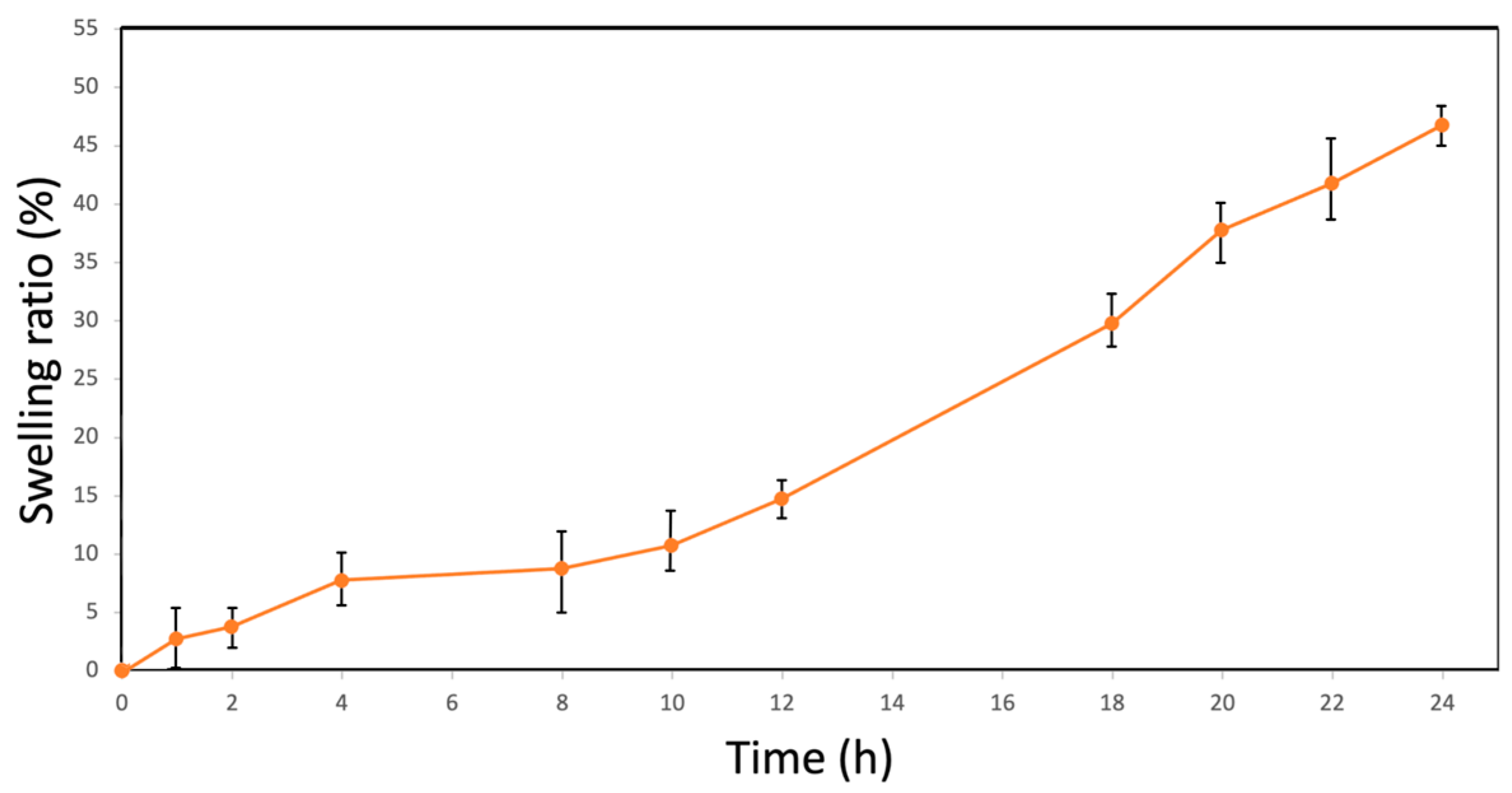
The SR of BC films in PBS was investigated to demonstrate their potential as responsive materials in moist environments as it measures the degree of expansion or swelling of a material when it absorbs a solvent. The SR increased somewhat steadily within the first few hours, with a noticeable rise from 3.2 ± 2.9% at 1 h to 15.1 ± 1.0% at 12 h. The SR continued to rise to 47.0 ± 1.7 at 24 h (Figure 2), a value within the higher range of values reported in the literature of 26.2 [64], 42.8 [65] and 45.9 [23]. The average SR of BC can vary depending on various factors such as the specific bacterial strain, growth conditions, film preparation methods, and specific characteristics of the BC membrane [66,67].
The calculated RR of the BC films was 3.0 ± 0.2, indicating that the BC material could absorb three times its own weight. This capability helps in maintaining a moist wound environment, essential for optimal healing. Additionally, it can prevent the accumulation of excess fluid, reducing the risk of maceration and promoting a cleaner and more conducive environment for tissue regeneration. Our obtained result surpasses that of previous studies utilizing native BC or composites, in the hopes of improving such properties, ranging from 2 to 10 [21,68,69]. This suggests a potentially superior performance in drug delivery and highlights the significance of our approach in improving the RR. Such variations can be attributed to differences in the degree of polymerization, crystallinity, and porosity of the BC films, as well as to variations in the composition of the liquid environment used for rehydration. These factors collectively influence the material’s capacity to absorb and retain moisture.
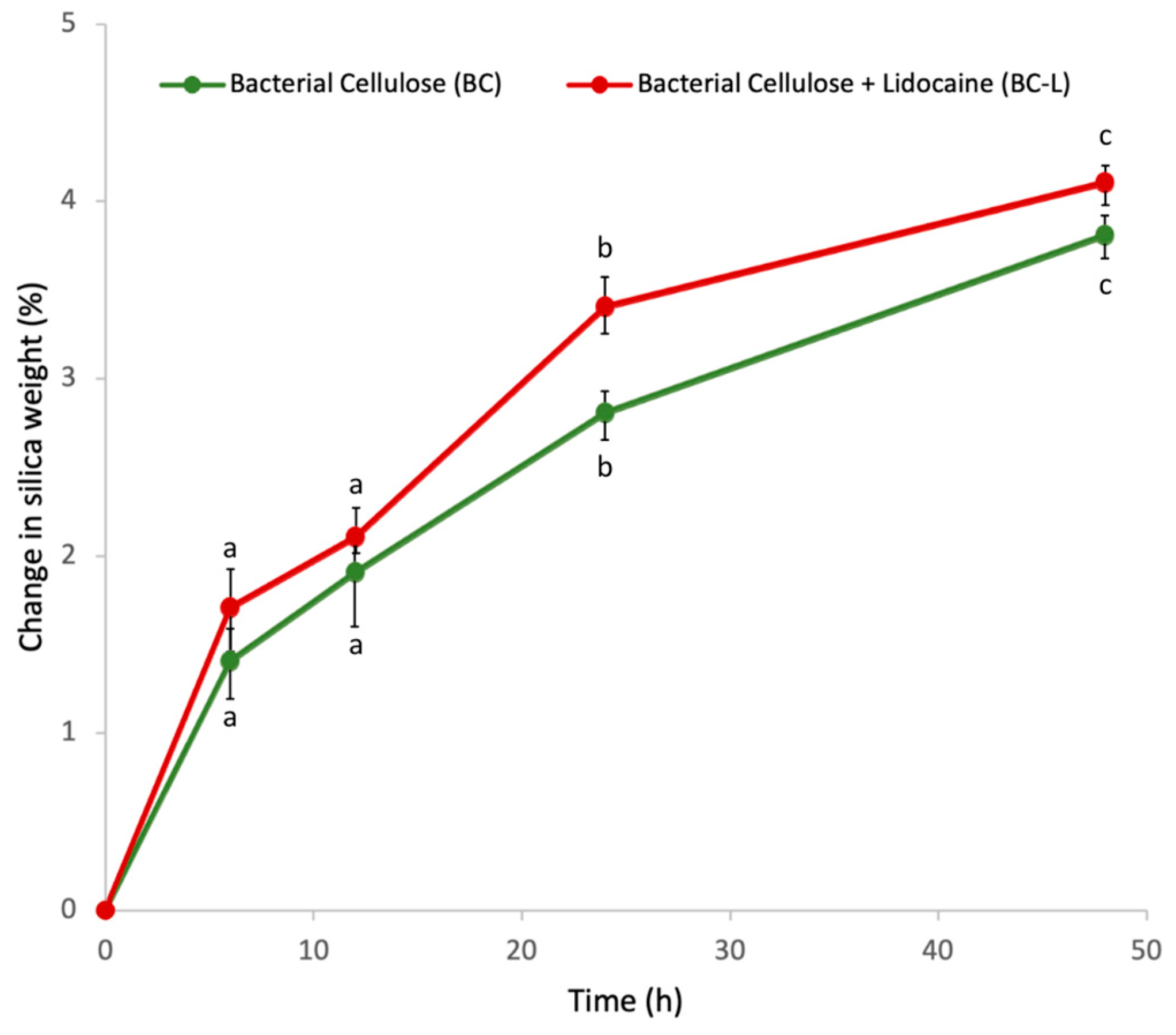
The WVP through the films was evaluated at 6, 12, 24, and 48 h, expressed as the percentage increase in weight of the initially dried silica gel. Both transparent films (BC and BC-L) exhibited similar profiles of water penetration over time (Figure 3). The presence of lidocaine was anticipated to lead to enhanced WVP due to the amount of hydrophilic amine groups and water adsorption properties [35,70]. Thus, a hydrated film should be able to facilitate vapor transfer from a moisture-rich environment to a dry environment. A wound dressing with the capacity to efficiently transfer water vapor helps to create a conducive environment for the wound, ensuring a balance between hydration and preventing excessive moisture buildup [71]. This property can contribute to a more favorable wound-healing environment by assisting in moisture management, which is essential for optimal tissue repair and regeneration [72].
According to the data obtained, the water contact angle measurement of 41.86 ± 3.10° indicates that the pure BC films exhibited a hydrophilic nature. The films exhibited a sorption index of 58.0 ± 3.0% for water and 52.0 ± 4.0% for PBS. These results align well with those reported in previous studies, such as 64% [73] and 40% [74], also in PBS. This consistency underscores the significant moisture-absorbing capacity of the BC films, highlighting their potential effectiveness in various applications. The strong attraction between the membranes and the substances can be attributed to the extensive formation of hydrogen bonds between them and the cellulose units within the membrane [32].
These results demonstrate properties that are important for various applications where moisture management and the controlled release of substances are critical factors for optimal performance. The ability of BC wound dressings to absorb and retain moisture helps to create and maintain a moist environment around the wound, which has been shown to promote wound healing by facilitating cell migration, reducing bacterial infection risk, and promoting tissue regeneration [12].

3.2. Bacterial Cellulose Film Performance Characterization

The porosity of the pure BC film was found to be 71.8 ± 4.2%, which aligns well with previous studies reporting high porosity values for BC films, typically ranging from 70% to 90% [62,75]. The high porosity of BC films is of great significance for surface modification and functionalization. BC’s porous structure provides a large surface area, allowing for efficient adsorption of various substances onto the film surface [12]. Furthermore, it also facilitates the diffusion of substances within the film matrix. This promotes the efficient transport of liquids, gases, and nutrients, which is advantageous for applications involving the exchange of substances between the film and its surroundings [33].
To validate the microbial resistance of the developed films, positive control flasks with BC-L films were employed to confirm the suitability of the nutrient broth for bacterial growth. Conversely, negative control flasks simulated conditions free from bacterial contamination. Results demonstrated that only the positive control flasks exhibited bacterial contamination (Figure 4a), while the films under investigation maintained clear solutions, signifying an absence of visible microbial contamination (Figure 4b).
The ex vivo adhesion strength was quantified as the force needed to separate the sample from the surface of the porcine skin. In our study, the adhesive strength of the developed films, referred to as BC and BC-L, was 3.2 ± 0.9 kPa and 4.0 ± 0.8 kPa, respectively, as seen in Figure 5. For comparison, we analyzed a commercially available lidocaine adhesive wound dressing (Toperma 5% 700 mg, Teikoku Seiyaku Co, Kagawa, Japan), which exhibited an adhesive strength of 8.9 ± 0.4 kPa. While our BC films demonstrated lower adhesive strengths, the observed values are still within the clinically relevant range for hydrogel wound dressings made from biomaterials like functionalized chitosan and alginate with 3.4–11.5 kPa [76,77,78].
The investigation into the folding endurance of the three distinct samples, BC, BC-L, and BC-RL, unveiled remarkably consistent outcomes. Each sample exhibited folding endurance values of 141 ± 09, 178 ± 12, and 189 ± 13 cycles, respectively. The obtained folding endurance outcomes underscore the polymer’s ability to endure folding and unfolding cycles, aligning with the findings reported in the literature of around 150 cycles [48,79]. The fatigue fracture surfaces displayed a gradual diffusion across both sides of all samples. The folding striations exhibited prominent growth, notably oriented approximately perpendicular to the direction of the fracture surface propagation. The comparable endurance values observed after lidocaine incorporation could be attributed to the molecular interactions between lidocaine and BC, resulting in a plasticized film structure or altered mechanical properties [80]. We hypothesize that these molecular interactions, which initially contribute to the plasticized film structure, do not significantly alter the folding endurance once the lidocaine has been released.
The flexibility of BC films allows them to conform closely to wound surfaces, promoting optimal contact between the drug-loaded film and the wound bed. This close contact enhances drug localization and effectiveness by facilitating efficient absorption and penetration into underlying tissues. Additionally, the enhanced mechanical properties of BC films, demonstrated by reduced fatigue fracture surfaces and increased folding striations, ensure durability and effectiveness during patient use across various body applications. Moreover, the material’s resistance to rupture enhances ease of handling, contributing to its practicality and reliability in medical settings.

3.3. Assessment of Lidocaine Incorporation and Release

Opting for in situ incorporation of lidocaine into the BC’s matrix can be challenging and often results in less predictable encapsulation efficiency. Consequently, the ex situ method emerges as a more favorable choice [81]. Therefore, the encapsulation of lidocaine was conducted through an ex situ process, where lidocaine was introduced into the pre-formed BC material, facilitating its diffusion and uniform distribution within the BC matrix. The extent of lidocaine adsorption is impacted by various factors such as the surface area of the adsorbent material and VDW forces. BC offers an ample surface area for adsorption processes and numerous sites for VDW interactions [82]. The amorphous regions of BC fibers, being less densely packed and more exposed to the surrounding aqueous environment, are inherently more susceptible to ionization and polarity effects. This makes them preferential sites for initial adsorption processes in aqueous systems [83]. This extensive surface area and the susceptibility of amorphous regions to adsorption facilitate the diffusion of lidocaine molecules into the BC matrix, enhancing the adsorption capacity of the material.
Lidocaine’s hydrophilic nature further facilitates its interaction with the cellulose network. Due to its ability to form hydrogen bonds, lidocaine can effectively penetrate the amorphous regions of BC fibers [33]. The hydrophilic groups present in lidocaine molecules, such as the amino and hydroxyl groups, are well-suited for establishing hydrogen-bonding interactions with the hydroxyl groups of cellulose [84]. Furthermore, the adsorption of lidocaine onto cellulose is governed by Coulombic forces between the positively charged lidocaine molecules and the negatively charged cellulose surface. In aqueous environments, the ionized hydroxyl groups on cellulose create negatively charged sites, attracting the positively charged lidocaine molecules [85]. These electrostatic interactions, play a significant role in facilitating the adsorption of lidocaine onto BC, further enhancing its EE. In this study, the EE of lidocaine within the BC matrix in this study was determined to be 39.86 ± 4.15%. This encapsulation efficiency is close to the average reported in the literature for similar drug-delivery systems of approximately 45% [81,86,87]. This close alignment with values in the literature further highlights the significant influence of lidocaine’s hydrophilic nature on its effective encapsulation within the BC matrix, contributing to the overall success of the drug-delivery system.
The release mechanism was characterized by a distinctive two-stage profile, as observed in the drug-release assay conducted on the BC-L films (Figure 6). During the initial stage (0–720 min), a lidocaine release of 24.6 ± 2.6% was observed. This behavior is indicative of an initial burst-release phase commonly observed in drug-delivery systems, where the surface-bound drug is rapidly released into the surrounding medium [88]. This phenomenon suggests that the release during this stage may be primarily controlled by the immediate availability of lidocaine molecules on the surface of the BC matrix.
Subsequently, between 720 and 2880 min, a slower cumulative release of 27.2 ± 2.1% of lidocaine was observed, indicating a sustained release profile. This release behavior is desirable for achieving prolonged therapeutic efficacy and minimizing the frequency of dosing in clinical applications [88] and can be attributed to the diffusion of lidocaine from deeper layers of the BC matrix [65]. Other wound-dressing materials utilizing BC as a matrix have demonstrated a rapid release of lidocaine, with over 90% of the drug being released within 20 min [89]. Even though the RR of this study could be further optimized, this sustained release profile indicates that our films show promise for prolonged treatments, ensuring a steady and extended delivery of the therapeutic agent. This characteristic is particularly advantageous for chronic wound management, where maintaining a consistent therapeutic level over an extended period is crucial for effective healing and patient comfort.
The hydrophilic nature of lidocaine and its interactions with the BC matrix significantly influence the diffusion kinetics and govern the release behavior [38]. The long, linear chains of cellulose molecules form an intricate network of interconnected pores and channels throughout the BC matrix. This network allows for the encapsulation and immobilization of lidocaine within the matrix [90]. Our experimental results underscore the importance of this molecular arrangement, as it facilitates the effective diffusion of lidocaine within the BC matrix. The binary release system employed in our BC-L films highlighted their potential for extended wound-management applications.
While our study has demonstrated the versatility and ease of functionalization of BC with lidocaine, it also underscores potential areas for further improvement. Although the films produced exhibit promising characteristics, such as flexibility and drug-delivery capabilities, further functionalization could enhance their performance in specific applications. Incorporating antimicrobial agents or growth factors into the BC matrix could augment its wound healing properties, addressing limitations related to thermal regulation, infection control, and tissue regeneration [91,92]. Similarly, the introduction of nanoparticles or bioactive compounds could impart additional functionalities, such as enhanced mechanical strength or targeted drug release, further expanding the potential applications of BC-based materials [93,94]. Integrating metal–organic frameworks (MOFs), which can be tailored to release antimicrobial agents or drugs in response to environmental triggers, could prevent infections and control drug release [41,42]. Future research efforts could focus on exploring these avenues for functionalization to optimize the properties and performance of BC films for various biomedical and pharmaceutical applications.

3.4. Fourier Transform Infrared Spectroscopy (FTIR)

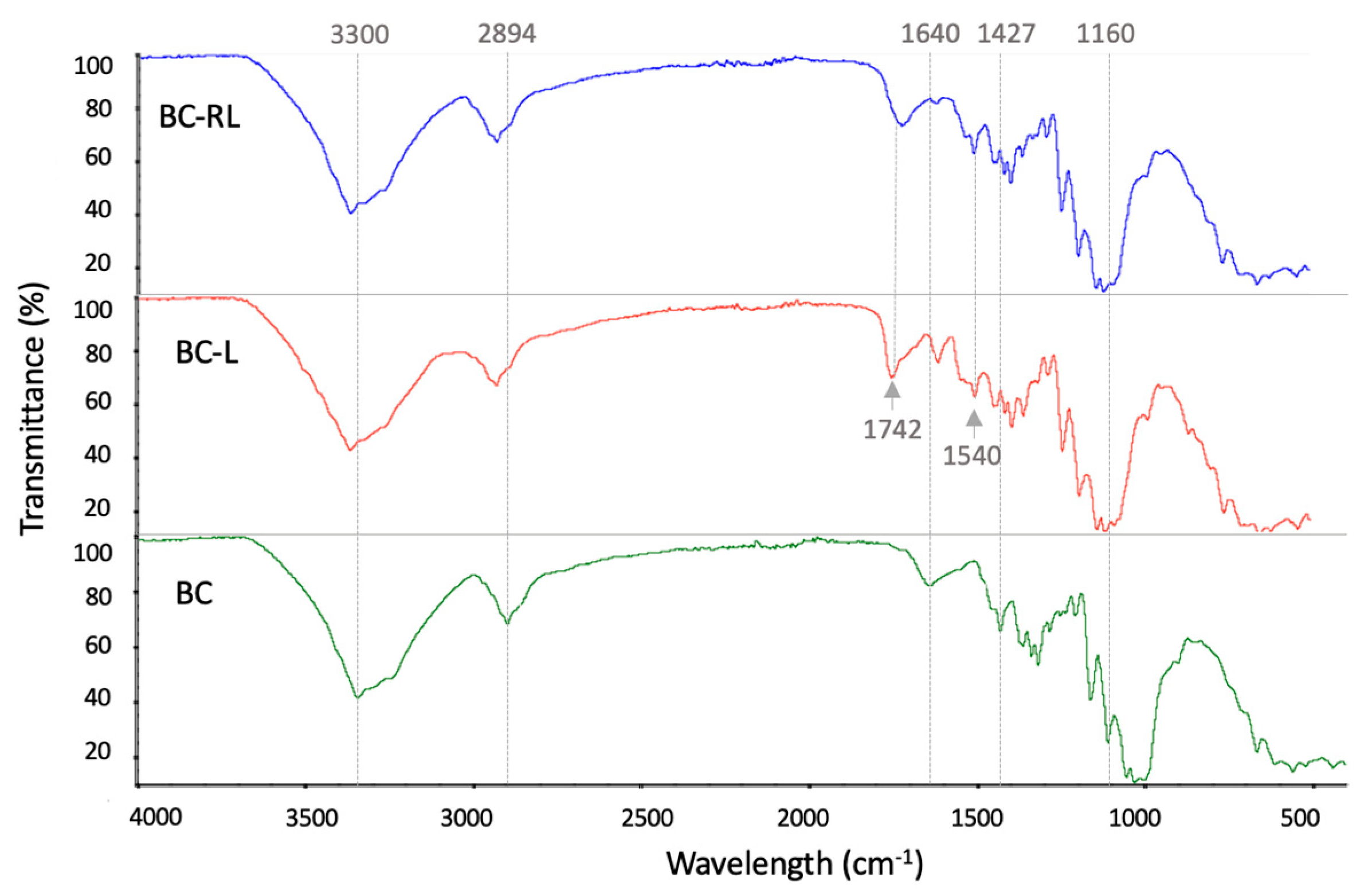
As depicted in Figure 7, the FTIR spectrum of the pure BC film reveals several distinctive peaks indicative of its molecular structure. A notable peak at 2894 cm−1 corresponds to the CH stretching vibrations at the C-6 position, suggesting the presence of alkane or alkene functional groups within the cellulose structure [21]. The spectrum also shows a range of peaks between 1427 and 1315 cm−1, which represent various functional groups and vibrations inherent to BC. These include CH2 symmetric bending, CH symmetric bending, OH in-plane bending, and CH2 wagging at the C-6 position, which are derived from the cellulose chains and their side groups [61,95]. The peaks in the 1280–1205 cm−1 range are associated with CO stretching vibrations, while those in the 1160–1029 cm−1 range are linked to COC glycosidic linkage vibrations [96].
When examining the BC-L and BC-RL films, distinct peaks corresponding to functional groups of both BC and lidocaine were observed. There was a shift of the peak to 2901 cm−1 after lidocaine incorporation and release, which indicates molecular interactions and structural changes within the BC matrix. This shift can be attributed to hydrophobic interactions and hydrogen bonding between lidocaine and cellulose, which alter the electronic environment around the C-H bonds [97]. Additionally, the incorporation and subsequent release of lidocaine may induce conformational changes or structural rearrangements in the cellulose matrix, impacting the vibrational energies of the C-H bonds. The persistence of the shifted peak after lidocaine release suggests that residual lidocaine or its effects remain in the BC matrix, continuing to influence the C-H stretching vibrations.
A sharp stretching vibration of the carbonyl (C=O) group was observed around 1742 cm−1 for both BC-L and BC-RL, which is a distinctive marker for the amide functional group present in lidocaine [97]. This is a key indicator of lidocaine’s incorporation into the BC matrix. Moreover, the peak, at 1540 cm−1, corresponds to aromatic ring vibrations, providing additional confirmation of lidocaine’s presence [98].
Changes in the intensity, broadening or position of the FTIR peaks suggest alterations in the molecular environment and potential hydrogen bonding or van der Waals interactions between the lidocaine and the cellulose fibers. The drug-release kinetics are significantly influenced by the interactions between lidocaine and the BC matrix, as evidenced by changes in the FTIR spectra [38].

3.5. X-ray Diffraction (XRD) and Crystallinity

The XRD pattern of BC and BC-RL samples exhibited diffraction peaks at 2θ angles around 14.9°, 16.6°, and 22.5°. These peaks correspond to the crystallographic planes (110), (200), and (004), respectively. However, in the BC-L sample, an additional peak was observed at 32°, which could indicate the presence of a crystalline phase, or a diffraction overlap between the two components [99]. Nonetheless, the obtained peaks in all samples are consistent with the characteristic peaks associated with the cellulose I crystal structure present in BC [38].
The crystallinity of BC plays a crucial role in its structural properties and functional behavior, such as the mechanical resistance, ultra-fine network of cellulose nanofibers, and water absorption capacity [100]. According to the obtained XRD data, the crystallinity of pure, transparent BC membranes was 45 ± 2%. However, upon the incorporation of lidocaine, an increase in crystallinity was observed, with the value reaching 50 ± 4% for BC-L. The observed rise in crystallinity is likely a result of the interactions occurring between lidocaine and the BC polymer chains. These interactions have the potential to enhance the arrangement and packaging of molecules within the film [32] (Figure 8).
Interestingly, even after subjecting the lidocaine-loaded BC films to release tests, the crystallinity remained constant at 50 ± 5% (BC-RL). The stability in crystallinity indicates that the BC films maintained their structural integrity throughout the drug-release process and is a significant indicator of its stability and durability. The sustained crystallinity observed in the lidocaine-loaded BC films is of great importance as it can influence various properties, such as mechanical strength, barrier properties, and drug-release kinetics, contributing to the overall performance of the material [100,101].

3.6. Thermogravimetric Analysis (TGA)

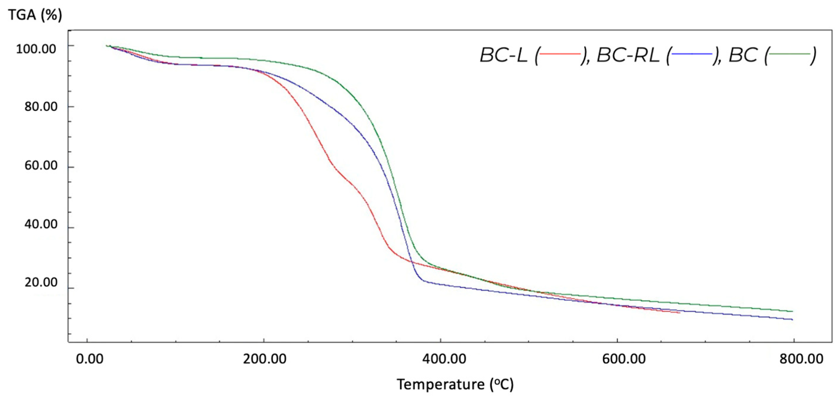
The thermal degradation of both BC and BC-RL films proceeded through three distinct stages, as outlined in Table 2, and illustrated in Figure 9.
In the initial stage (23–180 °C), moisture evaporation and the release of volatile compounds occurred in both samples. This stage primarily involves the loss of physically bound water. In the second stage (160–400 °C), the degradation of the BC matrix was observed. This stage is characterized by the thermal decomposition of the cellulose polymer, resulting in the breakdown of its molecular structure. The third stage (400–800 °C) involved further decomposition of the cellulose, leading to the generation of gases, such as CO2, CO, and other volatile compounds [95], marking the breakdown of the remaining cellulose material and the release of gaseous byproducts. This process led to the formation of residual ash of 12.38% and 9.85% of the total mass for the BC and BC-RL samples, respectively. These three degradation phases are attributed to the composition of the samples, including moisture, organic compounds, and the cellulose matrix [76].
In contrast, BC-L exhibited four stages of thermal degradation. The first stage occurred in the temperature range of 26–160 °C, corresponding to the slight weight loss attributed to moisture evaporation and the release of volatile compounds. The second stage, between 160–310 °C, showed a more pronounced weight loss associated with the decomposition of organic components, including the BC matrix and residual additives. The third stage, from 310–380 °C, resulted in significant weight loss due to the further degradation of organic compounds, and of the lidocaine [38,102]. The final stage, 380–800 °C, showed a stable weight, representing the residual ash content of the sample, of 19.42%.
The similarity in thermal degradation profiles between BC and BC-RL suggests that lidocaine release from the BC film in BC-RL does not significantly alter the overall thermal behavior of the material. However, the emergence of a distinct fourth degradation stage in the BC-L sample provides compelling evidence of the lidocaine incorporation into the film. This preservation of thermal stability ensures that the films maintain their integrity under various conditions, which is essential for applications that require exposure to high temperatures such as sterilization processes. Furthermore, the stability supports the films’ suitability for storage and transportation, particularly in the context of wound dressings, where maintaining the material’s properties is vital for effectiveness.

3.7. Scanning Electron Microscopy (SEM)

SEM analysis was conducted on the pure BC sample, BC-L, and BC-RL to examine morphological changes and the presence of lidocaine. The SEM images of the BC sample surface revealed a distinctive porous and interconnected network structure, consistent with the characteristic morphology of BC (Figure 10a,b). The fibers exhibited a smooth texture and uniform distribution, indicating a well-formed and homogeneous film structure. Additionally, the thickness of the film, measured from the cross-section (Figure 10c), was determined to be 10.5 ± 0.92 μm.
The SEM image of BC-L revealed the presence particles dispersed throughout the BC network, as depicted in Figure 11. These particles exhibit a distinct morphology consistent with lidocaine crystals, identifiable based on their size, shape, and characteristic structure [37,103]. The observation of lidocaine crystals confirms the successful incorporation of the drug into the BC film. It is noteworthy that the lidocaine crystals were only discernible under microscopic examination, being too small to be visible to the naked eye. Despite their presence, the film maintained its transparency, indicating the uniform dispersion of lidocaine within the film matrix.
Figure 12a displays the SEM image of BC-RL, which confirms the absence of visible lidocaine crystals, further attesting to the integrity of the film. In Figure 12b,c a porous network structure similar to the pure BC sample is evident. This observation suggests that lidocaine has either been released or dissolved from the porous BC matrix. This finding suggests that the BC matrix possesses the capability to accommodate the release of lidocaine while preserving its structural integrity.
The observed difference in pore dimensions between the BC films (mean pore diameter of 112.30 ± 25.5 nm) and BC-RL (mean pore diameter of 131.97 ± 29.1 nm) can suggest a correlation with the mechanism of incorporating and releasing lidocaine. The presence of lidocaine molecules influenced the arrangement and packing of cellulose fibrils within the BC matrix, resulting in alterations in the pore structure and an increase in the voids between the fibers. As lidocaine molecules diffused out of the BC matrix during the release process, they likely created pathways or channels within the film. This phenomenon contributed to further rearrangement of the pore dimensions, potentially enlarging the pores and altering their distribution [104]. These findings contribute to the understanding of the drug-delivery capabilities and structural changes in the BC film, supporting its potential applications in the biomedical field.

4. Conclusions

This research highlights a simple and efficient method for the incorporation of lidocaine into BC films by ex situ immersion and sonication methods. The characterization techniques employed, including SEM, FTIR, and XRD, confirmed the effective integration of lidocaine into the BC matrix without significantly affecting its crystalline nature. This study presents a practical solution for wound management. The obtained composite of BC and lidocaine presents itself as an environmentally friendly transparent film, which enables wound observation without the need for dressing removal, simplifying patient care in wound-healing practices.
The drug-release assay revealed a two-stage release profile with a rapid initial release followed by a secondary sustained release. These findings indicate the potential of BC films as a platform for controlled and prolonged drug delivery. The BC films maintained their thermal stability even after lidocaine incorporation, which is crucial for the sterilization, storage, and transportation of wound dressings. The study also highlighted the significant role of membrane permeability, BC properties, drug incorporation, and release behavior. These findings offer valuable insights into optimizing lidocaine-loaded BC films, showcasing their immense potential for medical applications, especially in skin condition treatments. The optimized samples show promise in improving wound care and delivering effective therapy. This technology shows promise in regulating drug availability for percutaneous administration, presenting opportunities for developing delivery systems adept at absorbing exudates and adhering to irregular skin surfaces.
However, it is important to acknowledge the inherent limitations of this proof-of-concept study. Generalizing these results to various types of wounds and clinical conditions may be constrained, necessitating further research to evaluate the effectiveness of the developed material across a broader range of scenarios. Future studies should explore additional functionalization of BC films, such as incorporating MOFs or nanoparticles, to enhance their properties and expand their applications. Moreover, comprehensive assessments, including cell attachment and viability assays, in vitro antibacterial tests, and in vivo animal testing, are essential to elucidate the wound-healing potential of the prepared dressings. Despite these limitations, this research advances the synthesis and application of BC films in wound management for drug delivery. It lays the groundwork for further exploration, optimization, and potential clinical translation, offering new possibilities for innovative therapeutic strategies and improved patient outcomes.

Author Contributions

Conceptualization, J.D.P.d.A. and L.A.S.; methodology, J.D.P.d.A.; software, J.D.P.d.A.; validation, J.D.P.d.A., A.F.d.S.C. and L.A.S.; investigation, J.D.P.d.A.; resources, L.A.S.; data curation, J.D.P.d.A.; writing—original draft preparation, J.D.P.d.A.; writing—review and editing, J.D.P.d.A., Y.d.F.C., A.D.M.d.M., C.J.G.d.S.J. and I.J.B.D.; supervision, A.F.d.S.C. and L.A.S.; project administration, A.F.d.S.C. and L.A.S.; funding acquisition, L.A.S. All authors have read and agreed to the published version of the manuscript.

Funding

This study was funded by the Coordenação de Aperfeiçoamento de Pessoal de Nível Superior (CAPES)—Finance Code 001.

Data Availability Statement

The original contributions presented in the study are included in the article, further inquiries can be directed to the corresponding author/s.

Acknowledgments

The authors are grateful to the Rede Nordeste de Biotecnologia (RENORBIO), Universidade Federal Rural de Pernambuco (UFRPE), Escola Icam Tech of Universidade Católica de Pernambuco (UNICAP), University of Washington (UW), United States of America, and Instituto Avançado de Tecnologia e Inovação (IATI), Brazil.

Conflicts of Interest

The authors declare no conflicts of interest.

References

  1. Falanga, V.; Isseroff, R.R.; Soulika, A.M.; Romanelli, M.; Margolis, D.; Kapp, S.; Granick, M.; Harding, K. Chronic wounds. Nat. Rev. Dis. Primers 2022, 8, 50. [Google Scholar] [CrossRef]
  2. Ahmed, A.; Getti, G.; Boateng, J. Medicated multi-targeted alginate-based dressings for potential treatment of mixed bacterial-fungal infections in diabetic foot ulcers. Int. J. Pharm. 2021, 606, 120903. [Google Scholar] [CrossRef]
  3. Firlar, I.; Altunbek, M.; McCarthy, C.; Ramalingam, M.; Camci-Unal, G. Functional hydrogels for treatment of chronic wounds. Gels 2022, 8, 127. [Google Scholar] [CrossRef]
  4. Khodabakhshi, D.; Eskandarinia, A.; Kefayat, A.; Rafienia, M.; Navid, S.; Karbasi, S.; Moshtaghian, J. In vitro and in vivo performance of a propolis-coated polyurethane wound dressing with high porosity and antibacterial efficacy. Colloids Surf. B Biointerfaces 2019, 178, 177–184. [Google Scholar] [CrossRef]
  5. Borges-Vilches, J.; Poblete, J.; Gajardo, F.; Aguayo, C.; Fernández, K. Graphene oxide/polyethylene glycol aerogel reinforced with grape seed extracts as wound dressing. J. Mater. Sci. 2021, 56, 16082–16096. [Google Scholar] [CrossRef]
  6. Varaprasad, K.; Jayaramudu, T.; Kanikireddy, V.; Toro, C.; Sadiku, E.R. Alginate-based composite materials for wound dressing application: A mini review. Carbohydr. Polym. 2020, 236, 116025. [Google Scholar] [CrossRef]
  7. Boateng, J.S.; Hafezi, F.; Tabriz, A.G.; Douroumis, D. 3D printed composite dressings loaded with human epidermal growth factor for potential chronic wound healing applications. J. Drug Deliv. Sci. Technol. 2023, 86, 104684. [Google Scholar] [CrossRef]
  8. Genesi, B.P.; de Melo Barbosa, R.; Severino, P.; Rodas, A.C.; Yoshida, C.M.; Mathor, M.B.; Lopes, P.S.; Viseras, C.; Souto, E.B.; da Silva, C.F. Aloe vera and copaiba oleoresin-loaded chitosan films for wound dressings: Microbial permeation, cytotoxicity, and in vivo proof of concept. Int. J. Pharm. 2023, 634, 122648. [Google Scholar] [CrossRef]
  9. Wang, X.; Zhao, D.; Li, Y.; Zhou, X.; Hui, Z.; Lei, X.; Qiu, L.; Bai, Y.; Wang, C.; Xia, J.; et al. Collagen hydrogel with multiple antimicrobial mechanisms as anti-bacterial wound dressing. Int. J. Biol. Macromol. 2023, 232, 123413. [Google Scholar] [CrossRef]
  10. Catanzano, O.; Docking, R.; Schofield, P.; Boateng, J. Advanced multi-targeted composite biomaterial dressing for pain and infection control in chronic leg ulcers. Carbohydr. Polym. 2017, 172, 40–48. [Google Scholar] [CrossRef]
  11. Graça, M.F.; Miguel, S.P.; Cabral, C.S.; Correia, I.J. Hyaluronic acid—Based wound dressings: A review. Carbohydr. Polym. 2020, 241, 116364. [Google Scholar] [CrossRef]
  12. Amorim, J.D.; da Silva Junior, C.J.; de Medeiros, A.D.; do Nascimento, H.A.; Sarubbo, M.; de Medeiros, T.P.; Costa, A.F.; Sarubbo, L.A. Bacterial Cellulose as a Versatile Biomaterial for Wound Dressing Application. Molecules 2022, 27, 5580. [Google Scholar] [CrossRef]
  13. Hou, S.; Xia, Z.; Pan, J.; Wang, N.; Gao, H.; Ren, J.; Xia, X. Bacterial cellulose applied in wound dressing materials: Production and functional modification—A review. Macromol. Biosci. 2024, 24, 2300333. [Google Scholar] [CrossRef]
  14. Rabin, N.; Zheng, Y.; Opoku-Temeng, C.; Du, Y.; Bonsu, E.; Sintim, H.O. Agents that inhibit bacterial biofilm formation. Future Med. Chem. 2015, 7, 647–671. [Google Scholar] [CrossRef]
  15. Piegerová, A.; Koščová, J.; Schusterová, P.; Nemcová, R.; Kryvtsova, M. In vitro inhibition of biofilm formation by Staphylococcus aureus under the action of selected plant extracts. Folia Vet. 2019, 63, 48–53. [Google Scholar] [CrossRef]
  16. Stanislaw, B.; Alina, K.; Marianna, T.; Halina, K. Bacterial Cellulose. In Biopolymers; Wiley-VCH Verlag GmbH: Weinheim, Germany, 2002; Volume 5, p. 37. [Google Scholar] [CrossRef]
  17. Yun, E.J.; Lee, J.; Kim, D.H.; Kim, J.; Kim, S.; Jin, Y.S.; Kim, K.H. Metabolomic elucidation of the effects of media and carbon sources on fatty acid production by Yarrowia lipolytica. J. Biotechnol. 2018, 272, 7–13. [Google Scholar] [CrossRef]
  18. Kim, J.; Kim, K.H. Effects of minimal media vs. complex media on the metabolite profiles of Escherichia coli and Saccharomyces cerevisiae. Process Biochem. 2017, 57, 64–71. [Google Scholar] [CrossRef]
  19. Choi, S.M.; Rao, K.M.; Zo, S.M.; Shin, E.J.; Han, S.S. Bacterial cellulose and its applications. Polymers 2022, 14, 1080. [Google Scholar] [CrossRef]
  20. Ahmed, J.; Gultekinoglu, M.; Edirisinghe, M. Bacterial cellulose micro-nano fibres for wound healing applications. Biotechnol. Adv. 2020, 41, 107549. [Google Scholar] [CrossRef]
  21. Balistreri, G.; Campbell, I.; Li, X.; Amorim, J.; Zhang, S.; Nance, E.; Roumeli, E. Bacterial cellulose nanoparticles as a sustainable drug delivery platform for protein-based therapeutics. RSC Appl. Polym. 2024, 2, 172–183. [Google Scholar] [CrossRef]
  22. Boyetey, M.J.B.; Torgbo, S.; Sukyai, P. Bio-scaffold for bone tissue engineering with focus on bacterial cellulose, biological materials for hydroxyapatite synthesis and growth factors. Eur. Polym. J. 2023, 194, 112168. [Google Scholar] [CrossRef]
  23. Chang, W.S.; Chen, H.H. Physical properties of bacterial cellulose composites for wound dressings. Food Hydrocoll. 2016, 53, 75–83. [Google Scholar] [CrossRef]
  24. Amorim, J.D.; Nascimento, H.A.; Silva Junior, C.J.; Medeiros, A.D.; Silva, I.D.; Costa, A.F.; Vinhas, G.M.; Sarubbo, L.A. Obtainment of bacterial cellulose with added propolis extract for cosmetic applications. Polym. Eng. Sci. 2022, 62, 565–575. [Google Scholar] [CrossRef]
  25. Atta, O.M.; Manan, S.; Ul-Islam, M.; Ahmed, A.A.; Ullah, M.W.; Yang, G. Development and characterization of plant oil-incorporated carboxymethyl cellulose/bacterial cellulose/glycerol-based antimicrobial edible films for food packaging applications. Adv. Compos. Hybrid Mater. 2022, 5, 973–990. [Google Scholar] [CrossRef]
  26. Zhang, X.; Kang, S.; Adstedt, K.; Kim, M.; Xiong, R.; Yu, J.; Chen, X.; Zhao, X.; Ye, C.; Tsukruk, V.V. Uniformly aligned flexible magnetic films from bacterial nanocelluloses for fast actuating optical materials. Nat. Commun. 2022, 13, 5804. [Google Scholar] [CrossRef]
  27. Zheng, L.; Li, S.; Luo, J.; Wang, X. Latest advances on bacterial cellulose-based antibacterial materials as wound dressings. Front. Bioeng. Biotechnol. 2020, 8, 593768. [Google Scholar] [CrossRef]
  28. Di, Z.; Shi, Z.; Ullah, M.W.; Li, S.; Yang, G. A transparent wound dressing based on bacterial cellulose whisker and poly (2-hydroxyethyl methacrylate). Int. J. Biol. Macromol. 2017, 105, 638–644. [Google Scholar] [CrossRef]
  29. Numata, Y.; Masaki, S.; Tajima, K. Mechanical properties of a bacterial cellulose/polyethylene glycol gel with a peripheral region crosslinked by polyethylene glycol diacrylate. Polym. J. 2016, 48, 317–321. [Google Scholar] [CrossRef]
  30. Sperotto, G.; Stasiak, L.G.; Godoi, J.P.; Gabiatti, N.C.; De Souza, S.S. A review of culture media for bacterial cellulose production: Complex, chemically defined and minimal media modulations. Cellulose 2021, 28, 2649–2673. [Google Scholar] [CrossRef]
  31. de Souza, S.S.; Berti, F.V.; de Oliveira, K.P.; Pittella, C.Q.; de Castro, J.V.; Pelissari, C.; Rambo, C.R.; Porto, L.M. Nanocellulose biosynthesis by Komagataeibacter hansenii in a defined minimal culture medium. Cellulose 2019, 26, 1641–1655. [Google Scholar] [CrossRef]
  32. Pandey, A.; Singh, M.K.; Singh, A. Bacterial cellulose: A smart biomaterial for biomedical applications. J. Mater. Res. 2023, 39, 2–18. [Google Scholar] [CrossRef]
  33. Trovatti, E.; Freire, C.S.; Pinto, P.C.; Almeida, I.F.; Costa, P.; Silvestre, A.J.; Neto, C.P.; Rosado, C. Bacterial cellulose membranes applied in topical and transdermal delivery of lidocaine hydrochloride and ibuprofen: In vitro diffusion studies. Int. J. Pharm. 2012, 435, 83–87. [Google Scholar] [CrossRef]
  34. Park, T.H. Topical Anesthetic versus Lidocaine Mixture for Pain Relief During Keloid Treatment: A Prospective, Split Study. J. Craniofacial Surg. 2023, 34, 10–97. [Google Scholar] [CrossRef]
  35. Karnina, R.; Arif, S.K.; Hatta, M.; Bukhari, A. Molecular mechanisms of lidocaine. Ann. Med. Surg. 2021, 69, 102733. [Google Scholar] [CrossRef]
  36. Gupta, A.K.; Sibbald, R.G. Eutectic lidocaine/prilocaine 5% cream and patch may provide satisfactory analgesia for excisional biopsy or curettage with electrosurgery of cutaneous lesions: A randomized, controlled, parallel group study. J. Am. Acad. Dermatol. 1996, 35, 419–423. [Google Scholar] [CrossRef]
  37. Yang, X.; Wei, X.; Mu, Y.; Li, Q.; Liu, J. A review of the mechanism of the central analgesic effect of lidocaine. Medicine 2020, 99, e19898. [Google Scholar] [CrossRef]
  38. Kim, H.Y.; Choi, J.B.; Min, S.K.; Chang, M.Y.; Lim, G.M.; Kim, J.E. A randomized clinical trial on the effect of a lidocaine patch on shoulder pain relief in laparoscopic cholecystectomy. Sci. Rep. 2021, 11, 1052. [Google Scholar] [CrossRef]
  39. Mick, G.; Correa-Illanes, G. Topical pain management with the 5% lidocaine medicated plaster–a review. Curr. Med. Res. Opin. 2012, 28, 937–951. [Google Scholar] [CrossRef]
  40. Ahmed, I.; Jhung, S.H. Adsorptive denitrogenation of model fuel with CuCl-loaded metal–organic frameworks (MOFs). Chem. Eng. J. 2014, 251, 35–42. [Google Scholar] [CrossRef]
  41. Sarker, M.; Jhung, S.H. Zr-MOF with free carboxylic acid for storage and controlled release of caffeine. J. Mol. Liq. 2019, 296, 112060. [Google Scholar] [CrossRef]
  42. Sarker, M.; Shin, S.; Jhung, S.H. Synthesis and functionalization of porous Zr-diaminostilbenedicarboxylate metal–organic framework for storage and stable delivery of ibuprofen. ACS Omega 2019, 4, 9860–9867. [Google Scholar] [CrossRef]
  43. Ferguson, A.; Khan, U.; Walsh, M.; Lee, K.Y.; Bismarck, A.; Shaffer, M.S.; Coleman, J.N.; Bergin, S.D. Understanding the dispersion and assembly of bacterial cellulose in organic solvents. Biomacromolecules 2016, 17, 1845–1853. [Google Scholar] [CrossRef]
  44. Gudin, J.; Nalamachu, S. Utility of lidocaine as a topical analgesic and improvements in patch delivery systems. Postgrad. Med. 2020, 132, 28–36. [Google Scholar] [CrossRef]
  45. Tse, C.H.; Comer, J.; Wang, Y.; Chipot, C. Link between membrane composition and permeability to drugs. J. Chem. Theory Comput. 2018, 14, 2895–2909. [Google Scholar] [CrossRef]
  46. Hamed, D.A.; Maghrawy, H.H.; Abdel Kareem, H. Biosynthesis of bacterial cellulose nanofibrils in black tea media by a symbiotic culture of bacteria and yeast isolated from commercial kombucha beverage. World J. Microbiol. Biotechnol. 2023, 39, 48. [Google Scholar] [CrossRef]
  47. Tabaii, M.J.; Emtiazi, G. Transparent nontoxic antibacterial wound dressing based on silver nano particle/bacterial cellulose nano composite synthesized in the presence of tripolyphosphate. J. Drug Deliv. Sci. Technol. 2018, 44, 244–253. [Google Scholar] [CrossRef]
  48. Galdino, C.J.G.; Amorim, J.D.P.; Medeiros, A.D.M.; Cavalcanti, A.K.L.H.; Nascimento, H.A.; Henrique, M.A.; Maranhão, L.J.C.N.; Vinhas, G.M.; Souto Silva, K.K.O.; Costa, A.F.S.; et al. Design of a Naturally Dyed and Waterproof Bio-technological Leather from Reconstituted Cellulose. J. Funct. Biomater. 2022, 13, 49. [Google Scholar] [CrossRef]
  49. Saska, S.; Teixeira, L.N.; de Oliveira, P.T.; Gaspar, A.M.; Ribeiro, S.J.; Messaddeq, Y.; Marchetto, R. Bacterial cellulose-collagen nano-composite for bone tissue engineering. J. Mater. Chem. 2012, 22, 22102–22112. [Google Scholar] [CrossRef]
  50. Shezad, O.; Khan, S.; Khan, T.; Park, J.K. Physicochemical and mechanical characterization of bacterial cellulose produced with an excellent productivity in static conditions using a simple fed-batch cultivation strategy. Carbohydr. Polym. 2010, 82, 173–180. [Google Scholar] [CrossRef]
  51. Padula, C.; Nicoli, S.; Colombo, P.; Santi, P. Single-layer transdermal film containing lidocaine: Modulation of drug release. Eur. J. Pharm. Biopharm. 2007, 66, 422–428. [Google Scholar] [CrossRef]
  52. Wiberg, K.; Hagman, A.; Burén, P.; Jacobsson, S.P. Determination of the content and identity of lidocaine solutions with UV–visible spectroscopy and multivariate calibration. Analyst 2001, 126, 1142–1148. [Google Scholar] [CrossRef]
  53. Hou, S.; Hsiu-Ying, Y. Comparison of absorption of aqueous lidocaine and liposome lidocaine following topical application on rabbit vessels. J. Orthop. Res. 1994, 12, 294–297. [Google Scholar] [CrossRef]
  54. Marin, E.; Rojas, J. Preparation and characterization of crosslinked poly (vinyl) alcohol films with waterproof properties. Int. J. Pharm. Pharm. Sci. 2015, 7, 242–248. [Google Scholar]
  55. Kamiński, K.; Jarosz, M.; Grudzień, J.; Pawlik, J.; Zastawnik, F.; Pandyra, P.; Kołodziejczyk, A.M. Hydrogel bacterial cellulose: A path to improved materials for new eco-friendly textiles. Cellulose 2020, 27, 5353–5365. [Google Scholar] [CrossRef]
  56. Wittaya-areekul, S.; Prahsarn, C. Development and in vitro evaluation of chitosan–polysaccharides composite wound dressings. Int. J. Pharm. 2006, 313, 123–128. [Google Scholar] [CrossRef]
  57. Asare, E.O.; Mun, E.A.; Marsili, E.; Paunov, V.N. Nanotechnologies for control of pathogenic microbial biofilms. J. Mater. Chem. B 2022, 10, 5129–5153. [Google Scholar] [CrossRef]
  58. Jin, S.G.; Kim, K.S.; Kim, D.W.; Kim, D.S.; Seo, Y.G.; Go, T.G.; Youn, Y.S.; Kim, J.O.; Yong, C.S.; Choi, H.G. Development of a novel sodium fusidate-loaded triple polymer hydrogel wound dressing: Mechanical properties and effects on wound repair. Int. J. Pharm. 2016, 497, 114–122. [Google Scholar] [CrossRef]
  59. Hu, Y.; Zhu, Y.; Zhou, X.; Ruan, C.; Pan, H.; Catchmark, J.M. Bioabsorbable cellulose composites prepared by an improved mineral-binding process for bone defect repair. J. Mater. Chem. B 2016, 4, 1235–1246. [Google Scholar] [CrossRef]
  60. Sehaqui, H.; Zhou, Q.; Ikkala, O.; Berglund, L.A. Strong and tough cellulose nanopaper with high specific surface area and porosity. Biomacromolecules 2011, 12, 3638–3644. [Google Scholar] [CrossRef]
  61. Fei, G.; Wang, Y.; Wang, H.; Ma, Y.; Guo, Q.; Huang, W.; Yang, D.; Shao, Y.; Ni, Y. Fabrication of bacterial cellulose/polyaniline nanocomposite paper with excellent conductivity, strength, and flexibility. ACS Sustain. Chem. Eng. 2019, 7, 8215–8225. [Google Scholar] [CrossRef]
  62. Medeiros, A.D.; Silva Junior, C.J.; Amorim, J.D.; Durval, I.J.; Damian, R.B.; Cavalcanti, Y.D.; Costa, A.F.; Sarubbo, L.A. Design and Modeling of a Biotechnological Nanofiltration Module Using Bacterial Cellulose Membranes for the Separation of Oily Mixtures. Water 2023, 15, 2. [Google Scholar] [CrossRef]
  63. van Zyl, E.M.; Kennedy, M.A.; Nason, W.; Fenlon, S.J.; Young, E.M.; Smith, L.J.; Bhatia, S.R.; Coburn, J.M. Structural properties of optically clear bacterial cellulose produced by Komagataeibacter hansenii using arabitol. Biomater. Adv. 2023, 148, 213345. [Google Scholar] [CrossRef] [PubMed]
  64. Wei, B.; Yang, G.; Hong, F. Preparation and evaluation of a kind of bacterial cellulose dry films with antibacterial properties. Carbohydr. Polym. 2011, 84, 533–538. [Google Scholar] [CrossRef]
  65. Juncu, G.; Stoica-Guzun, A.; Stroescu, M.; Isopencu, G.; Jinga, S.I. Drug release kinetics from carboxymethylcellulose-bacterial cellulose composite films. Int. J. Pharm. 2016, 510, 485–492. [Google Scholar] [CrossRef] [PubMed]
  66. Zhang, M.; Chen, S.; Sheng, N.; Wang, B.; Wu, Z.; Liang, Q.; Wang, H. Anisotropic bacterial cellulose hydrogels with tunable high mechanical performances, non-swelling and bionic nanofluidic ion transmission behavior. Nanoscale 2021, 13, 8126–8136. [Google Scholar] [CrossRef] [PubMed]
  67. Clasen, C.; Sultanova, B.; Wilhelms, T.; Heisig, P.; Kulicke, W.M. Effects of different drying processes on the material properties of bacterial cellulose membranes. In Macromolecular Symposia; WILEY-VCH Verlag: Weinheim, Germany, 2006; Volume 244, pp. 48–58. [Google Scholar] [CrossRef]
  68. Huang, H.C.; Chen, L.C.; Lin, S.B.; Hsu, C.P.; Chen, H.H. In situ modification of bacterial cellulose network structure by adding interfering substances during fermentation. Bioresour. Technol. 2010, 101, 6084–6091. [Google Scholar] [CrossRef] [PubMed]
  69. Chen, T.; Chen, Y.; Rehman, H.U.; Chen, Z.; Yang, Z.; Wang, M.; Li, H.; Liu, H. Ultratough, self-healing, and tissue-adhesive hydrogel for wound dressing. ACS Appl. Mater. Interfaces 2018, 10, 33523–33531. [Google Scholar] [CrossRef] [PubMed]
  70. Cazón, P.; Velázquez, G.; Vázquez, M. Bacterial cellulose films: Evaluation of the water interaction. Food Packag. Shelf Life 2020, 25, 100526. [Google Scholar] [CrossRef]
  71. Bainbridge, P.; Browning, P.; Bernatchez, S.F.; Blaser, C.; Hitschmann, G. Comparing test methods for moisture-vapor transmission rate (MVTR) for vascular access transparent semipermeable dressings. J. Vasc. Access 2023, 24, 1000–1007. [Google Scholar] [CrossRef]
  72. Shi, S.; Wu, H.; Zhi, C.; Yang, J.; Si, Y.; Ming, Y.; Fei, B.; Hu, J. A skin-like nanostructured membrane for advanced wound dressing. Compos. Part B Eng. 2023, 250, 110438. [Google Scholar] [CrossRef]
  73. Gama, M.; Gatenholm, P.; Klemm, D. (Eds.) Bacterial Nanocellulose: A Sophisticated Multifunctional Material; CRC Press: Boca Raton, FL, USA, 2012. [Google Scholar]
  74. Li, Y.; Jiang, H.; Zheng, W.; Gong, N.; Chen, L.; Jiang, X.; Yang, G. Bacterial cellulose–hyaluronan nanocomposite biomaterials as wound dressings for severe skin injury repair. J. Mater. Chem. B 2015, 3, 3498–3507. [Google Scholar] [CrossRef]
  75. Nascimento, H.A.; Amorim, J.D.; Mfilho, L.E.; Costa, A.F.; Sarubbo, L.A.; Napoleão, D.C.; Maria Vinhas, G. Production of bacterial cellulose with antioxidant additive from grape residue with promising cosmetic applications. Polym. Eng. Sci. 2022, 62, 2826–2839. [Google Scholar] [CrossRef]
  76. Yang, Y.; Liang, Y.; Chen, J.; Duan, X.; Guo, B. Mussel-inspired adhesive antioxidant antibacterial hemostatic composite hydrogel wound dressing via photo-polymerization for infected skin wound healing. Bioact. Mater. 2022, 8, 341–354. [Google Scholar] [CrossRef]
  77. He, J.; Shi, M.; Liang, Y.; Guo, B. Conductive adhesive self-healing nanocomposite hydrogel wound dressing for photothermal therapy of infected full-thickness skin wounds. Chem. Eng. J. 2020, 394, 124888. [Google Scholar] [CrossRef]
  78. Momoh, F.U.; Boateng, J.S.; Richardson, S.C.; Chowdhry, B.Z.; Mitchell, J.C. Development and functional characterization of alginate dressing as potential protein delivery system for wound healing. Int. J. Biol. Macromol. 2015, 81, 137–150. [Google Scholar] [CrossRef]
  79. Singh, B.; Abhishek, D. Design of acacia gum–carbopol–cross-linked-polyvinylimidazole hydrogel wound dressings for antibiotic/anesthetic drug delivery. Ind. Eng. Chem. Res. 2016, 55, 9176–9188. [Google Scholar] [CrossRef]
  80. Eleftheriadis, G.K.; Monou, P.K.; Bouropoulos, N.; Boetker, J.; Rantanen, J.; Jacobsen, J.; Vizirianakis, I.S.; Fatouros, D.G. Fabrication of mucoadhesive buccal films for local administration of ketoprofen and lidocaine hydrochloride by combining fused deposition modeling and inkjet printing. J. Pharm. Sci. 2020, 109, 2757–2766. [Google Scholar] [CrossRef]
  81. Franz-Montan, M.; Baroni, D.; Brunetto, G.; Sobral, V.R.V.; da Silva, C.M.G.; Venâncio, P.; Zago, P.W.; Cereda, C.M.S.; Volpato, M.C.; de Araújo, D.R.; et al. Liposomal lidocaine gel for topical use at the oral mucosa: Characterization, in vitro assays and in vivo an-esthetic efficacy in humans. J. Liposome Res. 2015, 25, 11–19. [Google Scholar] [CrossRef]
  82. Pahlevan, M.; Toivakka, M.; Alam, P. Mechanical properties of TEMPO-oxidised bacterial cellulose-amino acid biomaterials. Eur. Polym. J. 2018, 101, 29–36. [Google Scholar] [CrossRef]
  83. Ashaduzzaman, M.; Al-Rafin, A.; Mustary, N.; Shamsuddin, S.M. Studies on Interaction of Lidocaine Drug with Natural Cellulosic Fibres. Int. J. Pharmacol. Phytochem. Ethnomed. 2016, 4, 36–46. [Google Scholar] [CrossRef]
  84. Favatela, F.; Horst, M.F.; Bracone, M.; Gonzalez, J.; Alvarez, V.; Lassalle, V. Gelatin/Cellulose nanowhiskers hydrogels intended for the administration of drugs in dental treatments: Study of lidocaine as model case. J. Drug Deliv. Sci. Technol. 2021, 61, 101886. [Google Scholar] [CrossRef]
  85. Yang, D.; Chen, X.; Li, Z.; Yang, C. Mechanistic Study of Release Characteristics of Two Active Ingredients in Transdermal Patch Containing Lidocaine−Flurbiprofen Ionic Liquid. Pharmaceutics 2022, 14, 2158. [Google Scholar] [CrossRef] [PubMed]
  86. Babaei, S.; Ghanbarzadeh, Z.M.; Adib, M.; Kouhsoltani, S.; Davaran, H.; Hamishehkar, H. Enhanced skin penetration of lidocaine through encapsulation into nanoethosomes and nanostructured lipid carriers: A comparative study. Die Pharm. Int. J. Pharm. Sci. 2016, 71, 247–251. [Google Scholar] [CrossRef]
  87. Bakonyi, M.; Berkó, S.; Budai-Szűcs, M.; Kovács, A.; Csányi, E. DSC for evaluating the encapsulation efficiency of lidocaine-loaded liposomes compared to the ultracentrifugation method. J. Therm. Anal. Calorim. 2017, 130, 1619–1625. [Google Scholar] [CrossRef]
  88. Rodríguez, K.; Sundberg, J.; Gatenholm, P.; Renneckar, S. Electrospun nanofibrous cellulose scaffolds with controlled microar-chitecture. Carbohydr. Polym. 2014, 100, 143–149. [Google Scholar] [CrossRef] [PubMed]
  89. Trovatti, E.; Silva, N.H.; Duarte, I.F.; Rosado, C.F.; Almeida, I.F.; Costa, P.; Freire, C.S.; Silvestre, A.J.; Neto, C.P. Biocellulose membranes as supports for dermal release of lidocaine. Biomacromolecules 2011, 12, 4162–4168. [Google Scholar] [CrossRef] [PubMed]
  90. Müller, A.; Zink, M.; Hessler, N.; Wesarg, F.; Müller, F.A.; Kralisch, D.; Fischer, D. Bacterial nanocellulose with a shape-memory effect as potential drug delivery system. RSC Adv. 2014, 4, 57173–57184. [Google Scholar] [CrossRef]
  91. Zhang, Q.; Qi, C.; Wang, X.; Zhu, B.; Li, W.; Xiao, X.; Fu, H.; Hu, S.; Zhu, S.; Xu, W.; et al. Daytime radiative cooling dressings for accelerating wound healing under sunlight. Nat. Chem. Eng. 2024, 1, 301–310. [Google Scholar] [CrossRef]
  92. Loh, E.Y.; Mohamad, N.; Fauzi, M.B.; Ng, M.H.; Ng, S.F.; Mohd Amin, M.C. Development of a bacterial cellulose-based hydrogel cell carrier containing keratinocytes and fibroblasts for full-thickness wound healing. Sci. Rep. 2018, 8, 2875. [Google Scholar] [CrossRef]
  93. Mocanu, A.; Isopencu, G.; Busuioc, C.; Popa, O.M.; Dietrich, P.; Socaciu-Siebert, L. Bacterial cellulose films with ZnO nanoparticles and propolis extracts: Synergistic antimicrobial effect. Sci. Rep. 2019, 9, 17687. [Google Scholar] [CrossRef]
  94. Onyszko, M.; Markowska-Szczupak, A.; Rakoczy, R.; Paszkiewicz, O.; Janusz, J.; Gorgon-Kuza, A.; Wenelska, K.; Mijowska, E. The cellulose fibers functionalized with star-like zinc oxide nanoparticles with boosted antibacterial performance for hygienic products. Sci. Rep. 2022, 12, 1321. [Google Scholar] [CrossRef] [PubMed]
  95. Liu, X.; Cao, L.; Wang, S.; Huang, L.; Zhang, Y.; Tian, M.; Li, X.; Zhang, J. Isolation and characterization of bacterial cellulose produced from soybean whey and soybean hydrolyzate. Sci. Rep. 2023, 13, 16024. [Google Scholar] [CrossRef] [PubMed]
  96. Zeng, M.; Laromaine, A.; Roig, A. Bacterial cellulose films: Influence of bacterial strain and drying route on film properties. Cellulose 2014, 21, 4455–4469. [Google Scholar] [CrossRef]
  97. Rasool, A.; Ata, S.; Islam, A.; Rizwan, M.; Azeem, M.K.; Mehmood, A.; Khan, R.U.; Mahmood, H.A. Kinetics and controlled release of lidocaine from novel carrageenan and alginate-based blend hydrogels. Int. J. Biol. Macromol. 2020, 147, 67–78. [Google Scholar] [CrossRef] [PubMed]
  98. Brassolatti, P.; Kido, H.W.; Bossini, P.S.; Gabbai-Armelin, P.R.; Otterço, A.N.; Almeida-Lopes, L.; Zanardi, L.M.; Napolitano, M.A.; de Avó, L.R.; Forato, L.A.; et al. Bacterial cellulose membrane used as biological dressings on third-degree burns in rats. Bio-Med. Mater. Eng. 2018, 29, 29–42. [Google Scholar] [CrossRef] [PubMed]
  99. Ghozali, M.; Meliana, Y.; Chalid, M. Synthesis and characterization of bacterial cellulose by Acetobacter xylinum using liquid tapioca waste. Mater. Today Proc. 2021, 44, 2131–2134. [Google Scholar] [CrossRef]
  100. Ruan, C.; Zhu, Y.; Zhou, X.; Abidi, N.; Hu, Y.; Catchmark, J.M. Effect of cellulose crystallinity on bacterial cellulose assembly. Cellulose 2016, 23, 3417–3427. [Google Scholar] [CrossRef]
  101. Rodriguez-Chanfrau, J.E.; Santos, M.L.; Riccardi, C.D.; De Olyveira, G.M.; Hernandez-Escalona, M.; Basmaji, P.; Veranes-Pantoja, Y.; Guastaldi, A.C. Chemical modification of bacterial cellulose for use in regenerative medicine. Cellul. Chem. Technol. 2017, 51, 673–680. [Google Scholar]
  102. Verdugo-Escamilla, C.; Alarcón-Payer, C.; Acebedo-Martínez, F.J.; Fernández-Penas, R.; Domínguez-Martín, A.; Choquesillo-Lazarte, D. Lidocaine Pharmaceutical Multicomponent Forms: A Story about the Role of Chloride Ions on Their Stability. Crystals 2022, 12, 798. [Google Scholar] [CrossRef]
  103. Masaki, Y.; Tanaka, M.; Nishikawa, T. Physicochemical compatibility of propofol-lidocaine mixture. Anesth. Analg. 2003, 97, 1646–1651. [Google Scholar] [CrossRef]
  104. Li, G.; Nandgaonkar, A.G.; Habibi, Y.; Krause, W.E.; Wei, Q.; Lucia, L.A. An environmentally benign approach to achieving vectorial alignment and high microporosity in bacterial cellulose/chitosan scaffolds. RSC Adv. 2017, 7, 13678–13688. [Google Scholar] [CrossRef]
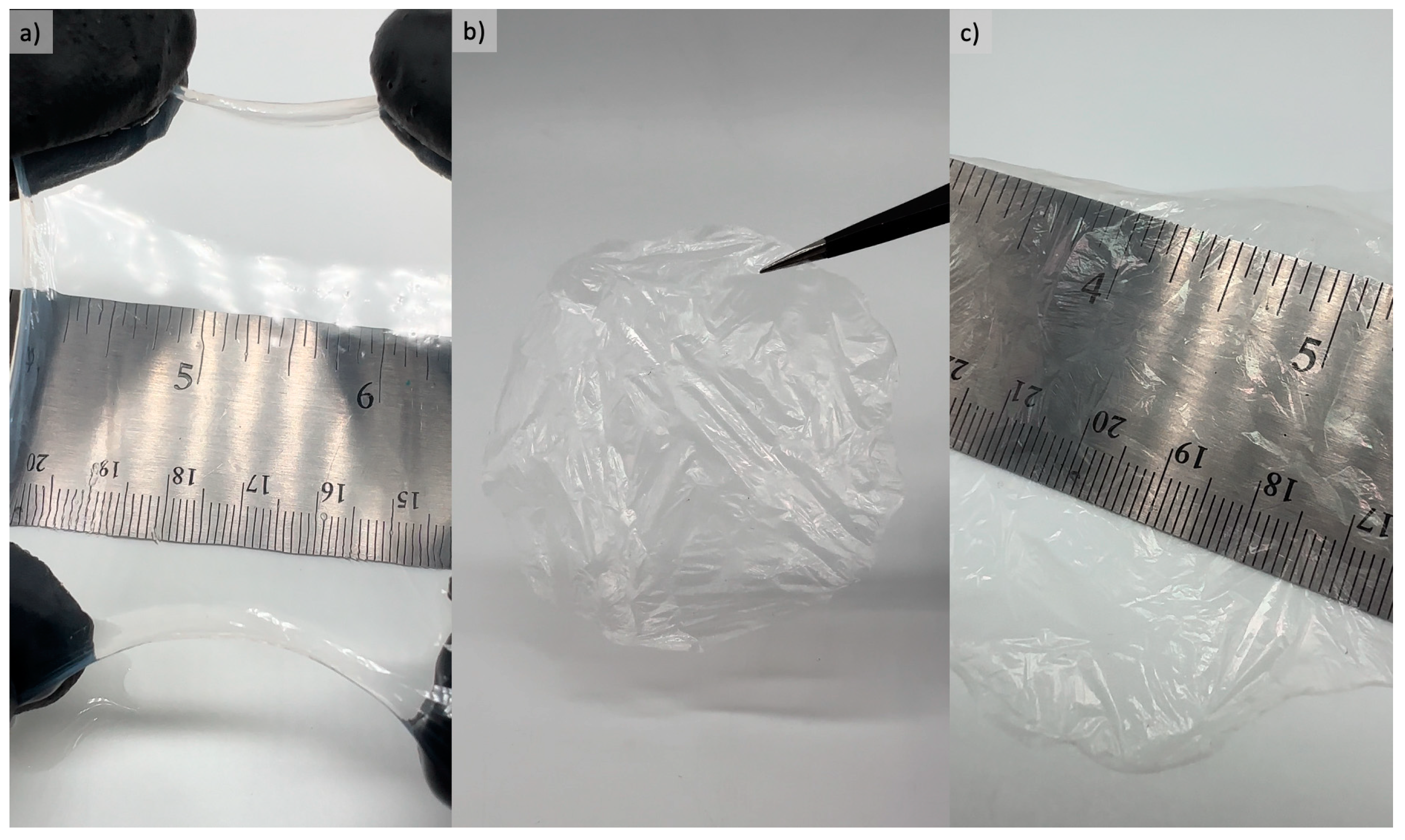
Figure 1. (a) Visual aspects of wet bacterial cellulose transparent film after purification process; and (b,c) dry bacterial cellulose transparent film.
Processes 12 01282 g001
Figure 2. Swelling ratio of the BC films from 0–24 h.
Processes 12 01282 g002
Figure 3. Vapor penetration through films at 6, 12, 24, and 48 h. Results correspond to the average of five replicates with standard deviations. a–c Values for each sample with different superscripts are significantly different (p < 0.05).
Processes 12 01282 g003
Figure 4. Microbial resistance of the developed BC-L films: (a) positive control flasks exhibiting bacterial contamination; and (b) negative control flasks maintained clear solutions, indicating the absence of visible microbial contamination.
Processes 12 01282 g004
Figure 5. Adhesive strength of the bacterial cellulose films (BC), BC films incorporated with lidocaine (BC-L) and commercial adhesive wound-dressing material. Results correspond to the average of five replicates with standard deviations. a–c Values for each sample with different superscripts are significantly different (p < 0.05).
Processes 12 01282 g005
Figure 6. Cumulative lidocaine release (%) over time. Results correspond to the average of five replicates with standard deviations.
Processes 12 01282 g006
Figure 7. FTIR spectra of bacterial cellulose films (BC), BC films incorporated with lidocaine (BC-L) and BC-L films after drug-release test (BC-RL).
Processes 12 01282 g007
Figure 8. X-ray diffractograms showing the highlighted amorphous halos of (a) bacterial cellulose films (BC), (b) BC films incorporated with lidocaine (BC-L) and (c) BC-L films after drug-release test (BC-RL).
Processes 12 01282 g008
Figure 9. Thermogravimetric curves of bacterial cellulose films (BC), BC films incorporated with lidocaine (BC-L) and BC-L films after drug-release test (BC-RL).
Processes 12 01282 g009
Figure 10. Scanning electron microscopy (SEM) images of the surface view of pure bacterial cellulose film (BC): (ac) cross-section view.
Processes 12 01282 g010
Figure 11. Scanning electron microscopy surface images of the bacterial cellulose film incorporated with lidocaine (BC-L).
Processes 12 01282 g011
Figure 12. (a) Macrograph of transparent BC film after lidocaine release test; (b,c) Scanning electron microscopy surface images of the bacterial cellulose film after the lidocaine release test with different magnifications (BC-RL).
Processes 12 01282 g012
Table 1. Bacterial cellulose yields and moisture content (MC) results of bacterial cellulose films (BC).
Wet Bacterial Cellulose YieldDry Bacterial Cellulose YieldMoisture Content (MC)
77.50 ± 3.71 g/L b1.24 ± 0.32 g/L a98.4 ± 0.3% c
a–c Values for each sample with different superscripts are significantly different (n = 5, p < 0.05).
Table 2. Thermogravimetric analysis results of bacterial cellulose films (BC), BC films incorporated with lidocaine (BC-L) and BC-L films after drug-release test (BC-RL).
SampleTemperature Range (°C)Weight Loss (%)Residue (%)
BC23–1804.2512.38
180–40068.99
400–80016.37
BC-L26–1606.1519.42
160–31046.51
310–38016.37
380–80011.55
BC-RL27–1606.469.85
160–39071.51
390–80012.15
Disclaimer/Publisher’s Note: The statements, opinions and data contained in all publications are solely those of the individual author(s) and contributor(s) and not of MDPI and/or the editor(s). MDPI and/or the editor(s) disclaim responsibility for any injury to people or property resulting from any ideas, methods, instructions or products referred to in the content.