Article

Biodecolorization and Biodegradation of Sulfur Black by the Strain Aspergillus sp. DS-28

1
SINOPEC Maoming Petrochemical Company Ltd., Maoming 525000, China
2
School of Biological and Food Engineering, Changzhou University, Changzhou 213164, China
*
Author to whom correspondence should be addressed.
Processes 2024, 12(9), 1818; https://doi.org/10.3390/pr12091818
Submission received: 28 July 2024 / Revised: 15 August 2024 / Accepted: 23 August 2024 / Published: 27 August 2024
(This article belongs to the Section Biological Processes and Systems)

Abstract

:
The textile industry significantly contributes to environmental pollution through its use of synthetic dyes, especially sulfur black, known for its toxicity and resistance to degradation. This research focuses on a fungal strain, Aspergillus sp. strain DS-28, isolated from activated sludge, which exhibits an exceptional ability to biodegrade sulfur black dye. This study systematically assessed the biodegradation capacity of this strain through a series of experiments conducted over a 7-day period. Analytical techniques including high-performance liquid chromatography time-of-flight mass spectrometry (HPLC-TOF/MS), scanning electron microscopy (SEM), X-ray diffraction (XRD), and Fourier-transform infrared spectroscopy (FTIR) were employed to monitor the degradation process. SEM showed a significant reduction in particle size, with surfaces becoming smoother and flatter post treatment. XRD indicated a decrease in the intensity of several chemical bonds, and FTIR analysis demonstrated the enhanced vibrational absorption peaks of benzene ring bonds, with the disappearance of -C-S- and -C-S-S-C- groups. The results demonstrate that Aspergillus sp. DS-28 degrades sulfur black by initiating the oxidative breakdown of its complex structures into simpler forms. This study not only elucidates the biodegradation pathway facilitated by Aspergillus sp. DS-28, but also highlights its potential application in developing eco-friendly waste management strategies for treating dye-contaminated wastewater.

1. Introduction

The textile industry is a significant contributor to environmental pollution in the current industrial landscape, mainly due to the widespread use of synthetic dyes [1]. Sulfur black is one of the most significant dyes used for cotton and cellulosic fibers, with an estimated annual production of approximately 100,000 tons [2]. Sulfur dyes provide excellent color depth and good fastness at a relatively low cost [3]. Despite these advantages, the environmental impact of sulfur black dye is a growing concern due to its recalcitrant nature and the challenges associated with its removal from industrial effluents [1]. This dye is known for its low degradability, which leads to persistent contamination in aquatic environments. The environmental impact of sulfur black extends to its potential toxicity to aquatic life, inhibitory effects on plant growth, and adverse impacts on soil microbiota, making it a pollutant of concern [4]. During traditional sulfur dyeing processes, approximately 10–40% of sulfur dyes are estimated to escape into effluents, resulting in high concentrations of sulfides (1.06–1.40 g/L) [4,5,6]. The presence of trace quantities of dyes in aqueous environments, even at concentrations as low as less than 1 ppm, markedly influences the visual quality and transparency of water bodies such as lakes, rivers, and streams. Furthermore, these concentrations can significantly alter the solubility of gases in these ecosystems [7]. To tackle the problem of pollution caused by sulfur black dye, various conventional wastewater treatment methods have been used, including adsorption, coagulation or precipitation processes, chemical oxidation, and membrane processes [8,9,10]. However, these approaches often suffer from significant drawbacks, including incomplete dye removal, high operational costs, and the generation of hazardous by-products, which further exacerbate environmental pollution [11]. In addition to chemical and physical methods for degrading sulfur black dye, microbial degradation has attracted the interest of researchers. Recently, the use of microbes has been considered as an eco-friendly alternative to degrade sulfur black dye. The ability of microorganisms to carry out dye decolorization has recently received much attention. The bacterial strain Bacillus cirulans W3 exhibits a high removal efficiency, with 10 mg of its dry cells capable of degrading up to 99% of sulfur black at a dye concentration of 200 mg/L [12]. More recently, the presence of Acinetobacter sp. DS-9 under the initial degradation conditions led to a 15.22% increase in sulfate (SO42–) content in the culture medium compared to the control test on the fifth day [13]. This suggests that Acinetobacter sp. DS-9 may contribute to the breakdown of sulfur black compounds, resulting in an increase in sulfate ions as a byproduct of the degradation process [13].
Numerous studies have been published on the degradation of synthetic dyes by various fungal species, indicating a broad interest and significant research activity in this area [14,15]. The capabilities of fungal species such as Aspergillus, Trichoderma, Phanerochaete, and Pleurotus in degrading various synthetic dyes, including Congo red, azo dye, Rubine GFL, and methylene blue, have been extensively investigated [16,17]. Fungi are active agents of ecosystems, functioning as saprophytes and producing a variety of enzymes, including laccase, lignin peroxidase, manganese peroxidase, and others. These enzymes have the potential to catalyze the conversion of diverse types of dye molecules, such as Congo red, malachite green, and methylene blue. Fungi have been discovered to play a role in the research on the decolorization of black sulfide dyes. Rhizopus oryzae and Anabaena variabilis decolorize black sulfide by biosorption, which involves physicochemical interactions such as adsorption, deposition, and ion exchange [18]. The Aspergillus terreus SA3 fungus strain was isolated from industrial textile wastewater and added to a 5 L reactor to remove sulfur black dyes from the wastewater. The total color removal was 84.53% at a dye concentration of 50 mg/L [7]. Fungi have attracted considerable attention due to their versatile enzymatic systems capable of breaking down complex organic molecules. However, the evidence regarding the capacity of fungi to break down sulfur black remains limited and lacks the detailed substantiation of their effectiveness in degrading this compound.
Here, Aspergillus sp. DS-28, isolated from a dye-contaminated environment, shows exceptional oxidation and biodegradation capabilities against sulfur black dye. The studies have revealed that Aspergillus sp. DS-28 can not only tolerate high concentrations of sulfur black, but also utilize it as a carbon source, leading to its significant degradation. The chemical and physical properties of degradation products, as well as the surface of the treated sulfur black, were characterized with a variety of means including high-performance liquid chromatography time-of-flight mass spectrometry (HPLC-TOF/MS), scanning electron microscopy (SEM), X-ray diffraction (XRD), and Fourier-transform infrared spectroscopy (FTIR). Meanwhile, a complete metabolic pathway for the degradation of black sulfide by fungi has been proposed. Through a comprehensive exploration of Aspergillus sp. DS-28’s properties and capabilities, this article aims to highlight the potential of fungal strains as eco-friendly solutions to dye pollution. The insights gained from this research may pave the way for the development of more effective and sustainable methods for managing industrial sulfur black dye effluents.

2. Materials and Methods

2.1. Materials

The powdered sulfur black dye was supplied by Peony (Group) Co., Ltd., located in Changzhou, China. A total of 200 mL of biological pond water sample and sludge–water mixture sample were collected on 4 March 2018 from the biological pond and aeration tank of the wastewater treatment plant of Guangdong Maoming Petrochemical Company, Maoming, China, located at latitude 21.679332362007763 and longitude 110.90319416749107. The water samples were stored in sealed bottles at 4 °C and reserved for subsequent screening of fungi that degrade sulfur black dye.

2.2. Enrichment and Isolation of Sulfur Black Degrading Fungi

The preliminary liquid enrichment medium was formulated with a concentration of 50 mg/L of sulfur black. Liquid enrichment media (LEM) per liter consist of: 4 g K2HPO4, 4 g KH2PO4, 0.4 g NH4Cl, 1.0 g NaCl, 0.2 g MgSO4, 0.002 g FeSO4·7H2O, 0.002 g ZnSO4·7H2O, and 0.001 g MnSO4·H2O, and the pH of the media was about 7.0–7.4. Samples of the soil and activated sludge (3 g) were combined in 250 mL Erlenmeyer flasks with 100 mL of LEM. The flasks were incubated at 28 °C and 120 rpm for 5 days on a rotary shaker. Subsequently, 5 mL of the inoculum was transferred to fresh LEM containing 50 mg/L of sulfur black for further subculturing. After 3 subcultures, 200 μL of the inoculum from the flask was streaked onto Potato Dextrose Broth (PDB) agar, which contained 100 mg/L of sulfur black as the sulfur source.
The positive strains were examined for the production of sulfate ions, a metabolite that results in the formation of sulfur black, in the liquid medium [13]. The genomic DNA was isolated using the Ezup Column Fungal Genomic DNA Extraction Kit (Sangon Biotech, Shanghai, China). The 18S rRNA of the fungal strain was amplified via PCR using the universal fungal primers NS1 and NS6 [19].

2.3. Evaluation of the Capabilities of Strain DS-28 for Degrading Sulfur Black

The Aspergillus sp. strain DS-28 was cultured in PDB medium at 28 °C and 200 rpm for 12 h. Subsequently, 5% (v/v) of the activated cells were inoculated into an LEM, with a blank control group (no inoculation), and incubated at 28 °C and 200 rpm for 5 days. After cultivation, the culture broth was centrifuged at 250 rpm for 5 min to remove the bottom fungi cells. The detection of sulfate concentration is referenced in the previously published method [13]. The decolorization percentage was quantified by the percentage of reduction in optical density (OD625nm) using the equation: D= [(C1−C2)/C1×100%], where D represents the percentage of dye decolorization, C1 is the initial OD625nm of the dye solution, and C2 is the OD625nm after incubation. The sulfur black precipitate was collected and processed by repeating the process three times, followed by centrifugation at 10,000 rpm for 10 min. The processed sulfur black was subjected to Scanning electron microscopy (SEM), X-ray powder diffraction (XRD), and Fourier-transform infrared (FTIR) for characterization.

2.4. Topographical, Physical, and Chemical Characterizations of Sulfur Black Surfaces by SEM, XRD, and FTIR

The surface morphology of the sulfur black was analyzed using SEM measurements (HITACHI, S3400II, Tokyo, Japan). Sulfur black was treated with 0.1 M phosphate buffer (pH 7.2) for 20 min to eliminate any excess biomass. Subsequently, the sulfur black was dried in an Automated Critical Point Dryer (Leica CPD 300) for 2 h and then coated with a 5 nm layer of gold using a high-vacuum sputter coater (Leica SCD050) at a sputtering time of 60 s before SEM analysis [20].
The structural analysis of sulfur black was conducted using XRD with Cu Kα radiation (D/max 2500 PC, Rigaku Corporation, Tokyo, Japan). It is important to note that XRD patterns of intermediate of sulfur black were measured within 1 min of their formation to prevent decomposition of the intermediates, and this analysis was carried out in a N2-filled glovebox [21]. A FTIR spectrophotometer was utilized to measure the sulfur black using FTIR and to identify alterations in their functional groups. Samples were prepared by grinding with potassium bromide (KBr) and pressing into translucent pellets. Spectra were collected in the range of 4000–500 cm−1 with a resolution of 4 cm−1. The spectrometer was continuously purged with dry nitrogen to minimize atmospheric interference.

3. Results and Discussion

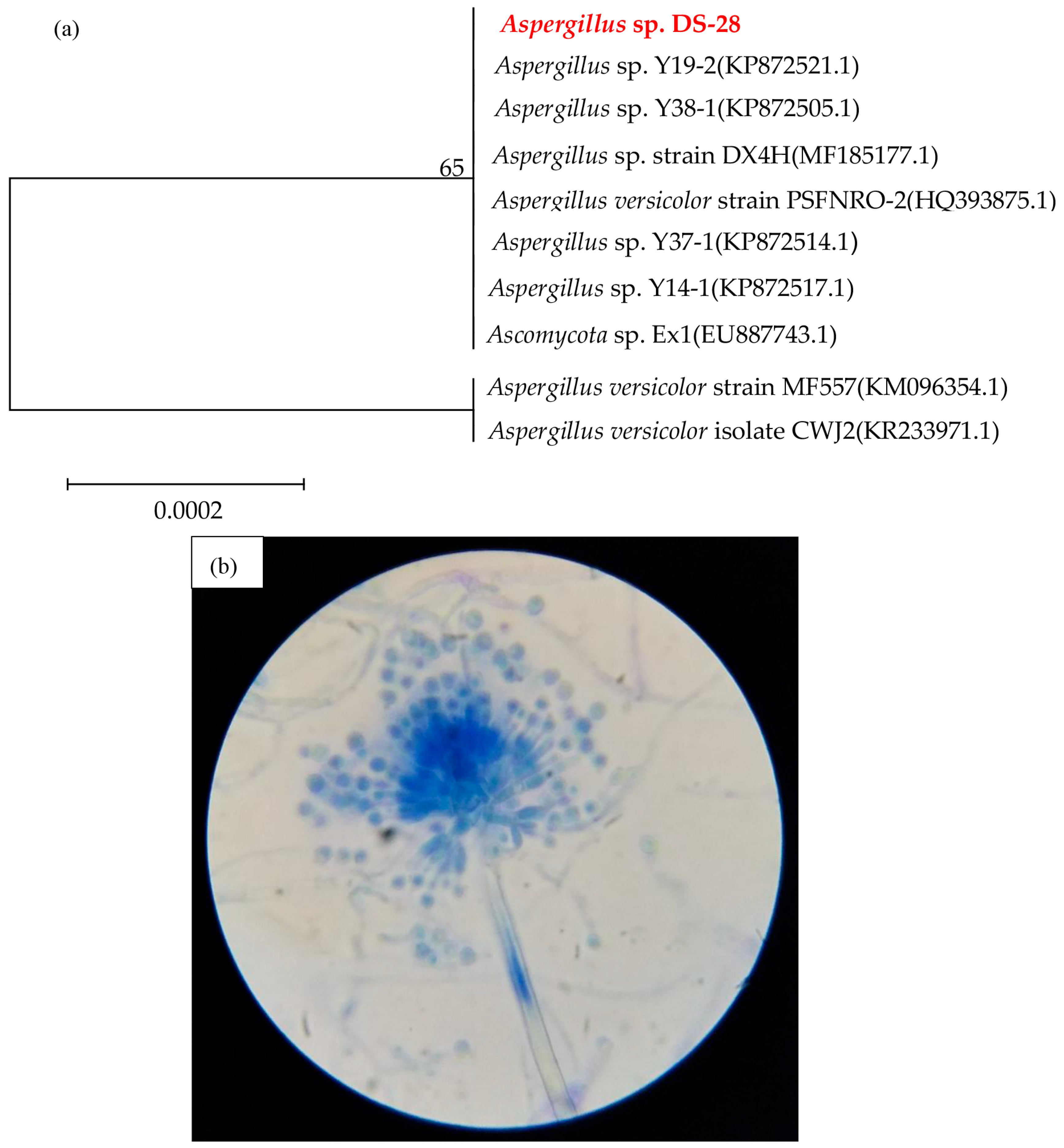
3.1. Isolation and Identification of Sulfur Black Degraders

The suspension was diluted appropriately and spread onto PDB plates to observe differences in size, morphology, and color among single colonies. Colonies with distinct differences were selected for streak purification. After the growth of single colonies, they were picked for further streak purification. The experiment was conducted multiple times, leading to the isolation and purification of 10 fungal strains. The strains in the well plate medium had to rely on the added sulfur black for growth since there were no carbon or sulfur sources present. To monitor the growth rate of the fungal strains, the optical density at 600 nm (OD600nm) was measured using a microplate reader. A total of 200 µL of inorganic salt medium containing 100 mg/L sulfur black was added to the wells of a 96-well plate. Each well plate was then inoculated with 20 µL of the initial screening fungal suspension. Additionally, two sets of blank controls were established, which did not contain sulfur black. The well plates were then incubated at 28 °C and 160 rpm for 5 days. After incubation, the OD600nm of the fungal suspensions was measured using a microplate reader. Various strains grew at different rates when sulfur black was the sole carbon and sulfur source, with some growing faster than others. The strains in the well plate medium had to rely on the added sulfur black for growth since there were no other carbon or sulfur sources present. This variability in growth rates might reflect differences in the enzymatic capabilities of the strains to process sulfur black, suggesting that some strains could be more efficient at biodegradation than others [22].
However, the growth rate of the strains using sulfur black did not conclusively demonstrate their efficiency in the desulfurization process. Therefore, the strains were further inoculated at 5% inoculum into the liquid enrichment media (LEM) containing sulfur black and cultured for 5 days. This experimental setup was designed to assess the ability of the strains to utilize sulfur black as their sole nutritional source. This is indicative of their metabolic flexibility and potential utility in environmental clean-up processes. This experimental setup provides a rapid screening method for black sulfide-degrading fungi.
The concentration of sulfate (SO42−) in the culture medium was measured. After following this process, a strain of fungus called DS-28 was selected. Figure 1a shows a phylogenetic tree analysis indicating that strain DS-28 has 100% similarity with Aspergillus sp. Y19-2 (Accession No.: KP872521.1). The image in Figure 1b under the optical microscope shows that the conidiophore of strain DS-28 is formed from an erect hypha. The terminal end of the hypha forms a spherical vesicle. Each phialide produces a chain of spherical, non-septate conidia. Combined with the morphological characteristics of the strain, it was identified as belonging to the genus Aspergillus, and thus it was named Aspergillus sp. DS-28. The fungus Aspergillus has been found to be capable of degrading various types of dyes. The extracellular enzymes secreted by Aspergillus can degrade Congo red, malachite green, Remazol Brilliant Blue R, Indigo carmine, and malachite green dyes [22]. The ease with which a dye can be degraded is influenced by a number of factors, including the molecular size of the dye, the presence of substituent groups, and the type of chromophoric groups (i.e., the part of the dye molecule responsible for its color) [23]. For instance, dyes with elevated molecular weights or those that are highly conjugated (characterized by alternating double and single bonds) tend to exhibit greater resistance to fungal degradation [24]. Conversely, dyes with more straightforward, less stable structures are more susceptible to breakdown. Fungi notably produce enzymes such as lignin peroxidase, manganese peroxidase, and laccase, which can degrade a wide range of aromatic compounds due to their non-specific activity [7]. Meanwhile, starting with an initial dye concentration of 100 mg/L, the Aspergillus strain was able to degrade 99% of Reactive Blue and 75% of Reactive Black [25]. This suggests that the Aspergillus strain is effective in decolorizing and potentially biodegrading these synthetic dyes, which are commonly used in textile industries and are known for their recalcitrance and environmental persistence. The difference in degradation percentages may be due to the structural differences between the two dyes, which can affect the efficiency of the biodegradation process.

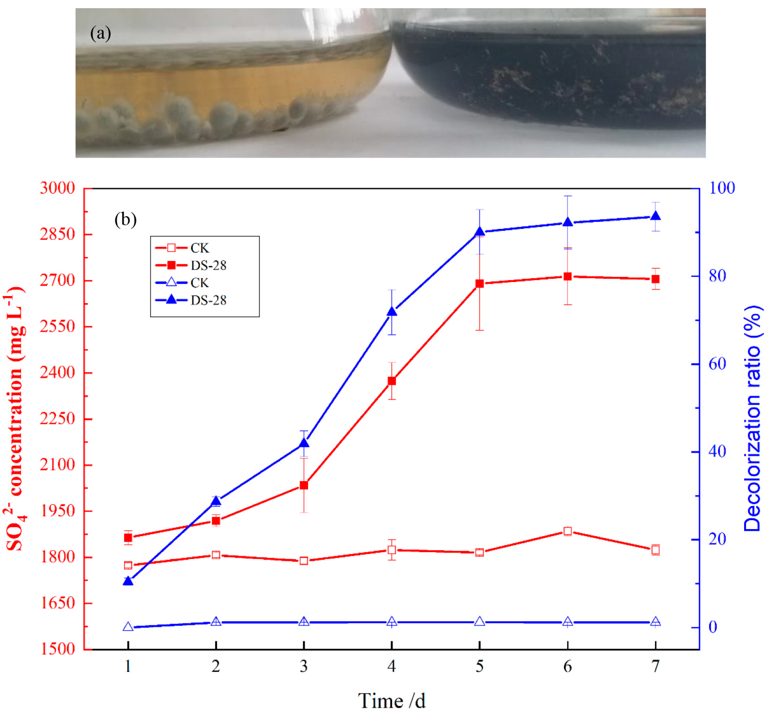
3.2. Sulfur Black Dye Removal Efficiency

Figure 2 shows the removal of sulfur black (50 mg/L) by the 7-day inoculated strain DS-28 compared to the control. Figure 2a demonstrates that, in comparison to the control, the solution became transparent after 7 days of treatment with strain DS-28, while the control solution remained black, similar to its initial state. Figure 2b shows that the SO42− content increased slowly in the early stage of culture. Starting from the third day, it rapidly increased and then gradually leveled off. By the seventh day, the SO42− content in the culture medium reached 2705.6 ± 34.6 mg/L, which was 48.4 ± 1.1% higher than the control. However, the bacterial strain Acinetobacter sp. DS-9 is also capable of oxidizing sulfur black, resulting in a 52.06% increase in SO42− concentration compared to the control [13]. Environmentally friendly reducing agents are utilized to degrade the sulfur in sulfur black dye wastewater, resulting in a 32% reduction in the mass fraction of reducing sulfur in the wastewater [26]. At the same time, the decolorization rate of the strain DS-28 can reach 93.6 ± 3.3% after seven days, while the decolorization rate of the control remained at the same level as the initial 1.2% after seven days. This result is consistent with the description in Figure 2a. Although the concentration of SO42− formed from the oxidation of sulfur black by fungi and the desulfurization oxidation by bacteria is similar, fungi have a stronger resistance to impact load in dye degradation compared to bacteria.

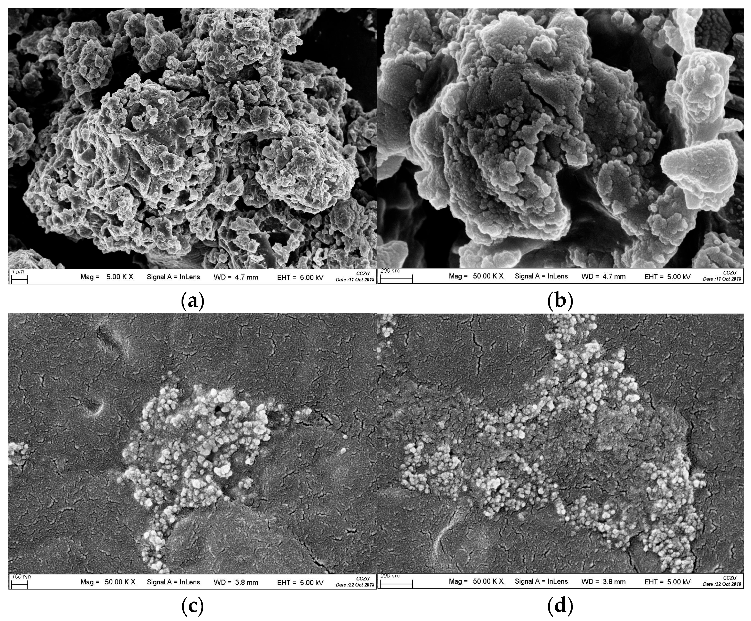
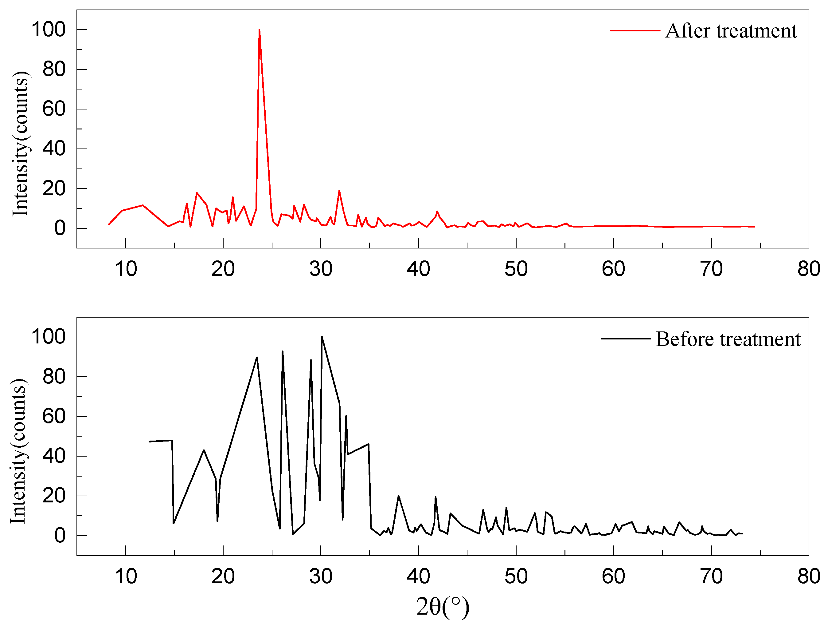
3.3. Changes in Physical, Chemical, and Topographical Properties of Sulfur Black Surface after Strain DS-28 Treatment

Figure 3a,b presents the SEM images of untreated sulfur black, where the powder particles are distinctly visible. These particles appear large, porous, and exhibit rough surfaces. However, a contrasting scenario is observed in Figure 3c,d, which depicts the SEM images of sulfur black post treatment with the fungus strain DS-28. Despite efforts to eliminate fungal cells from the culture medium, a few cells inevitably persist. These remaining fungal cells encapsulate the sulfur black particles, which results in a substantial reduction in particle size and a transformation of the surface to a smoother, more uniform texture. This suggests that, during the degradation process, the fungal cells fully interact with the sulfur black, leading to the decomposition of the sulfur black particles and the smoothing of the particle surfaces. This result was in accordance with the findings of the lignin biodegradation report [27,28]. Observation by SEM revealed that large organic particles were transformed into smaller particles, indicating microbial degradation.
The X-ray powder diffraction patterns of sulfur black before and after treatment are shown in Figure 4. The X-ray diffraction pattern of the untreated sulfur black reveals numerous diffraction peaks of high intensity at various angles, suggesting a sophisticated molecular structure with the existence of multiple chemical bonds and crystal planes. However, the post-treatment diffraction pattern of sulfur black demonstrates a notable reduction in the number of peaks, with a single high-intensity response at a diffraction angle (2θ) of 24°, a response that is also evident in the treated spectrum. This suggests that the sulfur black dye has undergone significant degradation by the strain DS-28 cells, leading to the disappearance or decrease in intensity of the diffraction angles associated with multiple chemical bonds and crystal planes. Due to the absence of standard reference cards for sulfur black, the precise nature of the peaks corresponding to the chemical bonds or crystal planes in the degraded sulfur black remains undetermined. Nevertheless, the X-ray diffraction findings clearly indicate the degradation of sulfur black upon interaction with the fungal cells.

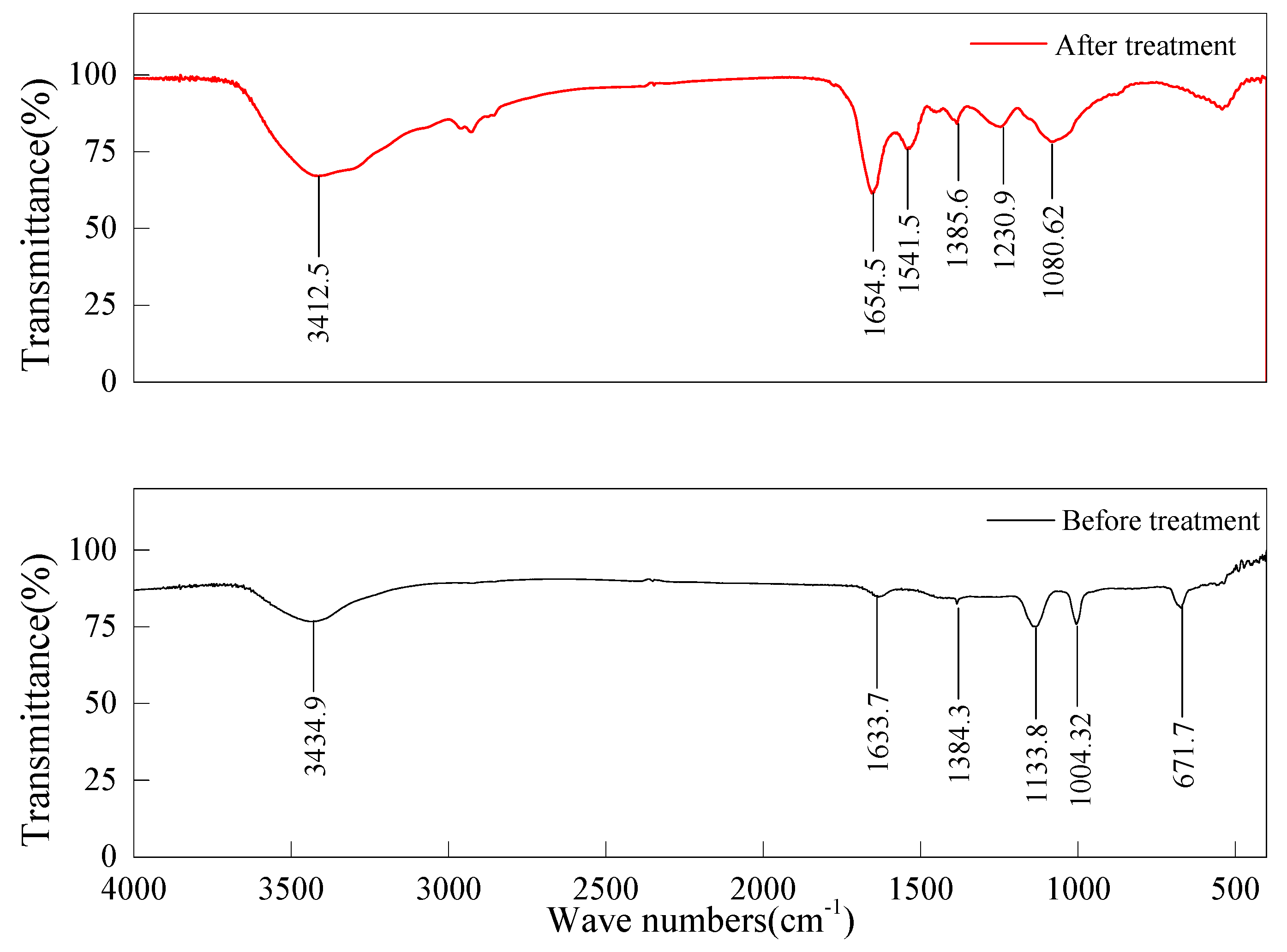
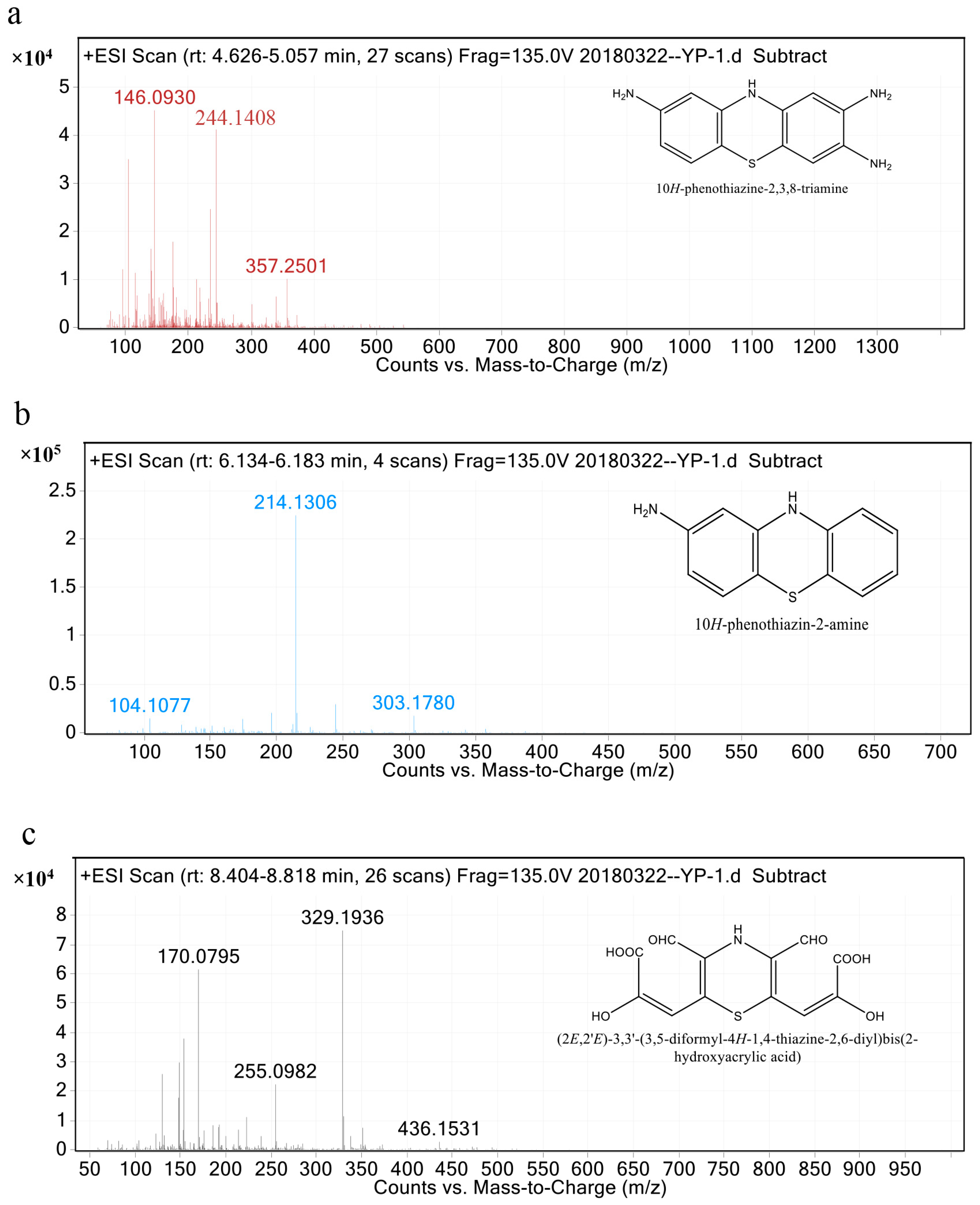
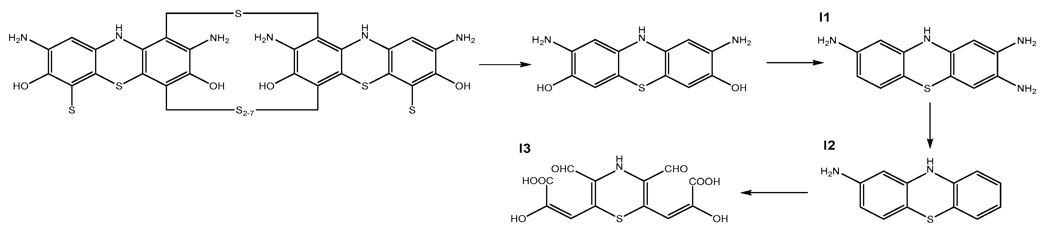
3.4. FTIR Analysis and Metabolite Identification

The infrared spectra of sulfur black before and after treatment are shown in Figure 5. The FTIR spectrum of the untreated sulfur black manifests a vibrational absorption peak at 3439.9 cm−1, which is indicative of the -OH group, and stretching vibrations ranging from 1633.7 to 1004.32 cm−1 associated with the -C=C- and -C=N- groups within the benzene ring. The vibrations of the various groups in the benzene ring are relatively weak due to the intact molecular structure of the untreated sulfur black. Furthermore, a distinct peak at 671.7 cm−1 marks the vibrational absorption of the -C-S- and -C-S-S-C- groups, confirming the presence of C-S and S-S bonds in the molecular structure prior to degradation. Conversely, the FTIR spectrum of the treated sulfur black exhibits a peak at 3412.5 cm−1, correlating to the vibrations of the -NH2 group. The vibrations within the 1654.5 to 1080.62 cm−1 range again pertain to the benzene ring. After degradation by the fungal cells, the bonds in the benzene ring are broken by the action of proteins, resulting in a significant enhancement of the vibration absorption peaks of various groups. Notably, the absorption peaks of the -C-S- and -C-S-S-C- groups vanish, signifying the disruption of carbon–sulfur and sulfur–sulfur bonds by the fungal cells, which then expel sulfur from the sulfur black’s molecular structure and further oxidize it to SO42−. These findings demonstrate that the Aspergillus sp. DS-28 strain can remove sulfur from sulfur black molecules.
Based on the data from the HPLC chromatogram and the LC-MS/MS spectrum presented in Figure 6, the degradation of sulfur black has yielded three distinct intermediate metabolites, labeled as I1, I2, and I3. The intermediates can be described as follows. Intermediate I1 has a retention time between 4.626 and 5.057 min, with a mass-to-charge ratio (m/z) of 244.0783. It has a chemical structure of C12H12N4S and is identified as 2,3,8-triamino-10H-phenothiazine. Intermediate I2 has a retention time ranging from 6.134 to 6.183 min, with an m/z of 214.0565. It has the chemical formula C12H10N4S and is named 2-amino-10H-phenothiazine. Intermediate I3 has a retention time between 8.404 and 8.818 min and an m/z of 327.0049. Its chemical structure is C12H9NO8S and it is recognized as (2E,2’E)-3,3’-(3,5-diformyl-4H-1,4-thiazine-2,6-diyl)bis(2-hydroxyacrylic acid). However, only I1, I2, and I3 have been definitively identified. Further analysis is required to accurately identify and characterize the remaining intermediate products.
Figure 7 shows the possible degradation pathway of sulfur black, inferred from the molecular structure of the degradation products. The degradation mechanism facilitated by strain DS-28 is preliminarily postulated. This pathway begins with the oxidative cleavage of polysulfide bonds within sulfur black’s symmetric structure, a reaction we attribute to the action of specific enzymes produced by the strain. Manganese peroxidase is an enzyme that catalyzes the oxidation of manganese(II) to manganese(III), which then acts as a mediator in the oxidation of various phenolic compounds [29,30]. It plays a pivotal role in the degradation of high-molecular-weight organic substances and sulfur-containing dyes through the oxidative cleavage of sulfur bonds [31,32]. The oxidative cleavage of polysulfide bonds in the symmetric structure of sulfur black leads to the formation of 2,8-diamino-3,7-dihydroxyphenothiazine, which then transforms into degradation product I1 under the influence of transaminase activity. Subsequently, degradation product I1 loses two amino groups to become degradation product I2. Laccase plays a crucial role in the oxidative cleavage of carbon–carbon bonds within aromatic rings, effectively initiating the fragmentation of complex dye molecules into simpler, less toxic compounds [33]. It is believed that I3 may result from the additional oxidation of the C-O bond in the benzene ring of I2 by laccase, leading to the formation of aldehyde (-CHO) and carboxylic acid (-COOH) functional groups. The fungus Aspergillus is capable of biodegrading aromatic hydrocarbons, resulting in the production of ring-opening compounds that are subsequently metabolized into the central metabolic system [28].

4. Conclusions

A strain of black-sulfide-degrading fungus was isolated from petrochemical wastewater and designated as Aspergillus sp. strain DS-28. The SEM analysis of black sulfide before and after treatment with strain DS-28 revealed a significant reduction in particle size and smoother, flatter particle surfaces. XRD analysis showed a disappearance or reduction in response intensity of several chemical bonds in the molecular structure of black sulfide and the crystal surface. The FTIR analysis revealed that the vibrational absorption peaks of the bonds and groups on the benzene ring were significantly enhanced after treatment with the strain DS-28; the vibrational absorption peaks of the -C-S- and -C-S-S-C groups disappeared, the carbon–sulfur and sulfur–sulfur bonds in the molecular formula of black sulfide were broken, and the S in the molecular formula of black sulfide was removed and further oxidized to SO42-. The HPLC and LC-MS/MS methods were used to identify the degradation products. Three possible degradation products of black sulfide, I1, I2, and I3, were identified. Based on the structural formulae of the degradation products, possible degradation pathways and metabolic mechanisms were briefly hypothesized.
Nevertheless, further investigation is required to elucidate the impact of factors such as pH, temperature, and the presence of other organic compounds on the degradation efficiency. Moreover, our study is primarily concerned with aerobic degradation processes; anaerobic pathways, which may play a significant role in natural sediment environments, have not yet been investigated. Future research should aim to elucidate these anaerobic degradation mechanisms and assess their feasibility for practical applications. Additionally, the potential for genetic or metabolic engineering to enhance the efficacy of the involved enzymes or to develop microbial consortia specifically tailored for efficient sulfur black degradation could be promising areas of research. This could lead to more robust and versatile biotechnological applications in industrial dye degradation processes.

Author Contributions

Z.G.: methodology, conceptualization, formal analysis, investigation, writing—original draft preparation. Y.W., W.C. and Y.L.: resources, formal analysis, software, validation. W.Y. and Z.C.: conceptualization, writing—review and editing, funding acquisition, project administration. All authors have read and agreed to the published version of the manuscript.

Funding

This study was supported by Maoming Branch Company, SINOPEC (MPBB220024).

Data Availability Statement

Data are contained within the article.

Conflicts of Interest

Author Zhipeng Guan was employed by Guangdong Maoming Petrochemical Company. The remaining authors declare that the research was conducted in the absence of any commercial or financial relationships that could be construed as a potential conflict of interest.

References

  1. Nguyen, T.A.; Juang, R.-S. Treatment of waters and wastewaters containing sulfur dyes: A review. Chem. Eng. J. 2013, 219, 109–117. [Google Scholar] [CrossRef]
  2. Lenninger, M.; Bechtold, T.; Pham, T. Improvement of wet rub fastness in continuous dyeing with c.i. Sulphur Black 1 by ultrasonic treatment. Ultrason. Sonochem. 2023, 99, 106558. [Google Scholar] [CrossRef]
  3. Bechtold, T.; Berktold, F.; Turcanu, A. The redox behaviour of CI Sulphur Black 1-a′basis for improved understanding of sulphur dyeing. Color. Technol. 2006, 116, 215–221. [Google Scholar] [CrossRef]
  4. Brown, K.A. Sulphur in the environment: A review. Environ. Pollut. Ser. B Chem. Phys. 1982, 3, 47–80. [Google Scholar] [CrossRef]
  5. Han, R.; Zhang, S.; Zhao, W.; Li, X.; Jian, X. Treating sulfur black dye wastewater with quaternized poly (phthalazinone ether sulfone ketone) nanofiltration membranes. Sep. Purif. Technol. 2009, 67, 26–30. [Google Scholar] [CrossRef]
  6. Ogunlaja, O.O.; Aemere, O. Evaluating the efficiency of a textile wastewater treatment plant located in Oshodi, Lagos. Afr. J. Pure Appl. Chem. 2009, 3, 189–196. [Google Scholar] [CrossRef]
  7. Andleeb, S.; Atiq, N.; Ali, M.; Razi-Ul-Hussnain, R.; Shafique, M.; Ahmad, B.; Ghumro, P.B.; Hussain, M.; Hameed, A.; Ahmad, S. Biological treatment of textile effluent in stirred tank bioreactor. Int. J. Agric. Biol. 2010, 12, 256–260. [Google Scholar] [CrossRef]
  8. Marmagne, O.; Coste, C.U. Color removal from textile plant effluents. Am. Dyest. Report. 1996, 85, 6. [Google Scholar] [CrossRef]
  9. Al-Hemiri, A.; Al-Anbari, H.; Shakir, I.K. Dye Removal from Wastewater Using Iron Salts. Iraqi J. Chem. Pet. Eng. 2008, 9, 1–7. [Google Scholar] [CrossRef]
  10. Blackburn, R.S. Natural Polysaccharides and Their Interactions with Dye Molecules:  Applications in Effluent Treatment. Environ. Sci. Technol. 2004, 38, 4905–4909. [Google Scholar] [CrossRef]
  11. Dasgupta, J.; Sikder, J.; Chakraborty, S.; Curcio, S.; Drioli, E. Remediation of textile effluents by membrane based treatment techniques: A state of the art review. J. Environ. Manag. 2015, 147, 55–72. [Google Scholar] [CrossRef] [PubMed]
  12. Keung, M.K. Bacterium Useful in the Removal of Sulphur Black Dye from a Substrate. U.S. Patent 5,610,064, 1997. [Google Scholar]
  13. Gu, Y.; Zhu, X.; Yu, W.; Zhong, W.; Guo, J.; Cai, Z. Oxidization and biodegradation of sulfur black by a newly isolated strain Acinetobacter sp. DS-9. Desalination Water Treat. 2021, 227, 321–329. [Google Scholar] [CrossRef]
  14. Sharma, B.; Tiwari, S.; Kumar, R.; Kumar, M.; Tewari, L. Eco-friendly detoxification of hazardous Congo red dye using novel fungal strain Trametes flavida WTFP2: Deduced enzymatic biomineralization process through combinatorial in-silico and in-vitro studies. J. Hazard. Mater. 2023, 455, 131503. [Google Scholar] [CrossRef]
  15. Coelho, G.D.; Silva, M.A.; de Melo Pinheiro, M.A.; Nadvorny, D.; Costa Amador, V.; Maia, R.T. In silico and in vitro assays suggests Congo red dye degradation by a Lentinus sp. laccase enzyme. J. Biomol. Struct. Dyn. 2024, 42, 3802–3813. [Google Scholar] [CrossRef]
  16. Alexander, D.G.S.; Thatheyus, A.J. Fungal bioremediation of toxic textile dye effluents. In Fungi Bio-Prospects in Sustainable Agriculture, Environment and Nano-Technology; Elsevier: Amsterdam, The Netherlands, 2021; pp. 353–380. [Google Scholar]
  17. Akansha, K.; Kaur, T.; Yadav, A.; Kour, D.; Kumar Rai, A.; Singh, S.; Mishra, S.; Kumar, L.; Miglani, K.; Singh, K.; et al. Microbe-mediated remediation of dyes: Current status and future challenges. J. Appl. Biol. Biotechnol. 2022, 11, 1–23. [Google Scholar] [CrossRef]
  18. Polman, J.; Breckenridge, C.R. Biomass-mediated binding and recovery of textile dyes from waste effluents. Text. Chem. Color. 1996, 28, 31–35. [Google Scholar]
  19. White, T.J. Amplification and direct sequencing of fungal ribosomal RNA genes for phylogenetics. In PCR Protocols a Guide to Methods and Applications; Academic Press: Cambridge, MA, USA, 1990. [Google Scholar] [CrossRef]
  20. Cai, Z.; Zhang, W.; Ma, J.; Cai, J.; Li, S.; Zhu, X.; Yang, G.; Zhao, X. Biodegradation of Azo Dye Disperse Orange S-RL by a Newly Isolated Strain Acinetobacter sp. SRL8. Water Environ. Res. 2015, 87, 516–523. [Google Scholar] [CrossRef]
  21. Xu, H.; Wang, K.; Jin, L.; Yang, L.; Yuan, J.; Zhang, W.; He, G.; Chen, H. Synergistically engineering of vacancy and doping in thiospinel to boost electrocatalytic oxygen evolution in alkaline water and seawater. J. Colloid. Interface Sci. 2023, 650, 1500–1508. [Google Scholar] [CrossRef]
  22. Klose, S.; Bilen, S.; Tabatabai, M.A.; Dick, W.A. Sulfur Cycle Enzymes. In Methods of Soil Enzymology; Soil Science Society of America: Madison, WI, USA, 2011; Volume 9, pp. 125–159. [Google Scholar] [CrossRef]
  23. Waring, D.R.; Hallas, G. The Chemistry and Application of Dyes; Springer: New York, NY, USA, 1990. [Google Scholar] [CrossRef]
  24. Chen, J.; Garcia, E.S.; Zimmerman, S.C. Intramolecularly Cross-Linked Polymers: From Structure to Function with Applications as Artificial Antibodies and Artificial Enzymes. Acc. Chem. Res. 2020, 53, 1244–1256. [Google Scholar] [CrossRef]
  25. Chen, S.H.; Yien Ting, A.S. Biodecolorization and biodegradation potential of recalcitrant triphenylmethane dyes by Coriolopsis sp. isolated from compost. J. Environ. Manag. 2015, 150, 274–280. [Google Scholar] [CrossRef]
  26. Li, R.; Wang, W.; Wei, B.; Zhou, C.; Zhang, S. Sulfur black dyeing process with environment friendly reducing agent. J. Text. Res. 2020, 41, 50–54. [Google Scholar] [CrossRef]
  27. Zhu, D.; Zhang, P.; Xie, C.; Zhang, W.; Sun, J.; Qian, W.-J.; Yang, B. Biodegradation of alkaline lignin by Bacillus ligniniphilus L1. Biotechnol. Biofuels 2017, 10, 44. [Google Scholar] [CrossRef] [PubMed]
  28. Arentshorst, M.; Falco, M.D.; Moisan, M.-C.; Reid, I.D.; Spaapen, T.O.M.; van Dam, J.; Demirci, E.; Powlowski, J.; Punt, P.J.; Tsang, A.; et al. Identification of a Conserved Transcriptional Activator-Repressor Module Controlling the Expression of Genes Involved in Tannic Acid Degradation and Gallic Acid Utilization in Aspergillus niger. Front. Fungal Biol. 2021, 2, 681631. [Google Scholar] [CrossRef] [PubMed]
  29. Zhou, H.; Fu, C. Manganese-oxidizing microbes and biogenic manganese oxides: Characterization, Mn(II) oxidation mechanism and environmental relevance. Rev. Environ. Sci. Bio/Technol. 2020, 19, 489–507. [Google Scholar] [CrossRef]
  30. Bilal, M.; Zdarta, J.; Jesionowski, T.; Iqbal, H.M.N. Manganese peroxidases as robust biocatalytic tool—An overview of sources, immobilization, and biotechnological applications. Int. J. Biol. Macromol. 2023, 234, 123531. [Google Scholar] [CrossRef]
  31. Bustamante-Torres, M.; Arcentales-Vera, B.; Abad-Sojos, S.; Torres-Constante, O.; Ruiz-Rubio, F.; Bucio, E. Lignin-Based Membrane for Dye Removal. In Membrane Based Methods for Dye Containing Wastewater; Springer: Berlin/Heidelberg, Germany, 2021; pp. 181–213. [Google Scholar] [CrossRef]
  32. Raychaudhuri, A.; Behera, M. Chapter 9-Microbial degradation of lignin: Conversion, application, and challenges. In Development in Wastewater Treatment Research and Processes; Shah, M.P., Rodriguez-Couto, S., Kapoor, R.T., Eds.; Elsevier: Amsterdam, The Netherlands, 2022; pp. 195–219. [Google Scholar] [CrossRef]
  33. Dhir, B. Degradation of Dyes Using Filamentous Fungi. In Dye Biodegradation, Mechanisms and Techniques; Springer: Berlin/Heidelberg, Germany, 2021; pp. 51–66. [Google Scholar] [CrossRef]
Figure 1. (a) Phylogenetic analysis of 18S rRNA genes from Aspergillus sp. strain DS-28 in this study. The nodes are labeled with bootstrap values expressed as percentages. The scale bars correspond to a substitution rate of 0.0002 per site. (b) Morphological structure of Aspergillus sp. strain DS-28 observed under light microscope.
Processes 12 01818 g001
Figure 2. (a) Observation of color changes in sulfur black culture liquid after 7 days of in-cubation with strain DS-28. The left graph shows the incubation with strain DS-28 and the right graph shows the control group without strain. (b) Over a period of 7 consecutive days, the SO42− and decolorization change curves of strain DS-28 in the liquid medium supplemented with black sulfide.
Processes 12 01818 g002
Figure 3. Scanning electron microscopy (SEM) images of the control sample (a,b) and sulfur black degradation by strain DS-28 for 7 days (c,d).
Processes 12 01818 g003
Figure 4. X-ray powder diffractograms of sulfur black before and after treatment with strain DS-28 for 7 days.
Processes 12 01818 g004
Figure 5. Fourier-transform infrared (FTIR) spectra of sulfur black treated with strain DS-28 after 7 days (upper panel) and control sample (low panel).
Processes 12 01818 g005
Figure 6. After 7 days of culture, strain DS-28 degraded sulfur black. LC-MS/MS graphs displayed intermediate degradation products of sulfur black. The mass spectra of metabolic intermediates I1, I2, and I3 are presented in Figures (a), (b) and (c), respectively.
Processes 12 01818 g006
Figure 7. Proposed pathway for the metabolism of black sulfide by strain DS-28.
Processes 12 01818 g007
Disclaimer/Publisher’s Note: The statements, opinions and data contained in all publications are solely those of the individual author(s) and contributor(s) and not of MDPI and/or the editor(s). MDPI and/or the editor(s) disclaim responsibility for any injury to people or property resulting from any ideas, methods, instructions or products referred to in the content.

