
Recent Developments in Application of Nanofibers

Department of Clothing and Textile Technology, Faculty of Engineering and the Built Environment, Cape Peninsula University of Technology, Bellville Campus, Cape Town 7535, South Africa
Processes 2024, 12(9), 1894; https://doi.org/10.3390/pr12091894
Submission received: 29 July 2024
/
Revised: 18 August 2024
/
Accepted: 2 September 2024
/
Published: 4 September 2024
(This article belongs to the Special Issue Application of Nanofibres in Sustainable Fashion and Textiles)
Abstract
:Technological advancements in nanofibers and production technologies have led to nanofibers being applied in various applications. Nanofibers are produced by a variety of techniques such as electrospinning, drawing, self-assembly, phase separation, and others. Electrospinning is widely used due to its versatility and scalability. Nanofiber production by other techniques is still limited to the laboratory scale, hence the dominance of electrospinning. The versatility of nanofibers has seen them being used in various applications such as health, protection, clothing, filtration, packaging, and electronics. Their large surface area, small diameters, and porous structures make them good materials in these diverse fields. Nanofibers are incorporated with nanoparticles to enhance stability. In biomedical applications, nanofibers are used in drug delivery systems, wound healing, and tissue engineering because of their biocompatibility and biodegradability. In fields like protection, clothing, and packaging, nanofibers are used due to their large surface area, porosity, and flexibility. These properties also make nanofibers highly effective in filtration, where their small size and large surface area allow them to efficiently remove a significant number of contaminants. Additionally, nanofibers are utilized in the production of flexible electronics, enhancing comfort in wearable devices. Biopolymers are being adopted to address the environmental and health concerns of traditional nanofiber materials. Biopolymers are biodegradable and biocompatible; however, their stability can be affected by production and environmental conditions. This work highlights the applications of nanofibers, especially the environmentally friendly nanofiber applications in health, packaging, water treatment, protection, electronics, clothing, and technical textiles.
1. Introduction
Nanofibers are increasingly receiving interest from researchers due to their unique properties. The fibers have diameters in the range of 1 nanometer to 100 nanometers [1]. Fibers above 100 nanometers have also been referred to as nanofibers. Nanofibers have a large surface area, small diameters, and porous structures [2,3]. These characteristics make nanofibers suitable for a wide range of applications in various fields, like health, cosmetics, protection, apparel, filtration, packaging, electronics, etc. Nanofibers are produced using various techniques such as electrospinning, drawing, self-assembly, and phase separation, among others. These techniques provide a variety of options for producing nanofibers. Unlike electrospinning, non-electrospinning techniques do not require conductive polymer materials or the application of high voltage. However, they are limited in terms of scalability and the quality of the nanofibers produced, and they often involve more complex processes. Together with material properties, process parameters are manipulated to produce nanofiber structures with specific properties [4,5,6,7].
Electrospinning is widely utilized for producing nanofibers with good uniformity. It involves applying a high voltage to a polymer solution, which results in the formation of an extruded fluid jet being elongated under the electric force to form a nanofiber on the collector [8,9]. Drawing is a non-electrospinning technique of creating nanofibers by mechanically stretching an elastic polymer using a rod to achieve the desired dimensions. The rotating rod is brought into contact with a droplet of the elastic polymer solution and then pulled away. When the solvent evaporates, the elongated droplet solidifies to form a nanofiber [10,11]. The template technique requires the creation of a template membrane with a porous layer. The polymer solution is poured into the pores of the template, and then the template is dissolved to recover the nanofibers [4,5]. Self-assembly is a process where nanoparticles organize themselves into a nanolayer due to intermolecular forces [10]. In phase separation techniques, the polymer solution separates into distinct phases, and the nanomembrane is freeze-dried to remove the solvents.
In health, nanofibers, especially biopolymers, are utilized in medical wound dressings, drug delivery systems, and tissue engineering. Biopolymers are biocompatible and biodegradable and have led to advancements in wound dressing, drug delivery, and tissue engineering without eliciting immune responses [9,10,12]. In tissue engineering, they regenerate tissues by mimicking extracellular matrix. Antimicrobials can be incorporated for targeted release at the wound site in nanofibers. Additionally, in wound dressings, the high porosity allows excess moisture to escape while keeping the wound area moist and thereby enhancing wound healing [9,10,12,13]. Mechanical stability and chemical stability of biopolymers can vary depending on the environment. The incorporation of other non-biopolymer nanoparticles to enhance stability may elicit an immune response [13,14].
Nanofiber membranes are used for filtering particles from liquids and gases [13,15]. Modified nanofibers can be utilized for the selective removal of heavy metals [8]. Treating an alumina hollow fiber membrane with polyacrylonitrile/graphitic carbon nanofibers produced a membrane that can degrade oil droplets when exposed to polyacrylonitrile/graphitic carbon nanofibers on an alumina hollow fiber membrane [13].
Nanofibers, especially the biopolymers in food packaging, have generated a lot of interest as they prolong the shelf life of food. Incorporating licorice and eugenol compounds in biopolymer zein nanofibers, extended the shelf life of grapes [16].
Nanofiber sensors are incorporated in clothing and technical textiles for detection and protection applications. Athletes wear to gather information that can be utilized to improve their performance and protection [17]. The utilization of wire in traditional electronics makes the product uncomfortable to wear as they are not flexible and soft [18], hence textile polymers are being explored. Janus nanofiber membrane-Cu-hemin metal organic framework has been investigated for detection of glucose from the sweat. The change in color of the membrane is proportional to the level of glucose detected [19]. A polyester/polyurethane membrane coated with vanadium dioxide creates a phase-changing material that can be utilized to regulate the temperature [20].
Electrospinning
The electrospinning technique is the most common due to its low production cost, easy adaptability, and simplicity. It utilizes high voltage to generate fine jets that solidify to form nanofibers on the collector [21,22]. A typical basic electrospinning setup has a high-voltage source that charges the extruded polymer solution, a collecting plate to collect the nanofibers, a syringe with a pump, and a polymer solution, as seen in Figure 1 [8]. Some of the factors (Table 1) that affect electro-spinnability include polymer type, molecular weight, concentration, solvent, surface tension, viscosity, solution conductivity, voltage, the distance between nozzle and collector, collector, flow rate, and surrounding environment (humidity and temperature). These factors are controlled to produce uniform nanofibers that have no beads. Beads change the morphology and diameters of nanofibers [12,22,23,24]. Collector’s speed is regulated to produce a uniform membrane; excessive speeds can lead to fiber breakages that cause the fibers to stick to the collector [25].
The electrospinning techniques based on spinneret configuration are categorized into two groups: needle-based and needleless [24,26]. Tayebi-Khorrami et al. [9] classify the electrospinning techniques into four classes based on how the nanofibers are produced, as shown in Figure 2. The heterogenous polymer solutions in B, C, and D produce core-sheath nanofibers in contrast to A, which has one homogenous polymer solution. A and B have single nozzles, whereas C and D have two [9]. Gavande et al. [21] classified electrospinning based on its eco-friendliness, which will depend on the types of materials, types of solvents, and the techniques applied. Their classification is important when one wants to address issues of protecting the environment when producing nanofibers.
Traditional plastic materials are not degraded and contaminate the environment [16,27]. Traditional solvents utilized to melt polymers are toxic. Natural materials and solvents are being investigated as they are biocompatible and biodegradable, and ones are being suggested to avoid contaminating the environment and promote sustainability as they are sourced from renewable sources [21,28]. Even though they are not considered ecofriendly, nanometals have high thermal and chemical stability and low cost [29]. Nanoparticles are incorporated into nanofibers by blending them with the nanopolymer solution. Nanofibers can also be coated with nanoparticles by applying the nanoparticle solution to the surface of the nanofibers. Nanoparticle solution can also be dried by evaporating the solvent, and the dried layer of nanoparticles is then applied to a fabric [24].
The production rate of electrospinning using a single needle range from about 0.01 to 0.4 g/h. This limitation led to the development of multiple needles and needleless spinning techniques. With multiple needles, the production rate increases to 0.7 g/h, and needleless spinning can achieve rates between 0.5 g/h and 600 g/h. However, challenges such as needle clogging persist [24,26]. Utilizing the needle-free electrospinning technique called the coil-based, a production rate of 23 g/h was achieved with polyacrylonitrile polymer solution. The electrical force pulls the polymers from the rotating drum saturated with the polymer solution [24].
Other modifications of the electrospinning technique are conical wire coil, spiral coil, rotary cone, sprocket wheel disc, rotating disc, metal dish, double-ring, curved slot, bowl, and tube [26]. To address these issues of high voltage and clogging of needle holes, alternative nanofiber production techniques are being explored, including drawing, phase separation, template synthesis, and self-assembly, which do not require the utilization of high voltage (Table 2). These non-electrospinning techniques are still limited to laboratory production; hence, the electrospinning technique is still widely used despite its challenges, such as the process parameters and material properties that must be maintained to produce finer nanofibers.
2. Non-Electrospinning Methods
- (i)
- Drawing
The drawing process, also referred to as dry spinning, involves drawing an elastic polymer using a rod or needle, which pulls the polymer droplet without breaking it. The rod or needle is placed in contact with a droplet of the elastic polymer and then pulled away. This technique is typically limited to laboratory-scale production. As the solvent evaporates, the viscosity of the polymer increases, potentially leading to variations in fiber diameter [10,11].
- (ii)
- Template
A template requires the creation of a template with a porous layer. The desired polymer solution is deposited into the nanoscale-diameter pores of the template. The surrounding template layer is then removed, leaving the short nanofibers [10,11].
- (iii)
- Self-assembly
Small molecular particles assemble to form a thin nanofiber layer. It is suitable for creating a thin layer for nanocoating [10].
- (iv)
- Phase separation
The polymer material is dissolved in a solvent, and when heat is applied or a nonsolvent is introduced, the mixture separates into two phases due to their physical incompatibility. Once the gel forms, it is frozen and then subjected to freeze-drying, which effectively removes the solvents [10,11].
2.1. Health Applications
In medical applications, nanofibers are also being investigated for tissue engineering and wound healing as they promote cell adhesion, proliferation, and wound repair [13,21,30]. Nanofiber scaffold structure is porous and has a large surface, and by mimicking the extracellular matrix of the bone, it promotes bone regeneration by allowing the tissues to grow on the nanofiber scaffold [31]. Wound dressings promote wound healing by keeping the wound area moist by absorbing excess moisture and allowing gaseous exchange. A dried wound would promote scar development [30].
Local delivery of the drug to the wound site is another area whereby a stimulus (changing surrounding environment) stimulates the nanofibers to release the drug [9,21]. However, there are still challenges in making selective nanofibers stimulated by a single stimulus [31]. Examples of stimuli that can stimulate nanofibers are temperature, pH, light, and magnetic fields. As seen in Figure 3, the delivery of the drug to the site is expected to greatly improve its effectiveness because oral drugs can be affected by intestinal pH and enzymes; furthermore, the drug can target other tissues. Controlled release of the drug components occurs on the site because the drug on the nanofibers’ surface is released quickly, whereas those incorporated in the nanofibers are released gradually when the chitosan is gradually degraded when it is dissolved [12].
Biopolymer nanocellulose aerogel can be utilized as adsorbents, drug delivery, tissue engineering, medicine, and wound cover applications. Traditional sources of cellulose are plants [32,33], even though nanocellulose has been found to tunicate marine animals and bacteria [31,34]. Nanocellulose has good moisture content, which makes it a good candidate for applications in tissue engineering and wound treatment [35,36]. A bacterial nanocellulose treated with fluorosilane and fluorocarbons performed better than the polytetrafluoroethylene (PTFE) membrane in decreasing plasma and blood clot formation due to its superhydrophobicity. A nanomembrane filter made from cellulosic nanocrystal and graphene oxide was effective in removing heavy metal ions from a contaminated effluent [27,31]. Talukder et al. (2024) [37] developed bead-containing hydrophobic superhydrophobic sulfonated polyethersulfone nanofiber membranes that had high 99% salt rejection efficiency [37]. The current extraction of nanocellulose from these new sources is not encouraging, as it cannot be upscaled for industrial-scale production.
Chitosan is a polysaccharide biopolymer derived from chitin. Its inherent antimicrobial activity expands a range of materials that can be utilized for wound dressings, tissue engineering, and drug delivery [12,15]. Because of its poor electro-spinnability, it is blended with other compounds [30]. A chitosan-silk fibroin nanocomposite membrane was reported to promote cell differentiation and proliferation [12]. Carbon nanotube-polyvinyl alcohol-chitosan nanocomposite membrane [30] and poly(lactic-co-glycolic acid) (PLGA) nanofibers containing mesoporous silica nanoparticles [38] are also reported to improve cell proliferation. The chitosan-PVA-ZnO membrane and chitosan-polyethylene glycol-Ag membrane showed excellent antimicrobial activity against bacteria. Chitosan-polycaprolactone nanofibers prepared with mupirocin and lidocaine hydrochloride and chitosan-polylactic acid nanofibers prepared with tetracycline hydrochloride showed good antimicrobial activity. Microbial activity was also reported for the polylactic acid/chitosan membrane and the chitosan–polylactate membrane [30,39,40]. The positively charged chitosan attaches to the oppositely charged membrane. Nanofiber membranes are utilized as skin care masks [30].
2.2. Wastewater Treatment
Nanofiber membranes are utilized for filtering particles from liquids and gases due to their extensive surface area, porous structure, and small pore size, which make them ideal for filtration applications [13,15]. A large surface area enables large quantities of components to be adsorbed and trapped. Membranes must maintain their porosity when the water passes through under pressure.
In desalination, nanomembranes can separate up to 100% of the salt components from water to produce potable water. Reverse osmosis membranes often suffer from fouling compared to forward osmosis, membrane filtration membranes. Incorporating metals into nanofibers improved anti-fouling and achieved separation [8,41,42]. Polyvinylidene fluoride membrane treated with TiO2, SiO2, and CuO achieved 99% separation of salt. A CF4 membrane modified by the plasma treatment achieved close to 100% separation of salt [8]. Nanofibers can be utilized to remove heavy metals from affluents. The heavy metals are absorbed into the nanofiber surfaces [43,44]. Nanofibers can be modified by incorporating functional groups for the selective removal of heavy metals. Incorporating MgS into cellulose nanofibers imparts selectivity of the nanofibers to Cd(II). Chitosan modified with polyacrylic acid sodium has selectively for Cr(VI). Polyvinyl alcohol-sodium alginate fibers have an affinity for Cd(II). Poly (vinyl alcohol-co-ethylene) modified with iminodiacetic acid has an affinity for Cd(II). CS–PGMA–PEI membrane has an affinity for Cr(IV), Cu(II), and Co(II) [8].
Modified nanofibers can also remove contaminants of emerging concern (CEC) pollutants. These pollutants are found in lower concentrations, making them difficult to remove using conventional effluent treatment techniques. Alkaline lignin-PVA removes fluoxetine. PAN-β-cyclodextrin removes atrazine. Polyamide-TiO2 removes atrazine, benzotriazole, caffeine, carbamazepine, DEET, naproxen, metoprolol, and sulfamethoxazole. ZnO–carbon removes caffeine and diclofenac. PAN-carbon nanotube removes atrazine and sulfamethoxazole. Fe/N-doped PAN removes Bisphenol A. ZnO-polystyrene removes caffeine [8].
Incorporating copper oxide nanoparticles into the PVDF–hexafluoropropylene (HFP) copolymer enhanced both its mechanical properties and hydrophilicity. Fabricating PAN/graphitic carbon nanofibers on an alumina hollow fiber membrane enhanced the membrane’s hydrophilicity. This modified membrane demonstrated the ability to degrade oil droplets when subjected to UV light. Incorporating TiO2 and silicon nanoparticles into a PAN polymer produced a membrane that was both superhydrophilic and superoleophobic, making it effective for filtering water contaminated with oil. A PAN polymer incorporated with silver nanoparticles demonstrated antimicrobial activity [13]. The weak mechanical properties of nanofibers are due to their low-molecular-chain orientation, which is not enhanced because they are not subjected to post-drawing processes that could improve their orientation and crystallinity. Improvement was reported when a polyacrylonitrile nanofiber membrane was subjected to post-drawing. Additionally, reducing the diameter of nanofibers has also been shown to increase their Young’s modulus [45].
2.3. Protection
Nanofibers exhibit weak mechanical and chemical stability, necessitating their utilization with a supporting layer or the incorporation of other nanomaterials [13].
Protective clothing is designed to protect the whole or some parts of the body from objects, radiant heat, flame, and chemical splashes to prevent or minimize injuries. Protective clothing does not provide absolute protection but only minimizes life-threatening hazards. Lightweight and low bulk are one of the requirements, as items must be carried by individuals or vehicles with minimal space available [46,47,48]. Protective clothing can be coated with chemical repellents to be superamphiphobic—phobic to both water and oil. Firefighter protective clothing has multiple layers to ensure adequate protection from the outside heat—usually has three or four layers that are separated by thin open-air spaces. Protective clothing for high-risk work can be equipped with sensors for the monitoring of the external risk factors and/or vital signs of the body. The alarm can be set to a defined level to activate it, helping the person to escape [49,50,51,52,53].
Nylon electrospun with magnesium oxide enhanced nylon flame retardancy and antimicrobial resistance against E coli and S aureus [24]. Modified nanofibers in protective clothing applications as phase-change fibers, thermofibers, and chromic fibers to sense and respond to the environment. A polyester yarn coated with polyurethane nanofiber coated with vanadium dioxide as a phase-changing material was able to regulate the temperature [20]. In defense applications, nanocarbon fibers can be used to create lightweight components for military aircraft. Moreover, these carbon nanofibers have the ability to absorb electromagnetic waves, making it harder for radars to detect the aircraft [54,55].
When polybenzimidazole nanofibers were added to rubber, it improved their mechanical performance more than that of pure rubber. Adding nylon nanofibers to dental composite resins improved the mechanical performance [15]. Almetwally et al. (2017) [11] reported that the strength of an electrospun polyurethane nanofiber membrane was reduced by half when compared to a cast polyurethane membrane.
2.4. Electronics
Digital electronic components are integrated into clothing and technical textiles to impart smartness [56]. Electronic textiles can be utilized to detect gas leakages. In hospitals, they can be utilized for monitoring and diagnosis [57]. Data collected can be analyzed to provide appropriate treatment.
Nanofibers are used for a wide range of soft sensor-based applications, ranging from pressure sensors, battery separators, supercapacitors, nanogenerators, strain and chemical sensors, transistors, conductors, etc. (Figure 4). A detailed review on electronics applications of nanofibers is covered by many authors [58,59,60].
Nanofibers can be used in the form of a sheet, yarn, or single fibers to make flexible electronic components. Nanofibers are used for stretchable, nontransparent conductors and transparent electrodes [58].
In sports, sensors can be utilized to enhance competitiveness. Components are attached inside or onto the surface of textile products. Electronics in textiles can be grouped into reversible and non-reversible groups. Reversible can be removed from textiles for charging or washing, which is not the case with the non-reversible group. Non-reversible are sewed or soldered permanently in the textiles [56].
The utilization of wire in electronics makes the product uncomfortable to wear because the wires are not soft and breathable [18]. Traditional clothing and technical textiles are not conductive, and to make them conductive, their surfaces are treated with nanometals such as silver, copper, and gold. Wires are utilized in traditional electronics to charge it; however, with a wireless energy transfer system, no wires are required. The receiver coils in A receive energy from the transmitter coils in the hanger, as shown in Figure 5. This type of shirt can be utilized to generate heat and keep the wearer warm. When utilized together with an energy-harvesting device that stores energy, the stored energy can be released when it is needed [56].
Nanofibers were fabricated from spatially confined MXene/polyvinylidene fluoride (PVDF) for piezoelectric application, with dual functions of pressure sensing and energy harvesting [59]. Mxene is defined as a class of 2D carbides, carbonitrides, and nitrides of transition metals [61]. The voltage and current generated by fabricated MXene/PVDF (0.8 wt%) nanofiber piezoelectric electronic devices are, respectively, 3.97 times and 10.1 times higher than those generated by pure PVDF nanofibers. They are used to monitor human movements and harvest energy.
With sustainability playing a role in many fields, alternative microbial protein nanowires are emerging as an alternative to silicon nanowires and carbon nanotubes for electronics applications [60]. Shapiro et al. (2022) created highly conductive protein nanowires by using E. coli bacteria to generate protein appendages on their surface, known as pili [62]. These pili were treated with gold nanoparticles, resulting in a protein nanowire with conductivity increased by 170-fold [62]. Lekbach et al. (2023) also used E. coli bacteria to develop a protein nanowire sensor that can sense ammonia [63]. These nanowires are produced from renewable feedstocks without requiring high energy inputs or toxic reagents. Sensors with high specificity and memory and electricity-generating devices have been fabricated with microbial nanowires.
2.5. Packaging
Packaging materials act as barriers to maintaining food quality and safety. Traditional plastic materials, such as polyethylene, polypropylene, and polystyrene, contaminate the environment as they are not biodegradable. They are still being utilized due to their low cost and good mechanical properties [64]. Nanofibers, especially the biopolymers in food packaging, have generated a lot of interest as they prolong the shelf life of food. Traditional packaging materials cannot deactivate microorganisms and oxidants. To overcome this, nanofibers are incorporated with antibacterial agents and antioxidants [8,16].
The formation of beads in nanofibers is unwanted as it changes the diameter and morphology of the nanofiber [65]. Zhang et al. (2024) reported that beads can be incorporated with antibacterials and antioxidants [16]. Natural active agents extracted from licorice have antimicrobial activity, whereas the eugenol compound extracted from clove has both antibacterial and antioxidant activities. Incorporating licorice and eugenol compounds in zein nanofibers extended the shelf life of grapes [16]. Zein is a protein fiber harvested from maize; it is insoluble in water but soluble in alcohol and some acids [30,66].
Biopolymeric nanofibers as an active biodegradable packaging system have attracted specific attention. In another work, researchers developed zein-based electrospun nanofibers (NFs) incorporated with geraniol-loaded nanoliposomes (G-loaded NLPs) for active packaging applications [67]. The incorporation of G-loaded NLPs in the zein NFs provided antibacterial and antioxidant activities. These nanofibers have suitable physicochemical properties and biological activity for application as an active packaging material in the food packaging industry.
Functionalized cellulose nanofibers (CNF) are used for active packaging applications [68]. CNF is developed in the form of films, membranes, hydrogels, aerogels, foams, and microcapsules. For developing CNF-based films/membranes, several manufacturing techniques, including electrospinning, can be used for active packaging applications. In CNF-based hydrogels, CNF is used as substrates, reinforcing agents, or cross-linkers in the manufacture of hybrid hydrogels [68]. CNF-based hydrogels were used as a carrier for pH-sensitive dyes to form an indicator that could change visual color based on the freshness of the packed chicken [69].
Gelatin and pectin are other biopolymers that are utilized to produce nanofibers [70]. Gelatin extracted from the animal collagen waste and pectin extracted from citrus skin waste are blended with nanocellulose to produce a biodegradable packaging membrane that can be incorporated with antimicrobials. Gelatin is blended with other compounds because it has poor mechanical strength and is soluble in water. Incorporating an antimicrobial agent in a gelatin nanocomposite membrane (G/CP/N/P) significantly extended the shelf life of cherries compared to a gelatin nanocomposite membrane (G/CP/N) without antimicrobials. The non-biopolymer polyethylene membrane, which also lacked antimicrobials, showed poor performance, as seen in Figure 6 [27].
Another biopolymer that can be utilized as an alternative is polylactic acid (PLA). It can be harvested from plants by microbial fermentation. Nanometals such as zinc oxide, magnesium oxide, and nickel oxide are added to improve its mechanical strength. However, excessive addition of the nanometals can decrease the strength. Halloysite nanotubes (HNTs) PLA/halloysite nanotubes-copper membrane extended the shelf life for strawberries to 8 days [71]. This supports the previously stated argument that incorporating antimicrobials into nanofibers extends the shelf life of foods.
Chromic nanofibers can be utilized to sense the accumulation of gases (CO2, NH3, H2S, dimethylamine, and trimethylamine) released by food contaminated with bacteria. Nanoparticles and chromic molecules are blended with nanofibers that change color when the released gases are absorbed. Cetinkaya et al. (2024) investigated the black elderberry extract/tin-oxide/gold/gelatin-based nanofibers, which change the color when trimethylamine, NH2, and H2S were released by a fillet hake fish [29].
CK group: no sealing film; PE group: polyethylene plastic; G/CP/N group: gelatin/citrus pectin/cellulose nanofibers film; G/CP/N/P group: gelatin/citrus pectin/cellulose nanofibers composite film containing antibacterial agent PHMB [27].
2.6. Apparel and Technical Textiles
The use of nanofibers in clothing and technical textiles is linked to many inter-disciplinary areas. Some of them were already discussed in the previous sections.
Nanofibers can be used for a range of applications in the clothing and textile areas [72]. Some of the application areas are highlighted in Figure 7. These are for filtration, comfort, smart textiles, antimicrobial, thermal, waterproof, UV protection, etc. [72]. Therefore, depending upon the application, nanofibers can be produced as per the requirements.
In a study, a composite nanofabric with exceptional water resistance and breathability using polyurethane (PU), fluorinated polyurethane (FPU), and polyvinyl butyral (PVB), namely, PU-FPU-PVB composite nanofabric, has been reported by Zhou et al., 2024 [73]. They reported that optimizing the TPU and PVB contents is crucial for obtaining PU-FPU-PVB composite nanofabrics. Increasing the PVB content from 0 wt% to 100 wt% led to decreased moisture permeability from 10.5 kg/m2/d to 3.0 kg/m2/d during thermal comfort testing. Furthermore, its air permeability decreased from 22.5 mm/s to 2.8 mm/s under these conditions. These findings indicate that the designed composite nanofabric exhibits excellent thermal comfort when incorporating appropriate levels of PVB into its composition, as indicated by decreased moisture permeability from 10.5 kg/m2/d to 3.0 kg/m2/d and decreased air permeability from 22.5 mm/s to 2.8 mm/s, making it an ideal high-performance material for waterproof and breathable fabrics [73].
In another study, authors reported the recycled polyethylene terephthalate nanocoated silk technical cloth embedded with green-synthesized silver nanoparticles using a solution electrospinning technique for advanced air filtration applications [74]. Hossain et al. (2024) reported enhanced filtration efficiency (96.58%) and low-pressure drop (29.1 Pa/cm2) in the developed samples [74].
Utilization of nanofibers in clothing and technical textiles has also received interest, especially in sports to improve performance and protection [17]. Sensors in clothing and technical textiles are utilized for detection. Muscles and joints of the lower limbs are particularly susceptible to injuries when exercising or playing sports. Wearing the correct protective wear can prevent or minimize injuries.
In athletes, if the injuries are not treated, their performance will decrease. The utilization of nanofiber products in the treatment of sports injuries helps athletes recover faster than those who did not utilize them [1]. Females can wear a smart bra that conforms to the movement of the breasts; it contracts and expands to accommodate the breast movement and thereby reduces bra discomfort [17,75]. In health, the incorporation of sensors in bras to detect breast cancer has already been investigated with promising results [75].
A wearable sweat monitor made from a Janus membrane detects the sweat released by the body. The Janus nanofiber membrane, unlike traditional membranes, has a polyurethane hydrophobic side and a polyacrylonitrile/silica hydrophilic side; this prevents the accumulation of sweat as it is transmitted only in one direction. Janus nanofiber membrane-Cu-hemin MOF was able to detect the glucose from the sweat. The green color deepens when the glucose increases. This opens the future possibility of using it as a non-invasive technique to detect glucose levels [19].
Wu et al. (2024) [20] developed stretchable carbon nanotube/thermoplastic polyurethane nanofibers for sports bandages. The coated sensor with conductive materials required pre-stretching to improve strain sensing. Because pre-stretching causes some damage as cracks develop, its application in highly physical activities is of concern [76].
Researchers are exploring the use of 3D printing to create an electrospun wearable garment directly on a mold, eliminating the need for supporting base materials for the delicate nanofiber membranes. Additionally, nanofibers are being developed for mattress coverings to produce lightweight, porous fabrics that can act as barriers against insects like dust mites [24].
2.7. Future Research
Due to their distinctive properties, nanofibers are being explored for utilization in various applications. Most nanofiber production methods are still confined to laboratory settings, which is why electrospinning remains the predominant technique. However, new non-electrospinning methods are emerging as more cost-effective and efficient [77]. Future applications of nanofiber materials will involve biopolymers, biosolvents, and recyclable traditional polymers [78,79]. Conventional nanoplastic fibers are made from non-biodegradable polymers and use environmentally harmful solvents. It is one of the main reasons for biopolymers being increasingly considered alternative materials. Additionally, recycled polycarbonate nanofibers have been combined with strontium aluminate nanoparticles to create a membrane that changes color [80]. Similarly, future research could focus on further improving the scalability of various techniques for producing nanofibers as well as improving their production rate. Another area that could be of significant interest is the toxicity effect of nanomaterials.
3. Conclusions
Nanofibers play a crucial role in various applications such as medicine, clothing and textiles, protection, filtration, packing, clothing, etc. Despite their potential, there are still production and material issues that are being investigated to enhance their adoption. The electrospinning technique remains a popular choice for producing nanofibers because it can create uniform fibers and is scalable. While non-electrospinning production techniques do not require high voltage, they come with drawbacks such as limited scalability, complex processes, and inconsistent nanofiber quality. There are environmental and health concerns about the nanoplastics they form when traditional non-biopolymers disintegrate. Furthermore, the utilization of toxic chemicals during the manufacturing processes is a concern. Addressing these challenges requires innovative manufacturing processes and materials.
While nanofiber materials offer remarkable properties such as high surface area, small diameter size, and high porosity structure, they also face several limitations. These include challenges in large-scale production, inconsistencies in fiber uniformity, environmental pollution, and the high cost associated with materials and production. Additionally, challenges related to the environmental and health impact of materials can limit their adoption, especially in medical applications; hence, biopolymers are increasingly being investigated as alternatives to traditional nanofiber materials in various applications as they are biodegradable and biocompatible.
In health and packaging, antimicrobials are incorporated to prevent microbial invasion, which further adds to the appeal. Nanofibers in filtration and wastewater systems are excellent at removing large quantities of contaminants due to their large surface area. Additionally, nanofibers can be utilized as sensors for monitoring. Furthermore, the mechanical stability and chemical stability of nanomaterials, especially biopolymers, can vary under varying environmental conditions, which limit their applications.
The electrospinning technique is still widely utilized to produce nanofibers, however, there are concerns about the use of high voltage, which poses a safety risk, and the high energy cost associated with it. Alternative non-electrospinning nanofiber production techniques reported in this review are not scalable for large nanofiber production. However, these non-electrospinning techniques offer the advantage of not requiring the utilization of high voltage. The production of nanofibers, while promising for various applications, faces several limitations. These include challenges in achieving consistent fiber diameters, difficulties in scaling up from laboratory to industrial production, and the need for precise control over environmental conditions to ensure uniformity. Functionalization of drugs in nanofibers is a complicated process. The choice of production techniques and materials often involves trade-offs between material properties and process efficiency. In electrospinning, an optimum voltage is applied to avoid producing nonuniform nanofibers. Addressing these limitations is crucial for advancing the commercial viability and broader adoption of nanofiber technologies in diverse fields.
The potential future solutions for nanofibers lie in improving their material properties, production techniques, scalability, and expanding their applications. Biopolymers will increasingly replace traditional polymer materials to meet environmental and health requirements. Additionally, the incorporation of functional nanomaterials into nanofibers may open new possibilities for utilization in various applications like health, packaging, filtration, electronics, protection, and clothing. As research continues to address the production and material properties challenges of controlling fiber diameter, alignment, and surface functionality, nanofibers are poised to play a crucial role in next-generation technologies, offering novel solutions to complex problems.
Funding
This research was funded by the National Research Foundation (NRF) South Africa, grant number 132153.
Conflicts of Interest
The author declares no conflicts of interest.
References
- Yang, G.; Yang, H.; Wang, M.; Sun, Y.; Wang, M. Application of nanofiber material based on electrospinning technology in sports rehabilitation of basketball player’s wrist joint. J. Nanomater. 2022, 2022, 1–9. [Google Scholar] [CrossRef]
- Yu, X.-C.; Li, B.-B.; Wang, P.; Tong, L.; Jiang, X.-F.; Li, Y.; Gong, Q.; Xiao, Y.-F. Single nanoparticle detection and sizing using a nanofiber pair in an aqueous environment. Adv. Mater. 2014, 26, 7462–7467. [Google Scholar] [CrossRef]
- Beachley, V.; Wen, X. Effect of electrospinning parameters on the nanofiber diameter and length. Mater. Sci. Eng. C 2009, 29, 663–668. [Google Scholar] [CrossRef]
- Rafiq, M.; Rather, A.H.; Khan, R.S.; Rehman, R.; Qureashi, A.; Khan, H.A.; Alhomida, A.S.; Triphathi, R.M.; Rather, S.; Majeed, S.; et al. Enhancing nanofiber characteristics through solvothermal approaches for environmental cleaning and energy-saving applications. Mater. Today Commun. 2024, 40, 110120. [Google Scholar] [CrossRef]
- Huang, Y.; Song, J.; Yang, C.; Long, Y.; Wu, H. Scalable manufacturing and applications of nanofibers. Mater. Today 2019, 28, 98–113. [Google Scholar] [CrossRef]
- Coleman, B.F.; Street, R.M.; Fitzsimons, T.; Schauer, C.L. Touchspinning: Mechanically drawing polyacrylonitrile nanofibers. J. Appl. Polym. Sci. 2022, 139, e52339. [Google Scholar] [CrossRef]
- Dahlin, R.L.; Kasper, F.K.; Mikos, A.G. Polymeric nanofibers in tissue engineering. Tissue Eng. Part Rev. 2011, 17, 349–364. [Google Scholar] [CrossRef]
- Agrawal, S.; Ranjan, R.; Lal, B.; Rahman, A.; Singh, S.P.; Selvaratnam, T.; Nawaz, T. Synthesis and water treatment applications of nanofibers by electrospinning. Processes 2021, 9, 1779. [Google Scholar] [CrossRef]
- Tayebi-Khorrami, V.; Rahmanian-Devin, P.; Fadaei, M.R.; Movaffagh, J.; Askari, V.R. Advanced applications of smart electrospun nanofibers in cancer therapy: With insight into material capabilities and electrospinning parameters. Int. J. Pharm. X 2024, 100265, 8. [Google Scholar] [CrossRef]
- Anusiya, G.; Jaiganesh, R. A review on fabrication methods of nanofibers and a special focus on application of cellulose nanofibers. Carbohydr. Polym. Technol. Appl. 2022, 4, 100262. [Google Scholar] [CrossRef]
- Almetwally, A.A.; El-Sakhawy, M.; Elshakankery, M.H.; Kasem, M.H. Technology of nanofibers: Production techniques and properties-Critical review. J. Text. Assoc. 2017, 78, 5–14. [Google Scholar]
- Chen, S.; Tian, H.; Mao, J.; Ma, F.; Zhang, M.; Chen, F. Preparation and application of chitosan-based medical electrospun nanofibers. Int. J. Biol. Macromol. 2023, 226, 410–422. [Google Scholar] [CrossRef]
- Toriello, M.; Afsari, M.; Shon, H.K.; Tijing, L.D. Progress on the fabrication and application of electrospun nanofiber composites. Membranes 2020, 10, 204. [Google Scholar] [CrossRef] [PubMed]
- Khandanga, V.; Mishra, P.C. A review on toxicity mechanism and risk factors of nanoparticles in respiratory tract. Toxicology 2024, 504, 153781. [Google Scholar] [CrossRef] [PubMed]
- Zamwar, K.P.; Tonge, S.; Raut, J.S.; Parshive, P.S.; Bharsakale, R.R.; Jain, N. A review on synthesis, advantages and disadvantages of nanofibers. Mukt Shabd J. 2020, 915, 2347–3150. [Google Scholar]
- Zhang, M.; Xu, L.; Liu, F. Batch preparation and characterization of zein-based beaded nanofiber membranes for active food packaging. Int. J. Biol. Macromol. 2024, 276, 133966. [Google Scholar] [CrossRef]
- Hassabo, A.G.; Elmorsy, H.M.; Gamal, N.; Sedik, A.; Saad, F.; Hegazy, B.M.; Othman, H.A. Applications of nanotechnology in the creation of smart sportswear for enhanced sports performance: Efficiency and comfort. J. Text. Color. Polym. Sci. 2022, 20, 11–28. [Google Scholar] [CrossRef]
- Liu, M.; Lake-Thompson, G.; Wescott, A.; Beeby, S.; Tudor, J.; Yang, K. Design and development of a stretchable electronic textile and its application in a knee sleeve targeting wearable pain management. Sens. Actuators A Phys. 2024, 369, 115102. [Google Scholar] [CrossRef]
- Mei, X.; Zhu, L.; Peng, X.; Yang, J.; Li, Y. Wearable Janus nanofiber membrane-based colorimetric sensors for directional transfer and analysis of sweat. Biomed. Anal. 2024, 1, 64–72. [Google Scholar] [CrossRef]
- Wu, H.; Yu, Y.; Yu, Y.; Shi, J.; Morikawa, H.; Zhu, C. Hyperthermal management core-sheath polyurethane nanofiber yarns hyperthermal management core-sheath polyurethane nanofiber yarns based on vanadium dioxide toward commercialization. J. Energy Storage 2024, 86, 111311. [Google Scholar]
- Gavande, V.; Nagappan, S.; Seo, B.; Lee, W. A systematic review on green and natural polymeric nanofibers for biomedical applications. Int. J. Biol. Macromol. 2024, 262, 130135. [Google Scholar] [CrossRef]
- Xue, J.; Xie, J.; Liu, W.; Xia, X. Electrospun nanofibers: New concepts, materials, and applications. Acc. Chem. Res. 2017, 50, 1976–1987. [Google Scholar] [CrossRef]
- Haider, A.; Haider, S.; Kang, I.-N. A comprehensive review summarizing the effect of electrospinning parameters and potential applications of nanofibers in biomedical and biotechnology. Arab. J. Chem. 2018, 11, 1165–1188. [Google Scholar] [CrossRef]
- Fadil, F.; Affandi, N.D.; Misnon, M.I.; Bonnia, N.N.; Harun, A.M.; Alam, M.K. Review on electrospun nanofiber-Applied products. Polymers 2021, 13, 2087. [Google Scholar] [CrossRef]
- Kailasa, S.; Reddy, M.S.; Maurya, M.R.; Rani, B.C.; Rao, K.V.; Sadasivun, K.K. Electrospun nanofibers: Materials, synthesis parameters, and their role in sensing applications. Macromol. Mater. Eng. 2021, 306, 2100410. [Google Scholar] [CrossRef]
- Song, J.; Kim, M.; Lee, H. Recent advances on nanofiber fabrications: Unconventional state-of-the-art spinning techniques. Polymers 2020, 12, 1386. [Google Scholar] [CrossRef] [PubMed]
- Lou, L.; Chen, H.; Zhang, L. Biodegradable gelatin/pectin films containing cellulose nanofibers and biguanide polymers: Characterization and application in sweet cherry packaging. Int. J. Biol. Macromol. 2024, 274, 133530. [Google Scholar] [CrossRef]
- Yusuf, J.; Sapuan, S.M.; Ansari, M.A.; Siddiqui, V.U.; Jamal, T.; Ilyas, R.A.; Hassan, M.R. Exploring nanocellulose frontiers: A comprehensive review of its extraction, properties, and pioneering applications in the automotive and biomedical industries. Int. J. Biol. Macromol. 2024, 255, 128121. [Google Scholar] [CrossRef]
- Cetinkaya, T.; Bildik, F.; Altay, F.; Ceylan, Z. Gelatin nanofibers with black elderberry, Au nanoparticles and SnO2 as intelligent packaging layer used for monitoring freshness of Hake fish. Food Chem. 2024, 437, 137843. [Google Scholar] [CrossRef]
- El-Seedi, H.R.; Said, N.S.; Yosri, N.; Hawash, H.B.; El-Sherif, D.M.; Abouzid, M.; Abdel-Daim, M.M.; Yaseen, M.; Omar, H.; Shou, Q.; et al. Gelatin nanofibers: Recent insights in synthesis, bio-medical applications and limitations. Heliyon 2023, 9, e26228. [Google Scholar] [CrossRef]
- Wu, X.; Cao, S.; Ghim, D.; Jiang, Q.; Singamaneni, S.; Jun, Y.-S. A thermally engineered polydopamine and bacterial nanocellulose bilayer membrane for photothermal membrane distillation with bactericidal capability. Nano Energy 2021, 79, 105353. [Google Scholar] [CrossRef]
- Seddiqi, H.; Oliaei, E.; Honarkar, H.; Jin, J.; Geonzon, L.C.; Bacabac, R.G.; Klein-Nulend, J. Cellulose and its derivatives: Towards biomedical applications. Cellulose 2021, 18, 1893–1931. [Google Scholar] [CrossRef]
- Pennels, J.; Godwin, I.D.; Amiralian, N.; Martin, D.J. Trends in the production of cellulose nanofibers from nonwood sources. Cellulose 2020, 27, 575–593. [Google Scholar] [CrossRef]
- Apelgren, P.; Samfors, S.; Saljo, K.; Molne, J.; Gatenholm, P.; Troedsson, C.; Thompson, E.M.; Kolby, L. Biomaterial and biocompatibility evaluation of tunicate nanocellulose for tissue engineering. Biomater. Adv. 2022, 137, 212828. [Google Scholar] [CrossRef]
- Patil, T.V.; Patel, D.K.; Dutta, S.D.; Ganguly, K.; Santra, T.S.; Lim, K.-T. Nanocellulose, a versatile platform: From the delivery of active molecules to tissue engineering applications. Bioact. Mater. 2022, 9, 556–589. [Google Scholar] [CrossRef] [PubMed]
- Ferreira, F.V.; Otoni, C.G.; De France, K.J.; Barud, H.S.; Lona, L.M.; Cranston, E.D.; Rojas, O.J. Porous nanocellulose gels and foams: Breakthrough status in the development of scaffolds for tissue engineering. Mater. Today 2020, 37, 126–141. [Google Scholar] [CrossRef]
- Talukder, E.; Talukder, R.; Pervez, N.; Song, H.; Naddeo, V. Bead-containing superhydrophobic nanofiber membrane for membrane distillation. Membranes 2024, 14, 120. [Google Scholar] [CrossRef] [PubMed]
- Mehrasa, M.; Doostmohamadi, M.; Forootanfar, H.; Amini, S.; Davari, N.; Salehi, H.; Amirpour, N. Silica nano particles embedded in random and aligned PLGA/gelatin electrospun nano fibers improve growth and differentiation of human adipose-derived stem cells into anterior neuroectodermal cells. Mater. Today Commun. 2022, 31, 103461. [Google Scholar] [CrossRef]
- Yan, T.; Wang, X.; Qiao, Y. Strategy to Antibacterial, High-Mechanical, and Degradable polylactic acid/chitosan composite film through reactive compatibilization via epoxy chain extender. ACS Omega 2024, 9, 27312–27320. [Google Scholar] [CrossRef]
- Chang, S.-H.; Chen, Y.-J.; Tseng, H.-J.; Hsiao, H.-I.; Chai, H.-J.; Shang, K.-C.; Pan, C.-L.; Tsai, G.-J. Antibacterial activity of chitosan–polylactate fabricated plastic film and its application on the preservation of fish fillet. Polymers 2021, 13, 696. [Google Scholar] [CrossRef]
- Osali, S.; Ghiyasi, Y.; Esfahani, H.; Jose, R.; Ramakrishna, S. Electrospun nanomembranes at the liquid–liquid and solid–liquid interface-a review. Mater. Today 2023, 76, 151–177. [Google Scholar] [CrossRef]
- Zhou, J.; Cui, Z.; Liu, X.; Wang, Q.; Chen, B.; Zeng, S.; Si, J. Multifunctional nanofiber membrane with antifouling properties for efficient seawater desalination and wastewater purification. Desalination 2024, 586, 117812. [Google Scholar] [CrossRef]
- Rasal, R.K.; Badsha, I.; Thiyagarajan, D.; Rajendran, N.; Sundaresan, M.B.; Nallathambi, G. Statistically engineered self-cleaning neoteric multifunctional PAN-GOCM and PAN-BDMCAQD electrospun nanofibers for ultrafiltration, heavy metal removal and corrosion mitigation. J. Ind. Eng. Chem. 2024, 138, 208–236. [Google Scholar]
- Radoor, S.; Karayil, J.; Jayakumar, A.; Siengchin, S. Efficient removal of dyes, heavy metals and oil-water from wastewater using electrospun nanofiber membranes: A review. J. Water Process Eng. 2024, 59, 104983. [Google Scholar] [CrossRef]
- Yao, J.; Bastiaansen, C.W.; Peijs, T. High strength and high modulus electrospun nanofibers. Fibers 2014, 2, 158–187. [Google Scholar] [CrossRef]
- Lu, X.; Meng, J.; He, J.; Qi, N.; Lu, Y. Numerical investigation of the effect of static and dynamic air gap on heat. Int. Commun. Heat Mass Transf. 2024, 156, 107665. [Google Scholar] [CrossRef]
- Colburn, D.; Russo, L.; Burkard, R.; Hostler, D. Firefighter protective clothing and self-contained breathing apparatus does not alter balance testing using a standard sensory organization test or motor control test in healthy, rested individuals. Appl. Ergon. 2019, 80, 187–192. [Google Scholar] [CrossRef]
- Abtew, M.A.; Boussu, F.; Bruniaux, P.; Loghin, C.; Cristian, I. Ballistic impact mechanisms–A review on textiles and fiber-reinforced composites impact responses. Compos. Struct. 2019, 223, 110966. [Google Scholar] [CrossRef]
- Su, Y.; Yao, J.; Li, J.; Tian, M. Numerical analysis of heat and moisture transfer in waterproof and breathable composite fabric used for steam protective clothing. Int. Commun. Heat Mass Transf. 2024, 152, 107336. [Google Scholar] [CrossRef]
- Kumar, V.; Bhanja, D.; Acharya, J. Performance assessment of firefighter protective clothing under different thermal environments: Impact of the variation of thickness in inter-layer airgap and microclimate. Int. J. Therm. Sci. 2024, 205, 109293. [Google Scholar] [CrossRef]
- Roossien, C.C.; Heus, R.; Reneman, M.F.; Verkerke, G.J. Monitoring core temperature of firefighters to validate a wearable non-invasive core thermometer in different types of protective clothing: Concurrent in-vivo validation. Appl. Ergon. 2020, 83, 103001. [Google Scholar] [CrossRef] [PubMed]
- Su, Y.; Li, R.; Yang, I.; Song, G.; Li, J. Developing a test device to analyze heat transfer through firefighter protective clothing. Int. J. Therm. Sci. 2019, 138, 1–11. [Google Scholar] [CrossRef]
- Kolka, M.A.; Levine, L.; Stephenson, L.A. Use of an ingestible telemetry sensor to measure core temperature under chemical protective clothing. J. Therm. Biol. 1997, 22, 343–349. [Google Scholar] [CrossRef]
- Abdalla, I.; Cai, J.; Lu, W.; Yu, J.; Li, Z.; Ding, B. Recent progress on electromagnetic wave absorption materials enabled by electrospun carbon nanofibers. Carbon 2023, 213, 118300. [Google Scholar] [CrossRef]
- Zheng, H.; Zhao, X.; Jiang, Q.; Duan, J.; Hou, B.; Jin, Z.; Ma, F.; Deng, J. Research progress in the preparation of electromagnetic wave absorbing and corrosion resistant nanofiber materials by electrospinning. J. Ind. Eng. Chem. 2024, 138, 34–48. [Google Scholar] [CrossRef]
- Rahman, S.; Al Haque, M.; Solaiman, M.; Ratul, R.H.; Ahmed, I.; Tabassum, S.; Ciesielska-Wrobel, I. Wireless power transfer using electronic textiles: A comparative review. J. Eng. Res. 2024. [Google Scholar] [CrossRef]
- Lang, K.; Liu, T.; Padilla, D.J.; Nelson, M.; Landorf, C.W.; Patel, J.; Ballentine, M.L.; Kennedy, A.J.; Shih, W.-S.; Scotch, A.; et al. Nanofibers enabled advanced gas sensors: A review. Adv. Sens. Energy Mater. 2024, 3, 1000. [Google Scholar] [CrossRef]
- Wang, Y.; Yokota, T.; Someya, T. Electrospun nanofiber-based soft electronics. NPG Asia Mater. 2021, 13, 22. [Google Scholar] [CrossRef]
- Zhang, J.; Yang, T.; Tian, G.; Lan, B.; Deng, W.; Tang, L.; Ao, Y.; Sun, Y.; Zeng, W.; Ren, X.; et al. Spatially Confined MXene/PVDF nanofiber piezoelectric electronics. Adv. Fiber Mater. 2024, 6, 133–144. [Google Scholar] [CrossRef]
- Guberman-Pfeffer, M.J.; Dorval Courchesne, N.M.; Lovley, D.R. Microbial nanowires for sustainable electronics. Nat. Rev. Bioeng. 2024, 1–18. [Google Scholar] [CrossRef]
- Oyehan, T.A.; Salami, B.A.; Abdulrasheed, A.A.; Hambali, H.U.; Gbadamosi, A.; Valsami-Jones, E.; Saleh, T.A. MXenes: Synthesis, properties, and applications for sustainable energy and environment. Appl. Mater. Today 2023, 35, 101993. [Google Scholar] [CrossRef]
- Shapiro, D.M.; Mandava, G.; Yalcin, S.E.; Arranz-Gibert, P.; Dahl, P.J.; Shipps, C.; Gu, Y.; Srikanth, V.; Salazar-Morales, A.I.; O’Brien, J.P.; et al. Protein nanowires with tunable functionality and programmable self-assembly using sequence controlled. Nat. Commun. 2022, 13, 1–10. [Google Scholar]
- Lekbach, Y.; Ueki, T.; Liu, X.; Woodard, T.; Yao, J.; Lovley, D.R. Microbial nanowires with genetically modified peptide ligands to sustainably fabricate electronic sensing devices. Biosens. Bioelectron. 2023, 226, 115147. [Google Scholar] [CrossRef]
- Jin, J.; Luo, B.; Xuan, S.; Shen, P.; Jin, P.; Wu, Z.; Zheng, Y. Degradable chitosan-based bioplastic packaging: Design, preparation and applications. Int. J. Biol. Macromol. 2024, 266, 131253. [Google Scholar] [CrossRef]
- Shahreen, L.; Chase, G.G. Effects of electrospinning solution properties on formation of beads in TIO2 fibers with PDO particles. J. Eng. Fibers Fabr. 2015, 10, 3. [Google Scholar] [CrossRef]
- Nanda, A.; Pandey, P.; Rajinikanth, P.S.; Singh, N. Revolution of nanotechnology in food packaging: Harnessing electrospun zein nanofibers for improved preservation-A review. Int. J. Biol. Macromol. 2024, 260, 129416. [Google Scholar] [CrossRef] [PubMed]
- Gholizadeh, S.; Almasi, H.; Amjadi, S.; Moradi, M.; Alamdari, N.G.; Salmasi, S.; Divsala, E. Development and characterization of active packaging system based on zein nanofibers mat incorporated with geraniol-loaded nanoliposomes. Food Sci. Nutr. 2024, 12, 5373–5387. [Google Scholar] [CrossRef] [PubMed]
- Lu, Z.; Zhang, H.; Toivakka, M.; Xu, C. Current progress in functionalization of cellulose nanofibers (CNFs) for active food packaging. Int. J. Biol. Macromol. 2024, 267, 131490. [Google Scholar] [CrossRef]
- Lu, P.; Yang, Y.; Liu, R.; Liu, X.; Ma, J.; Wu, M.; Wang, S. Preparation of sugarcane bagasse nanocellulose hydrogel as a colourimetric freshness indicator for intelligent food packaging. Carbohydr. Polym. 2020, 249, 116831. [Google Scholar] [CrossRef]
- Joshi, M.; Aayush, K.; Sharma, K.; Bose, I.; Khan, A.A.; Atanassova, M.; Yang, T.; Murariu, O.C.; Sharma, S.; Caruso, G. Fiber and nanofiber based edible packaging for enhancing the shelf life of food: A review. Food Biosci. 2024, 59, 103970. [Google Scholar] [CrossRef]
- Zhao, X.; Zou, D.; Liu, Y.; Xia, Y.; Tao, J.; Zeng, Q.; Hou, Y.; Liu, M. Electrospun polylactic acid nanofibers membrane with copper ion-loaded clay nanotubes for fresh-keeping packaging. Int. J. Biol. Macromol. 2024, 267, 131651. [Google Scholar] [CrossRef] [PubMed]
- Stramarkou, M.; Tzegiannakis, I.; Christoforidi, E.; Krokida, M. Use of electrospinning for sustainable production of nanofibers: A comparative assessment of smart textiles-related applications. Polymers 2024, 16, 514. [Google Scholar] [CrossRef] [PubMed]
- Zhou, Y.; Zhu, H.; Chao, Y. Innovative material applications in clothing design research. Mater. Express 2024, 14, 820–827. [Google Scholar] [CrossRef]
- Hossain, M.T.; Shahid, M.A. Hybrid structured silk-rPET nanotechnical cloth for advanced air purification. Discov. Appl. Sci. 2024, 6, 296. [Google Scholar] [CrossRef]
- Moreno, M.; Herrera, E. Evaluation on phantoms of the feasibility of a smart bra to detect breast cancer in young adults. Sensors 2019, 19, 5491. [Google Scholar] [CrossRef]
- Tang, J.; Wu, T.; Ma, S.; Yan, T.; Pan, Z. Flexible strain sensor based on CNT/TPU composite nanofiber yarn for smart sports bandage. Compos. Part B 2022, 232, 109605. [Google Scholar] [CrossRef]
- Kenry; Lim, C.T. Nanofiber technology: Current status and emerging developments. Prog. Polym. Sci. 2017, 70, 1–17. [Google Scholar] [CrossRef]
- Chen, Q.; Zhou, M.; Yuan, J.; Cai, J.; Xie, H.; Zhu, M.; Cai, L.; Wei, P.; Chang, C. High-strength and recyclable hydroplastic films from hydrophobic cellulose nanofibers produced via deep eutectic solvents. Chem. Eng. J. 2023, 476, 146771. [Google Scholar] [CrossRef]
- Nicolau, G.D.; Weber, R.P.; Monteiro, S.N.; Monsores, K.C.; da Silva, A.O. Influence of solution concentration on recycled pol-ycarbonate nanofibers produced by solution blow-spinning process: A short communication. J. Mater. Res. Technol. 2022, 21, 1454–1460. [Google Scholar] [CrossRef]
- Mogharbel, R.T.; Almahri, A.; Alaysuy, O.; Alzahrani, S.O.; Alorabi, A.Q.; Al-Qahtani, S.D.; El-Metwaly, N.M. Preparation of photochromic solution blow spun polycarbonate nanofibers from recycled plastic for optical anti-counterfeiting. Opt. Mater. 2023, 141, 113936. [Google Scholar] [CrossRef]
Figure 1.
Basic electrospinning set up [8].
Figure 1.
Basic electrospinning set up [8].

Figure 2.
Illustration of different electrospinning techniques. Electrospinning with a single nozzle (A). Emulsion electrospinning through a single nozzle (B). Side-by-side nozzle electrospinning (C). Electrospinning coaxial nozzles (D) [9].
Figure 2.
Illustration of different electrospinning techniques. Electrospinning with a single nozzle (A). Emulsion electrospinning through a single nozzle (B). Side-by-side nozzle electrospinning (C). Electrospinning coaxial nozzles (D) [9].
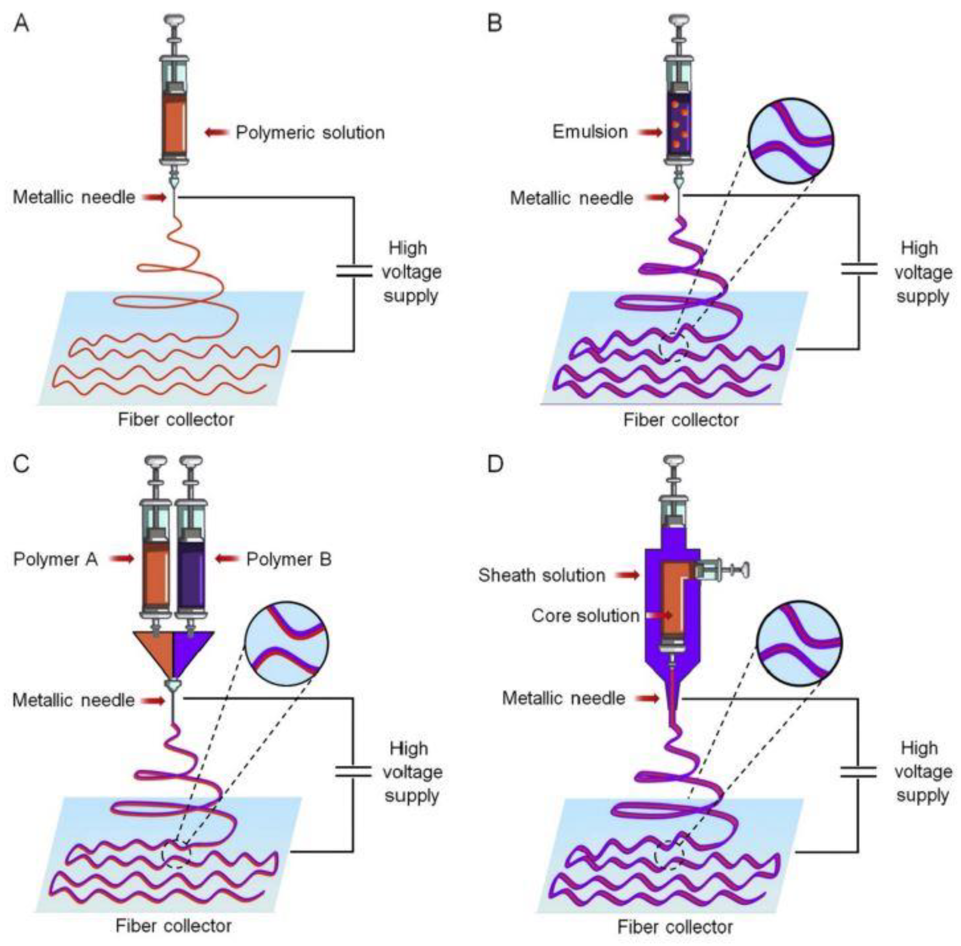
Figure 3.
Administration routes of drug-loaded gelatin nanofibers [30].
Figure 3.
Administration routes of drug-loaded gelatin nanofibers [30].
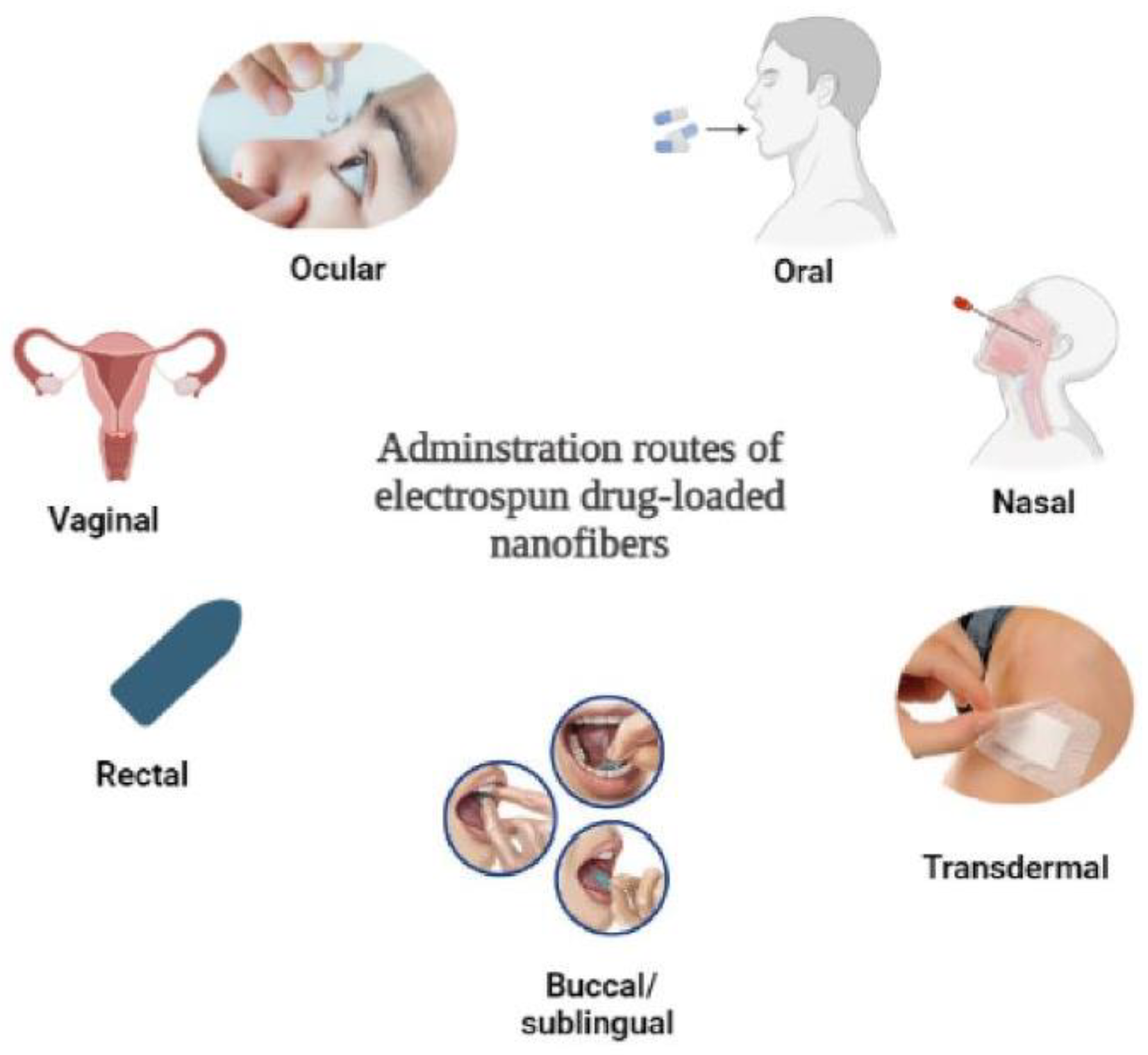
Figure 4.
Electrospun nanofiber-based soft electronics [58].
Figure 4.
Electrospun nanofiber-based soft electronics [58].

Figure 5.
Shirt powered by wireless energy transfer. (a) Receiver coils. (b) Smart T-shirt [56].
Figure 5.
Shirt powered by wireless energy transfer. (a) Receiver coils. (b) Smart T-shirt [56].

Figure 6.
Different packaging materials on sweet cherries [27].
Figure 6.
Different packaging materials on sweet cherries [27].
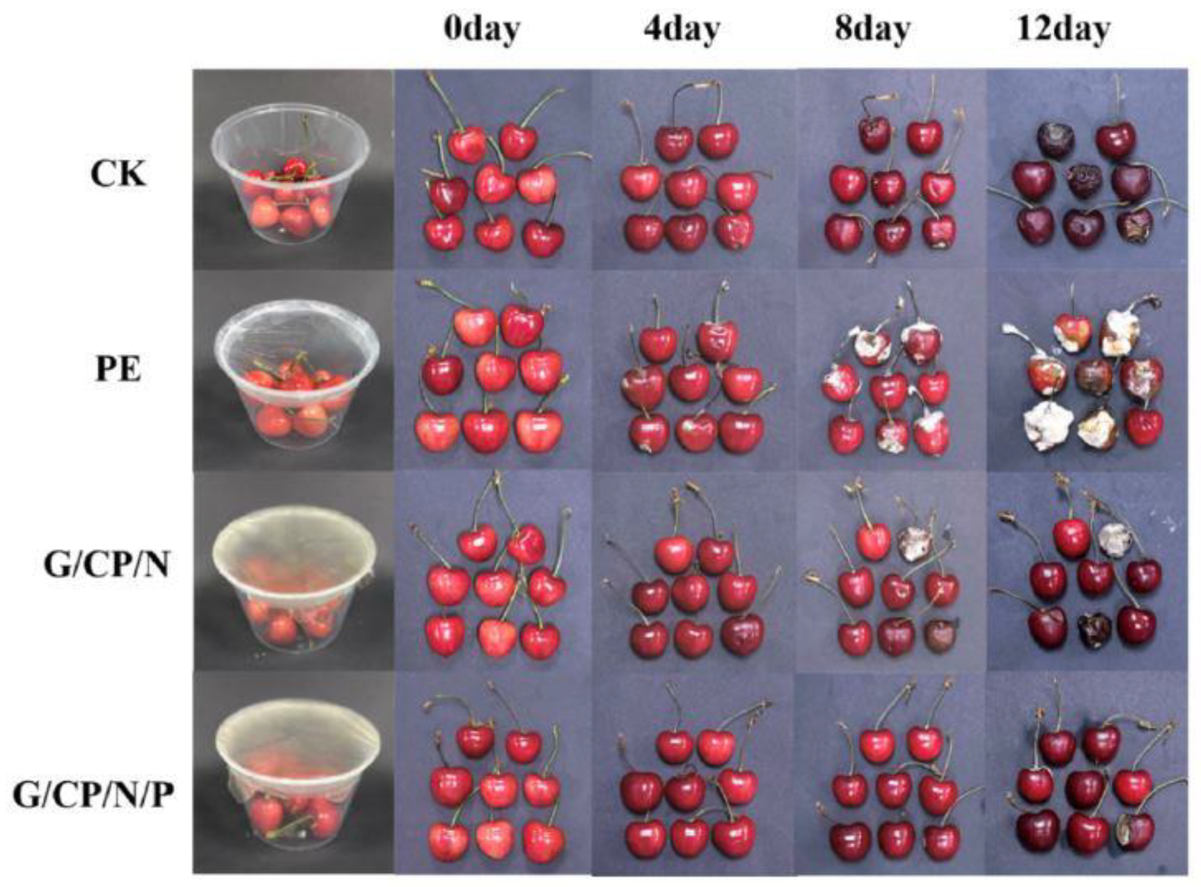
Figure 7.
Properties of electrospun textiles prepared by bio-based and biodegradable polymers [72].
Figure 7.
Properties of electrospun textiles prepared by bio-based and biodegradable polymers [72].


| Factors | Electrospinning Fiber Formation |
|---|---|
| Polymer type and concentration | Low concentrations lead to the formation of beads, whereas excessively high concentrations lead to the formation of coarse fibers or discontinuous fibers. The optimum feed rate is maintained to a stable Taylor cone shape, and the solvents have enough time to evaporate before the polymer is collected on the collector. |
| Polymer molecular weight | The entanglement of low-molecular-weight polymeric chains is not effective, which leads to the formation of beads. Both a dilute solution of a high-molecular-weight polymer and a concentrated solution of a low-molecular-weight polymer can produce fibers of identical fineness. |
| Solution viscosity | Different polymers have different viscosities. Viscosity is affected by the solvent used and the polymer concentration. Low solution viscosity promotes the formation of beads. When the viscosity is excessively high, coarse fibers are formed. |
| Solution conductivity | High conductivity promotes the formation of finer nanofibers, whereas a lower conductivity will promote beads formation. |
| Solvent | The solvent must have a good boiling point to evaporate from the polymers. Highly volatile solvents have a low boiling point and quickly evaporate, which can cause the jet to dry. Less volatile will not evaporate quickly to allow the jet to solidify. Precaution should be taken as most of the volatile solvents are toxic to the environment. |
| Voltage | The voltage applied to the solution differs from polymer to polymer. Excessive voltage can cause the formation of beads. |
| Collector type | The collector needs to have strong conductivity to effectively remove charges from the plate. If charges accumulate, they will repel incoming fibers and hinder membrane formation |
| Distance between nozzle and collector | The distance influences the fiber diameter. Shorter distances produce coarse fibers as the distance is too small to stretch the polymer to reduce its diameter |
| Surrounding conditions | High temperatures decrease viscosity, causing the formation of finer fibers. High humidity leads to the formation of coarse fibers. |
| Phase Separation | Drawing | Template | Self-Assembly | Electrospinning |
|---|---|---|---|---|
| No voltage is required. | No voltage is required. | No voltage is required. | No voltage is required. | High voltage is required. |
| Limited to certain polymers. | Limited to elastic polymers. | Production of continuous, long nanofibers is a challenge. | Limited to certain polymers. | Limited to conductive polymers, and solvent evaporation is required. |
| Low scalability and time-consuming | Limited to laboratory production | Limited to laboratory production. | Low scalability and complex processes | Can be scalable, but it also comes with its own challenges. |
Disclaimer/Publisher’s Note: The statements, opinions and data contained in all publications are solely those of the individual author(s) and contributor(s) and not of MDPI and/or the editor(s). MDPI and/or the editor(s) disclaim responsibility for any injury to people or property resulting from any ideas, methods, instructions or products referred to in the content. |
© 2024 by the author. Licensee MDPI, Basel, Switzerland. This article is an open access article distributed under the terms and conditions of the Creative Commons Attribution (CC BY) license (https://creativecommons.org/licenses/by/4.0/).
Share and Cite
MDPI and ACS Style
Patnaik, A. Recent Developments in Application of Nanofibers. Processes 2024, 12, 1894. https://doi.org/10.3390/pr12091894
AMA Style
Patnaik A. Recent Developments in Application of Nanofibers. Processes. 2024; 12(9):1894. https://doi.org/10.3390/pr12091894
Chicago/Turabian StylePatnaik, Asis. 2024. "Recent Developments in Application of Nanofibers" Processes 12, no. 9: 1894. https://doi.org/10.3390/pr12091894
Note that from the first issue of 2016, this journal uses article numbers instead of page numbers. See further details here.
Article Metrics
Article metric data becomes available approximately 24 hours after publication online.