Article

Antimicrobial and Anti-Biofilm Activities of Medicinal Plant-Derived Honey Against ESKAPE Pathogens: Insights into β-Lactamase Inhibition via Metabolomics and Molecular Modeling Studies

1
Department of Pharmacognosy, College of Pharmacy, King Saud University, Riyadh 11495, Saudi Arabia
2
Department of Pharmacognosy, Faculty of Pharmacy, Nahda University, Beni-Suef 62513, Egypt
3
Dental Health Department, College of Applied Medical Sciences, King Saud University, Riyadh 11433, Saudi Arabia
4
College of Pharmacy, Alfaisal University, Al Takhassusi Rd, Riyadh 11533, Saudi Arabia
5
Department of Pharmacognosy, Faculty of Pharmacy, Beni-Suef University, Beni-Suef 62514, Egypt
6
Department of Pharmacognosy, College of Pharmacy, University of Kut, Wasit 52001, Iraq
*
Authors to whom correspondence should be addressed.
These authors contributed equally to this work.
Processes 2025, 13(5), 1294; https://doi.org/10.3390/pr13051294
Submission received: 11 February 2025 / Revised: 13 April 2025 / Accepted: 22 April 2025 / Published: 24 April 2025
(This article belongs to the Special Issue Microbial Biofilms: Latest Advances and Prospects)

Abstract

:
The emergence of multidrug-resistant bacterial infections is a major global public health concern. Human health is in danger from microorganisms that have developed resistance to currently used drugs. Honey is well known for its significant activity against antibiotic-resistant bacteria. In this study, the antibacterial properties of honey from various botanical sources in Saudi Arabia against seven significant nosocomial and foodborne pathogens were investigated. The physicochemical properties of four Saudi honey samples—aloe honey (HO1) (Aloe vera L.), anise honey (HO2) (Pimpinella anisum L.), moringa honey (HO4) (Moringa oleifera Lam.), and acacia honey (HO5) (Acacia sp.)—were examined. In addition, they were screened for antibacterial activity against ESKAPE pathogens (Enterobacter faecalis, Staphylococcus aureus, Klebsiella pneumoniae, Pseudomonas aeruginosa, Salmonella Typhimurium, Escherichia coli, and Enterobacter sp.) and anti-biofilm activity against four pathogenic bacteria strains: S. aureus, P. aeruginosa, S. typhimurium, and E. coli. 1H NMR profiling and multivariate analysis (PCA and PLS-DA) were performed. Aloe honey (HO1) was the most distinct sample based on MVDA and its antibacterial activity, and it exhibited anti-biofilm activity against most biofilm-forming microorganisms. Its metabolic profile was deduced using LC-MS, and the resulting annotated compounds were docked against several β-lactamase enzyme classes. The results reveal the potential of honey-derived compounds to inhibit β-lactamases due to the presence of gallic acid hexoside and rosmarinic acid, suggesting their potential as competitive inhibitors. Our findings suggest that further honey antibacterial compounds could offer a novel approach to overcoming antibiotic resistance by targeting and inhibiting β-lactamase enzymes.

1. Introduction

Microorganisms adapt to survive in both favorable and unfavorable environments through a range of defense mechanisms, one of which is to reduce their susceptibility to antibiotics. This causes antibiotics to eventually lose their effectiveness and become unusable for treating bacterial infections, leading to a high mortality rate worldwide [1]. Among the most virulent multidrug-resistant bacteria are ESKAPE pathogens. The acronym ESKAPE, which stands for E. faecalis, S. aureus, K. pneumoniae, P. aeruginosa, S. Typhimurium, E. coli, and Enterobacter sp., describes a class of virulent bacteria recognized by the Infectious Diseases Society of America (IDSA) and the European Centre for Disease Prevention and Control (ECDC); this designation results from these microorganisms’ tendency to develop resistance to and “escape” from currently available antibiotics, raising the danger of nosocomial infections in hospitalized patients or individuals with impaired immune systems [2]. Although the ESKAPE pathogens are genetically diverse, they exhibit similar resistance mechanisms, including drug degradation or modification, antibiotic inflow prevention, efflux-pump-mediated antibiotic expulsion, antibiotic target alteration, and so forth [1]. Recently, the World Health Organization (WHO) added ESKAPE infections to its list of twelve germs that necessitate the immediate discovery of new antibiotics. Based on the urgency with which new antibiotics are needed to combat them, vancomycin-resistant E. faecium (VREF) and methicillin/vancomycin-resistant S. aureus (MRSA/VRSA) were classified as having high priority among ESKAPE organisms, whereas carbapenem-resistant A. baumannii and carbapenem-resistant P. aeruginosa were deemed as having critical priority [1,3].
Metabolomics is a field in chemistry that combines analytical metabolite measurement techniques, such as LC-HRMS and NMR, with chemometric statistical analysis. This method is widely applicable in many fields, such as plant sciences, food sciences, toxicology, and biochemistry [4]. For natural products, metabolomics studies can determine the complete chemical profile or fingerprint of an organism under particular conditions, which is essential for the discovery of new bioactive metabolites and natural drugs [5].
Nature is an endless source of bioactive secondary metabolites with a broad spectrum of pharmacological properties, such as antibacterial, antiviral, anticancer, antifungal, antihypertensive, and depressive properties. Honey, the sticky liquid that honey bees naturally produce, is recommended in the Holy Quran for healing diseases. Honey has long piqued human curiosity; it has been utilized as both a nutritious foodstuff and a medicinal remedy, and is the key product of the apiculture industry [6]. Honey’s beneficial bioactive ingredients are responsible for its many functional qualities, including anti-inflammatory, antioxidant, antiviral, and antibacterial effects, which help to preserve human health and treat certain ailments and diseases, as well as strengthen the immune system [7]. Various studies have revealed the antibacterial potential of honey, which is related to both enzymatic and non-enzymatic processes. One of the main antibacterial agents in honey, hydrogen peroxide (H2O2), is created by the enzyme glucose oxidase (GOX), which is derived from bees [8,9]. Furthermore, honey’s polyphenolic and phytochemical constituents, which vary based on their botanical and geographic origins, have shown non-enzymatic antibacterial effects [9]. Given that honey’s chemical and pharmacological activity depends on its botanical and geographical origins, we carried out the first comprehensive analysis of the ESKAPE profile of multi-floral honey from various floristic regions in Saudi Arabia. Four honey samples, aloe honey (HO1) (Aloe vera L.), anise honey (HO2) (Pimpinella anisum L.), moringa honey (HO4) (Moringa oleifera Lam.), and acacia honey (HO5) (Acacia sp.), were collected and evaluated for their antibacterial activity against ESKAPE pathogens. We subsequently applied 1H NMR-based metabolomic techniques, and multivariate data statistical analysis (MVDA) was then performed in order to minimize the large dataset obtained and correlate and differentiate the tested honey samples [10]. Moreover, LC-MS analysis, followed by chemotaxonomic categorization and molecular modeling tools, was applied to elucidate the bacterial targets of the compounds identified in aloe honey (HO1) (Aloe vera L.).

2. Materials and Methods

2.1. Honey Preparation and Collection

Four Saudi honey samples from different botanical sources were obtained: aloe honey (Aloe vera L.), anise honey (Pimpinella anisum L.), moringa honey (Moringa oleifera Lam.), and acacia honey (Acacia sp.), all samples were purchased from (Riyadh, Saudi Arabia). A reference number was given to each sample. Samples were authenticated in the Honey Quality Laboratory on Orooba Street, Oliaya, Riyadh. The samples of honey were kept in the dark and at room temperature.

2.2. 1H-NMR Analysis

1H-NMR (400 MHz) spectra were recorded (Bruker, Munich, Germany) with tetramethylsilane (TMS) as the internal standard and dimethyl sulfoxide (DMSO-d6) as the solvent. Chemical shift measurements are expressed in ppm.

2.3. LC-MS Metabolomic Analysis

An Accela HPLC (Thermo Fisher Scientific, Bremen, Germany), along with an Exactive (Orbitrap) mass spectrometer and an Accela UV/VIS, was used to analyze the whole extract (1 mg/mL in MeOH). The mobile phase comprised 0.1% formic acid in each of two solvents: acetonitrile (B) and water (A). Gradient elution was initiated at a flow rate of 300 μL/min, and after 30 min, 10% B increased linearly to 100% B. After that, it remained isocratic for 5 min, and then it decreased linearly to 10% B for 1 min. The next injection was made after the mobile phase had had nine minutes to acclimate. The mass range was set to m/z (mass-to-charge ratio) 100–2000 with the in-source collision-induced dissociation (CID) mechanism for ESI–MS, and to m/z 50–1000 with untargeted HCD (high-energy collision dissociation) for MS/MS. The raw data were loaded into MZmine 2.12, a framework for differential analysis of mass spectrometry data. Peak deisotoping was then carried out after chromatogram deconvolution. The retention time normalizer was utilized for gap-filling and chromatographic alignment purposes. Positive and negative ionization mode data files produced by MZmine were combined using Excel macros. The peaks that the sample produced were extracted. Using RT and a m/z threshold of ±5 ppm, an Excel macro was utilized to dereplicate each m/z ion peak with chemicals in a customized database. This yielded information on the probable identities of all metabolites in the extract as a whole. After making a list for the extract, the Excel macro was used to determine the top 20 characteristics (ordered by peak intensity) and the related putative identities. After that, the metabolites were determined by cross-referencing them with a few internal and online databases.

2.4. Multivariate Data Statistical Analysis

The web-based statistical analysis platform MetaboAnalyst 6.0 was utilized to analyze NMR data. An input file including a table of peak chemical shifts (ppm) and peak intensities exported as comma-separated values (.csv) is needed for this exploration. The MetaboAnalyst 6.0 server (https://www.metaboanalyst.ca) received the data uploaded in a single zip file. Initially, the raw data were normalized using the median, and then scaled using Pareto scaling. Subsequently, multivariate analysis was conducted using unsupervised principal component analysis (PCA) and supervised partial least-squares discriminant analysis (PLS-DA). In addition to the aforementioned experiments, a clustering analysis was also performed [11].

2.5. Antibacterial Activity

To measure the antibacterial activity of the tested compounds, Gram-negative bacteria and Gram-positive bacteria were used as test organisms [12]. The test was performed in 96-well flat polystyrene plates. A 10 µL sample of the test extract (final concentration of 250 µg/mL) was added to 80 µL of lysogeny broth (LB broth), followed by the addition of 10 µL of bacterial culture suspension (log phase), and then the plates were incubated overnight at 37 °C. After incubation, clearance in a well indicated a positive antibacterial effect of the tested compound, whereas a well with an opaque medium indicated that the compound did not affect the bacteria. Pathogens without any treatment were used as controls. The absorbance was measured after about 20 h at OD600 in a Spectrostar Nano Microplate Reader (BMG LABTECH GmbH, Allmendgrun, Germany).

2.6. Biofilm Inhibition Assay

Using 96-well flat polystyrene plates, different honey samples H (1, 2, 4, and 5) were tested for their ability to suppress biofilm formation in four pathogenic bacteria strains: S. aureus, P. aeruginosa, S. typhimurium, and E. coli. The experiment involved filling each well with 180 µL of LB broth, inoculating it with 10 µL of pathogenic bacteria, and then adding 10 µL of each sample and control (final concentration: 500 µg mL−2). For 24 h, the plates remained in a 37 °C incubator. The liquid was removed from the wells, which were then washed with 200 µL of pH 7.2 phosphate-buffered saline (PBS). This removed non-surface microorganisms. After 1 h of sterilized laminar flow drying, the plates were then stained with 200 µL of 0.1 percent weight/volume crystal violet solution per well. After 1 h, the excess pigment was removed, and the plates were allowed to dry. After the dried plates were cleaned with 95% ethanol, a Spectrostar Nano Microplate Reader was used to measure optical density at 570 nm. BMG LABTECH GmbH is in Allmendgrun, Germany [13]. HO1 and HO5 were the most consistent and effective among strains, whereas HO4 was less effective.

2.7. Molecular Docking

The LigandFit docking engine of Biovia Discovery Studio Client v16 was used for docking experiments. This software treats ligands as flexible and receptors as rigid structures. The active site was defined using the “Define and Edit Binding site” tool in Discovery Studio. Proteins and ligands were prepared using the “Prepare Protein” and “Prepare Ligand” tools of Discovery Studio. The default settings of LigandFit were applied in the docking experiments. The consensus scores of the docked ligands were calculated from 14 scoring functions: Goldscore, ASP, CHEMPLP, LigScore1, LigScore2, PMF, PMF04, PLP1, PLP2, Ludi Energy Estimate 1, Ludi Energy Estimate 2, Ludi Energy Estimate 3, Jain, and Chemscore [14,15]. Consensus scoring is a rapid method to classify ligands based on multiple scoring functions. The consensus score calculated for a ligand corresponds to the count of scores falling within the top rank percentile [16].

3. Results

3.1. Physicochemical Properties

The physicochemical properties of the honey samples under investigation were analyzed by Saudi Arabia honey quality laboratories. These characteristics included color, odor, taste, purity, moisture content, sugar composition, hydroxymethylfurfural (HMF), acidity, and diastase enzyme levels (Figures S1–S4). The results showed that the honey samples were high-quality and complied with both national and international standards. Table 1 shows the results of the physicochemical parameters examined.

3.2. Chemical Profiling

3.2.1. NMR Analysis

1H NMR analysis was performed for the four Saudi multi-floral honey samples, and the raw data were further processed using MestreNova v6.0.2-5475. The results show that while the generated spectra have similar aliphatic areas and sugar content, they are unique, especially in the aromatic region. The spectra are presented in Figures S5–S8.

3.2.2. Multivariate Data Statistical Analysis

In the principal component analysis (PCA) and partial least-squares discriminant analysis (PLS-DA) score plots (Figure 1), the samples are mainly distributed into segregated areas. The PCA score plot reveals the dispersal of the culture extracts HO1, HO2, HO4, and HO5 in four different regions between PC1 (45.3%) and PC2 (28.2%) (Figure 1A), which is confirmed by the PLS-DA score plot (Figure 1B). These findings indicate that the four Saudi honey samples have distinct chemical profiles. Furthermore, in PLS-DA, the model’s correlation coefficient, R2, and cross-validation correlation coefficient, Q2, are equal to 1.0 when using three components (Figure 1C).

3.3. Antibacterial Activity of Saudi Multi-Floral Honey Samples

In order to evaluate the antibacterial properties of the four Saudi multi-floral honey samples against ESKAPE pathogens, seven significant human and foodborne pathogens were tested (Enterobacter faecalis, Staphylococcus aureus, Klebsiella pneumoniae, Pseudomonas aeruginosa, Salmonella Typhimurium, Escherichia coli, and Enterobacter sp.). The samples were compared to cephalosporin as a positive control. Table 2 displays the bacterial inhibition rates, and Table 3 displays the values of the minimum inhibitory concentration (MIC). The findings demonstrate that, compared to cephalosporin, aloe honey (HO1) (Aloe vera L.) exhibited a potent inhibitory effect against S. aureus, K. pneumoniae, S. typhimurium, and Enterobacter sp., with inhibition rates of 93.3%, 81.10%, 82.9%, and 78.32, respectively.
Conversely, the majority of the pathogens under investigation were weakly inhibited by moringa honey (HO4), which is derived from Moringa oleifera Lam.

3.4. Anti-Biofilm Activity

The biofilm inhibition study showed that the different treatments worked to varying degrees against different bacterial strains. HO1 had the highest inhibitory activity against S. aureus ATCC 25923, at 89.26%, whereas HO5 had 84.25%. HO2 and HO4 had 45.2% and 50.23% inhibition, respectively, demonstrating a significant difference in their effects. The inhibition rates of HO2 and HO5 against P. aeruginosa ATCC 90902 were above 84%, but HO1 had a rate of only 55.5%. HO4 was ineffective against this strain, demonstrating its limits in treating P. aeruginosa biofilms. HO1 had the highest inhibition rate against S. typhimurium ATCC 14028, at 85.12%, slightly exceeding those of HO2 and HO5, which were 82.23% and 83.42%, respectively. With 15.2% inhibition, HO4 had no effect. All treatments moderately inhibited E. coli ATCC 8739, with HO1 inhibiting it the most at 45.32%, followed by HO5 (44.28%), HO2 (40.52%), and HO4 (35.23%). HO1 and HO5 were the most consistent and effective among strains, whereas HO4 was less effective (Figure 2).

3.5. LC-MS Metabolomic Profiling

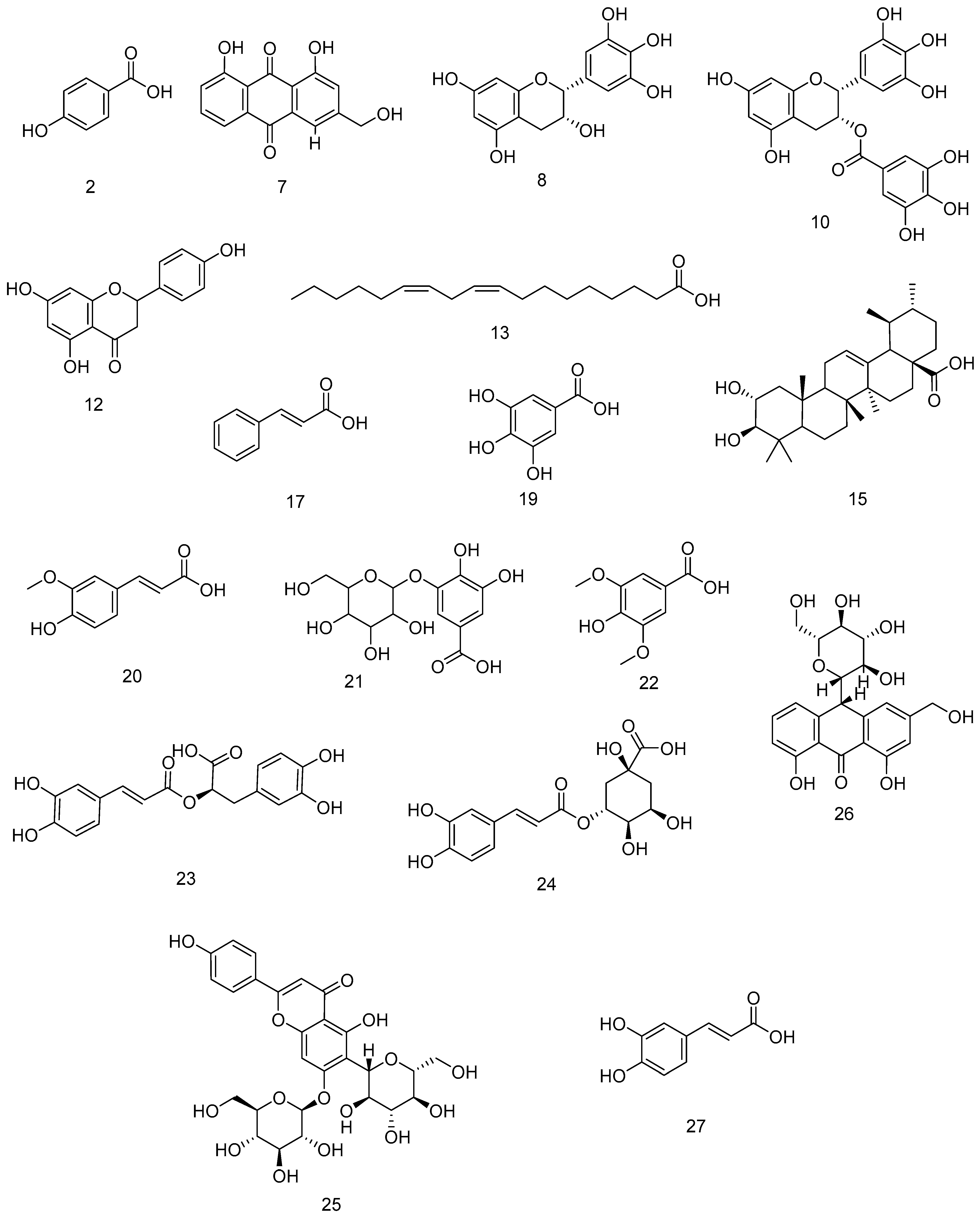
The aloe honey (HO1) sample was subjected to further analysis using LC-MS in positive and negative modes. Only low-molecular-weight (m/z < 1500) ionizable molecules were taken into consideration when profiling the metabolites using untargeted metabolomic methods. Dereplication was carried out using the DNP database. The number of obtained characteristics was then decreased by applying a chemotaxonomic filter, leading to the identification of twenty-seven metabolites (Table 4, Figure 3). Phenolic acid derivatives were found to prevail among the detected metabolites, which were primarily represented by sugars, flavonoids, vitamins, and fatty acids.

3.6. Molecular Docking

The aforementioned results prompted us to evaluate the potential of aloe honey-derived compounds to inhibit β-lactamases. According to the Bush–Jacoby group classification, β-lactamases are categorized into several groups based on the diversity and characteristics of these enzymes [33]. Therefore, the β-lactamase-inhibitory activities of honey-derived compounds were investigated using 10 classes of β-lactamases, as listed in Table 5. Each compound was docked inside the catalytic active site of each β-lactamase, and a consensus score of the 14 scoring functions (listed in the experimental section) was calculated. Consensus scoring involves combining multiple scoring functions to assess the binding affinity between a ligand and a target protein, which can improve the accuracy and reliability of the docking results by considering different aspects of the molecular interactions [34]. Afterward, the sum of the 10 consensus scores of each compound was calculated to prioritize the compounds according to their binding affinity across the 10 classes of β-lactamases (Table 6).
Two compounds, gallic acid hexoside and rosmarinic acid, demonstrated total consensus scores higher than that of avibactam, a reversible competitive β-lactamase inhibitor. Gallic acid hexoside, also known as 3-glucogallic acid, is a phenolic glycoside with reported antioxidant and anticancer activities. This compound demonstrated high docking scores with most classes of β-lactamases, with the highest scores observed for those with PDB codes 1SHV (a broad-spectrum beta-lactamase) and 1G68 (a carbenicillinase).
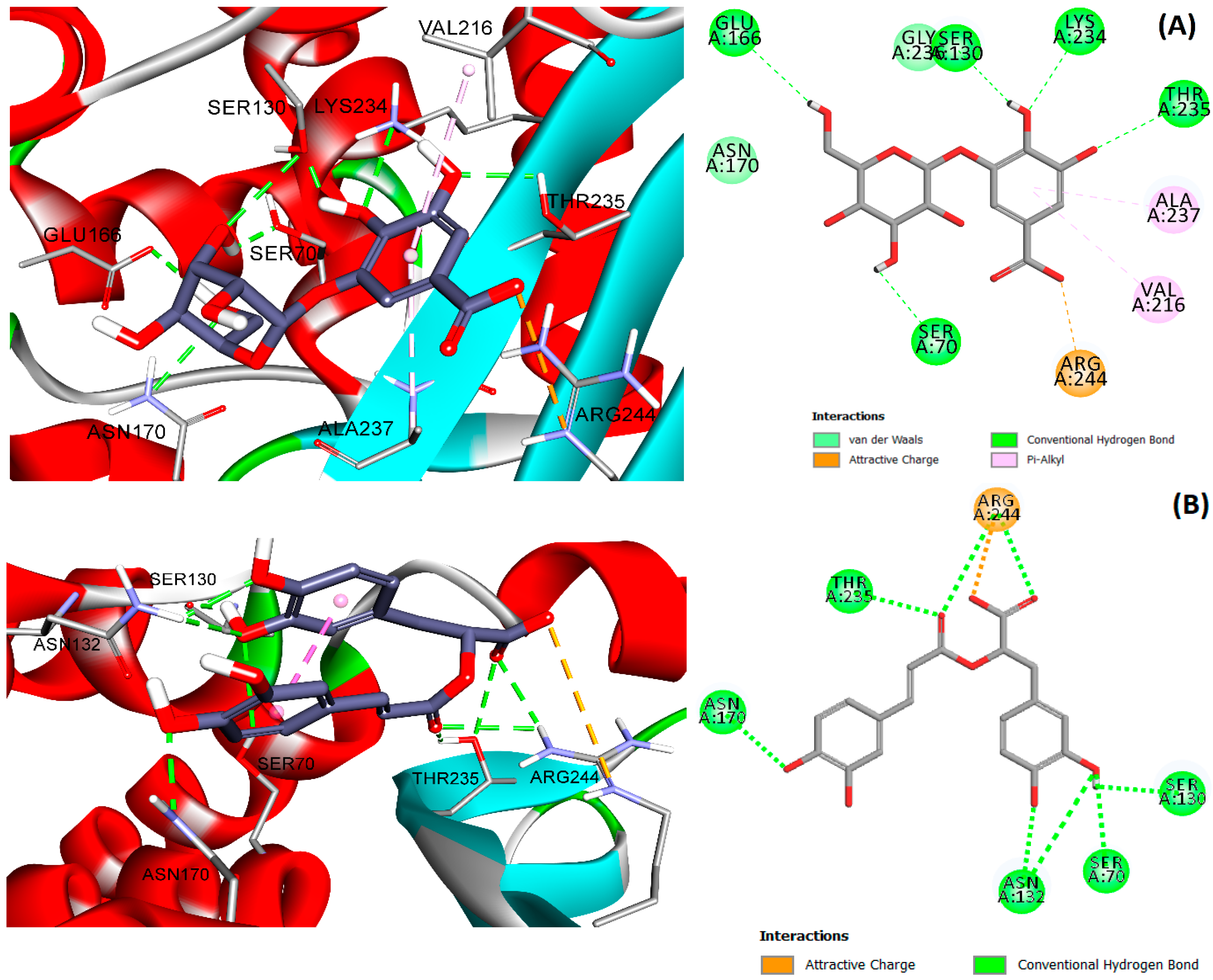
Docking studies of gallic acid hexoside with the active site of 1SHV showed strong ionic interaction with ARG244 and a network of hydrogen bonding interactions with THR235, ASN170, GLU166, SER130, SER70, and LYS234 (Figure 4A). Gallic acid hexoside blocks two important residues: SER70, the key catalytic residue of β-lactamase, and GLU166, which serves as the general base for the acylation and deacylation steps of β-lactam ring hydrolysis [35]. Additionally, gallic acid hexoside forms π–alkyl interactions with VAL216 and ALA237.
Rosmarinic acid is a naturally occurring polyphenolic compound found in various plants, particularly in the Lamiaceae family, with various biological activities. Similarly to gallic acid hexoside, rosmarinic acid demonstrated high docking scores with most classes of β-lactamases, with the highest scores observed for those with PDB codes 1SHV (a broad-spectrum beta-lactamase) and 1LHY (an inhibitor-resistant beta-lactamase). The docking of rosmarinic acid with 1SHV showed a strong ionic interaction with ARG244 and multiple hydrogen interactions with SER130, SER70 (the catalytic residue), ASN132, ASN170, and THR235 (Figure 4B). The docked pose of rosmarinic acid showed intramolecular π–π stacking between the two aromatic rings, which would enhance binding affinity by reducing the entropic penalty and stabilizing the binding conformation.

4. Discussion

The developing science of “metabolomics,” which integrates spectroscopy, metabolism, and multivariate statistical analysis (pattern recognition) techniques, entails the extraction of useful information from complex spectroscopic data of metabolite mixtures [36]. For metabolic profiling, nuclear magnetic resonance (NMR) and mass spectrometry (MS) are the most often utilized analytical methods. 1H-NMR spectroscopy is frequently utilized to develop metabolic profiles in metabolic investigations because it requires little sample preparation and yields quantifiable and repeatable information [37]. A series of procedures are involved in metabolomics, such as sample preparation, sample analysis (NMR), data acquisition, data analysis, and interpretation. Here, untargeted metabolomic techniques were used to profile the metabolites found in four investigated Saudi honey samples from different botanical origins, namely, aloe honey (HO1) (Aloe vera L.), anise honey (HO2) (Pimpinella anisum L.), moringa honey (HO4) (Moringa oleifera Lam.), and acacia honey (HO5) (Acacia sp.), thereby illuminating the variations in the metabolomes of the samples. The NMR spectra obtained from processing the four materials under investigation using MestreNova indicated that they are distinct, especially in the aromatic region (5–8 ppm) (Figures S5–S8).

4.1. Multivariate Data Analysis

In order to simplify the dataset, remove high dimensionality, correlate the results, and draw conclusions, the NMR analysis dataset was statistically processed using MetaboAnalyst 6.0. First to be used was the unsupervised PCA approach, which reduces the dimensions of multivariate data and highlights significant differences without needing prior knowledge of the dataset being analyzed [5]; the honey samples were primarily distributed to four segregated areas between PC1 and PC2, which indicated statistically significant differences between the extracts. The principal components (Figure 1A) distinguished from the various samples, i.e., PC1 and PC2, accounted for a total variance of 45.3% and 28.2%, respectively.
PLS-DA is a supervised approach that employs multivariate linear regression. The supervised PLS-DA analysis verified the differences between honey samples (Figure 1B). The model’s correlation coefficient, R2, and cross-validation correlation coefficient, Q2, were equal to 1.0 for three components, indicating the strong predictive performance of the model (Figure 1C). These findings indicate that differences in honey’s botanical and geographical origins have a significant impact on its chemical constituents and, as a result, its biological activity.
Honey’s antibacterial action is one of its best-known bioactivities, as documented in numerous studies. Honey impacts microorganism growth and survival through a variety of processes. For example, honey’s low pH, high sugar concentration, high osmolarity, and antibacterial compounds, including hydrogen peroxide and polyphenols, make it a hostile habitat for bacteria [8,9,38]. In the present investigation, the antibacterial activity of four Saudi honey samples showed varying degrees of potency against ESKAPE pathogens when compared to cephalosporin (Figure 2). Among these, aloe honey (HO1) demonstrated a strong inhibitory effect against S. aureus, K. pneumoniae, S. typhimurium, and Enterobacter sp., with inhibition rates of 93.3%, 81.10%, 82.9%, and 78.32, respectively. These findings inspired us to carry out more research on the aloe honey sample (HO1); LC-MS was employed to determine the chemical contents of aloe honey (HO1), followed by docking analysis. Following LC-MS analysis, dereplication investigations based on chemotaxonomic sorting suggested potential active metabolites that belonged to various chemical classes, primarily phenolic acids, flavonoids, and anthraquinones.

4.2. Molecular Docking

β-lactamases are enzymes produced by bacteria that hydrolyze the crucial beta-lactam ring found in penicillin, cephalosporins, and other antibiotics, effectively rendering them useless. This mechanism of resistance poses a significant threat to treatments for bacterial infections [39]. To combat this challenge, researchers have developed beta-lactamase inhibitors, which act by binding to and inhibiting the activity of these enzymes. These inhibitors, such as clavulanate, sulbactam, and tazobactam, are often combined with beta-lactam antibiotics to protect them from degradation, enhancing their effectiveness against resistant bacteria [40]. However, the constant emergence of novel and more potent beta-lactamases, such as extended-spectrum beta-lactamases (ESBLs) and carbapenemases, necessitates ongoing research to develop new and more effective inhibitors to counter the growing threat of antibiotic resistance.
Two compounds, gallic acid hexoside and rosmarinic acid, demonstrated total consensus scores higher than that of avibactam, a reversible competitive β-lactamase inhibitor. These findings shed light on the potential of honey-derived compounds to inhibit β-lactamases. Notably, the presence of gallic acid hexoside and rosmarinic acid suggests their roles as potential competitive inhibitors. These natural compounds could offer a novel approach to overcoming antibiotic resistance by targeting and inhibiting β-lactamase enzymes, which are responsible for antibiotic degradation. Further research into these compounds may reveal new strategies for enhancing the efficacy of β-lactam antibiotics.

5. Conclusions

Honey’s antimicrobial properties are well known, and it has been shown to be effective against a wide range of microorganisms, including bacteria. Honey’s antimicrobial properties are attributed to a variety of factors, including osmolarity, H2O2 content, low pH, phenolic acid levels, flavonoids, tetracycline, peroxides, amylase, fatty acids, phenols, ascorbic acid, terpenes, benzyl alcohols, and benzoic acid. Other phytochemical factors, including fatty acids, phenolics, ascorbic acid, terpenes, benzyl alcohols, and benzoic acid, make honey active against pathogenic bacteria and produce either bactericidal or bacteriostatic effects, depending on the nectar source, geographical origin, and storage conditions. In this study, NMR analysis and MVDA, in addition to antibacterial tests, revealed significant variability between the honey samples under investigation, which can be attributed to their varied botanical sources affecting their chemical makeup. Aloe honey showed high antibacterial potency against ESKAPE infections. Additionally, LC/MS analysis was carried out to analyze the aloe honey sample, and the resulting annotated compounds were docked against the β-lactamase enzyme. Based on their competitive inhibitory effects, gallic acid hexoside and rosmarinic acid, two substances identified in aloe honey, were found to be able to block β-lactamases. Our research indicates that by specifically targeting and blocking β-lactamase enzymes, natural substances may provide alternatives for combating antibiotic resistance. Further investigation is recommended to determine the exact dosage of aloe honey needed to yield the desired therapeutic effects on a range of illnesses caused by ESKAPE bacteria. Moreover, honey could be a potential alternative to antibacterials, with promising therapeutic potential in the medical field. The precise chemical composition of honey must be understood to produce artificial honey that can treat medical conditions. The need for an improved delivery method is crucial to enhancing its effectiveness, and its potential to be integrated into the clinical setting to provide innovative treatments should not be overlooked.

Supplementary Materials

The following supporting information can be downloaded at https://www.mdpi.com/article/10.3390/pr13051294/s1, Figure S1: Sample test report of Saudi Aloe Honey (HO1) analyzed by Honey Quality Laboratory, ap-proved by the Saudi Accreditation Committee; Figure S2: Sample test report of Saudi Anise Honey (HO2) analyzed by Honey Quality Laboratory, ap-proved by the Saudi Accreditation Committee; Figure S3: Sample test report of Saudi moringa Honey (HO4) analyzed by Honey Quality Laboratory, ap-proved by the Saudi Accreditation Committee; Figure S4: Sample test report of Saudi Acacia Honey (HO5) analyzed by Honey Quality Laboratory, ap-proved by the Saudi Accreditation Committee; Figure S5: 1H-NMR Spectrum of aloe honey sample (HO1) (500 MHz, DMSO-d6); Figure S6: 1H-NMR Spectrum of anise honey sample (HO2) (500 MHz, DMSO-d6); Figure S7: 1H-NMR Spectrum of moringa honey sample (HO4) (500 MHz, DMSO-d6); Figure S8: 1H-NMR Spectrum of acacia honey sample (HO5) (500 MHz, DMSO-d6).

Author Contributions

Conceptualization, H.A. and H.M.H.; methodology, M.A.K.; software, M.A.K. and H.S.B.; validation, S.Y.A. and N.M.L.; formal analysis, S.Y.A.; investigation, N.M.L.; resources, H.A.; data curation, H.M.H.; writing—original draft preparation, N.M.L., M.A.K. and H.S.B.; writing—review and editing, H.M.H.; visualization, N.M.L.; supervision, H.M.H.; project administration, H.M.H.; funding acquisition, H.A. All authors have read and agreed to the published version of the manuscript.

Funding

This research project was supported by a grant from the “Research Center of the Female Scientific and Medical Colleges”, Deanship of Scientific Research, King Saud University; also this project is partially funded by office of research at Alfaisal University.

Data Availability Statement

All data from this research are included in the manuscript.

Conflicts of Interest

The authors declare no conflicts of interest.

References

  1. Jadimurthy, R.; Mayegowda, S.B.; Nayak, S.C.; Mohan, C.D.; Rangappa, K.S. Escaping mechanisms of ESKAPE pathogens from antibiotics and their targeting by natural compounds. Biotechnol. Rep. 2022, 34, e00728. [Google Scholar] [CrossRef] [PubMed]
  2. Naz, S.; Ngo, T.; Farooq, U.; Abagyan, R. Analysis of drug binding pockets and repurposing opportunities for twelve essential enzymes of ESKAPE pathogens. PeerJ 2017, 5, e3765. [Google Scholar] [CrossRef] [PubMed]
  3. De Oliveira, D.M.P.; Forde, B.M.; Kidd, T.J.; Harris, P.N.A.; Schembri, M.A.; Beatson, S.A.; Paterson, D.L.; Walker, M.J. Antimicrobial resistance in ESKAPE pathogens. Clin. Microbiol. Rev. 2020, 33, e00181-19. [Google Scholar] [CrossRef]
  4. Sarkar, J.; Singh, R.; Chandel, S. Understanding LC/MS-Based Metabolomics: A Detailed Reference for Natural Product Analysis. Proteom. Clin. Appl. 2025, 19, e202400048. [Google Scholar] [CrossRef]
  5. El-Hawary, S.S.; Mohammed, R.; Bahr, H.S.; Attia, E.Z.; El-Katatny, M.H.; Abelyan, N.; Al-Sanea, M.M.; Moawad, A.S.; Abdelmohsen, U.R. Soybean-associated endophytic fungi as potential source for anti-COVID-19 metabolites supported by docking analysis. J. Appl. Microbiol. 2021, 131, 1193–1211. [Google Scholar] [CrossRef]
  6. Kumar, D.; Hazra, K.; Prasad, P.V.V.; Bulleddu, R. Honey: An important nutrient and adjuvant for maintenance of health and management of diseases. J. Ethn. Foods 2024, 11, 19. [Google Scholar] [CrossRef]
  7. Hajian-Tilaki, A.; Kenari, R.E.; Razavi, R.; Farahmandfar, R. Phenolic profile, antioxidant properties, and pollen spectra of Iranian-originated honeys. Eur. Food Res. Technol. 2024, 250, 2317–2329. [Google Scholar] [CrossRef]
  8. Abdelhady, A.S.M.; Darwish, N.M.; Abdel-Rahman, S.M.; El Magd, N.M.A. The combined antimicrobial activity of citrus honey and fosfomycin on multidrug resistant pseudomonas aeruginosa isolates. AIMS Microbiol. 2020, 6, 162–175. [Google Scholar] [CrossRef]
  9. Tsadila, C.; Nikolaidis, M.; Dimitriou, T.G.; Kafantaris, I.; Amoutzias, G.D.; Pournaras, S.; Mossialos, D. Antibacterial activity and characterization of bacteria isolated from diverse types of greek honey against nosocomial and foodborne pathogens. Appl. Sci. 2021, 11, 5801. [Google Scholar] [CrossRef]
  10. Raheem, D.J.; Tawfike, A.F.; Abdelmohsen, U.R.; Edrada-Ebel, R.A.; Fitzsimmons-Thoss, V. Application of metabolomics and molecular networking in investigating the chemical profile and antitrypanosomal activity of British bluebells (Hyacinthoides non-scripta). Sci. Rep. 2019, 9, 2547. [Google Scholar] [CrossRef]
  11. Demarque, D.P.; Dusi, R.G.; de Sousa, F.D.M.; Grossi, S.M.; Silvério, M.R.S.; Lopes, N.P.; Espindola, L.S. Mass spectrometry-based metabolomics approach in the isolation of bioactive natural products. Sci. Rep. 2020, 10, 1051. [Google Scholar] [CrossRef] [PubMed]
  12. Alhadrami, H.A.; Sayed, A.M.; Sharif, A.M.; Azhar, E.I.; Rateb, M.E. Olive-derived triterpenes suppress SARS-CoV-2 main protease: A promising scaffold for future therapeutics. Molecules 2021, 26, 2654. [Google Scholar] [CrossRef] [PubMed]
  13. Attea, S.A.; Ghareeb, M.A.; Kelany, A.K.; Elhakim, H.K.A.; Allemailem, K.S.; Bukhari, S.I.; Rashidi, F.B.; Hamed, A.A. Biosynthesis of Iron Oxide Nanoparticles by Marine Streptomyces sp. SMGL39 with Antibiofilm Activity: In Vitro and In Silico Study. Molecules 2024, 29, 4784. [Google Scholar] [CrossRef] [PubMed]
  14. Khanfar, M.A.; Taha, M.O. Elaborate ligand-based modeling coupled with multiple linear regression and k nearest neighbor QSAR analyses unveiled new nanomolar mTOR inhibitors. J. Chem. Inf. Model. 2013, 53, 2587–2612. [Google Scholar] [CrossRef]
  15. Khanfar, M.A.; Salaas, N.; Abumostafa, R. Discovery of natural-derived Mpro inhibitors as therapeutic candidates for COVID-19: Structure-based pharmacophore screening combined with QSAR analysis. Mol. Inform. 2023, 42, 2200198. [Google Scholar] [CrossRef]
  16. Nandigrami, P.; Fiser, A. Assessing the functional impact of protein binding site definition. Protein Sci. 2024, 33, e5026. [Google Scholar] [CrossRef]
  17. Ricochon, G.; Paris, C.; Girardin, M.; Muniglia, L. Highly sensitive, quick and simple quantification method for mono and disaccharides in aqueous media using liquid chromatography–atmospheric pressure chemical ionization–mass spectrometry (LC–APCI–MS). J. Chromatogr. B 2011, 879, 1529–1536. [Google Scholar] [CrossRef]
  18. Wang, Y.; Xing, L.; Zhang, J.; Ma, X.; Weng, R. Determination of endogenous phenolic compounds in honey by HPLC-MS/MS. LWT 2023, 183, 114951. [Google Scholar] [CrossRef]
  19. Li, T.; Zhao, T. Analysis of Raffinose Metabolism-Related Sugar Components in Seeds. In Seed Dormancy: Methods and Protocols; Springer: Berlin/Heidelberg, Germany, 2024; pp. 73–80. [Google Scholar]
  20. Soyseven, M.; Sezgin, B.; Arli, G. A novel, rapid and robust HPLC-ELSD method for simultaneous determination of fructose, glucose and sucrose in various food samples: Method development and validation. J. Food Compos. Anal. 2022, 107, 104400. [Google Scholar] [CrossRef]
  21. Zhou, J.; Qi, Y.; Ritho, J.; Duan, L.; Wu, L.; Diao, Q.; Li, Y.; Zhao, J. Analysis of maltooligosaccharides in honey samples by ultra-performance liquid chromatography coupled with evaporative light scattering detection. Food Res. Int. 2014, 56, 260–265. [Google Scholar] [CrossRef]
  22. Valverde, S.; Ares, A.M.; Elmore, J.S.; Bernal, J. Recent trends in the analysis of honey constituents. Food Chem. 2022, 387, 132920. [Google Scholar] [CrossRef] [PubMed]
  23. Yan, Y.; Abdulla, R.; Ma, Q.; Aisa, H.A. Comprehensive Identification of Chemical Fingerprint and Screening of Potential Quality Markers of Aloe vera (L.) Burm. f. from Different Geographical Origins via Ultra-High-Performance Liquid Chromatography Hyphenated with Quadrupole–Orbitrap-High-Resolu. J. Chromatogr. Sci. 2023, 61, 312–321. [Google Scholar] [CrossRef] [PubMed]
  24. Susanti, E.; Ratnawati, R.; Rudijanto, A. Qualitative analysis of catechins from green tea GMB-4 clone using HPLC and LC-MS/MS. Asian Pac. J. Trop. Biomed. 2015, 5, 1046–1050. [Google Scholar] [CrossRef]
  25. Sorucu, A.; Oruç, H.H. Determination of biologically active phenolic compounds in propolis by LC–MS/MS according to seasons and altitudes. J. Food Meas. Charact. 2019, 13, 2461–2469. [Google Scholar] [CrossRef]
  26. An, N.; Cai, W.-J.; Zhu, Q.-F.; Wang, W.; Hussain, D.; Feng, Y.-Q. Metabolic profiling of organic acids in honey by stable isotope labeling assisted liquid chromatography-mass spectrometry. J. Food Compos. Anal. 2020, 87, 103423. [Google Scholar] [CrossRef]
  27. El-Wahed, A.A.A.; Rashwan, E.H.; AlAjmi, M.F.; Khalifa, S.A.M.; Saeed, A.; Zhao, C.; Naggar, Y.A.; Guo, Z.; Musharraf, S.G.; Wang, K. Sidr Honeys Physical and Chemical Characterization, a Comprehensive Approach through LC-MS/MS, NMR, and GC-MS Analysis. Separations 2023, 10, 372. [Google Scholar] [CrossRef]
  28. Wang, Y.; Suo, Y.; Sun, Y.; You, J. Determination of triterpene acids from 37 different varieties of raspberry using pre-column derivatization and HPLC fluorescence detection. Chromatographia 2016, 79, 1515–1525. [Google Scholar] [CrossRef]
  29. Tedesco, R.; Barbaro, E.; Zangrando, R.; Rizzoli, A.; Malagnini, V.; Gambaro, A.; Fontana, P.; Capodaglio, G. Carbohydrate determination in honey samples by ion chromatography–mass spectrometry (HPAEC-MS). Anal. Bioanal. Chem. 2020, 412, 5217–5227. [Google Scholar] [CrossRef]
  30. Duckstein, S.M.; Stintzing, F.C. Investigation on the phenolic constituents in Hamamelis virginiana leaves by HPLC-DAD and LC-MS/MS. Anal. Bioanal. Chem. 2011, 401, 677–688. [Google Scholar] [CrossRef]
  31. Sundaram, R.S.; Ramanathan, M.; Rajesh, R.; Satheesh, B.; Saravanan, D. LC-MS quantification of rosmarinic acid and ursolic acid in the Ocimum sanctum Linn. leaf extract (Holy basil, Tulsi). J. Liq. Chromatogr. Relat. Technol. 2012, 35, 634–650. [Google Scholar] [CrossRef]
  32. Keyhanian, S.; Stahl-Biskup, E. Phenolic constituents in dried flowers of Aloe vera (Aloe barbadensis) and their in vitro antioxidative capacity. Planta Med. 2007, 73, 599–602. [Google Scholar] [CrossRef] [PubMed]
  33. Bush, K. Classification for β-lactamases: Historical perspectives. Expert Rev. Anti. Infect. Ther. 2023, 21, 513–522. [Google Scholar] [CrossRef] [PubMed]
  34. Sahu, M.K.; Nayak, A.K.; Hailemeskel, B.; Eyupoglu, O.E. Exploring recent updates on molecular docking: Types, method, application, limitation & future prospects. Int. J. Pharm. Res. Allied Sci. 2024, 13, 24–40. [Google Scholar]
  35. He, Y.; Lei, J.; Pan, X.; Huang, X.; Zhao, Y. The hydrolytic water molecule of Class A β-lactamase relies on the acyl-enzyme intermediate ES* for proper coordination and catalysis. Sci. Rep. 2020, 10, 10205. [Google Scholar] [CrossRef]
  36. Weljie, A.M.; Newton, J.; Mercier, P.; Carlson, E.; Slupsky, C.M. Targeted Profiling: Quantitative Analysis of 1H NMR Metabolomics Data. Anal. Chem. 2006, 78, 4430–4442. [Google Scholar] [CrossRef]
  37. Gu, H.; Pan, Z.; Xi, B.; Hainline, B.E.; Shanaiah, N.; Asiago, V.; Gowda, G.A.N.; Raftery, D. 1H NMR metabolomics study of age profiling in children. NMR Biomed. 2009, 22, 826–833. [Google Scholar] [CrossRef]
  38. Naama, R.T.A. Evaluation of in-vitro inhibitory effect of honey on some microbial isolate. African J. Bacteriol. Res. 2009, 1, 064–067. [Google Scholar]
  39. Bush, K. Past and present perspectives on β-lactamases. Antimicrob. Agents Chemother. 2018, 62, 10–1128. [Google Scholar] [CrossRef]
  40. Tooke, C.L.; Hinchliffe, P.; Bragginton, E.C.; Colenso, C.K.; Hirvonen, V.H.A.; Takebayashi, Y.; Spencer, J. β-Lactamases and β-Lactamase Inhibitors in the 21st Century. J. Mol. Biol. 2019, 431, 3472–3500. [Google Scholar] [CrossRef]
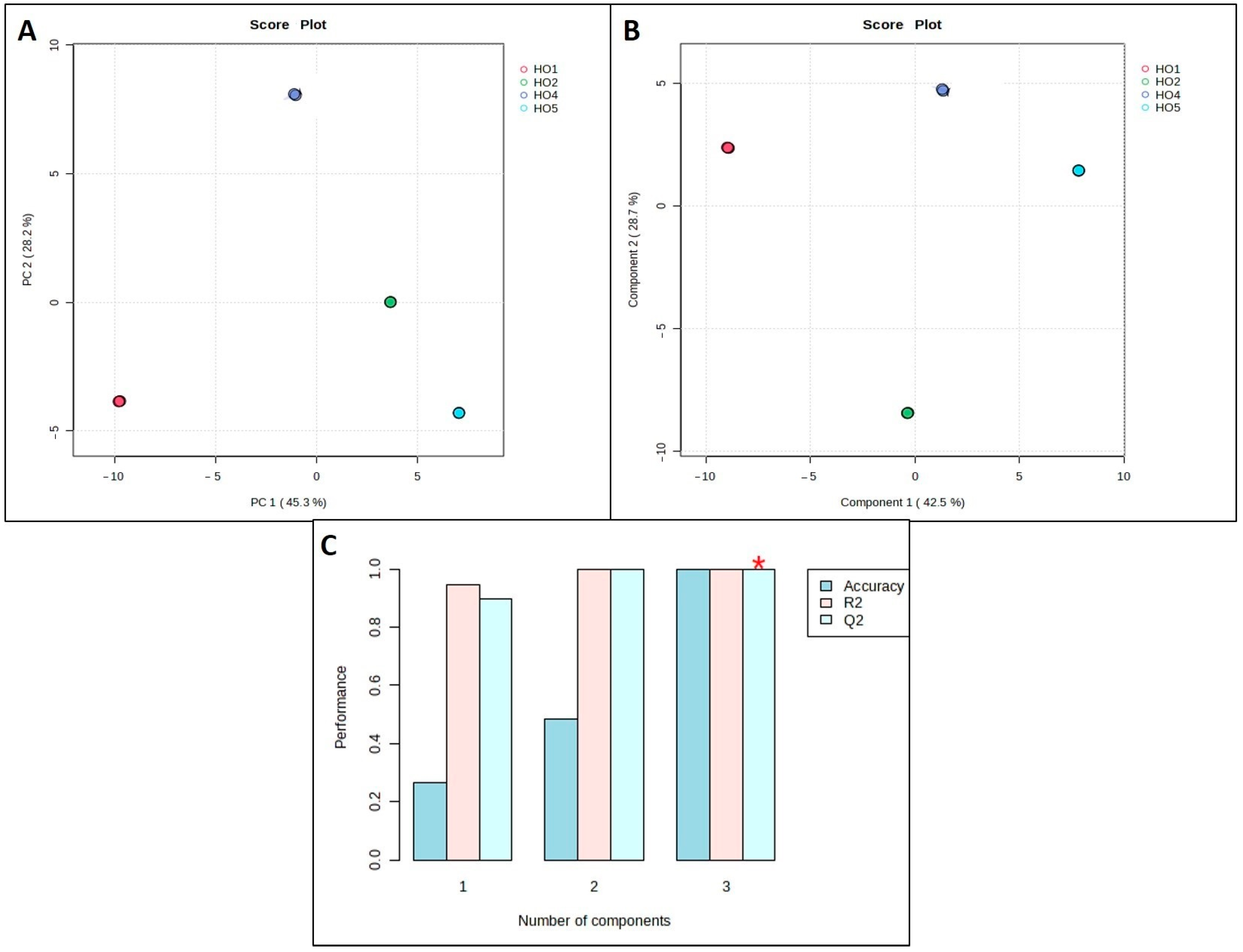
Figure 1. Multivariate data analysis of 1H NMR data of Saudi multi-floral honey samples: (A) 2D PCA score plot with unsupervised method, (B) PLS-DA score plot with supervised method, and (C) cross-validation model of PLS-DA. * component 3 is the most accurate one.
Processes 13 01294 g001
Figure 2. Biofilm inhibition by Saudi multi-floral honey samples, where (A) shows biofilm growth reduction after exposure to HO1, HO2, HO4, and HO5, as indicated by changes in the crystal violet intensity, and (B) indicates the biofilm inhibition percentage.
Processes 13 01294 g002
Figure 3. Dereplicated metabolites from Saudi Aloe honey sample. (2) 4-Hydroxybenzoic acid; (7) Aloe-emodin; (8) Epigallocatechin; (10) Epigallocatechin gallate; (12) Naringin; (13) Linoleic acid; (15) Corosolic acid; (17) Cinnamic acid; (19) Gallic acid; (20) Ferulic acid; (21) Gallic acid hexoside; (22) Syringic acid; (23) Rosmarinic acid; (24) Chlorogenic acid; (25) Saponarin; (26) Aloin; (27) Caffeic acid.
Processes 13 01294 g003
Figure 4. The 3D and 2D binding interactions of gallic acid hexoside (A) and rosmarinic acid (B) within the binding site of β-lactamase (PDB code 1SHV).
Processes 13 01294 g004
Table 1. Physicochemical properties of Saudi honey samples.
TestsResultsStandards
HO1HO2HO4HO5Limitations of Saudi and Gulf Standard Specifications
Physical tests
1. ColorDark amberLight amberLight amberDark amber
2. OdorOdorlessOdorlessOdorlessOdorlessFree from foreign odors
3. TasteSweetSweetSweetSweet
4. PurityPurePurePurePureFree from impurities
Chemical tests
1. Moisture 15.8015.2017.2018.40Calluna and clover honeys: 23% max.; other kinds: 21% max.
2. Reducing sugars74.2074.4777.2072.90Nectar honey: 60% min.
Honey dew honey: 45% min.
     - Glucose 34.2031.9638.4035.60
     - Fructose 40.0042.5138.8037.30
     - Fructose/glucose ratio1.171.331.011.05
3. Sucrose 1.501.031.50.505% max.
4. Hydroxymethylfurfural (HMF)53.7021.1015.4069.1080 mg/kg max.
5. Acidity 14.006.4012.0032.0050 meq/kg max.
6. Diastase enzyme ----1216.63 Goth Scal min.
HO1: aloe honey; HO2: anise honey; HO4: moringa honey; HO5: acacia honey.
Table 2. Bacterial inhibition rates (percentage) of Saudi multi-floral honey samples against ESKAPE pathogens.
Staphylococcus aureus ATCC 25923Klebsiella pneumoniae ATCC 700603Pseudomonas auruginodse ATCC 90902Salmonella typhimurium ATCC 14028Escherichia coli ATCC8739 lot 03801105Enterococcus faecalisEnterobacter sp.
HO249.711.685.985.538.547.80
HO590.718.585.685.449.859.76.5
HO458.7006.533.538.40
HO193.381.150.582.965.071.178.3
Cephalosporin (10 ug/mL)98.297.4-98.498.2-
Table 3. Minimum inhibitory concentration (MIC) values (ug/mL) of Saudi multi-floral honey samples against ESKAPE pathogens.
BacteriaStaphylococcus aureus ATCC 25923Klebsiella pneumoniae ATCC 700603Pseudomonas auruginose ATCC 90902Salmonella typhimurium ATCC 14028Escherichia coli 0157 ATCC 700728Enterococcus faecalisEnterobacter sp.
HO225-1.56251.562512.525-
HO51.5625251.56251.562512.53.125-
HO412.5---12.512.5-
HO11.56251.562512.51.562512.512.51.5625
Cephalosporin (10 ug/mL)1.56251.5625-1.56251.5625--
Table 4. Metabolites dereplicated from Saudi aloe honey sample.
N.CompoundModeFormulaClassm/zRt% vol.References
1Arabinose+C5H10O5Sugar151.03570.2650.09%[17]
24-Hydroxybenzoic acid+C7H6O3Phenolic acid139.00680.30.13%[18]
3RaffinoseC18H32O16Sugar503.16662.3240.83%[19]
4SucroseC12H22O11Sugar341.10982.363%[20]
5MaltotetraoseC24H42O21Sugar665.21052.3860.19%[21]
6RiboflavinC17H20N4O6Vitamin375.10892.3950.08%[22]
7Aloe-emodin+C15H10O5Anthraquinone271.08342.4250.09%[23]
8Epigallocatechin+C15H14O7Flavonoid307.10382.4340.08%[24]
9GlucoseC6H12O6Sugar179.05602.45519.81%[20]
10Epigallocatechin gallate+C22H18O11Flavonoid459.13782.5040.05%[24]
11Lactic acidC3H6O3Organic acid89.024522.5599.43%[22]
12Naringenin+C15H12O5Flavonoid273.08372.620.28%[25]
13Linoleic acid+C18H32O2Fatty acid281.07242.6840.62%[26]
14Biotin+C10H16N2O3SVitamin245.06432.6920.05%[27]
15Corosolic acidC30H48O4Triterpenoid 472.20123.1260.11%[28]
16FructoseC6H12O6Sugar179.05573.1357.99%[20]
17Cinnamic acid+C9H8O2Phenolic acid149.02104.8720.57%[25]
18LactoseC12H22O11Sugar341.11565.160.72%[29]
19Gallic acid+C7H6O5Phenolic acid171.09968.1070.38%[18]
20Ferulic acid+C10H10O4Phenolic acid195.063112.8230.23%[18]
21Gallic acid hexosideC13H16O10Phenolic glycoside331.249319.5770.54%[30]
22Syringic acid+C9H10O5Phenolic acid199.094520.410.09%[18]
23Rosmarinic acidC18H16O8Phenolic acid359.125422.6470.23%[31]
24Chlorogenic acidC16H18O9Phenolic acid353.211925.8260.27%[18]
25Saponarin+C27H30O15Flavonoid595.383129.3980.04%[32]
26Aloin+C21H22O9Anthraquinone419.278929.8230.04%[23]
27Caffeic acid+C9H8O4Phenolic acid181.986040.3370.29%[18]
Table 5. Classifications of and information about the β-lactamases that were utilized to study the binding affinity of honey-derived compounds using molecular docking.
Bush–Jacoby Group ClassificationNameProtein Data Bank (PDB) CodeBacteria
Group 1: CephalosporinasesAmpC-type beta-lactamases1KVLEscherichia coli
Group 2: Serine beta-lactamases
2a: PenicillinasesTEM-1 beta-lactamase1FQGEscherichia coli
2b: Broad-spectrum beta-lactamasesSHV-1 beta-lactamase1SHVKlebsiella pneumoniae
2be: Extended-spectrum beta-lactamases (ESBLs)CTX-M-14 beta-lactamase4HBUEscherichia coli
2br: Inhibitor-resistant beta-lactamasesTEM-30 beta-lactamase1LHYEscherichia coli
2c: CarbenicillinasesPSE-4 beta-lactamase1G68Pseudomonas aeruginosa
2d: Cloxacillin-hydrolyzing beta-lactamases (OXA-type)OXA-10 beta-lactamase1FOFPseudomonas aeruginosa
2f: CarbapenemasesKPC-2 beta-lactamase4ZBEKlebsiella pneumoniae
Group 3: Metallo-beta-lactamases (MBLs)NDM-1 beta-lactamase4EXSKlebsiella pneumoniae
Group 4: Penicillinases that do not fit into other groupsDouble mutation, E166D:N170Q, of class A enzyme1GHIStaphylococcus aureus
Table 6. Consensus docking score of each compound with each class of β-lactamase.
Compound Name1KVL1FQG1SHV4HBU1LHY1G681FOF4ZBE4EXS1GHITotal Consensus Score a
4-Hydroxybenzoic acid00000001102
Aloe-emodin302211039021
Aloin147706435239
Caffeic acid00100200003
Chlorogenic acid262194722843
Cinnamic acid01010100014
Corosolic acid310330068327
Epigallocatechin gallate680700594645
Epigallocatechin904267327040
Ferulic acid00010311017
Gallic acid hexoside04119710811354
Gallic acid00000100001
Linoleic acid01103010107
Naringenin002031214114
Rosmarinic acid4810495731859
Saponarin590500390940
Syringic acid01000000001
Avibactam345455663445
a The total consensus score is the summation of the 10 individual consensus scores calculated from 14 scoring functions.
Disclaimer/Publisher’s Note: The statements, opinions and data contained in all publications are solely those of the individual author(s) and contributor(s) and not of MDPI and/or the editor(s). MDPI and/or the editor(s) disclaim responsibility for any injury to people or property resulting from any ideas, methods, instructions or products referred to in the content.