Article

Manipulation of Culture Conditions: Tool for Correlating/Improving Lipid and Carotenoid Production by Rhodotorula glutinis

1
School of Bioengineering, Dalian University of Technology, Dalian 116024, China
2
Faculty of Science, Botany Department, Menoufia University, Shebin El-koom 32511, Egypt
3
School of Food and Environment Science and Engineering, Dalian University of Technology, Panjin 124221, China
*
Author to whom correspondence should be addressed.
Processes 2020, 8(2), 140; https://doi.org/10.3390/pr8020140
Submission received: 23 December 2019 / Revised: 14 January 2020 / Accepted: 16 January 2020 / Published: 21 January 2020
(This article belongs to the Special Issue Model Lipid Membranes)

Abstract

:
The coproduction of lipid and carotenoid by red yeasts in one cycle is more convenient and economical for the industrial sectors, while the kinetics correlation between both products under different culture conditions has been scarcely studied. This study is aiming to correlate the impact of different carbon sources, carbon to phosphorus ratio (C/P), temperature, aeration, pH, and metals on dry cell weight, lipid (GC and fluorescence microscope), and carotenoid (HPLC) production by Rhodotorula glutinis, and applying a novel feeding approach using a 5 L bioreactor to enhance carotenoid and unsaturated fatty acid production by R. glutinis. Whatever the culture condition is, the reversible correlation between lipid and carotenoid production was detected. Remarkably, when adding 0.1 mM BaCl2, cellular lipid was significantly increased 14% more than the control, with 79.3% unsaturated fatty acid (46% C18:2 and C18:3) and 50% γ-carotene, while adding 1 mM NiSO4, cellular carotenoid was enhanced around 53% than the control (torulene 88%) with 81% unsaturated fatty acid (61% oleic acid). Excitingly, 68.8 g/l biomass with 41% cellular lipid (79% unsaturated fatty acid) and 426 µgpigment/gdcw cellular carotenoid (29.3 mg/L) (71% torulene) were obtained, when the pH-temperature dual controlled process combined with metallo-sulfo-phospho-glucose feeding approach in the 5 L bioreactor during the accumulation phase was conducted. This is the first study on the kinetic correlation between lipid and carotenoid under different C/P ratio and the dual effect of different metals like NiSO4 on lipid and carotenoid production by red oleaginous yeasts, which in turn significant for enhancing the coproduction of lipid and carotenoid by R. glutinis.

1. Introduction

Red oleaginous yeasts are a broad group of yeasts, which are characterized by producing many valuable metabolites such as lipids and carotenoid [1], one of the main representative genus of those colored yeasts is Rhodotorula glutinis. R. glutinis can accumulate lipid up to 60% of its dry cell [2]. This yeast primarily synthesizes palmitic acid (C16:0), oleic acid (C18:1), linoleic acid (C18:2), and α-linolenic acid (C18:3) [1].
Based on a nutritional classification, fatty acids that are not synthesized by humans and are indispensable for development and health are known as essential, while those produced by humans are classified as non-essential fatty acids [3]. In this context, linoleic and alpha-linolenic acids are polyunsaturated fatty acids (PUFA) classified as essential, while monounsaturated fatty acids (MUFA) like oleic acid are classified as a non-essential fatty acid that has been recently described as a regulator of immune function and health [4]. Although oleic acid is a non-essential fatty acid, it has many human health advantages. Oleic acid represents 70%–80% of olive oil composition, besides minor phenolic compounds [5]. In the last few years, many studies have described the contribution of olive oil to general health, partly due to its high oleic acid content [6,7], which was demonstrated to lead to a reduction in cholesterol levels, atherogenesis risk [5,8,9], blood pressure, and daily antihypertensive drug intake [9]. In addition, oleic acid was demonstrated to induce beneficial anti-inflammatory effects on autoimmune diseases [10], protective effects on breast cancer, and improvement of immune system function [11,12].
Under certain conditions, R. glutinis can accumulate oleic acid in a ratio similar to olive oil [13], besides the variable accumulated amount from polyunsaturated fatty acid, i.e., linoleic and linolenic acid depending on either culture conditions or yeast strains [14]. Accordingly, this study aimed to investigate the possibilities to enhance and or improve the lipid profile of R. glutinis through the manipulation of culture conditions.
In addition to lipid production, R. glutinis can also produce carotenoids with variable quantities, mainly dependent on the yeast strains and culture conditions. Carotenoid are natural pigments, which received increased interest during the few last years due to the growing general concern for using organic pigment in the industrial and pharmaceutical sectors instead of synthetic ones. Four different individual carotenoids produced by R. glutinis, β-carotene, γ-carotene, torulene, and torularhodin were detected in the previous report [15].
Carotenoid is the primary source of vitamin A in the human diet, and they have health-promoting properties. Carotenoids reinforce the immune system of the body, accelerates the healing of wounds [16], and counteracts eye conditions, such as cataracts [17] or age-related macular degeneration [18]. Carotenoid supplements are commonly used as agents protecting the skin against harmful ultraviolet radiation [19,20]. Carotenoids may also be used in cancer prevention owing to their anti-oxidative properties [20]. Among carotenoid, β-carotene and γ-carotene have already been transferred industrial production, while torulene and torularhodin are still in the development stage, and more studies still need to be conducted to clarify their production conditions and maximize the yield to be suitable for upgrading to an industrial scale.
The biotechnological synthesis of both lipid and carotenoid is influenced by many factors involved in the processes that can affect yields and operation costs [14,21]. The type of species and growth conditions, i.e., temperature, pH, type of substrate, variation in carbon to phosphorous (C/P) ratio, and oxygen not only influence the efficiency of either lipid or carotenoid accumulation but also their profile inside the yeast cell [14,21].
The kinetics correlation studies between lipid and carotenoid production by red oleaginous yeasts under different carbon to nitrogen (C/N) molar ratios showed confusing results, as some authors reported a positive correlation between high C/N and lipid and carotenoid production with a parallel increase of both lipid and carotenoid kinetics till the end of incubation time [22], while another study reported a negative correlation between lipid and carotenoid production under high C/N ratio, as the highest carotenoid accumulation was observed under low C/N ratio [23]. While our recent lab work showed that both lipid and carotenoid production was enhanced under high C/N during the growth phase, then the negative correlation was detected to favor lipid accumulation during the accumulation phase [24]. The same study detected the effect of different carbon to sulfur (C/S) ratios on the kinetics of lipid and carotenoid production by R. glutinis, and cellular carotenoid was enhanced over cellular lipid under high C/N ratio with decreasing C/S ratio [24].On the other hand, the effect of different C/P molar ratio on carotenoid biosynthesis, thus as well the correlation between lipid and carotenoid production by red oleaginous yeasts under different C/P ratios, were never studied before, although the role of phosphorus deficiency for enhancing lipid accumulation by oleaginous yeasts has been confirmed [25].
Metals, even at very low concentrations, pose threats to human and environmental health for their hazardous effects, persistence, and accumulation. Metals, such as copper, zinc, manganese, nickel, and iron, despite being toxic at a high concentration, they are essential for the growth of microorganisms in trace amounts acting as micronutrients contributing to the yeast’s growth [26]. Only very few studies have been thus far focused on the effect of metals on either lipid or carotenoid production by the colored oleaginous microorganism, especially yeasts [27,28,29,30]. Meanwhile, no attempts have been made to evaluate the dual effects of the heavy metals on the simultaneous production of lipid and carotenoid by red oleaginous yeast. Our previous work stated the effect of the combined cultivation mode of low carbon to sulfur ratio (C/S) with high carbon to nitrogen molar ratio (C/N) in the presence of 0.7 mM of aluminum sulfate for enhancing carotenoid production, specifically torulene by R. glutinis [24].
During the last 20 years, only a few studies have been conducted on the simultaneous production of lipid and carotenoid by red oleaginous yeasts [2,13,22,23,24]. Most authors stated the suitability of the produced lipid for biodiesel production, except for a recent study carried by Kot et al. [10], which stated the similarity of the produced lipid with olive oil composition. The carotenoid profile of R. glutinis reported by the most previously reported studies showed the dominance of β-carotene, while few authors [23,24] reported the dominance of torularhodin and torulene. However, to make the coproduction of lipid and carotenoid from the red oleaginous yeasts a more economical process, the accumulated lipid should be more lavish with monounsaturated fatty acid (USFA) like oleic acid and polyunsaturated fatty acid (PUFA) instead of saturated fatty acid. As a result, a highly valuable colored fat can be produced at the end of the process, which can be further processed in the human health-related industries and food industry.
Continuously, this study aims to correlate between lipid and carotenoid production by R. glutinis and investigate the possibilities of improving the yield and profile of both lipid and carotenoid produced by R. glutinis through manipulation of culture conditions. Also, detect the dual role of some metals as a stress factor on lipid and carotenoid production. Followed by applying a novel fed-batch approach, which depends on the combined dual controlled of different physical and chemical conditions, as an attempt to improve the fatty acid and carotenoid profile produced by R. glutinis for the potential purpose of human consumption.

2. Materials and Methods

2.1. The Yeast Strain and Media Compositions

The oleaginous red yeast Rhodotorula glutinis (AS 2.703), which was obtained from the China General Microbiological Culture Collection Center (CGMCC) (Beijing, China), was used in this study. The yeast strain was cultivated on yeast extract peptone dextrose (YPD) agar slants (glucose 20 g/L, yeast extract 10 g/L, peptone 10 g/L, agar 15 g/L), then incubated at 28 °C for 3 days and then preserved at 4 °C for further experiments.
Three different media were prepared, the seed culture media and the YPD broth media. Fermentation medium A (FMA), which contained (per liter) glucose (60 g), (NH4)2SO4 (0.5 g), yeast extract (0.75 g), KH2PO4 (1.5 g), and MgSO4.7H2O (1 g), FMA was used to detect the effect of different culture conditions on R. glutinis growth, lipid, and carotenoid production (consider this basal condition as control C1). Fermentation medium B (FMB), which contained (per liter) glucose (60 g), (NH4)2SO4 (0.9 g), KH2PO4 (1.5 g), and MgSO4.7H2O (1.2 g). This medium was used to detect the effect of different heavy metal stress on R. glutinis growth, lipid, and carotenoid production. FMB media was used as an optimized medium after optimizing the C/N ratio, nitrogen source, and C/S ratio for enhancing carotenoid production under a high C/N ratio (consider this basal condition as control C2) [24]. The pH of the 3 culture media was adjusted at pH 5, then 100 mL was distributed in 250 mL flasks followed by sterilization at 121 °C for 20 min after inoculation with yeast cells, then the flasks were incubated at 28 °C and 180 rpm. Otherwise, the different conditions were stated. Seed culture preparation and inoculation in the fermentation medium was fully explained in our previous work [24].

2.2. Effect of Different Culture Conditions and Metals Stress on Growth, Lipid, and Carotenoid Production by R. glutinis

Herein, sterilized FMA was prepared, replacing the carbon source (glucose C1) (Glu) with its equivalent amount of maltose (Mal), sucrose (Suc), lactose, starch, and glucose:sucrose (1:1) (Glu: Suc) to fix the initial C/N molar ratio at 146 with all treatments and to study the effect of different carbon source on growth, lipid, and carotenoid production by R. glutinis.
To study the effect of different pH degree on growth, lipid, and carotenoid production by R. glutinis, the sterilized FMA was prepared while adjusting pH at different degrees; 3, 4, 5 (C1), 6, 7, and 8 using 6N HCl for decreasing pH and 6N NaOH for increasing pH.
To study the effect of different temperature degrees on growth, lipid, and carotenoid production by R. glutinis, after inoculating the sterilized FMA with the seed culture; the flasks were incubated at different temperature degrees of 25 °C, 28 °C (C1), 30 °C, and 32 °C under the same agitation condition.
To study the effect of different agitation speeds on growth, lipid, and carotenoid production by R. glutinis, after inoculating the sterilized FMA by the seed culture; the flasks were incubated at different agitation speeds of 150, 180 (C1), 200, and 220 rpm.
Continuously, the effect of different C/P molar ratios on growth, lipid, and carotenoid production by R. glutinis was studied. The sterilized FMA was prepared with different phosphorus concentration 0.5 g/L, 1.5 g/L (C1), 3 g/L, and 4.5 g/L and fixed glucose concentration (60 g/L) to prepare different initial C/P molar ratio 527, 176, 88, and 59, respectively.
Finally, the effect of different metal types and concentrations on growth, lipid, and carotenoid production by R. glutinis were studied as follows; a new fermentation medium FMB was prepared. After sterilization, different metals ZnCl2, MnCl2, BaCl2, FeCl3, Fe2(SO4)3, CuCl3, Cu2(SO4)3, NiSO4, and LiCl were added separately to the culture medium to prepare two initial concentrations of 0.1 mM and 1 mM from each metal. Afterward, the steps proceeded normally.

2.3. Fed-Batch Cultivation of R. glutinis under Optimized Condition

For fed-batch cultivation of R. glutinis, a 5 L bioreactor (biotech-5BG-700A, China, www.bxbio.com) was used using with the following medium: Glucose 30 g/L, sucrose 15 g/L; yeast extracts 15 g/L, peptone 8 g/L, ammonium sulfate 10 g/L, MgSO4 3 g/L, KH2PO4 3g/L. To reduce the lag phase, the seed culture was prepared from the same medium, 150 mL in 500 mL flasks, after sterilization, 3 discs from 24 h R. glutinis plate were added to each flask then incubated at 30 °C and 180 rpm for 36 h (Mid exponential phase). The seed culture was added to the bioreactor vessel to start the experiment with 2.5 L as an initial volume. The dissolving oxygen (DO) was fixed above 40%, pH was adjusted automatically. Herein, we conducted 2 separate experiments; the first one was the control at which a fixed temperature of 30 °C, pH 6, and agitation speed 400 rpm was used during the whole experiment. Glucose solution (100 g/ 100 mL distilled water) was continuously fed at the rate of 4.5 g/h before the exhaustion of carbon source during the growth phase then to 5.5 g/h during the accumulation phase till 128 h. The residual glucose in the growth media was kept below 25 g/L.
On the other hand, a second experiment was carried out that was similar to the first one until 20 h (end of the growth phase), after that Al2SO4, NiSO4, MnCl2, LiCl, and BaCl2 solutions were added separately to the bioreactor to prepare the following concentrations of 0.7 mM, 0.5 mM, 0.01 mM, 0.01 mM, and 0.01 mM, respectively. In addition, the temperature and pH were changed to 23 °C and 4, respectively. The feeding solution consisted of glucose: MgSO4: KH2PO4 with a ratio of 100:0.2:0.2 (g/ 100 mL distilled water) was used to feed during the accumulation phase. The feeding rate was 5 g/h till 128 h to keep the residual glucose below 25 g/L in the medium. The samples were withdrawn every 12 h to detect dry cell weight (DCW), lipid, and carotenoid production by R. glutinis.

2.4. Detection of Dry Cell Weight (DCW) and Reducing Sugar in the Culture Media

From the withdrawn samples, 5 mL were centrifuged at 10,000 rpm for 10 min; the cells were washed twice with the same volume of sterilized distilled water, then freeze-dried (lyophilized) and weighted for the determination of dry cell weight (DCW) [31]. The supernatant was used for measuring residual sugar using the 3, 5-dinitrosalicylic acid method [32]. For non-reducing sugars like sucrose, firstly, the acid was hydrolyzed, followed by incubating at 90 °C in a hot water bath for 15 min and then neutralized to pH 7 by NaOH 6N. Then, the reducing sugar detected by the DNS method [33].

2.5. Fluorescence Microscopic Examination of Nile-Red Treated Cells

After the incubation period, the cells collected from 100 µL were washed twice and suspended in 10 mM phosphate buffer with 0.15 M potassium hydroxide, pH 7, then mixed with 10 µL of Nile red solution (1 mg of Nile-red in 1 mL of acetone and kept in the dark at 4 °C). After 5 min in the dark, the cells were viewed using an Olympus IX71 fluorescence microscope equipped with a blue fluorescence cube and IX71 frame camera port (Tokyo, Japan) to visualize the cell shape and golden fluorescent lipid bodies inside the cell [34]. The corrected total cell fluorescence (CTCF) for each treatment was calculated using image j software by using the following equation [35]:
C T C F = I n t e g r a t e d   d e n s i t y ( M e a n   A r e a   o f   s e l e c t e d   C e l l s   × M e a n   f l o u r e s c e n c e   o f   b a c k g r o u n d   r e a d i n g s )  
The area of 5 random cells was chosen from each figure, and their mean was used for the calculations.

2.6. Total Lipid Detection and GC Analysis of Fatty Acid Methyl Ester

The total lipids in the yeast cells were detected using the sulfo-phospho vanillin method [36], as briefly described at Elfeky et al. [24]. For determining the fatty acid composition, transesterification of the samples was carried out according to Van Wychen et al. [37]. The freeze-dried cells (10 mL yeast suspension) were added to vials previously heated at 85 °C, then 200 µl of chloroform: methanol (2:1 v/v) with 300 µl of 0.6 M HCl: Methanol was added. All vials were sealed with caps and vortexed well to mix the contents and then heated again at 85 °C for 60 min. After heating, the vials were removed and allowed to cool for 15 min at room temperature. One milliliter of hexane was added and vortexed well to mix the content, then 1 ml of 0.1% NaOH solution was added to wash the acid. This was centrifuged at 2000 rpm for 5 min to allow phase separation. The hexane layer was transferred to a new GC vial for GC analysis. The fatty acid methyl ester was analyzed by using an Agilent 7890A GC equipped with an autosampler and FID. The capillary column HP-FFAP (25 m length, 0.2 mm ID, 0.33 µm film thickness) was used for analytical separation. The temperatures of the injector and detector were set at 240 °C. The column temperature was kept at 180 °C for 2 min and then raised to 240 °C for 2 min at a rate of 7 °C/min. Fatty acid was identified by a comparison of their retention times with those of standard ones, quantified as a percent of the total of the FAMES content.

2.7. Total Carotenoid Extraction, Quantifying, and Identification

The extraction of carotenoid, quantifying, and identification was explained by Elfeky et al. [24] according to Frengova et al. [38] and Weber et al. [39].

2.8. Kinetics Analysis of Fermentation

According to Certik and Shimizu [40], the primary parameters determined in this study were to be: Dry cell weight (DCW) g/L, total lipid (TL) g/L, and total pigment (TP) mg/L. The relative productivity (RP) represents the cellular lipid (L-RP) and carotenoid ((gLipid/gDCW × 100)% and mgpigment/100 gDCW, respectively). The yield of product (Y) represents the entire amount of DCW (DCW-Y), TL (TL-Y), and TP (TP-Y) formation from the total consumed substrate gDCW/100 gglucose, glipid/100 gglucose, and mgpigment/100 gglucose, respectively. The rate of product synthesis (P-SR) was the average speed of total production of TL (L-SR) g/L·day and TP (Car-SR) µg/L·day and calculated using the following equation:
L S R   ( L i p i d   s y n t h e s i s   r a t e ) = d T L d t = T L 2 T L 1 t 2 t 1
C a r S R   ( C a r o t e n o i d   s y n t h e s i s   r a t e ) =   d T P d t = T p 2 T p 1 t 2 t 1
where TL1 and TL2 are the lipid concentrations (g/L) at times t1 and t2, respectively, and TP1 and TP2 are the carotenoid concentrations (µg/L) at time t1 and t2, respectively [24].

2.9. Statistical Analysis

All experiments were repeated 3 times. The data were expressed as the mean ± SD. Statistical analysis was carried out using Origin 8.6 (Northampton, MA USA), and comparisons of each group were evaluated using one-way analysis of variance (ANOVA), multivariate, and correlation coefficient using SPSS 19 software (IBM, Chicago, IL, USA). The results were considered statistically significant at p < 0.05.

3. Results and Discussion

3.1. Effect of Different Culture Conditions on Growth, Lipid, and Carotenoid Production by R. glutinis

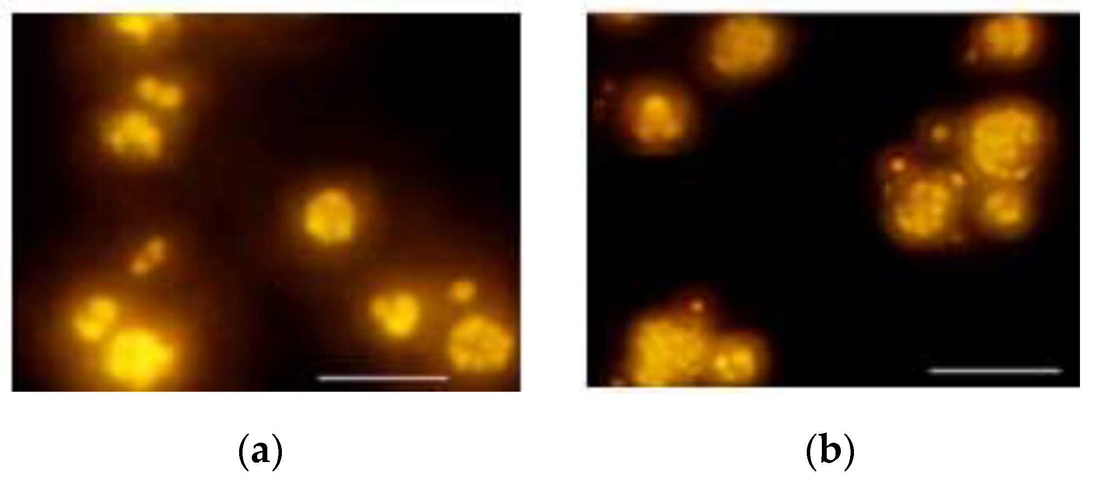
3.1.1. Microscopic Examination of R. glutinis Cellular Shapes and Lipid Bodies

The effect of different carbon sources on the lipid bodies accumulated inside the Nile red-stained yeast cells and examined under fluorescence microscopy using blue fluorescence mode is shown in Figure 1. The highly accumulated lipid bodies depend on the CTCF values that were observed with the combination of glucose and sucrose as a carbon source (Figure 1b and Figure S1a) followed by glucose as a carbon source (Figure 1a and Figure S1a). While using sucrose as a sole carbon source, there was a considerable reduction in the lipid bodies (Figure 1c). In contrast to sucrose, the lipid bodies were scarcely observed when maltose was used as a carbon source, which showed the lowest CTCF value (Figure 1d and Figure S1a). Studying the lipid bodies accumulation after growing R. glutinis under different initial pH levels is represented in Figure 2 and Figure S1b. As observed in Figure 2a–c, the acidic medium enhanced lipid accumulation and lipid droplets enlargement as well; the best accumulation condition with the highest CTCF value was provided at pH 4. Increasing pH to 8 was led to a significant decrease in the lipid bodies with decreasing CTCF value in turn, Figure 2d–f and Figure S1b.
The temperature could be an essential physical factor that significantly affects the lipid body accumulation. As represented in Figure 3, the lowest incubating temperature of 25 °C (Figure 3a) enhanced the lipid bodies accumulation compared to the highest temperature of 32 °C (Figure 3d), which showed the lowest CTCF value (Figure S1c). On the other hand, the highest CTCF value was detected at 30 °C, Figure 3c and Figure S1c.
Continuously, the impact of different agitation speeds on the lipid bodies accumulation under a fluorescence microscope is represented in Figure 4. Firstly, the low agitation speed of 150 rpm showed a few lipid bodies with the lowest CTCF compared to other groups, Figure 4a, Figure S1d, and the highest CTCF value was detected with 200 rpm treatment, Figure 4c and Figure S1d.
The earlier studies stated the enhancement role of phosphorus deficiency on lipid accumulation and lipid droplets formation [25]. The microscopic examination of the Nile-red fluorescence yeast cells after growing on different C/P ratio media showed a great decrease in lipid bodies number and size, thus as well the CTCF value as the phosphorus concentration gradually increased from 0.5 g/L (Figure 5a and Figure S1e) to 1.5 g/L (Figure 5b) then 3 g/L (Figure 5c) and finally 4.5 g/L (Figure 5d).

3.1.2. The Growth, Lipid, and Carotenoid Production by R. glutinis under Different Culture Conditions

The effect of manipulating different culture conditions on the growth, lipid, and carotenoid production by R. glutinis is represented in Table 1.
Firstly, the effect of different carbon sources on the growth, lipid, and carotenoid production showed a significant variation in the detected parameters between different carbon sources. The yeast strain was unable to grow on neither starch nor lactose (data are not shown), while a low growth rate was observed with maltose and sucrose when used as a sole carbon source, which was statistically significant with glucose after 3 days (p = 0.008, Table S1). Using a combined carbon source from glucose and sucrose did not significantly increase the growth compared with glucose but was significant with sucrose (p = 0.01, Table S1). On the same pattern, the growth efficiency (DCW-Y) was almost the same with glucose and Glu + Suc and was decreased with sucrose and maltose.
On the other hand, a combination of glucose and sucrose efficiently improved the lipid and carotenoid production compared with the individual sources; glucose and sucrose. Around 5% (not significant p >0.05) and 26.5% (p = 0.021) increase was observed for lipid production, respectively, while a 15% and 30% increase was detected for carotenoid production, respectively, after 6 days. Thus, the highest value of TL-RP, L-SR, Car-RP, and Car-SR was observed with the combination of glucose and sucrose as carbon sources recording around 49.4% (after 6 days), 0.89 g/L.day (after 6 days), 92.3 µg/g (after 3 days), and 221.8 µg/L.day (after 3 days), respectively, shown in Table 1 and Table S1. The multivariate analysis showed a significant difference between the dependent variable DCW after 3 and 6 days with glucose (p = 0.05) and Glu + Suc (p = 0.004), while TL was significant with all carbon sources after 3 and 6 days (p < 0.003), and carotenoid was only significant with carbon sources after 6 days (p < 0.003, Table S2).
The enhancement of the growth rate and lipid yield by Rhodosporidium kratochvilovae HIMPA1 was also detected when the mixture glucose + fructose + sucrose was used as a carbon source. The DCW and TL was 15.56 g/L and 9.26 g/L, respectively, compared with 14.15 g/L and 8.0 g/L, respectively, when glycerol was used as a sole carbon source [41], which supported our suggestion that points to the combination of different carbon sources at the culture media, which may enhance the growth and metabolite production by microorganisms.
Secondly, the studied yeast strain showed the ability to grow at a wide range of pH degrees from 3 to 8, Table 1 and Table S3. The highest growth rate was observed at pH 5 and 6, as the growth was almost the same around 10.4 and 10.5 g/L, respectively. Thus, as well the growth yield, which was above 47%, the growth decreased around 6% with pH 3 (p = 0.01) and pH 4 (p = 0.027), while recording around a 14% decrease with alkaline pH 8 (p = 0.001), shown in Table 1 and Table S3. This result indicates the ability of this yeast to grow well, even under a highly acidic environment. The same result was reported by Karamerou [42] when R. glutinis was able to grow well on glycerol at pH 2.6.
The statistical analysis of total lipid showed significant differences between all treatments with pH 8 (p < 0.01) after 3 and 6 days (Table S3). The TL for acidic pH was almost the same between 4.9 and 5.2 g/L, even the differences in the total biomass. Therefore, the Lower pH was observed to accelerate lipogenesis more than high pH; the TL-RP was recorded around 42.9% and 40.4% after 3 days with pH 3 and pH 4, respectively, which represented around an 18% increase with pH 5, and a 41% increase with alkaline pH 8 (Table 1). In addition, the reduction in the total lipid with pH 3 and pH 4 may be related to the considerable reduction of pH after growing and producing acid, which subsequently led to a rapid pH decrease compared with the other higher pH levels, and the negative effect on the yeast metabolic activity. Similarly, the optimal medium pH to produce lipid by the red yeast Rhodosporidium toruloides NCYC 921 and Rhodotorula glutinis IIP-30 was 4.0 with glucose feeding in fed-batch fermentation [43] and [44], respectively. Another recent study recorded an optimum pH for lipid production at 5.5 when R. kratochvilovae was cultivated on a medium supplemented with glucose [45]. In contrast to lipids, the gradual increase in the total pigment production and Car-RP was observed with further increasing of pH, recording the highest value at pH 6, 0.894 mg/L at the 6th day, and 90.1 µg/gdcw at the 3rd day, respectively, which represented around a 30 µg/gdcw increase than pH 3 and around 9 µg/gdcw than pH 8 (Table 1). Other strains like Rhodosporidium toruloides NCYC 921 and Rhodotorula mucilaginosa showed the optimum pH at 5 and 6.6 when cultivated on glucose and salted cheese whey, respectively [43,46].
The multivariate test showed a significant difference between the DCW after 6 days and TL with different pH treatments, while the significance of carotenoid was only detected with pH 3 after 6 days (Table S4).
Thirdly, the temperature had a great effect on the yeast growth rate, lipid and carotenoids production. The highest growth rate was observed at 30 °C after 6 days 10.9 g/L, which represents a significant increase around 24.7% (p = 0.007), and 32% (p = 0.000) than 25 and 32 °C, respectively. While the highest DCW-Y was observed with a lower incubating temperature than higher temperature treatments, shown in Table 1 and Table S5.
Lipid as a function of different temperatures treatments, the highest TL, TL-RP, and TL-SR was observed at 30 °C, which was 5.3 g/L (±0.37), 49.1%, and 0.98 g/L.day, respectively, at 6 days. And decreased significantly with 32 and 25 °C, around 27% (±0.33, p = 0.001) and 34% (±0.35, p = 0.001), respectively, compared with 28 °C treatment (Table 1 and Table S5). Other yeast strains showed different optimum temperatures for biomass and lipid production; the optimum temperature of Rhodotorula kratochvilovae was 30 °C, producing around 56% cellular lipid [45].
On the same pattern, the best temperature for carotenoid production was 28 °C, TP was 0.81 mg/L (±0.09) on the 6th day with cellular carotenoid around 84.2 µg/gdcw on the 3rd day, this result was statistically significant with 25 and 32 °C, which represented around an 8% (p = 0.042), and 11% (p = 0.014) increase than TP at 25 and 32 °C, respectively. Similarly, the optimum temperature for either biomass or carotenoid production by Rhodotorula sp. RY1801 was 28 °C when glucose was used as a carbon source [47]. Another study investigating the effect of different temperature degrees on Sporobolomyces ruberrimus carotenoid using glycerol as a carbon source, the result revealed that the carotenoid production occurred whenever the temperature was kept between 19 °C and 27 °C, but at 31 °C the cells remained colorless, and no pigment production was seen [48]. The multivariate test showed a significance of the DCW and TL with different temperature treatments; 28 °C (p = 0.000) and 30 °C (p = 0.000), while the significance of carotenoid was only detected with 28 °C (p = 0.014) and 30 °C (p = 0.01) after 6 days (Table S6).
It has been suggested that oleaginous yeasts require substantial oxygen supply for energy and biosynthesis [49]. One factor that influences the oxygen levels in the medium is the agitation rate. By increasing the agitation rate, the dissolved oxygen in the medium increases, which enhances the growth and lipid content [45]. The effect of different agitation speeds on growth, lipid, and carotenoid production by Rhodotorula glutinis is represented in Table 1.
Increasing agitation speed from 150 rpm to 200 and 220 increased the growth rate significantly by around 16% (p = 0.011), while no differences in biomass were observed between 200 rpm and 220 rpm. The highest growth efficiency was observed with low agitation speed around 48.3%, whereas, the highest agitation speed decreased growth efficiency to 43.9% after 3 days, shown in Table 1 and Table S7. The decrease in the growth efficiency with increasing agitation speed may be related to the unbearable shear force, which caused the cell damage [50]. Or it may be due to the depression of the metabolic activity as reported by Vlaev et al. [51]. The authors found that the effect of the high agitation speed on Sporobolomyces salmonicolor AL was attributed to depressed metabolic activity at the evolving dissolved oxygen tension rather than to the direct effect of hydrodynamics.
Similarly, lipid production by R. glutinis was enhanced significantly with increasing agitation up to 200 rpm, which showed around 30% (p = 0.000), 35% (p = 0.005), and 32.6% (p = 0.007) compared with 180, 200, and 220 rpm, respectively, shown in Table 1 and Table S7. The highest TL-RP was observed at 200 rpm, around 48.8%, after 6 days. R. glutinis is an aerobic organism in which oxygen is a critical factor for enhancing biomass and lipid production, thus that increasing agitation speed to the suitable speed 200 rpm led to lipid and biomass production enhancement. Other studies reported similar results regarding the enhancement role of high aeration or agitation on lipid and biomass production. The optimum agitation rate for the production of single-cell oil (SCO) by R. glutinis was 180 rpm [52].
In addition, El-Fadaly et al. [53] investigated the effect of agitation speed on the SCO production by C. curvatus NRRLY-1511 and found that the maximum dry cell mass and lipid yield were obtained at 200 rpm. Milkessa et al. [45] observed the optimum agitation speed for the maximum lipid production by R. kratochvilovae SY89 at 225 rpm.
Additionally, the carotenoid production increased with a gradual increase of agitation speed from 150 rpm to 200 rpm, which showed around a 23.6% increased (insignificant p >0.05), while the further agitation speed showed no further increase in total pigment. Contrasting to the low agitation speed, the carotenoid relative productivity (Car-RP) of 200, and 220 rpm at the 6th day was almost the same like the 3rd day, and this may be related to the reduction in the lipid accumulation with 220 rpm and the excess of dissolved oxygen supply in case of 200 and 220 rpm (Table 1). The multivariate test showed the significance only with DCW and TL with low agitation speed 150 rpm (p < 0.008) after 3 and 6 days (Table S8).
When the growth rate was expressed as a function of phosphate concentration, the growth medium increased, and it was observed that both DCW and DCW-Y were enhanced with the further increase of phosphate, Table 1. Around an 11% (p = 0.001) increase was noticed when the DCW of C/p 527 (0.5 g/L) and C/P 59 (4.5 g/L) after 6 days were compared. Statistically, the dry cell weight of the highest phosphate concentrations (4.5 g/L) treatment was significant with the other lower concentration (p < 0.01), shown in Table 1 and Table S9. As has been mentioned, when the yeast usually grows under nutrient-rich conditions, it contains around 4%–5% total phosphorus as P2O5, on a dry-weight basis [25]. This is why its presence in the growth media, with a reasonable supply, fits with the available nutrient content in the growth media and will positively affect the growth rate and glucose assimilation.
On the contrary, both total lipid and lipid relative productivity (TL-RP) were decreased with increasing phosphorus concentration in the growth medium, the highest TL-RP was achieved at C/P 527 (0.5 g/L) 52.1% (TL was 5.3 ± 0.08 g/L), which represents around a 39% (p = 0.000) and 63% (p = 0.001) increase compared with C/P 88 (3 g/L) (3.5 ± 0.04 g/L TL) and C/P 59 (4.5 g/L) (2.3 ± 0.06 g/L TL) after 6 days. The statistical analysis revealed that the 4.5 g/L phosphorus group was significant within all groups (Table 1 and Table S9). The enhancement role of phosphorus deficiency to fat accumulation in yeast cells was reported earlier by Schulze [25] when different starter phosphorus was supplied in the form of ammonium pyrophosphate to the yeast continuously growing in the bioreactor. A reverse relationship between fat content and protein content depending on phosphorus concentration was observed, as the high phosphorus concentration led to rapid yeast reproduction, producing cells rich in protein and low in fat. An interesting recent study [54] emphasized that the role of phosphorus deficiency for enhancing lipid production by R. toruloide depends on the multi-omic study. The results showed that the cells accumulated less than 10% lipid when growing on nutrient-rich media, and the cellular lipid increased significantly to above 40% when the studied yeast was grown on phosphate deficient condition.
The carotenoid production under different starter phosphate concentration is represented in Table 1 and Table S9, where the increase of phosphate concentration in the growth media from 0.5 g/L to 4.5 g/L led to a significant increase in both volumetric and cellular pigment production by R. glutinis, which represented around a 37% (p = 0.001) and 11% increase, respectively. The one-way ANOVA analysis showed that the 0.5 g/L phosphorus group was statistically significant with all higher group only on the 6th day (p < 0.025). The multivariate test showed the significant effect of the low C/P molar ratio on DCW. All C/P treatments were significant with TL (3rd and 6th day) and carotenoid (6th day only), as shown in Table S10.
Notably, the yield of carotenoid was enhanced with increasing phosphate concentration in the growth media. These data indicated the role of phosphate to enhance the efficiency of glucose uptake from the growth media by the studied yeast strain and its further conversion to either biomass or carotenoid. Although several studies were conducted to study the effect of different culture condition on carotenoid production by red yeasts [29,45,47,55,56,57,58], as far as is known, there is no study that investigated the effect of phosphate on carotenoid production by red oleaginous yeast until now. A similar study conducted on algae [59] reported that both growth, chlorophyll, and beta carotene were significantly enhanced with increasing phosphorus concentration in the growth medium of Dunaliella salina. On the other hand, a study carried out on marine microalga Nannochloropsis gaditan observed the enhancement of carotenoid production under both phosphate and sulfur deficiency [60].
All the above-mentioned experiments showed the domination of carotenoid synthesis rate, yield, and cellular carotenoid on the 3rd day while the highest yield of lipid was observed after the 6th day. This result confirmed the reversible relationship between lipid and carotenoid production under high C/N through the manipulation of the culture conditions.
The Pearson correlation analysis (two-tailed) between the different studied factors—carbon sources, temperature, pH, agitation speed, and C/P molar ratio—and the different dependent variables of DCW, TL, and TP are represented at Supplementary Materials 2. The factors of carbon sources, temperature, pH, C/P molar ratios showed a significant correlation (p < 0.05) between the different dependent variables, which indicates their significance as a tool for enhancing biomass, lipid, and carotenoid production by R. glutinis.

3.1.3. Lipid and Carotenoid Profile of R. glutinis under Different Culture Conditions

The variation of the fatty acid methyl esters and carotenoid profile produced by R. glutinis due to manipulation of culture conditions is represented in Table 2 and Figure 6, respectively.
Looking to the carotenoid profile in Figure 6a as a response to different carbon source treatments, torulene was dominant with a ratio of more than 65% with glucose, sucrose, and a combination of glucose and sucrose. Using maltose reduce the torulene significantly to 57% (p = 0.004) to the favour of γ-carotene 32.5%. Shifting the carotenogenesis pathway toward torulene accumulation was also observed with Elbana et al. [29] when R. glutinis var. glutinis was grown on sucrose as a sole carbon source. The statistical analysis showed a significant difference in the torularhodin and γ-carotene ratio of glucose sets with other carbon sources treatments (p < 0.029), while the torulene and β-carotene ratio of glucose sets were only significant with maltose sets (p < 0.006). On the other hand, the lipid profile responded similarly to the different carbon sources except with maltose. Using glucose, sucrose, and glu+suc as a carbon source led to the dominance of oleic acid on the 3rd day, with 68.8%, 61.3%, and 69.6%, respectively, which was reduced to favor linoleic acid (C18:2) with further incubation. The oleic acid ratio of glucose treatment was significant with the oleic acid ratio of sucrose (p = 0.000), and maltose (p = 0.000) treatments, and not significant with the Glu+suc treatment (p >0.05) on the 3rd day, while the further incubation caused significant differences between all treatments (p = 0.000) (Supplementary Materials 2). Although the total lipid was greatly reduced when maltose was used as a sole carbon source, its lipid profile showed the dominance of polyunsaturated fatty acid compared with the other carbon sources treatments, Table 2. Worthy to note, the lipid profile of the studied yeast with glu + suc and maltose as a carbon source after 3 and 6 days, respectively, was considered as a suitable source for human dietary consumption due to the domination of unsaturated fatty acids compared with other treatments.
As a factor of different pH treatments, the carotenoid profile showed the dominance of torulene at low pH, above 58%, then it decreased gradually with increasing pH to alkalinity in favor of γ-carotene, showing around a 36.8% (p = 0.000) decrease in torulene, and around a 40% increase in the β-carotene and γ-carotene ratios (p = 0.000). When the carotenoid profile of pH 8 and pH 3 treatments were compared (Figure 6e and Supplementary Materials 2), C18:1 was the dominant fatty acid overall, but the pattern of the fatty acid profile was slightly variable with different pH degrees. The highest ratio of oleic acid was observed after 3 days with acidic pH (3–6), above 64%, while the alkaline pH (7,8) showed an oleic acid ratio below 58% on the 3rd day. The statistical analysis showed significant differences between the oleic acid ratio of the pH 3 treatment and other treatments on the 3rd day (p < 0.001). The further incubation led to a decrease in the oleic acid ratio to be between 49% and 44% with different pH treatments and insignificant when the pH 3 oleic acid ratio compared with pH4, 5, and 6 (p >0.05). Remarkably, the highest ratio of polyunsaturated fatty acid was observed at a low pH of 4 and 3, which counted above 30% of the total fatty acid on the 6th day and it was statistically significant with most pH treatments compared with pH 3 (p < 0.009), as shown in Table 2 and Supplementary Materials 2. This result indicates the possibility of increasing the nutritional value of the lipid produced by the studied yeast strain by adjusting the initial pH degree. Increasing torulene and the oleic acid ratio at low acidic pH was also observed when R. glutinis was grown on the culture medium with glycerol and deproteinized potato wastewater at pH 3 [61].
Additionally, the temperature factor showed an alteration in the profile of fatty acids. The polyunsaturated fatty acids after 3 days were above 35% at 25 °C, which represented around twice the ratio at the other temperature groups, Table 2. The higher temperatures showed the dominance of oleic acid even though a slight increase in the saturated fatty acid around 2% and 8% was observed with the highest temperature treatment (30 °C and 32 °C), respectively, on the 6th day. The statistical results showed a significant difference between the oleic acid ratio of 25 °C treatment and 30 and 32 °C treatments (p = 0.19, p = 0.03, respectively) after the 3rd day and the ratio of oleic acid of 25 °C group became significant with the three other treatments at the end of incubation (p < 0.000). Whereas, the total polyunsaturated fatty acid was almost the same with a slight insignificant variation at the end of cultivation compared with 28 °C, as shown in Table 2, and Supplementary Materials 2. Regarding the fatty acid profile, the dominance of polyunsaturated fatty acid was also observed at the low-temperature degree when Lipomyces starkey was incubated at different temperatures 10, 20, and 30 °C [62].
While the carotenoid profile showed the dominance of torulene above 67% at 25 °C and 28 °C at the 6th day with gradual shifting toward γ-carotene when the temperature was increased to 30 °C and 32 °C , representing around a 15% (p = 0.000) and 25% (p = 0.001) decrease in torulene and 22% (p = 0.000) and 31% (p = 0.000) increase in γ-carotene compared with 28 °C , respectively, in Figure 6c and Supplementary Materials 2. A significant increase in torulerhodin at a low-temperature of 19 °C was reported when Sporobolomyces ruberrimus was grown on glycerol under different temperatures [48]. This result showed the possibility of producing healthy oil with highly valuable carotenoid by R. glutinis when the low incubated temperature was considered.
The profile of fatty acids in respect to different agitation speeds showed the dominance of oleic acid for more than 68% with the lowest agitation speed 180 and 150 rpm on the 3rd day. While the highest agitation speeds of 200 and 220 rpm showed around 60.8% and 56.8% oleic acid dominancy, respectively, on the 3rd day. The oleic acid ratio was significant, with the highest agitation speed of 200 (p = 0.000) and 220 rpm (p = 0.000) compared with 150 rpm. Worthy to note, the polyunsaturated fatty acid, linolenic acid, increased by more than 12% when the agitation speed increased from 150 to 220 rpm on the 6th day, in Table 2 and Supplementary Materials 2. In addition, the highest ratio of saturated fatty acid was observed at 150 and 180 rpm, which was statistically significant after 6 days with the highest agitation speed (p < 0.01).
The further increase of agitation speed decreased the saturated fatty acid around 7% to the favour of unsaturated fatty acid. The increase of unsaturated fatty acids may be related to the activation of desaturase enzymes, which are responsible for the formation of oleic acid, linoleic acid, and linolenic acid through consecutive series of desaturation steps as a response to increasing dissolved oxygen supply, which comes from the increasing agitation speed. Similar to our results, when Aspergillus oryzae was grown with different agitation speeds (70, 120, and 170 rpm), the polyunsaturated fatty acid ratio in the lipid profile increased with 120 rpm to be twice as much, compared with 70 rpm, and a further increase to 170 rpm, led to a decrease in linoleic acid of around 5% compared with 120 rpm [63]. While other studies carried on the algal strain Porphyridium cruentum [64] reported the opposite, as the unsaturation level of the fatty acid, especially the long-chain polyunsaturated fatty acid, was enhanced under low agitation speed. The differences in result may rely on the other factor variations like the strains and other culture conditions.
Davoli et al. [65] grown R. glutinis and Sporobolomyces roseus under different aeration regimes; the result showed a differential response in their carotenoid content. At higher aeration, the concentration of total carotenoids increased relative to the biomass and total fatty acids in R. glutinis, but the composition of carotenoids (torulene > β-carotene > γ-carotene > torularhodin) remained unaltered. In contrast, S. roseus responded to enhanced aeration by a shift from the predominant β -carotene to torulene and torularhodin, indicating that the torulene production preferred the high aeration rate. This report is contrasting to our result, as the high agitation, 220 rpm, was favorable for γ-carotene and β-carotene accumulation over torulene production by R. glutinis, as the ratio of torulene produced at 220 rpm decreased around 10% compared with 150 rpm (Figure 6b). The statistical analysis showed a significant variance between the distribution of different individual carotenoid as a response to different agitation speeds (p < 0.05, Supplementary Materials 2).
Finally, the different starter phosphate concentrations did not greatly affect the oleic and stearic acid content. It was observed that during the first 3 days oleic acid was dominant with a ratio range between 60% to 69% with all treatments (p < 0.002), when the oleic acid ratio of C/P 527 treatment compared with 88 and 59 C/P treatments after 3 days (Supplementary Materials 2), then decreased with further incubation to the favor of saturated fatty acid with high C/P ratio, and polyunsaturated fatty acid with low C/P ratio, although the difference was only around a 13% increase than the 527 C/P ratio, Table 2.
Increasing C/P ratio was accompanied with the domination of torulene over other carotenoid fractions, and decreasing significantly with decreasing C/P ratio from 70% at C/P 527 to 40% at C/P 59 (p = 0.000) with shifting the carotenoid production toward γ-carotene (p = 0.000), shown as Figure 6d and Supplementary Materials 2.

3.2. Effect of Different Metal Types and Concentrations on R. glutinis Growth, Lipid, and Carotenoid Production

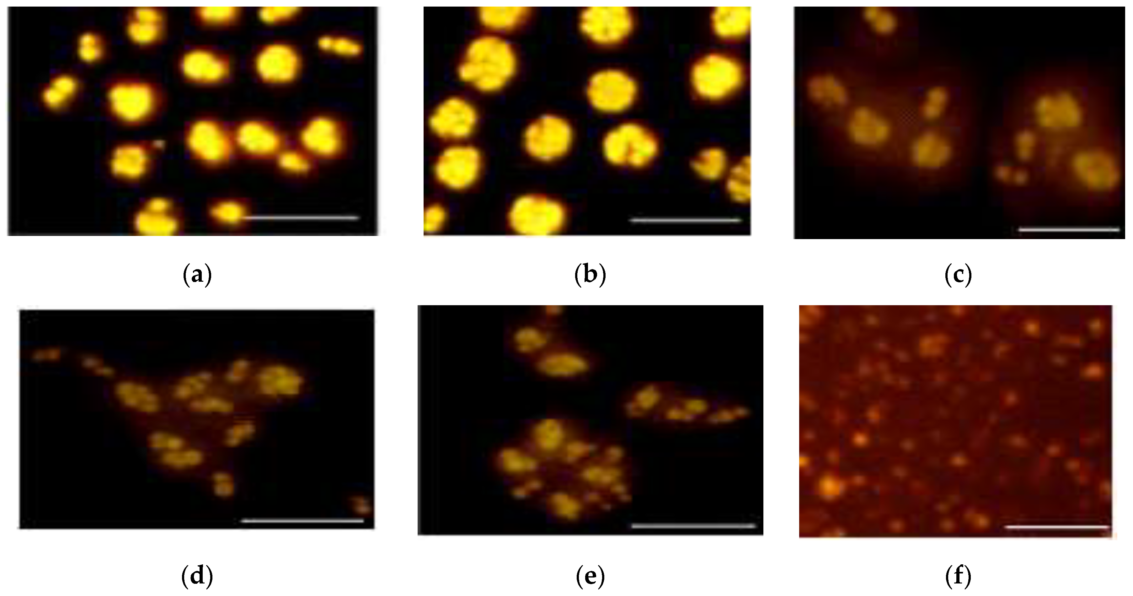
3.2.1. Microscopic Examination of Cell and Lipid Bodies Accumulation of R. glutinis under Different Metal Treatments

Differences in the cellular morphology and the lipid bodies accumulation after growing the yeast cells in media supplemented with different metal treatments, and their estimated corrected total cell fluorescence (CTCF) value is represented in Figure 7 and Figure S1. Changing the cultivation medium to medium with C/S ratio 120 (C2), led to reducing lipid bodies numbers inside the cells compared with the above-described treatment (C1) (Figure 7a).
The effect of metal supplementation is varied depending on the concentrations and the type of metal stress. Usually, the low concentration of CuSO4 0.1 mM (Figure 7b), ZnCl2 0.1 mM (Figure 7c), and NiSO4 0.1 mM (Figure 7e) showed a varied number of lipid bodies, the CTCF value of those metals was slightly more or similar to the CTCF of the control (Figure S1f, Supplementary Materials 1). However, increasing the previously mentioned metal to 1 mM led to a reduction in both cell size and lipid bodies and CTCF value, besides the appearance of the pseudo mycelium, which represented elongated cells that remained attached to the mother cells as observed with NiSO4 1 mM, which showed the lowest CTCF value (Figure 7f and Figure S1f). Mainly, the blue fluorescence was used for the examination of Nile red-stained lipid bodies, but the white light was used shortly with blue fluorescence at the time of imaging to detect the shape of the cells. 0.1 mM FeSO4 was affected on lipid accumulation as the cells kept a small oval-shape with the appearance of psedumycelium that carried small buddy cells on it. In contrast to the pseudomycelium formed with NiSO4 1 mM, there were no lipid bodies observed at all inside the psedumycelium of the iron treatment and the buddy cells carried on it (Figure 7g).
While using barium (Figure 7l,m) and lithium (Figure 7h,i) either in low or high concentrations, showed an observable increase in the cell size, lipid bodies as well as CTCF value, specifically with BaCl2 0.1 mM, which showed the highest CTCF value (Figure S1f). Regarding the microscopic analysis, the 0.1 mM and 1 mM BaCl2 could enhance the production of lipid by R. glutinis, even increasing the C/S ratio in the culture medium.

3.2.2. Effect of Different Metal Stress on Lipid and Carotenoid Production by R. glutinis

Metals can have a significant effect on the enzymatic activity of different biological pathways inside the microbial cells. The studied yeast strain showed a variable response in growth, lipid, and carotenoid production toward different metal treatments, as shown in Figure 8. The growth rate of R. glutinis reduced significantly after exposure to FeSO4 0.1 mM (p = 0.000), FeCl3 0.1 mM (p = 0.000), CuSO4 0.1 mM (p = 0.000), MnCl2 1 mM (p =0.000), NiSO4 1 mM (p = 0.003), and ZnCl2 0.1 and 1 mM (p = 0.000). While other metal treatments were showed insignificant DCW increase like BaCl2 0.1 mM, NiSO4 0.1 mM and LiCl compared with control, Figure 8a.
Focusing on lipid production under different metal stress, some metal reduced the lipid production to more than 80% like ZnCl2 1 mM (p = 0.000), FeSO4 0.1 mM (p = 0.000), and FeCl3 0.1 mM (p = 0.000). On the other hand, some metals enhanced cellular lipid accumulation such as BaCl2 (0.1 mM) (p = 0.05), MnCl2 (p >0.05), and LiCl (p >0.05). The cellular lipid increased significantly from 44.9% (Control) to 50% with BaCl2 supplementation, Figure 8a.
This points to the enhancement role of barium, manganese, and lithium-ion for lipid production by the studied yeast strain, even the low C/S ratio. Contrasting to the few studies, which investigated the effect of metal stress on lipogenesis of yeasts, several studies were carried out on algae.
A recent study investigated the effect of different metal stress on the growth and lipid production by Chlorella minutissima UTEX 2341 [27]. All the studied metals showed a considerable increase in the total lipid production by C. minutissima UTEX 2341, 6 mM Zn, 0.4 and 1 mM Cu significantly increased the algae lipid content by 18.30%, 21.07%, 19.87%, respectively.
The effect of metals stress on R. glutinis carotenoid production was also detected, Figure 8b. Surprisingly, some metals like 0.1 mM iron and copper salts blocked the carotenoid pathway, which reflected as the disappearance of the distinct red or radish color of the cells and appeared as a white biomass. Generally, metals like 0.1 mM of MnCl2, BaCl2, LiCl, and NiSO4 significantly increased carotenoid production of R. glutinis from 0.93 mg/L to 1.01 (p = 0.01), 1.05 (p = 0.001), 1.11 (p = 0.000), and 1.09 (p = 0.009) mg/L, respectively. In addition, cellular carotenoid increased from 86.2 to 100.3, 95.1, 101.7, and 102.4 µg/g, respectively. However, the further increase of the former metals to 1 mM led to a decrease of both cellular and total carotenoid significantly (p < 0.05) except with nickel sulfate, which increased cellular carotenoid to 132 µg/g and represented around 53% increase than the control.
The previously reported studies of the effect of metal ion on the carotenoid productivity by red yeasts stated the enhancement role of metal ion on carotenogenesis [24,29,30]. The enhancement role of nickel sulfate on the carotenogenesis pathway of red oleaginous yeasts was not reported before. While other metals like Ba, Zn, Fe, Cu, and Mn were reported to enhance carotenoid production by Rhodotorula mutant 32 [30]. In addition, Elbana et al. [29] stated a similar result for the enhancing role of Fe, Zn, Cu, and Mn on the carotenoid production by R. glutinis var. glutinis. While our results showed the inhibitory role of iron and copper on carotenoid production by R. glutinis, these differences may be mainly related to the genus sensitivity to the metal stress. The individual carotenoid profile of R. glutinis as a response factor to different metal treatments is represented in Figure 8c. The control groups detected the dominance of torulene 63%, followed by γ-carotene 30%. The addition of Mn+2, Ba+2, and Li+ led to enhance the production of γ-carotene significantly over torulene. The γ-carotene: Torulene ratio for 1 mM Mn+2, 1 mM Ba+2, and 1 mM Li+1 were 62:28, 50:37, and 50:44, respectively. While 1 mM NiSO4 and 0.1 mM ZnCl2 increased torulene significantly to 88.9% (p = 0.000) and 69.9% (p = 0.000), respectively.
R. glutinis var. glutinis showed the dominance of torulene when different heavy metal was introduced to the cultured media, the highest ratio observed with MnSO4 (80%), while the lowest ratio was observed with FeSO4 (56%) [29]. On the other hand, β-carotene was dominant when FeSO4, CuSO4, MnSO4, BaCl2, and ZnSO4 were introduced to the culture media of R. glutinis [30].
The response of the fatty acid profile to the different metal treatment was exciting (Table 3). Iron and copper supply to the culture media were led to reducing polyunsaturated fatty acid for the favor of saturated fatty acid; palmitic acid recorded the highest ratio 31% with 0.1 mM CuSO4 (p = 0.000). On the other hand, MnCl2, BaCl2 and LiCl enhance the production of unsaturated fatty acid significantly (p < 0.001), especially polyunsaturated fatty acid on the favor of oleic acid, whereas there was no alteration was observed with the profile of saturated fatty acid compared with the control.
This result indicated that the addition of low concentration of either MnCl2, BaCl2, and LiCl to the growth media could enhance the production of colored fats riches with unsaturated fatty acid, especially linolenic acid and linoleic acid, Table 3.
The addition of NiSO4 led to increasing oleic acid to 60%, while the linoleic and linolenic ratio was 15.6% and 3%, respectively, which represented only the half value compared with the control profile. The total unsaturated fatty acid produced by R. glutinis after adding NiSO4 was above 81%.

3.3. Fed-Batch Cultivation of R. glutinis Using Metallo-Sulfo-Phospho-Glucose Feeding Approach

As an attempt to enhance the biomass production, which accumulated colored fats riches with the unsaturated fatty acid, a two feeding stage fed-batch cultivation strategy was applied. The strategy was dependent on conducting two groups: In the first group, the culture conditions were fixed along with the whole experiment (control group). Glucose solution (100 g/100 mL) was used for feeding during the accumulation phase. While the second group, the temperature and pH were changed with metal supplementation after the growth phase (optimized group) and a mixture from glucose-sulfur-phosphorous solution at a ratio of 100:0.2:0.2 (g/100 mL distilled water) was used for feeding during the accumulation phase. Both groups showed almost the same DCW, TL, and TP production by R. glutinis during the first 20 h of cultivation and produced around 41 g/L DCW (Figure 9a) with a DCW synthesis rate 4.3 g/l/h and DCW-Y reached to 48%, (Figure 10a). At 32 h, the DCW, TL, and TP of the control group were higher than the optimized group. The decrease in the biomass, lipid, and carotenoid of the optimized group was mainly related to the change in the environmental conditions, as well as adding the metal solution, which decreased the cell activity to adapt to the new culture conditions. With the experiment progression, the lipid production, as well as the total lipid productivity in the control group, were greatly enhanced compared with the optimized group, representing around a 42% and 26.4% increase, respectively. Due to the increase of lipid bodies inside the yeast cells of the control group, the DCW showed around a 21.3% increase compared to the optimized media. Interestingly, the carotenoid of the optimized group was significantly increased by around 50% compared with the control, which emphasized the role of our strategy to enhance carotenoid production. The remarkable increase in carotenoid production could be related to the use of metal supplementation such as Al+3 and Ni+2, which proved to enhance carotenoid production by reducing lipid production in the flask fermentation experiment, besides the continuous supply of phosphorous and sulfate, which restricted the lipid accumulation. In addition, the shifting of the culture conditions played a critical role in this increase. The highest production of carotenoid was 29.3 mg/L with a maximum cellular carotenoid of 426 µgpigment/gDCW that was achieved at the end of cultivation with the optimized group, while the highest DCW, with the highest lipid production and productivity, were recorded with the control group 87.4 g/L, 49.2 g/L and 56.4%, respectively, Figure 9a,b.
The kinetic analysis of the process is represented in Figure 10. The maximum of TL-Y and TL-SR were detected with the control group, 21.3 (g/100g glucose) (time = 44 h) and 0.82 g/L/h (time = 44), respectively, which decreased with the optimized group to be 18.32 g/100 g glucose (time = 68 h), and o.77 g/L/h (time = 20), respectively, Figure 10b. The opposite was observed with the carotenoid kinetics, the maximum Car-Y and Car-SR were detected with the optimized group 0.17 g/100 g glucose (time = 56 h) and 0.67 mg/L/h (time = 56), respectively, Figure 10c. This result indicates the reversible relationship between lipid and carotenoid production during the same process.
Looking at the lipid and carotenoid profile under different cultivating strategies (Figure 9c), both torulene and unsaturated fatty acids, mainly C18:2, increased with the optimized group compared to the control. The torulene ratio was around 70% compared with 46% in the case of the control group. The increase in C18:2 in the optimized group may be mainly due to low incubating temperature and Ba+2 and Mn+2 supplementation, as observed in the flask fermentation experiment. In addition, increasing the torulene ratio may be related to metal supplementation, low pH, and temperature. Honestly, the ratio of torulene in the flask fermentation was higher than the bioreactor fermentation, and this result may be related to the agitation speed beside the phosphorous concentration in the culture media, as the increase of those two parameters enhanced the γ-carotene accumulation inside the yeast cells. The differences in the biomass color under the different cultivation strategy is represented in Figure 9d,e. The intense red color in the case of the optimized group was mainly related to the increase of the torulene ratio compared with the control group.
Several fed-batch studies were done to coproduce lipid and carotenoid from red oleaginous yeasts. Dias et al. [43] used pH control fed-batch cultivation strategy for the co-production of lipids and carotenoids by R. toruloides NCYC 921, the pH change step-wise strategy, setting another Rushton impeller to the bioreactor rotor shaft led to an increase in the biomass to 127 g/L, which was higher than this study, which was mainly due to the nitrogen feeding with glucose solution during the growth phase. The carotenoid content was 0.29 mg/g, which was lower than our result (0.426 mgpigment/g DCW with the optimized group).
Saenge et al. [2] used R. glutinis TISTR 5159 to coproduce lipids and carotenoids grown on palm oil mill effluent. Similarly, the two-stage process was attempted as an optimal way for cell growth in the first stage and product accumulation in the second stage. The lipid yield and carotenoid production obtained in the two-stage process were higher than those in the one-stage process. Although the total carotenoid yield was higher than our result, the biomass, lipid, and polyunsaturated fatty acid were lower than the presented study.
In addition, Zhang et al. [66] used a two-stage cultivation strategy for lipid and carotenoid by the strain R. glutinis CGMCC No. 2258: The first grow step was conducted under irradiation/high temperature; the second step (product accumulation) was conducted at dark/low temperature conditions in order to induce products accumulation. The biomass, lipid content, and carotenoid reached 86.2 g/L, 26.7%, and 4.2 mg/L, respectively, which was lower than our results. Lipids contained 22.8% saturated fatty acid, 51.7% monounsaturated fatty acids, and 24.8% polyunsaturated fatty acid. HPLC quantified the main carotenoid to be β-carotene 68.4%. Compared with our work, torulene had an intense antioxidant activity than β-carotene due to the presence of 13 double bonds [14], besides the higher ratio of polyunsaturated fatty acid, which made our colored lipid highly nutritive than Zhang et al. [66].

4. Conclusions

From this study, different culture conditions were investigated to correlate the production of lipid and carotenoid production by R. glutinis, as well as investigating the possibilities to improve the lipid and carotenoid profile of R. glutinis through culture conditions manipulation. The results revealed that all the studied conditions showed a reversible relationship between lipid and carotenoid production under the studied culture conditions. Studying the effect of different heavy metal stress on carotenoid and lipid production and profile was interesting. The addition of copper 0.1 mM, leading to preventing carotenoid production with increasing the saturation level of lipid profile producing around 70% palmitic and oleic acid, making it suitable for biodiesel production. While 0.1 mM of Mn+2, Ba+2, and Li+ led to the production of fat with the γ-carotene dominant carotenoid and unsaturated fatty acid domination, especially polyunsaturated fatty acid. 1 mM NiSO4 enhanced both the cellular carotenoid as well as torulene domination by R. glutinis, which accompanied 81% unsaturated fatty domination, where oleic acid represented 61% of the total lipid. Finally, the metallo-sulfo-phospho-glucose feeding approach combined with the pH-temperature shifting strategy, proved to be an effective approach for the production of carotenoid (29.3 mg/L), specifically torulene (71%) from R. glutinis. This study provides a significant understanding of the relationship between lipid and carotenoid production by R. glutinis under various culture conditions. This has led to a potential fed-batch strategy to enhance carotenoid, especially torulene production, by R. glutinis to be competitive among other relative studies and could be useful for the industrial production of healthy nutritive pigment.

Supplementary Materials

The following are available online at https://www.mdpi.com/2227-9717/8/2/140/s1, Supplementary material 1: Figure S1. Different corrected total cell fluorescence (CTCF) value calculated by using image j software; (a) CTCF values of different carbon sources` Nile-red stained cells, (b) CTCF values of different pH degrees` Nile-red stained cells, (c) CTCF values of different temperature` Nile-red stained cells, (d) CTCF values of different agitation` Nile-red stained cells, (e) CTCF values of different phosphorous concentration` Nile-red stained cells, (f) CTCF values of different metal treated Nile-red stained cells. Table S1. Significance of different carbon sources on R. glutinis growth, lipid, and carotenoid production after 3 and 6 days. Table S2. Multivariate test to detect the significance of different carbon sources on R. glutinis growth, lipid, and carotenoid production after 3 and 6 days. Table S3. Significance of different pH degree on R. glutinis growth, lipid, and carotenoid production. Table S4. Multivariate test to detect the significance of different pH degrees on R. glutinis growth, lipid, and carotenoid production after 3 and 6 days. Table S5. Significance of different incubation temperatures on R. glutinis growth, lipid, and carotenoid production. Table S6. Multivariate test to detect the significance of different Temperature degrees on R. glutinis growth, lipid, and carotenoid production after 3 and 6 days. Table S7. Significance of different agitation speed on DCW, lipid and carotenoid production by R. glutinis. Table S8. Multivariate test to detect the significance of different agitation speed on R. glutinis growth, lipid, and carotenoid production after 3 and 6 days. Table S9. Significance of different phosphorus concentrations (initial C/P molar ratio) on R. glutinis growth, lipid and carotenoid production after 3 and 6-day incubation at 28 °C and 180 rpm. Table S10. Multivariate test to detect the significance of different carbon to phosphorous ratio (C/P) on R. glutinis growth, lipid, and carotenoid production after 3 and 6 days. Supplementary Material 2; Excel sheet 1. correlation coefficient statistic. Excel sheet 2. Statistical analysis of the detection of individual carotenoid by HPLC. Excel sheet 3. Statistical analysis of the detection of fatty acid methyl esters by GC.

Author Contributions

Conceptualization, Y.B.; experimentation and writing—original draft preparation, N.E.; data analysis and investigation, N.E., M.E.; review and editing, Y.B., N.E. All authors have read and agree to the published version of the manuscript.

Funding

This research received no external funding.

Conflicts of Interest

The authors declare no conflict of interest.

References

  1. Kot, A.; Błażejak, S.; Kurcz, A.; Gientka, I.; Kieliszek, M. Rhodotorula glutinis—Potential source of lipids, carotenoids, and enzymes for use in industries. Appl. Microbiol. Biotechnol. 2016, 100, 6103–6117. [Google Scholar] [CrossRef] [PubMed] [Green Version]
  2. Saenge, C.; Cheirslip, B.; Suksaroge, T.T.; Bourtoom, T. Potential use of oleaginous red yeast Rhodotorula glutinis for the bioconversion of crude glycerol from biodiesel plant to lipids and carotenoids. Process Biochem. 2011, 46, 210–218. [Google Scholar] [CrossRef]
  3. Miles, E.A.; Calder, P.C. Modulation of immune function by dietary fatty acids. Proc. Nutr. Soc. 1998, 57, 277–292. [Google Scholar] [CrossRef] [PubMed] [Green Version]
  4. Sales-Campos, H.; Reis de Souza, P.; Peghini, C.B.; Da Silva, J.S.; Cardoso, C.R. An Overview of the Modulatory Effects of Oleic Acid in Health and Disease. Mini. Rev. Med. Chem. 2013, 13, 201–210. [Google Scholar] [CrossRef]
  5. Owen, R.W.; Mier, W.; Giacosa, A.; Hull, W.E.; Spiegelhalder, B.; Bartsch, H. Phenolic compounds and squalene in olive oils: The concentration and antioxidant potential of total phenols, simple phenols, secoiridoids, lignans and squalene. Food Chem. Toxicol. 2000, 38, 647–659. [Google Scholar] [CrossRef]
  6. Panagiotakos, D.B.; Dimakopoulou, K.; Katsouyanni, K.; Bellander, T.; Grau, W.K.; Lanki, T.; Pistelli, R.; Schneider, A.; Peters, A. Mediterranean diet and inflammatory response in myocardial infarction survivors. Int. J. Epidemiol. 2009, 38, 856–866. [Google Scholar] [CrossRef] [Green Version]
  7. Tere’s, S.; Barcelo’-Coblijn, G.; Benet, M.; A’lvarez, R.; Bressani, R.; Halver, J.E.; Escriba, P.V. Oleic acid content is responsible for the reduction in blood pressure induced by olive oil. Proc. Natl. Acad. Sci. USA 2008, 37, 13811–13816. [Google Scholar] [CrossRef] [Green Version]
  8. Besler, H.T.; Grimble, R.F. Comparison of the modulatory influence of maize and olive oils and butter on metabolic responses to endotoxin in rats. Clin. Sci. 1995, 66, 59–66. [Google Scholar] [CrossRef]
  9. Ferrara, L.A.; Raimondi, A.S.; Episcopo, L.; Guida, L.; Dello Russo, A.; Marotta, T. Olive oil and reduced need for antihypertensive medications. Arch. Intern. Med. 2000, 160, 837–842. [Google Scholar] [CrossRef] [Green Version]
  10. Kremer, M.; Lawrence, D.A.; Jubiz, W.; Digiacomo, R.; Rynes, R.; Bartholomew, L.E.; Sherman, M. Dietary Fish Oil And Olive Oil Supplementation In Patients With Rheumatoid Arthritis. Arthritis Rheumatol. 1990, 33, 810–820. [Google Scholar] [CrossRef]
  11. Martin-Moreno, J.M.; Willett, W.C.; Gorgojo, L.; Banegas, J.R.; Rodriguez-Artalejo, F.; Fernandez-Rodriguez, J.C.; Maisonneuve, P.; Boyle, P. Dietary fat, olive oil intake and breast cancer risk. Int. J. Cancer 1994, 58, 774–780. [Google Scholar] [CrossRef] [PubMed]
  12. Lipworth, L.; Martinez, M.E.; Angell, J.; Hsieh, C.C.; Trichopoulos, D. Olive Oil and Human Cancer: An Assessment of the Evidence. Prev. Med. 1997, 26, 181–190. [Google Scholar] [CrossRef] [PubMed]
  13. Kot, A.M.; Błazejak, S.; Kieliszek, M.; Gientka, I.; Bry’s, J. Simultaneous Production of Lipids and Carotenoids by the Red Yeast Rhodotorula from Waste Glycerol Fraction and Potato Wastewater. Appl. Biochem. Biotechnol. 2019, 1–19. [Google Scholar] [CrossRef] [PubMed]
  14. Ageitos, J.M.; Vallejo, J.A.; Veiga-crespo, P.; Villa, T.G. Oily yeasts as oleaginous cell factories. Appl Microbiol. Biotechnol. 2011, 90, 1219–1227. [Google Scholar] [CrossRef]
  15. Kot, A.M.; Błazejak, S.; Gientka, I.; Kieliszek, M.; Bry’s, J. Torulene and torularhodin: “New” fungal carotenoids for industry? Microbl. Cell Fact. 2018, 100, 6103–6117. [Google Scholar] [CrossRef] [Green Version]
  16. Krinsky, N.I.; Johnson, E.J. Carotenoid actions and their relation to health and disease. Mol. Asp. Med. 2005, 26, 459–516. [Google Scholar] [CrossRef]
  17. Brown, L.; Rimm, E.B.; Seddon, J.M.; Giovannucci, E.L.; Chasan-Taber, L.; Spiegelman, D.; Willett, W.C.; Hankinson, S.E. A prospective study of carotenoid intake and risk of cataract extraction in US men. Am. J. Clin. Nutr. 1999, 70, 517–524. [Google Scholar] [CrossRef] [Green Version]
  18. Beatty, S.; Nolan, J.; Kavanagh, H.; Donovan, O.O. Macular pigment optical density and its relationship with serum and dietary levels of lutein and zeaxanthin. ABB 2004, 430, 70–76. [Google Scholar] [CrossRef]
  19. Stahl, W.; Sies, A.H. Carotenoids and Flavonoids Contribute to Nutritional Protection against Skin Damage from Sunlight. Mol. Biotechnol. 2007, 37, 26–30. [Google Scholar] [CrossRef]
  20. Fraser, P.D.; Bramley, P.M. The biosynthesis and nutritional uses of carotenoids. Prog. Lipid Res. 2004, 43, 228–265. [Google Scholar] [CrossRef]
  21. Mata-gómez, L.C.; Montañez, J.C.; Méndez-zavala, A.; Aguilar, C.N. Biotechnological production of carotenoids by yeasts: An overview. Microbl. Cell Fact. 2014, 13, 1–11. Available online: http://www.microbialcellfactories.com/content/13/1/12 (accessed on 1 January 2014). [CrossRef] [PubMed] [Green Version]
  22. Braunwald, T.; Schwemmlein, L.; Graeff-Hönninger, S.; French, W.T.; Hernandez, R.; Holmes, W.E.; Claupein, W. Effect of different C/N ratios on carotenoid and lipid production by Rhodotorula glutinis. Appl. Microbiol. Biotechnol. 2013, 97, 6581–6588. [Google Scholar] [CrossRef]
  23. Tkáčová, J.; Čaplová, J.; Klempová, T.; Čertík, M. Correlation between lipid and carotenoid synthesis in torularhodin-producing Rhodotorula Glutinis. Ann. Microbiol. 2017, 67, 541–551. [Google Scholar] [CrossRef]
  24. Elfeky, N.; Elmahmoudy, M.; Zhang, Y.; Guo, J.; Yongming, B. Lipid and Carotenoid Production by Rhodotorula glutinis with a Combined Cultivation Mode of Nitrogen, Sulfur and Aluminium Stress. Appl. Sci. 2019, 9, 2444. [Google Scholar] [CrossRef] [Green Version]
  25. Schulze, K.L. The Effect of Phosphate Supply on the Rate of Growth and Fat Formation in Yeasts. Appl. Microbiol. 1956, 4, 207–210. [Google Scholar] [CrossRef] [Green Version]
  26. Baldrian, P.; Gabriel, I. Influence of Cadmium and Mercury on Activities of Ligninolytic Enzymes and Degradation of Polycyclic Aromatic Hydrocarbons by Pleurotus ostreatus in Soil. Appl. Environ. Microbiol. 2000, 66, 2471–2478. [Google Scholar] [CrossRef] [Green Version]
  27. Yang, J.; Cao, J.; Xing, G.; Yuan, H. Lipid production combined with biosorption and bioaccumulation of cadmium, copper, manganese and zinc by oleaginous microalgae Chlorella minutissima UTEX2341. Bioresour. Technol. 2015, 175, 537–544. [Google Scholar] [CrossRef]
  28. Goswami, L.; Manikandan, N.A.; Pakshirajan, K.; Pugazhenthi, G. Simultaneous heavy metal removal and anthracene biodegradation by the oleaginous bacteria Rhodococcus opacus. 3 Biotech 2017, 7, 1–9. [Google Scholar] [CrossRef]
  29. El-Banna, A.A.; Abd El-Razek, A.M.; El-Mahdy, A.R. Some Factors Affecting the Production of Carotenoids by Rhodotorula glutinis var. glutinis. Food Nutr. Sci. 2012, 3, 64–71. [Google Scholar] [CrossRef]
  30. Bhosale, P.B.; Gadre, V. Production of β-carotene by a mutant of Rhodotorula glutinis. Appl. Microbiol. Biotechnol. 2001, 55, 423–427. [Google Scholar] [CrossRef]
  31. Sitepu, R.; Sestric, R.; Ignatia, L.; Levin, D.; German, J.; Gillies, L.A.; Almada, L.A.; Boundy-Mills, K.L. Manipulation of culture conditions alters lipid content and fatty acid profiles of a wide variety of known and new oleaginous yeast species. Bioresour. Technol. 2013, 144, 360–369. [Google Scholar] [CrossRef] [PubMed] [Green Version]
  32. Miller, G.L. Use of Dinitrosalicylic Acid Reagent for Determination of Reducing Sugar. Anal. Chem. 1959, 31, 426–428. [Google Scholar] [CrossRef]
  33. Miloski, K.; Wallace, K.; Fenger, A.; Schneider, E.; Bendinskas, K. Comparison of Biochemical and Chemical Digestion and Detection Methods for Carbohydrates. Am. J. Undergrad. Res. 2019, 7, 7–18. [Google Scholar] [CrossRef]
  34. Kimura, K.; Yamaoka, M.; Kamisaka, Y. Rapid estimation of lipids in oleaginous fungi and yeasts using Nile red fluorescence. J. Microbiol. Methods 2004, 56, 331–338. [Google Scholar] [CrossRef]
  35. Available online: https://theolb.readthedocs.io/en/latest/imaging/measuring-cell-fluorescence-using-imagej.html (accessed on 1 January 2014).
  36. Mishra, S.K.; Suh, W.I.; Farooq, W.; Moon, M.; Shrivastav, A.; Park, M.S.; Yang, J.W. Rapid quantification of microalgal lipids in aqueous medium by a simple colorimetric method. Bioresour. Technol. 2014, 155, 330–333. [Google Scholar] [CrossRef] [PubMed]
  37. Van Wychen, S.; Ramirez, K.; Laurens, L.M. Determination of Total Lipids as Fatty Acid Methyl Esters (FAME) by in situ Transesterification. NREL 2013, 303, 275–3000. [Google Scholar]
  38. Frengova, G.; Sirnova, E.; Pavlova, K.; Beshkova, D. Formation of Carotenoids by Rhodotorula glutinis in whey ultrafiltrate. Biotechnol. Bioeng. 1994, 44, 888–894. [Google Scholar] [CrossRef]
  39. Weber, R.W.; Anke, H.; Davoli, P. Simple method for the extraction and reversed-phase high-performance liquid chromatographic analysis of carotenoid pigments from red yeasts (Basidiomycota, Fungi). J. Chromatogr. A 2007, 1145, 118–122. [Google Scholar] [CrossRef]
  40. Certik, M.; Shimizu, S. Kinetic analysis of oil biosynthesis by an arachidonic acid-producing fungus, Mortierella alpina 1S-4. Appl. Microbiol. Biotechnol. 2000, 54, 224–230. [Google Scholar] [CrossRef]
  41. Patel, A.; Pruthi, V.; Singh, R.P.; Pruthi, P.A. Synergistic effect of fermentable and non-fermentable carbon sources enhances accumulation in oleaginous yeast Rhodosporidium kratochvilovae HIMPA1. Bioresour. Technol. 2015, 188, 136–144. [Google Scholar] [CrossRef]
  42. Karamerou, E. Bioprocessing Strategies for the Cultivation of Oleaginous Yeasts on Glycerol. PhD Thesis, School of Chemical Engineering and Analytical Science, University of Manchester, UK, 2016. [Google Scholar]
  43. Dias, C.; Sousa, S.; Caldeira, J.; Reis, A. New dual-stage pH control fed-batch cultivation strategy for the improvement of lipids and carotenoids production by the red yeast Rhodosporidium toruloides NCYC 921. Bioresour. Technol. 2015, 189, 309–318. [Google Scholar] [CrossRef] [PubMed] [Green Version]
  44. Johnson, V.; Singh, M.; Saini, V.S.; Sista, V.R.; Yadav, N.K. Effect of pH on lipid accumulation by an oleaginous yeast: Rhodotorula glutinis IIP-30. World J. Microbiol. Biotechnol. 1992, 8, 382–384. [Google Scholar] [CrossRef]
  45. Milkessa, T.; Marizeth, J.; Carolina, G.; Laurinda, P.; Pohl, C. Optimization of cultivation conditions for biotechnological production of lipid by Rhodotorula kratochvilovae (syn, Rhodosporidium kratochvilovae) SY89 for biodiesel preparation. 3 Biotech 2017, 7, 1–11. [Google Scholar]
  46. Dhaliwal, M.K.; Chandra, N. Optimization of carotenoids production by Rhodotorula mucilaginosa. IJPSR 2015, 3, 1161–1165. [Google Scholar]
  47. Zhao, Y.; Guo, L.; Xia, Y.; Zhuang, X.; Chu, W. Isolation, Identification of Carotenoid-Producing Rhodotorula sp. from Marine Environment and Optimization for Carotenoid Production. Mar. Drugs 2019, 17, 161. [Google Scholar] [CrossRef] [PubMed] [Green Version]
  48. Hadi, S. Effect of Temperature and pH on the Growth Kinetics and Carotenoid Production by Sporobolomyces ruberrimus H110 Using Technical Glycerol as Carbon Source. Iran. J. Chem. Chem. Eng. 2006, 25, 59–64. [Google Scholar]
  49. Saad, N.; Abdeshahian, P.; Kalil, M.S.; Wan Yusoff, W.M.; Abdul Hamid, A. Optimization of Aeration and Agitation Rate for Lipid and Gamma Linolenic Acid Production by Cunninghamella bainieri 2A1 in Submerged Fermentation Using Response Surface Methodology. Sci. World J. 2014, 1–12. [Google Scholar] [CrossRef] [Green Version]
  50. Zhou, Y.; Han, L.; He, H.; Yu, D.; Feng, J.; Zhang, X. Effects of agitation, aeration and temperature on production of a novel glycoprotein gp-1 by streptomyces kanasenisi zx01 and scale-up based on volumetric oxygen transfer coefficient. Molecules 2018, 23, 125. [Google Scholar] [CrossRef] [Green Version]
  51. Vlaev, S.; Pavlova, K.; Rusinova-Videva, S.; Georgieva, K.; Georgiev, D. Agitation effects and kinetic constants of exoglucomannan production by Antarctic yeast strain in a stirred tank bioreactor. Chem. Biochem. Eng. 2016, 30, 393–400. [Google Scholar] [CrossRef]
  52. Dai, C.; Tao, J.; Xie, F.; Dai, Y.J.; Zhao, M. Biodiesel generation from oleaginous yeast Rhodotorula glutinis with xylose assimilating capacity. Afr. J. Biotechnol. 2007, 6, 2130–2134. [Google Scholar]
  53. El-Fadaly, A.H.; El-Naggar, N.; El-Sayed, M. Single Cell Oil Production by an Oleaginous Yeast Strain in a Low cost cultivation medium. Res. J. Microbiol. 2009, 4, 301–313. [Google Scholar] [CrossRef] [Green Version]
  54. Wang, Y.; Zhang, S.; Zhu, Z.; Shen, H.; Lin, X.; Jin, X.; Jiao, X.; Zhao, Z. Biotechnology for Biofuels Systems analysis of phosphate-limitation-induced lipid accumulation by the oleaginous yeast Rhodosporidium toruloides. Biotechnol. Biofuels 2018, 1–15. [Google Scholar] [CrossRef] [Green Version]
  55. Buzzini, P.; Martini, A.; Gaetani, M.; Turchetti, B.; Pagnonib, U.; Davoli, P. Optimization of carotenoid production by Rhodotorula graminis DBVPG 7021 as a function of trace element concentration by means of response surface analysis. Enzym. Microb. Technol. 2005, 36, 687–692. [Google Scholar] [CrossRef]
  56. Bhosale, P. Environmental and cultural stimulants in the production of carotenoids from microorganisms. Appl. Microbiol. Biotechnol. 2004, 63, 351–361. [Google Scholar] [CrossRef] [PubMed]
  57. Latha, B.V.; Jeevaratnam, K.; Murali, H.S.; Manja, K.S. Influence of growth factors on carotenoid pigmentation of Rhodotorula glutinis DFR-PDY from natural source. Indian J. Biotechnol. 2004, 4, 353–357. [Google Scholar]
  58. Frengova, G.I.; Beshkova, D.M. Carotenoids from Rhodotorula and Phaffia: Yeasts of biotechnological importance. J. Ind. Microbiol. Biotechnol. 2009, 36, 163–180. [Google Scholar] [CrossRef]
  59. Wongsnansilp, T.; Juntawong, N.; Wu, Z. Effects of phosphorus on the growth and chlorophyll fluorescence of a Dunaliella salina strain isolated from saline soil under nitrate limitation. J. Biol. Res. 2016, 89, 51–55. [Google Scholar] [CrossRef] [Green Version]
  60. Forján, E.; Garbayo, I.; Bejarano, C.; Vílchez, C. Enhancement of carotenoid production in Nannochloropsis by phosphate and sulphur limitation. In Communicating Current Research and Educational Topics and Trends in Applied Microbiology; Microbiology Book Series; Formatex: Badajoz, Spain, 2007; Volume 1, pp. 356–364. ISBN 978-84-611-9421-6. [Google Scholar]
  61. Kot, A.M.; Błazejak, S.; Kurcz, A.; Bry’s, J.; Gientka, I.; Bzducha-Wróbel, A.; Maliszewska, M.; Reczek, L. Effect of initial pH of medium with potato wastewater and glycerol on protein, lipid and carotenoid biosynthesis by Rhodotorula glutinis. Electron. J. Biotechnol. 2017, 27, 25–31. [Google Scholar] [CrossRef]
  62. Suutari, M.; Rintamliki, A.; Laakso, S. The Effect of Temperature on Lipid Classes and Their Fatty Acid Profiles in Lipomyces starkeyi. JAOCS 1996, 73, 1071–1073. [Google Scholar] [CrossRef]
  63. Miranti, A.; Arbianti, R.; Utami, T. Effect of pH, temperature and medium agitation rate in production of AA, DHA, EPA from Aspergillus oryzae with submerged fermentation. IOP Conf. Ser. Earth Environ. Sci. 2018, 105, 012113. [Google Scholar] [CrossRef]
  64. Hu, H.; Wang, H.; Li, J.; Ma, L.; Shen, X.; Zeng, R. Evaluation of the effect of agitation speed on the growth and high-value LC-PUFA formation of Porphyridium cruentum based on basic rheological analysis. J. Chem. Technol. Biotechnol. 2019, 7, 2158–2166. [Google Scholar] [CrossRef]
  65. Davoli, P.; Mierau, V.; Weber, R.W.S. Carotenoids and fatty acids in red yeasts Sporobolomyces roseus and Rhodotorula glutinis. Appl. Biochem. Microbiol. 2004, 40, 392–397. [Google Scholar] [CrossRef]
  66. Zhang, Z.; Zhang, X.; Tan, T. Lipid and carotenoid production by Rhodotorula glutinis under irradiation/high-temperature and dark/low-temperature cultivation. Bioresour. Technol. 2014, 157, 149–153. [Google Scholar] [CrossRef] [PubMed]
Figure 1. Fluorescence microscopy examination of Nile red-stained yeast cells after growing on lipid accumulating media supplemented with different carbon sources after the 6th day: (a) Glucose; (b) glucose and sucrose (1:1); (c) sucrose; (d) maltose. The magnification bar is equal to 10 µm.
Processes 08 00140 g001aProcesses 08 00140 g001b
Figure 2. Fluorescence microscopy examination of Nile red-stained yeast cells after growing on lipid accumulating media with different pH for 6 days: (a) 3; (b) 4; (c) 5; (d) 6; (e) 7; (f) 8. The magnification bar is equal to 10 µm.
Processes 08 00140 g002
Figure 3. Fluorescence microscopy examination of Nile red-stained yeast cells after growing on lipid accumulating media at different temperatures degrees for 6 days: (a) 25 °C. (b) 28 °C. (c) 30 °C. (d) 32 °C. The magnification bar is equal to 10 µm.
Processes 08 00140 g003
Figure 4. Fluorescence microscopy examination of Nile red-stained yeast cells after growing on lipid accumulating media under different agitation speed for 6 days: (a) 150 rpm. (b) 180 rpm. (c) 200 rpm. (d) 220 rpm. The magnification bar is equal to 10 µm.
Processes 08 00140 g004
Figure 5. Fluorescence microscope examination of Nile red-stained yeast cells after growing on lipid accumulating media with different starter phosphorus concentrations for 6 days; (a) 0.5 g/L, (b) 1.5 g/L, (c) 3 g/L, (d) 4.5 g/L. The magnification bar is equal to 10 µm.
Processes 08 00140 g005
Figure 6. Identification and quantification of the individual carotenoid of R. glutinis as a response to different culture conditions using high-performance liquid chromatography (HPLC), the Y-axis represents the ratio of the individual carotenoid, X-axis represents the factor groups ; (a) different carbon sources; Glu: Glucose, Suc: Sucrose, Gluc + Suc: Glucose + sucrose, Mal: Maltose. (b) Different agitation speed. (c) Different incubation temperatures. (d) Different initial C/P molar ratio. (e) Different pH degree. The data are the mean of two replicas, the standard deviation represented as the error bar. The significance was calculated by using the multivariate test and represented at Supplementary Materials 2.
Processes 08 00140 g006
Figure 7. Fluorescence microscopy examination of Nile-red stained yeast cells after growing on medium supplemented with different concentration of heavy metal: (a) Control; (b) CuSO4 0.1 mM; (c) Zncl2 0.1 mM; (d) ZnCl2 1 mM; (e) NiSO4 0.1 mM; (f) NiSO4 1 mM; (g) FeSO4 0.1 mM; (h) Licl 0.1 mM; (i) LiCl 1 mM; (j) MnCl2 0.1 mM; (k) MnCl2 1 mM; (l) BaCl2 0.1 mM; (m) BaCl2 1 mM. The magnification bar is equal to 10 µm.
Processes 08 00140 g007
Figure 8. Effect of different metal ion stress on growth, lipid, and carotenoid production of R. glutinis on the 6th day: (a) Effects of different metal ion stress on dry cell weight (DCW), total lipid (TL), and total lipid-relative productivity (TL-RP). * and ** mean DCW of metal ion treated groups are statistically significant with DCW of control group p < 0.05 and p < 0.01, respectively. # and ## mean TL of metal ion treated groups are statistically significant with TL of control group p < 0.05 and p < 0.01, respectively; (b) effects of different metal ion stress on TP and Car-RP. + and ++ mean TP of metal ion treated groups are statistically significant with TP of control group p < 0.05 and p < 0.01, respectively; (c) individual carotenoid profile of R. glutinis after treatment with different metal ion stress, the same letters means the individual carotenoids were statistically significant with its candidate at the control group (p < 0.05). The data are the mean of three separate biological samples. The error bar represents the standard deviation.
Processes 08 00140 g008aProcesses 08 00140 g008b
Figure 9. Effect of different fed-batch cultivation strategies on R. glutinis growth, lipid, and carotenoid production: (a) The control and optimized treatment DCW, TL and TP; (b) the control and optimized treatment TL-RP and Car-RP; (c) lipid and carotenoid profile; (d) control`biomass color at the end of cultivation; (e) Optimized` biomass color at the end of cultivation.
Processes 08 00140 g009aProcesses 08 00140 g009b
Figure 10. Kinetics parameters of different fed-batch cultivation strategies on R. glutinis growth, lipid, and carotenoid production: (a) Dry cell weight yield (DCW-Y) and dry cell weight synthesis rate (DCW-SR). (b) Total lipid yield (TL-Y) and total lipid synthesis rate (TL-SR). (c) Carotenoid yield (Car-Y) and carotenoid synthesis rate (Car-SR).
Processes 08 00140 g010aProcesses 08 00140 g010b
Table 1. The effect of manipulating culture conditions on R. glutinis growth, lipid, and carotenoid production.
FactorT (Day)DCWLipidCarotenoid
DCW g/LDCW-YTL g/LTL-Y %TL-RPTP mg/LCar-YCar-RP
g/g glug/gdcwmg/100 g gluµg/g dcw
Carbon sourcesGlu3 rd6.71 ± 0.0647.32.40 ± 0.2016.835.60.56 ± 0.1539.884.2
C16 th 10.5 ± 0.1026.15.00 ± 0.1418.047.70.81 ± 0.0917.377.8
Suc3 rd5.60 ± 0.0434.41.80 ± 0.0511.031.90.45 ± 0.0827.379.4
6 th 8.70 ± 0.0824.04.00 ± 0.1016.945.50.70 ± 0.0319.980.6
Glu + Suc3 rd7.20 ± 0.2047.62.60 ± 0.0517.336.40.67 ± 0.0344.092.3
6 th 10.7 ± 0.5021.15.30 ± 0.2016.049.40.96 ± 0.0717.989.9
Mal3 rd6.00 ± 0.0936.60.64 ± 0.203.9010.60.33 ± 0.0620.455.7
6 th 9.30 ± 0.1035.02.70 ± 0.5022.529.50.54 ± 0.1121.858.0
pH33 rd6.50 ± 1.7033.02.80 ± 0.0214.242.90.39 ± 0.0619.759.5
6 th 9.90 ± 2.2035.74.90 ± 0.5522.149.30.46 ± 0.157.8046.8
43 rd6.50 ± 1.7040.72.60 ± 0.3816.440.40.44 ± 0.1027.467.3
6 th 10.0 ± 1.0030.25.20 ± 0.2521.851.60.59 ± 0.1612.858.6
53 rd6.71 ± 0.0647.32.40 ± 0.2016.835.60.56 ± 0.1539.884.2
C16 th 10.5 ± 0.1026.15.00 ± 0.1418.047.70.81 ± 0.0917.377.8
63 rd6.60 ± 0.5347.61.97 ± 0.0614.330.20.59 ± 0.0242.990.1
6 th 10.5 ± 1.0027.64.74 ± 0.4619.245.10.89 ± 0.1021.185.0
73 rd6.40 ± 1.4046.71.86 ± 0.3713.528.90.56 ± 0.0440.887.5
6 th 9.50 ± 0.8026.84.04 ± 0.3618.942.30.80 ± 0.0620.784.2
83 rd6.10 ± 1.1046.30.55 ± 0.314.209.000.48 ± 0.0236.478.5
6 th 9.00 ± 1.1021.42.30 ± 0.3012.625.30.73 ± 0.0118.380.9
Temp (°C)253 rd5.70 ± 0.3447.61.36 ± 0.1111.424.00.44 ± 0.1036.777.1
6 th 8.20 ± 0.3024.73.63 ± 0.3322.144.20.63 ± 0.1219.077.0
283 rd6.71 ± 0.0647.32.40 ± 0.2016.835.60.56 ± 0.1539.884.2
C16 th 10.5 ± 0.126.15.00 ± 0.1418.047.70.81 ± 0.0917.377.8
303 rd6.90 ± 0.1143.82.39 ± 0.1815.134.50.58 ± 0.1536.883.9
6 th 10.9 ± 0.324.45.33 ± 0.3718.249.10.83 ± 0.1015.576.6
323 rd5.00 ± 0.1145.41.29 ± 0.1111.725.70.37 ± 0.1033.874.5
6 th 7.41 ± 0.1820.23.29 ± 0.3516.944.40.58 ± 0.1017.277.9
Agitation (rpm)1503 rd6.30 ± 0.2748.31.77 ± 0.3513.628.10.47 ± 0.1036.475.3
6 th 9.50 ± 0.4826.43.50 ± 0.3314.637.20.68 ± 0.1017.171.7
1803 rd6.70 ± 0.0547.52.40 ± 0.2016.635.00.57 ± 0.2039.883.8
C16 th 10.4. ± 0.125.65.00 ± 0.1618.247.80.81 ± 0.1016.777.2
2003 rd7.50 ± 0.5047.22.60 ± 0.1216.635.30.61 ± 0.2038.581.6
6 th 11.0 ± 0.7024.95.40 ± 0.3019.348.80.89 ± 0.2019.981.0
2203 rd7.60 ± 0.5043.92.60 ± 0.3215.034.20.61 ± 0.2035.480.6
6 th 11.0 ± 0.8023.35.20 ± 0.2217.747.10.89 ± 0.0319.281.2
C/P5273 rd6.56 ± 1.6041.52.61 ± 0.0416.539.80.50 ± 0.1731.676.0
6 th10.2 ± 1.9021.65.30 ± 0.0816.152.10.62 ± 0.117.3360.9
1763 rd6.80 ± 2.2047.52.36 ± 0.2016.635.00.57 ± 0.1639.883.8
C16 th10.4 ± 2.6025.35.10 ± 0.1619.049.10.80 ± 0.1016.677.4
883 rd7.60 ± 2.6051.21.60 ± 0.0410.821.20.65 ± 0.0243.585.0
6 th11.0 ± 0.6025.93.50 ± 0.0414.531.80.94 ± 0.0622.385.4
593 rd8.09 ± 1.7053.70.64 ± 0.054.207.900.69 ± 0.0445.885.3
6 th11.5 ± 1.7026.72.30 ± 0.0612.619.50.99 ± 0.0423.385.9
The data are the mean ± SD of three separate biological samples. The significance of each condition is represented separately in the Supplementary Materials Tables S1–S5.
Table 2. The profile of fatty acid methyl esters (FAMES) of R. glutinis as a factor of culture conditions manipulation.
FactorTime (Day)C16:0C16:1C18:0C18:1C18:2C18:3
Carbon sourcesGlu C13 rd12.2 ± 1.4nd8.80 ± 9.968.8 ± 5.75.90 ± 8.04.30 ± 8.5
6 th 20.4 ± 3.0nd11.7 ± 2.848.8 ± 7.110.4 ± 4.98.70 ± 3.1
Suc3 rd12.7 ± 6.4nd5.50 ± 2.961.3 ± 1015.2 ± 2.85.20 ± 6.2
6 th 17.1 ± 2.0nd2.40 ± 1.446.6 ± 5.79.10 ± 5.015.7 ± 2.0
Glu+Suc3 rd7.20 ± 4.0nd5.10 ± 5.069.6 ± 4.213.5 ± 7.14.60 ± 5.0
6 th 11.4 ± 2.6nd8.00 ± 6.455.5 ± 5.614.6 ± 1210.4 ± 4.9
Mal3 rd8.40 ± 3.50nd15.3 ± 4.248.1 ± 1.012.7 ± 1315.6 ± 4.9
6 th 10.1 ± 8.0nd6.10 ± 6.252.2 ± 6.421.1 ± 9.210.5 ± 11
pH33 rd10.1 ± 4.2nd4.80 ± 9.960.7 ± 3.516.1 ± 9.28.40 ± 11
6 th 13.6 ± 2.1nd10.3 ± 1.449.3 ± 9.219.9 ± 7.06.90 ± 2.8
43 rd10.9 ± 4.1nd5.80 ± 4.567.8 ± 5.410.5 ± 5.45.00 ± 5.6
6 th 14.5 ± 2.51.9 ± 2.38.30 ± 4.248.5 ± 2.920.1 ± 6.26.60 ± 6.4
5 C13 rd12.2 ± 1.4nd8.80 ± 9.968.8 ± 5.75.90 ± 8.04.30 ± 8.5
6 th 20.4 ± 3.0nd11.7 ± 2.848.8 ± 7.110.4 ± 4.98.70 ± 3.1
63 rd14.9 ± 4.52.5 ± 3.53.50 ± 3.764.1 ± 4.512.0 ± 3.03.00 ± 4.7
6 th 20.3 ± 4.42.3 ± 4.86.70 ± 3.648.4 ± 3.011.4 ± 4.311.0 ± 2.5
73 rd18.4 ± 2.31.0 ± 2.61.60 ± 2.257.6 ± 3.011.2 ± 5.710.2 ± 6.7
6 th 20.5 ± 5.83.1 ± 3.37.80 ± 6.046.3 ± 3.915.2 ± 4.67.20 ± 4.6
83 rd13.7 ± 4.01.3 ± 1.47.70 ± 4.758.0 ± 5.217.8 ± 4.21.40 ± 3.3
6 th 24.5 ± 4.9nd12.0 ± 2.444.0 ± 3.114.3 ± 4.05.30 ± 5.1
Temp (°C)253 rd7.8 ± 0.40nd4.60 ± 1.066.4 ± 4.315.8 ± 6.25.30 ± 8.5
6 th 10.4 ± 4.4nd3.80 ± 2.856.3 ± 7.120.6 ± 4.98.90 ± 3.1
28 C13 rd12.2 ± 1.4nd8.80 ± 9.968.8 ± 5.75.90 ± 8.04.30 ± 8.5
6 th 20.4 ± 3.0nd11.7 ± 2.848.8 ± 7.110.4 ± 4.98.70 ± 3.1
303 rd16.8 ± 0.3nd3.10 ± 4.362.6 ± 4.912.2 ± 4.25.20 ± 5.7
6 th 23.8 ± 3.5nd10.3 ± 4.946.5 ± 4.914.9 ± 6.44.50 ± 7.1
323 rd17.4 ± 3.2nd9.40 ± 0.759.8 ± 2.111.1 ± 7.12.20 ± 10
6 th 25.0 ± 5.7nd12.5 ± 2.145.7 ± 9.210.2 ± 7.86.50 ± 2.8
Agitation (rpm)1503 rd7.10 ± 2.10nd6.40 ± 5.069.5 ± 11.010.9 ± 2.86.10 ± 8.5
6 th 21.2 ± 10.0nd8.40 ± 1.054.3 ± 1.010.1 ± 8.55.80 ± 5.7
180 C13 rd12.2 ± 1.4nd8.80 ± 1068.8 ± 5.75.90 ± 8.04.30 ± 8.5
6 th 20.4 ± 3.0nd11.7 ± 3.048.8 ± 7.110.4 ± 4.98.70 ± 3.1
2003 rd10.2 ± 1.4nd5.70 ± 4.060.8 ± 1212.1 ± 6.411.2 ± 5.7
6 th 14.2 ± 3.5nd5.80 ± 2.045.9 ± 2.120.3 ± 6.313.7 ± 7.0
2203 rd10.7 ± 1.0nd5.20 ± 8.056.8 ± 5.016.4 ± 7.110.9 ± 9.9
6 th 15.3 ± 5.0nd9.10 ± 9.046.6 ± 9.221.7 ± 7.57.30 ± 2.9
C/P5273 rd15.4 ± 5.0nd3.40 ± 3.269.8 ± 137.20 ± 144.20 ± 7.9
6 th 19.7 ± 4.90.90 ± 0.713.10 ± 1.359.6 ± 4.212.1 ± 2.34.60 ± 2.1
176 C13 rd12.2 ± 1.4nd8.80 ± 1068.8 ± 5.75.90 ± 8.04.30 ± 8.5
6 th 20.4 ± 3.0nd11.7 ± 3.048.8 ± 7.110.4 ± 4.98.70 ± 3.1
883 rd16.7 ± 6.41.0 ± 4.58.90 ± 1.660.1 ± 3.811.3 ± 7.12.00 ± 4.9
6 th 15.1 ± 7.03.0 ± 1.110.2 ± 3.044.6 ± 2.517.0 ± 7.110.0 ± 6.2
593 rd16.0 ± 110.8 ± 1.45.30 ± 3.360.9 ± 4.112.3 ± 214.60 ± 7.6
6 th 17.3 ± 0.81.2 ± 0.710.5 ± 4.043.6 ± 1.817.1 ± 1.610.3 ± 7.1
The data are the mean of three separate biological samples (± SD). nd means not detected. The result of the multivariate test to compare between different dependent variables under different culture conditions is represented at Supplementary Materials 2.
Table 3. The lipid profile of R. glutinis after treatment with different heavy metal stress.
FactorC12:0C14:0C16:0C16:1C18:0C18:1C18:2C18:3
Control0.1 ± 0.20.1 ± 0.0313.2 ± 6.20.9 ± 6.75.8 ± 5.644.1 ± 4.729.3 ± 4.06.40 ± 6.0
FeSO4 0.1 mM1.2 ± 1.2 **2.4 ± 3.5 **23.9 ± 6.3 **1.2 ± 6.5 *6.0 ± 1.955.3 ± 5.7 **9.60 ± 2.1 **0.40 ± 6.9 **
FeCl3 0.1 mM1.1 ± 0.3 **2.3 ± 3.5 **19.1 ± 5.5 **1.1 ± 5.0 *7.5 ± 5.1 **56.2 ± 6.7 **11.0 ± 6.7 **1.70 ± 3.3 **
CuSO4 0.1 mM2.3 ± 5.6 **2.1 ± 1.8 **31.3 ± 6.6 **2.6 ± 4.5 **6.1 ± 4.538.5 ± 6.6**14.5 ± 5.9 **2.60 ± 7.5 **
CuCl2 0.1 mM2.1 ± 4.0 **1.3 ± 1.2 **26.4 ± 3.8 **1.7 ± 5.1 **7.5 ± 2.6 **43.0 ± 7.9 **14.8 ± 8.9 **3.10 ± 4.0 **
MnCl20.1 mM0.1 ± 0.80.04 ± 0.7 **15.2 ± 7.0 **1.1 ± 3.0 *3.4 ± 6.6 **29.5 ± 3.6 **36.3 ± 5.8 **14.3 ± 6.7 **
1 mM0.9 ± 0.5 **1.7 ± 0.4 **14.1 ± 2.0 *1.1 ± 2.0 *3.3 ± 4.8 **34.3 ± 6.0 **34.7 ± 5.0 **9.90 ± 3.9 **
BaCl20.1 mM0.5 ± 0.5 **0.1 ± 0.214.2 ± 9.0 *1.1 ± 1.8 *2.7 ± 10 **33.1 ± 3.0 **35.5 ± 3.8 **12.7 ± 8.4 **
1 mM0.02 ± 0.5 **0.2 ± 0.4 *15.2 ± 1.5 **1.2 ± 4.0 *0.3 ± 5.0 **35.0 ± 7.0 **35.7 ± 2.8 **12.3 ± 4.0 **
LiCl0.1 mM0.04 ± 0.3 **0.1 ± 1.514.6 ± 5.4 *1.2 ± 4.5 *2.8 ± 4.0 **36.1 ± 4.0 **35.1 ± 6.8 **10.0 ± 5.5 **
1 mMnd0.1 ± 0.714.6 ± 6.0 *1.2 ± 5.0 *3.0 ± 6.0 **33.0 ± 8.0 **36.3 ± 3.9 **11.9 ± 6.8 **
NiSO40.1 mMnd0.1 ± 0.912.3 ± 5.0 *1.5 ± 6.0 **6.0 ± 5.060.7 ± 3.8 **16.2 ± 2.0 **3.20 ± 5.5 **
1 mM0.2 ± 0.7 **0.1 ± 0.813.6 ± 8.01.6 ± 5.0**6.0 ± 8.860.0 ± 7.0 **15.6 ± 2.8 **3.00 ± 1.2 **
ZnCl20.1 mM0.1 ± 1.3**0.04 ± 0.1 **13.2 ± 5.70.6 ± 4.2 *11.2 ± 4.2 **40.5 ± 6.6 **27.8 ± 2.1 *6.60 ± 2.6
1 mM0.1 ± 0.6 **0.05 ± .3.0 **13.2 ± 4.20.7 ± 2.0 *12.2 ± 2.1 **40.4 ± 3.3 **27.3 ± 1.0 **6.10 ± 3.3
The data are the mean of two separate biological treatments ±SD. nd; not detected. * and ** mean the different fatty acid methyl esters of metal ion treated groups are statistically significant with their candidate of the control group p < 0.05 and p < 0.01, respectively.

