Review

Liposomes: From Bangham to Supercritical Fluids

by
Paolo Trucillo
1,2,
Roberta Campardelli
3,* and
Ernesto Reverchon
2
1
Department of Chemical, Material and Industrial Production Engineering (DICMAPI), University of Naples Federico II, Piazzale Vincenzo Tecchio 90, 80125 Napoli, NA, Italy
2
Department of Industrial Engineering (DIIN), University of Salerno, Via Giovanni Paolo II 132, 84084 Fisciano, SA, Italy
3
Department of Civil, Chemical and Environmental Engineering (DICCA), University of Genoa, Via Opera Pia 15, 16145 Genova, GE, Italy
*
Author to whom correspondence should be addressed.
Processes 2020, 8(9), 1022; https://doi.org/10.3390/pr8091022
Submission received: 14 June 2020 / Revised: 20 July 2020 / Accepted: 18 August 2020 / Published: 21 August 2020
(This article belongs to the Section Biological Processes and Systems)

Abstract

:
Liposomes are spherical vesicles made up of an aqueous core surrounded by phospholipids. These delivery systems (DS) are largely employed as drug carriers in several industrial fields, such as pharmaceutical and nutraceutical fields. The aim of this short review is to provide a fast overview on the main fundamentals of liposomes, thought as a compact guide for researchers and students that want to approach this topic for the first time. The mini-review will focus on the definitions, production methods and characterization protocols of the liposomes produced, making a critical comparison of the main conventional and supercritical based manufacturing methods available. The literature was analyzed deeply from the first works by Dr. Bangham in 1965 to the most recent supercritical fluid applications. The advantages and disadvantages of conventional and high-pressure processes will be described in terms of solvent elimination, production at the nanometric (50–300 nm) and micrometric level (1–100 μm) and encapsulation efficiency (20–90%). The first proposed methods were characterized by a low encapsulation efficiency (20–40%), resulting in drug loss, a high solvent residue and high operating cost. The repeatability of conventional processes was also low, due to the prevalent batch mode. Supercritical-assisted methods were developed in semi-continuous layouts, resulting in an easy process scale-up, better control of liposome dimensions (polydispersity index, PDI) and also higher encapsulation efficiencies (up to 90%).

Graphical Abstract

1. Introduction

Several drug delivery systems (DDS) have been developed for the treatment of human illnesses [1], such as polymeric and lipidic particles [2], hydrogels [3], emulsions [4], membranes [5], microspheres [6], dendrimers [7] and other molecular complexes. The development of these novel drug formulations contributed to scientific progress in the drug therapy field. For this reason, the introduction of novel drug carriers is considered a promising strategy to enhance drug bioavailability and administration effectiveness. Among DDS, the attention of industries on liposome production has increased significantly because of their high potential benefits to human body [8]. Moreover, these DDS can be programmed to reach specific tissues. For example, some techniques have been used to target drugs selectively, enhancing their blood half-life and circulation time and address the DDS to the specific target site of action [9].
Liposomes are spherical vesicles characterized by an inner aqueous core surrounded by one or more layer of phospholipids (Figure 1a); the latter are amphiphilic compounds formed by a hydrophilic polar head and two lipophilic tails [10]. For this reason, when phospholipids are in an aqueous environment, they self-assemble into spherical objects including a water volume [11]. Liposomes are used as drug carriers because they can include in their interior compartment water in which a hydrophilic compound can be dissolved. Moreover, lipophilic drugs can be entrapped within the space between the two lipidic layers, i.e., in the lipidic compartment [12]. Liposomes can entrap a wide variety of molecules, such as antibiotics [13], proteins [14], peptides [15], dyes [16], nucleic acids [17], antioxidants [18] or enzymes [19], studying in each case the interactions among lipids and drugs [20]. These vesicles are generally recognized as being biocompatible with the human body since they are easily uptaken by cells that have a similar structure. Laterally thinking, liposomes are very good artificial imitations of nature; moreover, they are biodegradable and characterized by a low toxicity and nonimmunogenicity for systemic and nonsystemic administrations. Lipidic vesicles can entrap unstable compounds and shield their functional properties [21].
Liposome-based DDS can have many applications in different industrial fields such as cosmetics [22], food [23], farming industries [24], and pharmaceutics [25]. They also have many advantages over conventional dosage formulations because they can protect the entrapped molecules after drug administration. Moreover, liposomes act as drug reservoirs and local depots for sustained, targeted or delayed releases [26]. They can be applied for antimicrobial [27], antifungal [28], antiviral [29], antitumor therapy [30], gene delivery [31], and immunological [32] purposes. In addition, liposomes can be programmed as targets for macrophages for blood cleaning [33]. Moreover, they can be used to protect antioxidants in dermatological applications [34] and anti-aging therapies [35]; they are involved as additives of flavors or as dietary supplements and food bioactive ingredients [36], such as iron-loaded liposomes [37] to increase hemoglobin levels [38]. Liposomes are involved in enzyme immobilization, bioreactor technology and the deposition of dyes in textile applications. A highly interesting application is the production of lipid oxygen-containing microparticles (LOMs) to deliver oxygen to O2-deprived hemoglobin after prolonged oxygen deprivation.
The aim of the present short review is to summarize the fundamentals on the topic of liposomes, from the first researcher who discovered them to the most recent supercritical-assisted methods of production. The idea is to create a compact guide for researchers and students who want to approach this topic for the first time. The readers of this mini-guide will have the possibility to rapidly acquire a basic knowledge of the topic, obtaining information to start different kinds of research related to the use of liposomes.

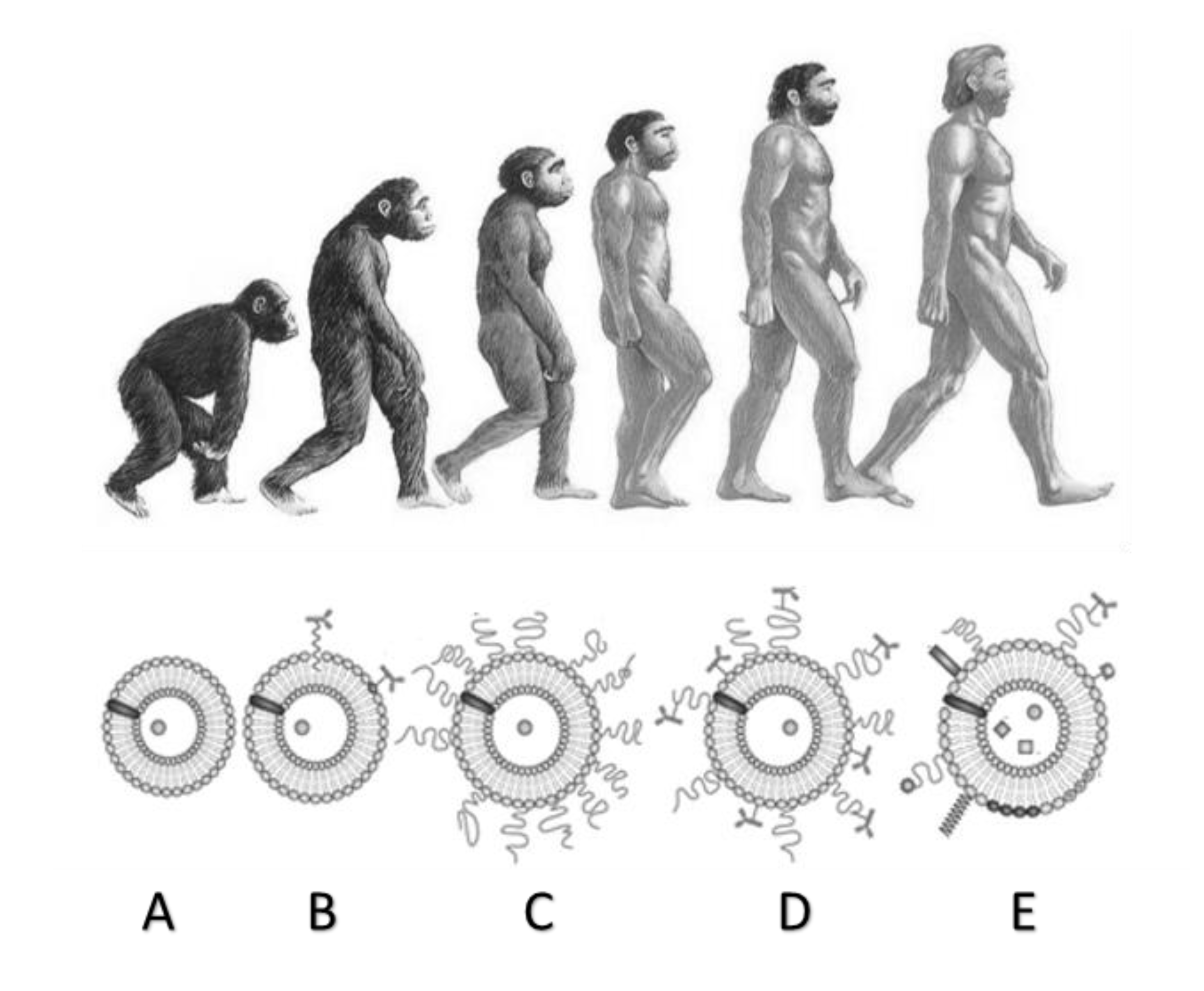
2. Liposomes: From History to Modern Times

Liposomes were first discovered by Dr. Alec D. Bangham, a hematologist of Cambridge Babraham Institute in 1965 [39,40], whose work was then followed by Dr. Gregoriadis’ studies [41,42]. Dr. Bangham understood that dried lipids rearrange spontaneously if put in contact with a sufficient amount of water, demonstrating that the spontaneous rearrangement of lipids is guided by unfavorable interactions between lipids and water which generate repulsion effects. This fact contributes to the distribution of amphiphilic molecules in the space, describing a spherical shape and minimizing the molecular interactions and Gibbs free energy of the system [43]. According to this explanation, a thin layer of lipids, when hydrated, starts to fold assuming a fire-balloon shape. At the end, the lipid layer closed, entrapping water in the inner core and assuming a spherical shape [44]. Therefore, Bangham defined liposomes as perfect thermodynamic models [45]. However, convinced by the scientific editor Gerald Weissmann to find a more suitable definition, the term liposomes was chosen, with it being made up of the two Greek words lipos (fat) and soma (body) [46]. Thanks to his open-mindedness, Bangham furthered his studies, producing interesting works that opened several fields of research with high industrial impacts. Nowadays, among a great variety of drug carrier systems, the field of liposomes is one of the fastest growing scientific topics worldwide. Nowadays, the literature about liposomes counts about 100 thousand papers; in particular, in the last ten years, more than 2000 papers per year were published (see Figure 1b).
Looking at Figure 1a, it is possible to see that the greatest progress in the field of research started from 1970, with particular attention to the problem of the physical and chemical stability of liposomes, connected to intravenous administration. In 1980, an important antitumor drug, doxorubicin, was encapsulated for the first time, in the context of the study of liposomes for therapeutic purposes [47,48,49].
Liposome morphologies and properties can be of fundamental importance, depending on the kind of application. Liposomes have been produced in different sizes and compositions, with different electrical charges and lamellarity, as shown in Table 1. Liposomes mean size can be controlled by different operating conditions; whereas, the lamellarity indicates the number of double layers of phospholipids that surround the inner aqueous core containing the hydrophilic drug.
Among these, single unilamellar vesicles (SUVs) are the most powerful vesicles since they can be employed in every kind of human tissue having nanometric interstices in which nanosomes can accumulate and be taken up by cells. Medium unilamellar vesicles (MVVs) could be used for the simultaneous entrapment of drugs with different targets.
Different kinds of phospholipids are generally used for the preparation of liposomes: phospholipids from natural sources, phospholipids from modified natural sources, semi-synthetic phospholipids, synthetic phospholipids, and phospholipids with modified head groups. Phosphatidylcholine (lecithin), phosphatidylethanolamine (cephalin) and phosphatidylserine are the most used for the preparation of liposomes [55]. Cholesterol can be added to the mixture to reduce the permeability of the lipid bilayer. This molecule plays a very important role in the behavior of liposomes. Cholesterol, in fact, is by its nature an amphoteric molecule, so it fits well in the double layer of the phospholipid structure. Alec Bangham already produced, with cholesterol, spherulites with a lipid layer 44.2 Å in thickness [56]. Other used phospholipids are phosphatidyl inositol, phosphatidyl glycerol, and lysophospholipids capable of losing a chain of fatty acids by chemical or enzymatic hydrolysis [31]. However, this class of lipids does not form membranes autonomously and, if implanted in large concentrations, they can damage the lipid bilayer, with toxicity problems for cells and organs [57]. In general, the hydrophobic tail is made up of two chains of fatty acids containing 10 to 24 carbon atoms and 0 to 6 double bonds in each chain. Phospholipids are therefore characterized by a glycerol bridge that connects the two hydrophobic tails with the hydrophilic polar head.
The primordial structure of the liposomes with a simple lipidic layer was then enriched, over time, with elements capable of adding or improving the functions of the administration or transmission of biochemical signals. The simple vesicle with a double lipid layer research moved on to decorated structures [58]. Scheme A of Figure 2 shows the first liposomes created in the laboratory, which were of a simpler nature. In these first spontaneous reorganizations of phospholipids, water-soluble drugs were encapsulated, trapped inside the aqueous core or in the double lipid layer. Scheme B shows the next step in the evolution of liposomes. In fact, liposomes’ surfaces have been modified to improve their properties. Long-circulating liposomes, the “grandchildren” of Bangham liposomes, are shown in Scheme C. They are characterized by a protective, inert and biocompatible polymer, such as polyethylene glycol (PEG), which preserves the surface of the vesicle from interactions with opsonizing proteins. In Scheme D, the long-circulating liposome possesses both a protective polymer and antibodies attached on the modified surface combining the effects of long-circulating liposomes with those of immunoliposomes. The antibody can be bonded both directly on the surface of the vesicles and at the end of the polymer bonded to the surface. The new generation of liposomes (Scheme E) is characterized by a surface that can be modified in various ways. These include the attachment of a protective polymer (with an antibody on the termination of the protective polymer), the possibility to fix a diagnostic label, the introduction of positively charged lipids, the introduction of stimuli-sensitive lipids, the introduction of stimuli-sensitive polymers, peptides penetrating cells, and viral components. In addition to the active ingredients already mentioned, magnetic particles or gold or silver particles can also be included in the new generation liposomes to carry out magnetic targeting and imaging.

3. Methods of Production

Liposomes joined the market with many difficulties, such as low drug encapsulation efficiencies, low process reproducibility and high production costs. These were the main problems of conventional methods developed from 1965 to 2000. Then, the necessity of strict production criteria related to size, zeta potential, morphology and stability was recognized. Supercritical-assisted technologies were proposed to overcome the problems previously found.

3.1. Conventional Methods

Conventional techniques generally consist of four main steps: the drying of lipids from organic solvents, dispersion of lipids in an aqueous medium, purification of obtained liposomes and eventual post-processing steps such as probe or bath sonication or extrusion.
Thin layer hydration (TLH), also known as the Bangham method [40], is considered to be the earliest liposome preparation method, performed at room temperature and pressure. According to TLH, lipids are first dissolved in a chloroform solution, and the hydrophilic drug is dissolved into an aqueous bulk. The organic solvent is evaporated under vacuum at 30 °C for 30 min, and a thin lipid layer is formed on the walls of the flask. The lipidic layer is then hydrated using the aqueous solution. After 1 h of contact, liposomes are formed. This method produces liposomes of 10–100 µm, not easily absorbable by human cells; for this reason, TLH is coupled with sonication to obtain smaller mean dimensions. The encapsulation efficiency (EE) is as low as 20–30% and the shape of the liposome is not always spherical.
The ethanol injection (EI) method involves the dissolution of lipids into an organic phase, followed by a dropwise injection of the lipid solution into an aqueous media [59]. A needle is used to inject the lipidic solution into the water bulk containing a hydrophilic drug. This method is simple because it works in absence of pressure, but a high solvent residue is present in the final suspension, and the removal of ethanol is difficult since it forms an azeotropic mixture in direct contact with water. Moreover, the evaporation step risks damaging the vesicle structure and altering its dimensions. Therefore, the liposome population is generally heterogeneous and its particle size distribution is difficult to control. The EE is lower than 20% for hydrophilic compounds and around 60% for lipophilic molecules.
The emulsion method implies that phospholipids are dissolved into an organic solvent and mixed with a solution to form a water in oil (W/O) emulsion [60]. The mixture is then added to another water solution to form a double emulsion w/o/w. The organic solvent is then evaporated, generating liposomal aqueous suspension. This method does not work continuously, and the production volumes are limited. Micrometric and multivesicular liposomes are obtained in this way; but, large amounts of organic solvent need to be eliminated.
Reverse phase evaporation (RPE) is based on the formation of water droplets surrounded by lipids and dispersed in an organic solvent to form a water in oil emulsion [61]. Since phospholipids behave as surfactants, they rearrange at the interface between oil and water, creating inverted micelles. Then, the solvent is eliminated slowly and the micelles are converted into a gel form. The gel state collapses and the excess of phospholipids in the environment creates a second layer of lipids around the first one. The formation of a complete bilayer around the residual micelles results in the formation of liposomes. However, long-lasting contact between drug and organic solvent can lead to drug denaturation in the case of fragile compounds such as proteins and peptides. An EE among 30 and 65% is obtained using this technique [62].
Freeze drying consists of the formation of a dispersion of lipids in water-soluble carrier materials [63]. The first step is the dissolution of the lipids in tert-butyl alcohol and water–sucrose to form an isotropic monophase solution; then, the freeze dying of the solution is performed. Freeze drying is characterized by two main steps—first, the sample is frozen at −40 °C and then dried at room temperature. Upon adding water, the lyophilized system spontaneously forms lipidic spherical vesicles. The presence of sucrose can prevent the formation of aggregated minidomains during the drying step. The mean dimensions of liposomes are 100–300 nm; the EE is 40–60%, but the time stability is lower than 60 days.
The microfluidic channel method consists of the development of a rigid silicon support in which microfluidic channels of 100 µm in depth have been previously designed and then 3D-printed, with a low stability due to the aggregation tendency [64]. The production cost of the micro-circuit is higher than other conventional methods. A lipidic solution is injected into the central feeding channel, whereas aqueous solutions are fed from the side channels, which intersect the main channel at the center of the support. In the outer channels, liposomes are created. Monodisperse nanometric suspensions are practically obtained, optimizing the water to lipid ratio. This method suffers high solvent residue levels in the final liposomes suspensions and a difficulty in reaching high production levels.
The heating method does not involve the use of organic solvents [65]. Phospholipids are added to the aqueous solution together with 3% v/v glycerol, working at a bath temperature of 120 °C. Glycerol is a water-soluble and isotonic agent able to increase vesicles’ stability, preventing the coagulation phenomena and assuring sterilization. Produced liposomes have nanometric dimensions and a long-term stability (12–14 months) but a low EE (12–54%).

3.2. Supercritical-Assisted Methods

After the diffusion of green chemistry concepts in 1990s, the interest in supercritical fluids (SCFs) grew exponentially—new technologies were developed, transferring academics’ knowledge to industrial enterprises for vesicle productions at a large scale. These methods have been proposed trying to take advantage of the enhanced mass transfer coefficient of SCF. However, working at high pressures involves an increase in the equipment cost [66].
According to the depressurization of an expanded solution into an aqueous medium (DESAM), lipids are first dissolved in ethanol; then, working at of 35–55 bar, the solution is expanded into CO2, injected into a water bath in a vessel and left bubbling for 1 h at 75 °C [67]. In these conditions, part of ethanol is evaporated. Lipids transported by bubbles and water containing drugs are put in contact, producing liposomes. The ethanol content is reduced to 2.2% v/v, which is still too high to guarantee the safety of the drug carrier. The nanometric dimensions of the liposomes can be obtained with a good stability (8 months).
The supercritical fluid method [68] is characterized by two parts: the high-pressure part is used to dissolve lipids and cholesterol in supercritical carbon dioxide at the pressure of 250 bar; then, in the following step, the homogeneous supercritical solution is expanded with the addition of 7% v/v ethanol at the temperature of 60 °C. The expanded liquid is then mixed with a water phase to obtain liposomes encapsulating water-soluble compounds. Only 3% of the vesicles are subjected to degradation and the mean diameter is about 200 nm, whereas the particle size distribution is generally bimodal. The total amount of ethanol content is 15 times lower than it is in the ethanol injection method.
According to supercritical antisolvents (SAS), the organic solvent containing phospholipids is spayed continuously in supercritical CO2, acting as antisolvent [69]. The dissolution of supercritical CO2 in the liquid phase and the subsequent extraction of the organic solvent leads to the precipitation of the lipidic particles. A washing step with pure CO2 is performed to remove any organic solvent in excess. The particles are hydrated in an aqueous buffer to obtain liposomes at the nanometric or micrometric level, depending on the kind of application needed. The encapsulation efficiency is similar to the TLH method, but the sphericity of the lipidic particles is improved. The particle size distribution is more controlled and repeatable.
In the rapid expansion of a supercritical solution (RESS), lipids are dissolved in supercritical CO2 plus 5–10% of v/v ethanol [70]. The solution is then released through a small orifice and mixed with an aqueous solution containing the drug. A rapid depressurization follows, and the pressure drop results in the lipids’ desolvation, forming layers around the droplets. However, this method shows problems such as the difficult separation between vesicles and co-solvents during depressurization. Even if the mean diameters of liposomes are 130–190 nm and the particle size distributions are monodispersed, the EEs are still lower than 60%.
Supercritical reverse phase evaporation (SCRPE) is similar to the conventional-related method [61], but this technique differs in the use of supercritical CO2 in substitution of the organic solvent. Lipids are put in contact with supercritical CO2 in a view cell in batch mode, working at 60 °C and 10–30 bar. After reaching the equilibrium, an aqueous solution including glucose is slowly inserted into the vessel, obtaining large unilamellar vesicles. Liposomes produced in this manner have a mean diameter between 200 and 1200 nm. With a decreasing lipid concentration, the mean size decreases to 100–250 nm.
Particles from gas-saturated solutions (PGSS) consist of dissolving a bioactive compound in an organic solvent; then, a supercritical carbon dioxide is introduced into the solution [70,71]. After saturation is reached, this solution is sprayed through a nozzle into a high-pressure vessel. Liposomes are formed and a rapid depressurization of CO2 eliminates the solvent, leaving supercritical conditions. The saturation temperature is 100–130 °C and the pressure is 80–100 bar. The liposomes produced have a mean diameter of 1–5 µm and an EE of about 60%.
Liposomes can be prepared using the supercritical-assisted liposome formation (SuperLip) [72]. It consists of three different feeding lines for the delivery of CO2, water and ethanol–phospholipid solutions, respectively [73]. The ethanol solution and CO2 are continuously fed to a homogenizer forming an expanded liquid, which is then delivered to a precipitation vessel. In the same vessel, the water + drug droplets are atomized; liposomes are created since the lipids capture water droplets forming a double lipidic layer around. SuperLip successfully demonstrated that it could overcome the drawbacks of the other methods [74]. Liposomes of nanometric dimensions were obtained, with a better control of the particle size distributions and a significant reduction in the solvent residue [75]. A high biocompatibility of the vesicles was guaranteed for biomedical applications, achieving encapsulation efficiencies of up to 99% for hydrophilic compounds [76].
A comparison of the main advantages of the SuperLip process against the major drawbacks of the other production techniques is reported in Table 2.

4. Characterization of Liposomes

Liposomes need to be characterized before administration [8]. For this reason, quality controls must be performed after production [31], according to several strict characterization protocols [12]. Liposome suspensions can be translucent or milky, depending on the size of vesicles and concentration of lipids. For this reason, the first analysis to perform is the visual appearance, to define the turbidity of the sample.
The second step consists of measuring the dimension in terms of the mean size and standard deviation of the liposomes produced, which is performed using dynamic light scattering (DLS). The liposome’s size also determines the extravasation, meaning the leakage of fluids from blood streams. Moreover, liposomes considered for inhalation need a strict control for the mean dimension, since it influences the in vivo fate of liposomes together with the encapsulated drug. Small-sized liposomes resulted in an increased blood circulation ability [77]. Another method to characterize liposomes’ dimensions is nanoparticle tracking analysis (NTA), which exploits the properties of light scattering and the Brownian motion of colloidal particles in a background medium. Every moving element is detected with a laser and appears as a white spot followed in its motion.
The lamellarity represents the number of double lipidic layers of the vesicles observed. This analysis contributes to classify liposomes (see Table 1) as well as determines their potential application and administration efficacy. This operation is performed using a transmission electron microscope (TEM). The thickness of each double layer is approximately 5 nm [68].
The surface charge is generally used to predict the colloidal stability of liposomes suspended in the external medium. This measurement, named ζ-potential, is performed using dynamic light scattering [78], and defines liposomes’ intramolecular interactions. According to this value, liposomes are classified as cationic [53], anionic or neutral [46]. The particle net surface charges affect the distribution of ions within the external double lipidic layer of liposomes, determining their affinity with entrapped drugs [79].
The morphology and shape are detected using scanning electron microscopy (SEM), which also gives a confirmation of liposomes’ sizes and distributions (see Figure 3).
Particularly, TEMs can be used to observe liposome sections to check the circularity of vesicles. The encapsulation efficiency (EE) is the percentage of active compound that is effectively entrapped in liposomes’ inner volumes; of course, its complement to 100 represents a not-entrapped compound, that is lost in the aqueous external bulk. This amount of drug does not take part in drug administration; for this reason, it is necessary to entrap as much of the drug as possible to reduce the loss of the active principle. This feature is taken into particular consideration when administering highly expensive drugs. The most direct method for measuring the EE consists of centrifuging liposome suspensions and separate supernatants from the vesicles. Then, the lipidic layers are dissolved in an organic solvent [80]; the absorbance of the amount of drug diffused in the solvent is measured using an ultraviolet-visible (UV-Vis) spectrophotometer. The EE is calculated by Equation (1):
EE   [ % ] =   Entrapped   Drug   Concentration Total   Drug   Concentration × 100
The stability of liposomes is measured, controlling for their mean size, using a scanning electron microscope and encapsulation efficiency tests during fixed time intervals. The storage temperature mostly affects the stability of lipidic vesicles: the particles stored at room temperature degradate, whereas a storage at −20 °C results in an increase in the mean diameter and polydispersity index. A refrigerator temperature (2–4 °C) maintains liposomes’ properties which are unaltered for months [81].

5. Natural and Artificial Release Mechanisms

Liposomes’ release mechanisms can be natural or artificial. The natural mechanism is strictly linked to the similarity between the vesicle and the cell membranes. Liposomes are attracted to the cell membrane and become part of the cellular barrier. For this reason, the drug can arrive directly inside the cell cytoplasm.
Liposome drug delivery can be performed through oral administration, skin penetration and systemic delivery. Liposomes administrated per os are highly unstable since the vesicles are subjected to the physiological conditions of the human gastrointestinal tract (GI) [82,83], resulting in a low drug bioavailability and short half-life, which leads to the necessity of multiple administrations per day [84]. Skin penetration is one of the most effective methods for drug delivery using liposomes, and it can be applied to most parts of hydrophilic and lipophilic compounds [85]. An intramuscular and subcutaneous administration (systemic) represents a direct method for liposomal drug delivery—in this case, SUV liposomes show a fast diffusion into the lymphatic capillaries, whereas, larger liposomes remain confined in the site of injection [86]. Moreover, the lymphatic system is highly exploited by tumor cells for the creation of secondary tumors [87]; for this reason, the subcutaneous injection of antitumoral drugs mediated by liposomes could be effective for these pathologies [33].
One of the most important aims of pharmaceutical industries is to increase the drug therapeutic index, minimizing side effects to normal tissues. This is extremely important for chemotherapy, which involves tissue treatment with very potent drugs. Normally, only 1% of the intravenously administered drug effectively reaches tumor tissue; the remaining part is dispersed throughout the whole body. The solution to this problem consists of the creation of “intelligent” liposomes, that can circulate in the blood streams for a longer time [26].
Artificial release mechanisms are induced by external stimuli; for example, a sudden increase in temperature. Some lipids are sensitive to temperature variation and can be activated even with a slight increase (e.g., 0.1 °C). Working with temperature-sensitive liposomes, the lipidic membrane can open and release the entrapped drug. In this way, it is possible to activate and stop release according to patient necessities.
Another external stimulus is the pH variation, since the formation of necrotic tissues due to the presence of cancer leads to a different pH value of the cellular environment [88]. In fact, pH-sensitive liposomes can accumulate in the site of action and release their content directly on the necrotic surface tissues. Artificial releases are also induced by ultrasound stimuli. This can induce release by the opening of the lipidic barrier.
It is possible to deliver a liposome in which a drug and triggering agent, such as hollow gold nanoshells (HGN), have been entrapped. HGNs are reactive to near-infrared radiation [21], i.e., when they are reached by this stimulus (trigger), the liposome double layer opens and releases the drug. When the irradiation is stopped, the structure closes and the drug is no longer released (pulsed drug release).
Recently, the fabrication of the surface-modified lipidic vesicles was also proposed, decorating the external surfaces with labels such as peptides [89], opsonines, antibodies [90] or polymer fragments [91] to obtain a specific long-lasting drug release to target tissues, creating special bindings with cell receptors [30].
One of the most important drawbacks of liposomes is their fast elimination from blood, since they are captured by the reticuloendothelial system tissues: the first of them being the liver. Liposomes have been improved to overcome this problem. Immunoliposomes are vesicles with a modified surface programmed to be digested by macrophages [92]. This plays a fundamental role in keeping human tissues clean. Since artificial liposomes are recognized to be external elements, macrophages eliminate them. For this reason, liposomes were used as Trojan horses to be digested by macrophages. During the digestion, the vesicles are dissolved and the drug is transferred to the target tissues by macrophages, exploiting the immune response of the human body. Long circulating immunoliposomes are able to recognize and blind target cells with great specificity, especially in anticancer therapies [6].
Long-circulating liposomes can be prepared by coating their surface with polyethylene glycol (PEG). PEG filaments improve the drug distribution in target tissues, avoiding vesicle aggregation phenomena and improving their stability [93]. The PEG-link improves liposomes’ half-lives, resulting in longer circulating times in humans. Long circulating vesicles can detect occult inflammations, demonstrating a higher potential than the alternative DDS [15,32].

6. Conclusions

Liposomes are produced on an industrial level in several manufacturing fields. The earliest methods of production were characterized by a very low average encapsulation efficiency (around 20–40%), resulting in drug loss and an increase in the operating costs. The second main problem was the high solvent residue, which resulted in a low biocompatibility with human tissues. The decision to work at an ambient pressure and temperature resulted in the necessity to add post-processing steps, to obtain the nanometric dimensions of vesicles. These methods were essentially developed in batch mode, causing a low repeatability.
The aim of supercritical methods was to work at the nanometric level in a repeatable manner; for this reason, (semi)continuous methods were proposed. Conventional solvents were partially or totally substituted with supercritical carbon dioxide, resulting in the increase in biocompatibility for drug administration. Post-processing steps were eliminated in almost all cases, obtaining techniques able to produce vesicles in one-shot, working at high pressures (70–100 bar) and mild temperatures (40–60 °C). For these reasons, high pressure methods were considered more efficient processes for the large scale production of lipidic vesicles.
SuperLip successfully demonstrated that it could overcome the limitations linked to the traditional production methods of liposomes. By exploiting the high diffusion coefficient of supercritical carbon dioxide, liposomes of nanometric dimensions were obtained, with a better control of the particle size distributions and encapsulation efficiencies of drugs higher than 90%.

Author Contributions

Conceptualization, P.T. and R.C.; Investigation, R.C. and E.R.; Data Curation, P.T.; Writing—Original Draft Preparation, P.T.; Writing—Review and Editing, R.C. and E.R. All authors have read and agreed to the published version of the manuscript.

Funding

This research received no external funding.

Conflicts of Interest

The authors declare no conflict of interest.

Abbreviations

DSDelivery Systems
DDSDrug Delivery Systems
LOMLipid Oxygen Containing Microparticles
SUVSingle Unilamellar Vesicles
MUVMedium Unilamellar Vesicles
LUVLarge Unilamellar Vesicles
GUVGiant Unilamellar Vesicles
OLVOligo Lamellar Vesicles
MLVMulti Lamellar Vesicles
MVVMulti Vesicular Vesicles
NTANanoparticle Tracking Analysis
SEMScanning Electron Microscope
EEEncapsulation Efficiency
UV-VisUltraviolet-Visible
GIGastrointestinal Tract
HGNHollow Gold Nanoshells
MagMagnification
PEGPolyethylene Glycol
TLHThin Layer Hydration
RPEReverse Phase Evaporation
DESAMDepressurization of an Expanded Solution into an Aqueous Medium
EIEthanol Injection
W/OWater in Oil emulsion
SCFSuperCritical Fluids
SCRPESuperCritical Reverse Phase Evaporation
SASSuperctitical Anti-Solvent
SuperLipSupercritical-Assisted Liposome Formation
RESSRapid Expansion of a Supercritical Solution
PGSSParticles from Gas-Saturated Solutions
PDIPolydispersity Index

References

  1. Martinho, N. Recent Advances in Drug Delivery Systems. J. Biomater. Nanobiotechnol. 2011, 02, 510–526. [Google Scholar] [CrossRef] [Green Version]
  2. Kaur, I.P.; Bhandari, R.; Bhandari, S.; Kakkar, V. Potential of solid lipid nanoparticles in brain targeting. J. Control. Release 2008, 127, 97–109. [Google Scholar] [CrossRef] [PubMed]
  3. Hoffman, A.S. Hydrogels for biomedical applications. Adv. Drug Deliv. Rev. 2012, 64, 18–23. [Google Scholar] [CrossRef]
  4. Lu, G.W.; Gao, P. Emulsions and Microemulsions for Topical and Transdermal Drug Delivery. In Handbook of Non-Invasive Drug Delivery Systems; William Andrew Publishing: Norwich, NY, USA, 2010; pp. 59–94. [Google Scholar]
  5. Peetla, C.; Stine, A.; Labhasetwar, V. Biophysical Interactions with Model Lipid Membranes: Applications in Drug Discovery and Drug Delivery. Mol. Pharm. 2009, 6, 1264–1276. [Google Scholar] [CrossRef]
  6. Jain, N.K.; Mishra, V.; Mehra, N.K. Targeted drug delivery to macrophages. Expert Opin. Drug Deliv. 2013, 10, 353–367. [Google Scholar] [CrossRef]
  7. Dufès, C.; Uchegbu, I.F.; Schätzlein, A.G. Dendrimers in gene delivery. Adv. Drug Deliv. Rev. 2005, 57, 2177–2202. [Google Scholar] [CrossRef] [PubMed] [Green Version]
  8. Shaheen, S.M.; Shakil Ahmed, F.; Hossen, M.N.; Ahmed, M.; Amran, M.S.; Ul-Islam, M. Liposome as a carrier for advanced drug delivery. Pak. J. Biol. Sci. 2006, 9, 1181–1191. [Google Scholar]
  9. Lian, T.; Ho, R.J.Y. Trends and developments in liposome drug delivery systems. J. Pharm. Sci. 2001, 90, 667–680. [Google Scholar] [CrossRef]
  10. Li, J.; Wang, X.; Zhang, T.; Wang, C.; Huang, Z.; Luo, X.; Deng, Y. A review on phospholipids and their main applications in drug delivery systems. Asian J. Pharm. Sci. 2015, 10, 81–98. [Google Scholar] [CrossRef]
  11. Bangham, A.D. Review of Lasic, Liposomes: From Physics to Applications. Biophys. J. 1994, 67, 1358–1359. [Google Scholar] [CrossRef] [Green Version]
  12. Perkins, W.R.; Minchey, S.R.; Ahl, P.L.; Janoff, A.S. The determination of liposome captured volume. Chem. Phys. Lipids 1993, 64, 197–217. [Google Scholar] [CrossRef]
  13. Abed, N.; Couvreur, P. Nanocarriers for antibiotics: A promising solution to treat intracellular bacterial infections. Int. J. Antimicrob. Agents 2014, 43, 485–496. [Google Scholar] [CrossRef] [PubMed]
  14. Colletier, J.-P.; Chaize, B.; Winterhalter, M.; Fournier, D. Protein encapsulation in liposomes: Efficiency depends on interactions between protein and phospholipid bilayer. BMC Biotechnol. 2002, 2, 9. [Google Scholar] [CrossRef] [PubMed] [Green Version]
  15. Crommelin, D.J.A.; van Rensen, A.J.M.L.; Wauben, M.H.M.; Storm, G. Liposomes in autoimmune diseases: Selected applications in immunotherapy and inflammation detection. J. Control. Release 1999, 62, 245–251. [Google Scholar] [CrossRef]
  16. Barani, H.; Montazer, M. A Review on Applications of Liposomes in Textile Processing. J. Liposome Res. 2008, 18, 249–262. [Google Scholar] [CrossRef]
  17. Can, S.; Giuseppina, S.; Vladimir, P.T. Recent advances in siRNA delivery. Biomol. Concepts 2015, 6, 321–341. [Google Scholar]
  18. Paiva-Martins, F.; Gordon, M.H.; Gameiro, P. Activity and location of olive oil phenolic antioxidants in liposomes. Chem. Phys. Lipids 2003, 124, 23–36. [Google Scholar] [CrossRef]
  19. Keller, B.C. Liposomes in Nutrition. Trends Food Sci. Technol. 2001, 12, 25–31. [Google Scholar] [CrossRef]
  20. Kluczyk, D.; Matwijczuk, A.; Górecki, A.; Karpińska, M.M.; Szymanek, M.; Niewiadomy, A.; Gagoś, M. Molecular Organization of Dipalmitoylphosphatidylcholine Bilayers Containing Bioactive Compounds 4-(5-Heptyl-1,3,4-thiadiazol-2-yl) Benzene-1,3-diol and 4-(5-Methyl-1,3,4-thiadiazol-2-yl) Benzene-1,3-diols. J. Phys. Chem. B 2016, 120, 12047–12063. [Google Scholar] [CrossRef]
  21. Tila, D.; Ghasemi, S.; Yazdani-Arazi, S.N.; Ghanbarzadeh, S. Functional liposomes in the cancer-targeted drug delivery. J. Biomater. Appl. 2015, 30, 3–16. [Google Scholar] [CrossRef]
  22. Lasic, D.D. Novel applications of liposomes. Trends Biotechnol. 1998, 16, 307–321. [Google Scholar] [CrossRef]
  23. Gibbs, B.F.; Kermasha, S.; Alli, I.; Mulligan, C.N. Encapsulation in the food industry: A review. Int. J. Food Sci. Nutr. 1999, 50, 213–224. [Google Scholar] [PubMed]
  24. Ozkan, G.; Franco, P.; De Marco, I.; Xiao, J.; Capanoglu, E. A review of microencapsulation methods for food antioxidants: Principles, advantages, drawbacks and applications. Food Chem. 2019, 272, 494–506. [Google Scholar] [CrossRef] [PubMed]
  25. Laouini, A.; Jaafar-Maalej, C.; Blouza, I.; Sfar, S.; Charcosset, C.; Fessi, H. Preparation, Characterization and Applications of Liposomes: State of the Art. J. Colloid Sci. Biotechnol. 2012, 1, 147–168. [Google Scholar] [CrossRef]
  26. Voinea, M.; Simionescu, M. Designing of ‘intelligent’ liposomes for efficient delivery of drugs. J. Cell. Mol. Med. 2002, 6, 465–474. [Google Scholar] [CrossRef] [Green Version]
  27. da Silva Malheiros, P.; Daroit, D.J.; Brandelli, A. Food applications of liposome-encapsulated antimicrobial peptides. Trends Food Sci. Technol. 2010, 21, 284–292. [Google Scholar] [CrossRef]
  28. Kaur, I.P.; Kakkar, S. Topical delivery of antifungal agents. Expert Opin. Drug Deliv. 2010, 7, 1303–1327. [Google Scholar] [CrossRef]
  29. Sharma, D.; Aara, A.; Ali, E.; Trivedi, L. An Updated Review On:Liposomes as Drug Delivery System. Pharmatutor 2018, 6, 50–62. [Google Scholar] [CrossRef]
  30. Karanth, H.; Murthy, R.S.R. pH-Sensitive liposomes-principle and application in cancer therapy. J. Pharm. Pharmacol. 2007, 59, 469–483. [Google Scholar] [CrossRef]
  31. Anwekar, H.; Patel, S.; Singhai, A. Liposome-as Drug Carriers. Int. J. Pharm. Life Sci. 2011, 2, 945–951. [Google Scholar]
  32. Immordino, M.L.; Dosio, F.; Cattel, L. Stealth liposomes: Review of the basic science, rationale, and clinical applications, existing and potential. Int. J. Nanomed. 2006, 1, 297–315. [Google Scholar]
  33. Oussoren, C.; Storm, G. Liposomes to target the lymphatics by subcutaneous administration. Adv. Drug Deliv. Rev. 2001, 50, 143–156. [Google Scholar] [CrossRef]
  34. Ahmed, K.S.; Hussein, S.A.; Ali, A.H.; Korma, S.A.; Lipeng, Q.; Jinghua, C. Liposome: Composition, characterisation, preparation, and recent innovation in clinical applications. J. Drug Target. 2019, 27, 742–761. [Google Scholar] [CrossRef] [PubMed]
  35. Lundberg, B.B.; Griffiths, G.; Hansen, H.J. Specific binding of sterically stabilized anti-B-cell immunoliposomes and cytotoxicity of entrapped doxorubicin. Int. J. Pharm. 2000, 205, 101–108. [Google Scholar] [CrossRef]
  36. Dutta, S.; Moses, J.A.; Anandharamakrishnan, C. Encapsulation of Nutraceutical Ingredients in Liposomes and Their Potential for Cancer Treatment. Nutr. Cancer 2018, 70, 1184–1198. [Google Scholar] [CrossRef]
  37. Illes, B.; Wuttke, S.; Engelke, H. Liposome-Coated Iron Fumarate Metal-Organic Framework Nanoparticles for Combination Therapy. Nanomaterials 2017, 7, 351. [Google Scholar] [CrossRef]
  38. Tao, Z.; Ghoroghchian, P.P. Microparticle, nanoparticle, and stem cell-based oxygen carriers as advanced blood substitutes. Trends Biotechnol. 2014, 32, 466–473. [Google Scholar] [CrossRef]
  39. Bangham, A.D. Surrogate cells or Trojan horses. The discovery of liposomes. BioEssays News Rev. Mol. Cell. Dev. Biol. 1995, 17, 1081–1088. [Google Scholar] [CrossRef]
  40. Meure, L.A.; Foster, N.R.; Dehghani, F. Conventional and Dense Gas Techniques for the Production of Liposomes: A Review. AAPS PharmSciTech 2008, 9, 798. [Google Scholar] [CrossRef] [Green Version]
  41. Gregoriadis, G. Immunological adjuvants: A role for liposomes. Immunol. Today 1990, 11, 89–97. [Google Scholar] [CrossRef]
  42. Gregoriadis, G.; Wills, E.J.; Swain, C.P.; Tavill, A.S. Drug-carrier potential of liposomes in cancer chemotherapy. Lancet 1974, 1, 1313–1316. [Google Scholar] [CrossRef]
  43. Sopyan, I.; Gozali, D. A Review: A Novel of Efforts to Enhance Liposome Stability as Drug Delivery Approach. Syst. Rev. Pharm. 2020, 11, 555–562. [Google Scholar]
  44. Bozzuto, G.; Molinari, A. Liposomes as nanomedical devices. Int. J. Nanomed. 2015, 10, 975–999. [Google Scholar] [CrossRef] [PubMed] [Green Version]
  45. Bangham, A.D. Liposomes and the physico-chemical basis of unconsciousness. FASEB J. 2005, 19, 1766–1768. [Google Scholar] [CrossRef]
  46. Patil, Y.P.; Jadhav, S. Novel methods for liposome preparation. Chem. Phys. Lipids 2014, 177, 8–18. [Google Scholar] [CrossRef]
  47. Herman, E.H.; Rahman, A.; Ferrans, V.J.; Vick, J.A.; Schein, P.S. Prevention of Chronic Doxorubicin Cardiotoxicity in Beagles by Liposomal Encapsulation. Cancer Res. 1983, 43, 5427–5432. [Google Scholar]
  48. Balazsovits, J.A.E.; Mayer, L.D.; Bally, M.B.; Cullis, P.R.; McDonell, M.; Ginsberg, R.S.; Falk, R.E. Analysis of the effect of liposome encapsulation on the vesicant properties, acute and cardiac toxicities, and antitumor efficacy of doxorubicin. Cancer Chemother. Pharmacol. 1989, 23, 81–86. [Google Scholar] [CrossRef]
  49. Bally, M.B.; Nayar, R.; Masin, D.; Hope, M.J.; Cullis, P.R.; Mayer, L.D. Liposomes with entrapped doxorubicin exhibit extended blood residence times. Biochim. Biophys. Acta (BBA) Biomembr. 1990, 1023, 133–139. [Google Scholar] [CrossRef]
  50. Fu, M.; Tang, W.; Liu, J.-J.; Gong, X.-Q.; Kong, L.; Yao, X.-M.; Jing, M.; Cai, F.-Y.; Li, X.-T.; Ju, R.-J. Combination of targeted daunorubicin liposomes and targeted emodin liposomes for treatment of invasive breast cancer. J. Drug Target. 2020, 28, 245–258. [Google Scholar] [CrossRef]
  51. Laouini, A.; Jaafar-Maalej, C.; Sfar, S.; Charcosset, C.; Fessi, H. Liposome preparation using a hollow fiber membrane contactor--application to spironolactone encapsulation. Int. J. Pharm. 2011, 415, 53–61. [Google Scholar] [CrossRef]
  52. Liu, Y.; Kim, Y.J.; Siriwon, N.; Rohrs, J.A.; Yu, Z.; Wanga, P. Combination drug delivery via multilamellar vesicles enables targeting of tumor cells and tumor vasculature. Biotechnol. Bioeng. 2018, 115, 1403–1415. [Google Scholar] [CrossRef] [PubMed]
  53. Barenholz, Y.; Bombelli, C.; Bonicelli, M.G.; di Profio, P.; Giansanti, L.; Mancini, G.; Pascale, F. Influence of lipid composition on the thermotropic behavior and size distribution of mixed cationic liposomes. J. Colloid Interface Sci. 2011, 356, 46–53. [Google Scholar] [CrossRef] [PubMed]
  54. Walde, P.; Cosentino, K.; Engel, H.; Stano, P. Giant Vesicles: Preparations and Applications. ChemBioChem 2010, 11, 848–865. [Google Scholar] [CrossRef] [PubMed]
  55. Guráň, R.; Kominkova, M.; Kopel, P.; Chudobova, D.; Zitka, O.; Adam, V.; Kizek, R. Liposomes as Drug Carriers and Their Characterization Using Different Analytical Methods. Ph.D. Thesis, Mendel University, Brno, Czech Republic, 2013. [Google Scholar]
  56. Duzgunes, N.; Gregoriadis, G. Introduction: The Origins of Liposomes: Alec Bangham at Babraham. Methods Enzymol. 2005, 391, 1–3. [Google Scholar]
  57. Gregoriadis, G. Overview of liposomes. J. Antimicrob. Chemother. 1991, 28 (Suppl. B), 39–48. [Google Scholar] [CrossRef]
  58. Torchilin, V.P. Recent advances with liposomes as pharmaceutical carriers. Nat. Rev. Drug Discov. 2005, 4, 145–160. [Google Scholar] [CrossRef]
  59. Jaafar-Maalej, C.; Diab, R.; Andrieu, V.; Elaissari, A.; Fessi, H. Ethanol injection method for hydrophilic and lipophilic drug-loaded liposome preparation. J. Liposome Res. 2010, 20, 228–243. [Google Scholar] [CrossRef]
  60. Otake, K.; Imura, T.; Sakai, H.; Abe, M. Development of a New Preparation Method of Liposomes Using Supercritical Carbon Dioxide. Langmuir 2001, 17, 3898–3901. [Google Scholar] [CrossRef]
  61. Imura, T.; Otake, K.; Hashimoto, S.; Gotoh, T.; Yuasa, M.; Yokoyama, S.; Sakai, H.; Rathman, J.F.; Abe, M. Preparation and physicochemical properties of various soybean lecithin liposomes using supercritical reverse phase evaporation method. Coll. Surf. B Biointerfaces 2003, 27, 133–140. [Google Scholar] [CrossRef]
  62. Szoka, F.; Papahadjopoulos, D. Procedure for preparation of liposomes with large internal aqueous space and high capture by reverse-phase evaporation. Proc. Natl. Acad. Sci. USA 1978, 75, 4194–4198. [Google Scholar] [CrossRef] [Green Version]
  63. Li, C.; Deng, Y. A novel method for the preparation of liposomes: Freeze drying of monophase solutions. J. Pharm. Sci. 2004, 93, 1403–1414. [Google Scholar] [CrossRef] [PubMed]
  64. Jahn, A.; Vreeland, W.N.; DeVoe, D.L.; Locascio, L.E.; Gaitan, M. Microfluidic Directed Formation of Liposomes of Controlled Size. Langmuir 2007, 23, 6289–6293. [Google Scholar] [CrossRef] [PubMed]
  65. Mozafari, M.R. Liposomes: An overview of manufacturing techniques. Cell. Mol. Biol. Lett. 2005, 10, 711–719. [Google Scholar] [PubMed]
  66. Campardelli, R.; Baldino, L.; Reverchon, E. Supercritical fluids applications in nanomedicine. J. Supercrit. Fluids 2015, 101, 193–214. [Google Scholar] [CrossRef]
  67. Meure, L.A.; Knott, R.; Foster, N.R.; Dehghani, F. The Depressurization of an Expanded Solution into Aqueous Media for the Bulk Production of Liposomes. Langmuir 2009, 25, 326–337. [Google Scholar] [CrossRef]
  68. Tsai, W.-C.; Rizvi, S.S.H. Liposomal microencapsulation using the conventional methods and novel supercritical fluid processes. Trends Food Sci. Technol. 2016, 55, 61–71. [Google Scholar] [CrossRef]
  69. Lesoin, L.; Crampon, C.; Boutin, O.; Badens, E. Preparation of liposomes using the supercritical anti-solvent (SAS) process and comparison with a conventional method. J. Supercrit. Fluids 2011, 57, 162–174. [Google Scholar] [CrossRef]
  70. Soh, S.H.; Lee, L.Y. Microencapsulation and Nanoencapsulation Using Supercritical Fluid (SCF) Techniques. Pharmaceutics 2019, 11, 21. [Google Scholar] [CrossRef] [Green Version]
  71. de Paz, E.; Martín, Á.; Cocero, M.J. Formulation of β-carotene with soybean lecithin by PGSS (Particles from Gas Saturated Solutions)-drying. J. Supercrit. Fluids 2012, 72, 125–133. [Google Scholar] [CrossRef]
  72. Trucillo, P.; Campardelli, R.; Reverchon, E. Supercritical CO2 assisted liposomes formation: Optimization of the lipidic layer for an efficient hydrophilic drug loading. J. CO2 Util. 2017, 18, 181–188. [Google Scholar] [CrossRef]
  73. Campardelli, R.; Trucillo, P.; Reverchon, E. Supercritical assisted process for the efficient production of liposomes containing antibiotics for ocular delivery. J. CO2 Util. 2018, 25, 235–241. [Google Scholar] [CrossRef]
  74. Trucillo, P.; Campardelli, R.; Reverchon, E. A versatile supercritical assisted process for the one-shot production of liposomes. J. Supercrit. Fluids 2019, 146, 136–143. [Google Scholar] [CrossRef]
  75. Trucillo, P.; Campardelli, R.; Scognamiglio, M.; Reverchon, E. Control of liposomes diameter at micrometric and nanometric level using a supercritical assisted technique. J. CO2 Util. 2019, 32, 119–127. [Google Scholar] [CrossRef]
  76. Trucillo, P.; Campardelli, R.; Reverchon, E. Production of liposomes loaded with antioxidants using a supercritical CO2 assisted process. Powder Technol. 2018, 323, 155–162. [Google Scholar] [CrossRef]
  77. Woodle, M.C.; Lasic, D.D. Sterically stabilized liposomes. Biochim. Biophys. Acta (BBA) Rev. Biomembr. 1992, 1113, 171–199. [Google Scholar] [CrossRef]
  78. Gibis, M.; Zeeb, B.; Weiss, J. Formation, characterization, and stability of encapsulated hibiscus extract in multilayered liposomes. Food Hydrocoll. 2014, 38, 28–39. [Google Scholar] [CrossRef]
  79. Disalvo, E.A.; Bouchet, A.M. Electrophoretic mobility and zeta potential of liposomes due to arginine and polyarginine adsorption. Coll. Surf. A Physicochem. Eng. Asp. 2014, 440, 170–174. [Google Scholar] [CrossRef]
  80. Lopes, L.B.; Scarpa, M.V.; Silva, G.V.J.; Rodrigues, D.C.; Santilli, C.V.; Oliveira, A.G. Studies on the encapsulation of diclofenac in small unilamellar liposomes of soya phosphatidylcholine. Coll. Surf. B Biointerfaces 2004, 39, 151–158. [Google Scholar] [CrossRef]
  81. Ball, R.L.; Bajaj, P.; Whitehead, K.A. Achieving long-term stability of lipid nanoparticles: Examining the effect of pH, temperature, and lyophilization. Int. J. Nanomed. 2017, 12, 305–315. [Google Scholar] [CrossRef] [Green Version]
  82. Rogers, J.A.; Anderson, K.E. The potential of liposomes in oral drug delivery. Crit. Rev. Ther. Drug Carr. Syst. 1998, 15, 421–480. [Google Scholar] [CrossRef]
  83. Li, X.; Chen, D.; Le, C.; Zhu, C.; Gan, Y.; Hovgaard, L.; Yang, M. Novel mucus-penetrating liposomes as a potential oral drug delivery system: Preparation, in vitro characterization, and enhanced cellular uptake. Int. J. Nanomed. 2011, 6, 3151–3162. [Google Scholar]
  84. Franzé, S.; Marengo, A.; Stella, B.; Minghetti, P.; Arpicco, S.; Cilurzo, F. Hyaluronan-decorated liposomes as drug delivery systems for cutaneous administration. Int. J. Pharm. 2018, 535, 333–339. [Google Scholar] [CrossRef] [PubMed]
  85. Doppalapudi, S.; Jain, A.; Chopra, D.K.; Khan, W. Psoralen loaded liposomal nanocarriers for improved skin penetration and efficacy of topical PUVA in psoriasis. Eur. J. Pharm. Sci. 2017, 96, 515–529. [Google Scholar] [CrossRef] [PubMed]
  86. Oussoren, C.; Zuidema, J.; Crommelin, D.J.A.; Storm, G. Lymphatic uptake and biodistribution of liposomes after subcutaneous injection.: II. Influence of liposomal size, lipid composition and lipid dose. Biochim. Biophys. Acta (BBA) Biomembr. 1997, 1328, 261–272. [Google Scholar] [CrossRef] [Green Version]
  87. Poste, G.; Fidler, I.J. The pathogenesis of cancer metastasis. Nature 1980, 283, 139–146. [Google Scholar] [CrossRef] [PubMed] [Green Version]
  88. Bertrand, N.; Bouvet, C.; Moreau, P.; Leroux, J.-C. Transmembrane pH-Gradient Liposomes To Treat Cardiovascular Drug Intoxication. ACS Nano 2010, 4, 7552–7558. [Google Scholar] [CrossRef]
  89. Wu, G.; Mikhailovsky, A.; Khant, H.A.; Fu, C.; Chiu, W.; Zasadzinski, J.A. Remotely Triggered Liposome Release by Near-Infrared Light Absorption via Hollow Gold Nanoshells. J. Am. Chem. Soc. 2008, 130, 8175–8177. [Google Scholar] [CrossRef] [Green Version]
  90. Ravikumar, M.; Modery, C.L.; Wong, T.L.; Sen Gupta, A. Peptide-Decorated Liposomes Promote Arrest and Aggregation of Activated Platelets under Flow on Vascular Injury Relevant Protein Surfaces in Vitro. Biomacromolecules 2012, 13, 1495–1502. [Google Scholar] [CrossRef]
  91. Papadia, K.; Markoutsa, E.; Antimisiaris, S.G. A simplified method to attach antibodies on liposomes by biotin-streptavidin affinity for rapid and economical screening of targeted liposomes. J. Biomed. Nanotechnol. 2014, 10, 871–876. [Google Scholar] [CrossRef]
  92. Paleos, C.M.; Tsiourvas, D.; Sideratou, Z. Triphenylphosphonium Decorated Liposomes and Dendritic Polymers: Prospective Second Generation Drug Delivery Systems for Targeting Mitochondria. Mol. Pharm. 2016, 13, 2233–2241. [Google Scholar] [CrossRef]
  93. Benech, R.-O.; Kheadr, E.E.; Laridi, R.; Lacroix, C.; Fliss, I. Inhibition of Listeria innocua in Cheddar Cheese by Addition of Nisin Z in Liposomes or by In Situ Production in Mixed Culture. Appl. Environ. Microbiol. 2002, 68, 3683–3690. [Google Scholar] [CrossRef] [PubMed] [Green Version]
Figure 1. An original sketch of liposome compartments (a) and the trend of the published papers/year (b) on liposomes (source: www.scopus.com, 2019).
Processes 08 01022 g001
Figure 2. Evolution of liposomes.
Processes 08 01022 g002
Figure 3. An example of an SEM image obtained using the SuperLip technique (Mag: magnification).
Processes 08 01022 g003
Table 1. Size and lamellarity classification of liposomes.
DimensionsLamellarity
NameSize Range [µm]NameNumber of Layers
Single Unilamellar Vesicles
(SUV) [50]
0.02–0.20Oligo Lamellar Vesicles [51]
(OLV)
<5
Medium Unilamellar Vesicles
(MUV) [51,52]
0.20–0.50Multi Lamellar Vesicles [46]
(MLV)
5–20
Large Unilamellar Vesicles
(LUV) [53]
0.50–10Multi Vesicular Vesicles
inner core disjointed vesicles [52] (MVV)
>50
Giant Unilamellar Vesicles
(GUV) [54]
100–200
Table 2. Advantages of SuperLip against the major drawbacks of other processes.
Other Methods DrawbacksSuperLip Advantages
Micrometric dimensionsNanometric dimensions
High solvent residueLow solvent residue
Low encapsulation efficienciesEncapsulation efficiencies
Post-processing steps1-shot production
Batch layoutContinuous and replicable

