Article

Development and Optimization of Chitosan-Hydroxypropyl Methylcellulose In Situ Gelling Systems for Ophthalmic Delivery of Bupivacaine Hydrochloride

by
Lăcrămioara Popa
,
Mihaela Violeta Ghica
*,
Roxana Popescu
,
Teodora Irimia
and
Cristina-Elena Dinu-Pîrvu
Department of Physical and Colloidal Chemistry, Faculty of Pharmacy, University of Medicine and Pharmacy “Carol Davila”, 020956 Bucharest, Romania
*
Author to whom correspondence should be addressed.
Processes 2021, 9(10), 1694; https://doi.org/10.3390/pr9101694
Submission received: 3 September 2021 / Revised: 18 September 2021 / Accepted: 19 September 2021 / Published: 22 September 2021
(This article belongs to the Special Issue Pharmaceutical Development and Bioavailability Analysis)

Abstract

:
The aim of this study was the development and optimization of chitosan and hydroxypropyl methylcellulose (HPMC) in situ gelling systems, loaded with bupivacaine hydrochloride for topical ocular administration. This study is based on the properties of two polymers: chitosan, which has mucoadhesive action and is a pH-sensitive polymer, but also the cellulose derivative hydroxypropyl methylcellulose, a thermosensitive polymer which has mucoadhesive properties and increases the viscosity of systems. The analysis and optimization of in situ gelling systems were performed based on an experimental design and response surface methodology. The following formulation parameters were considered: X1 = chitosan concentration (0.5%, 1%), X2 = HPMC E 5 LV concentration (2%, 5%) and X3 = Chitosan/HPMC E 5 LV ratio (1/1, 2/1). In addition, the parameters to be optimized were represented by the contact angle (CA (°)), viscosity and cumulative percentage of bupivacaine hydrochloride released in vitro. The results indicate that the designed in situ gelling systems are suitable for bupivacaine prolonged ophthalmic release and overcome the principal disadvantages of the liquid’s ocular formulations. An immediate therapeutic effect corresponding to ocular anesthetic installation was assured in the first stage: burst bupivacaine release. In the second phase, the gradual drug release was assured for over 6 h. This drug release profile, together with the corresponding rheological profile and a collection of superficial properties for good ocular adhesion balanced with an adequate hydrophilic character, assured the desired quality of the attributes for the proposed systems. The system, based on chitosan 1%, HPMC E 5 LV 5% and a 1/1 polymer ratio, could be a solution for the proposed formulation of in situ gelling colloidal systems, since the viscosity of the system was within the range of the optimal viscosity of the eye, and the amount of bupivacaine hydrochloride released after 6 h was the highest at 69.55%.

1. Introduction

The human eye is a complex system, so the administration of drugs to the eye is in continuous research. Studies are designed to develop modern drug delivery systems that increase the delivery of the active pharmaceutical ingredient to the ocular system [1], such as inserts [2,3], contact lenses [4], hydrogels [5], nanostructured lipid carriers, nanoparticles, liposomes [1] and so on. Certain systems may cause discomfort to the patient in the eye system, and in some situations, the method of administration is invasive. Instead, topical ocular administration is a simple method that can be performed at home without the need for medical care, thus improving patient adherence to the treatment [1].
Chitosan is a biopolymer with many useful properties for ophthalmic use; it is biocompatible, biodegradable, non-toxic and has its own antibacterial and antifungal actions [6]. Chitosan has mucoadhesive action on the mucous membranes, enhances permeability through them and helps to heal corneal wounds [6,7]. However, it has some limitations that are mainly due to its low solubility and low mechanical strength of the gels obtained from it due to the lack of control over the pore size of the network and the toxicity of crosslinking agents, respectively [8]. The mechanical strength of chitosan gels can be improved by being combined with other polymers [9].
Hydroxypropyl methylcellulose (HPMC) is a non-ionic, non-toxic, water-soluble polymer with good swelling capabilities. Through hydration, it forms a gelatinous layer and ensures the controlled release of drugs. By association with the chitosan biopolymer, it increases its mechanical strength and offers increased retention properties [9]. Viscous gels based on HPMC 2% are used for the lubricating effect during cataract surgery without affecting the optical clarity [10,11]. It is also known that HPMC promotes wound healing of the ocular surface [12]. Gels formed in situ increase the retention time of the active pharmaceutical ingredients in the eye, preventing drainage of the drug and thus increasing its bioavailability [5,13].
Bupivacaine is a local anesthetic from the amide class [14] with high potency [15] and a long duration of action [16], which is also notable for its analgesic and antibacterial effects [14,16]. Animal studies have shown that bupivacaine accelerates the vascularization and healing of surgical wounds [17]. Analysis performed on rats demonstrated the analgesic action of bupivacaine by reducing pain in burn wounds [14,18]. At high doses of anesthetic, it has toxic effects, but when applied topically, the risk of toxicity is low [14]. In studies performed on local anesthetics with intraocular administration, the concentration of bupivacaine used was 0.5% [19,20].
Ocular procedures require the administration of a topical anesthetic such as bupivacaine hydrochloride being used before and after cataract surgery, which has an anesthetic role but also a prolonged analgesic effect [21]. In addition, bupivacaine hydrochloride has antimicrobial properties, which is a benefit for its use in ocular interventions [22,23]. Eye drops require repeated administration, but the gel has a longer residence time and thus decreases the number of administrations [19].
The principles of Quality by Design (QbD) are used to obtain optimized systems, offering a holistic approach to the entire formulation process [24]. The experimental plan, as an organized and structured method, aims at the interactions between the independent variables (formulation factors), which have a role in optimizing the system responses [25].
Response surface methodology (RSM) is a useful approach in designing, developing and optimizing a process when its responses are influenced by certain independent variables [26]. Thus, this technique represents a combination of statistical and mathematical tools with a role in the experimental design and optimization of the effect of the process variables [27].
A major advantage of response surface methodology is that, along with the statistical design of the experiments (i.e., experimental design) is that it leads to a substantial reduction in the number of experiments, with a significant impact on the reduction of the working time and materials required [28].
The development of chitosan-HPMC in situ gelling systems aims to highlight the mucoadhesive character and the potentiating action of the permeability at the level of the mucous membranes of chitosan, but HPMC is designed to increase the viscosity, thus obtaining systems with improved mechanical properties and prolonged action for the eye.
The objective of this study is to apply the QbD principles in the development of systems with in situ gelling of chitosan and HPMC E 5 LV and optimize their characteristics in order to obtain a suitable system for the ocular mucosa. The first research regarding the development process of in situ gelling systems is carried out through an experimental design (Design of Experiments). The paper further presents a study of formulation and optimization of Chitosan-based gels in association with the cellulose derivative–hydroxypropyl methylcellulose loaded with bupivacaine hydrochloride.

2. Materials and Methods

2.1. Materials

For the preparation of colloidal gelling systems in situ with chitosan and HPMC E 5 LV, the following substances were used: a chitosan base with an average Mw of 190,000–310,000 and DD 75–85% (Sigma-Aldrich, Saint Louis, MO, USA), hydroxypropyl methylcellulose grade E 5 LV Premium (Loba Chemie, Mumbai, India) and bupivacaine hydrochloride (Sigma-Aldrich, Saint Louis, MO, USA). The other reagents and solvents used (1% acetic acid solution, a 10% NaOH solution, distilled water and sodium chloride) had analytical purity.

2.2. Preparation of In Situ Gelling Systems with Chitosan and HPMC E 5 LV

Polymeric chitosan solutions (0.5% and 1%) were prepared by weighing the appropriate amount of chitosan (0.5 g and 1 g) and dissolving in 100 mL of 1% acetic acid by continuous homogenization for 1 h at room temperature.
Colloidal solutions of HPMC E 5 LV (2% and 5%) were obtained by dissolving the appropriate amount (2 g and 5 g) in about 80 mL of distilled water heated to 80–85 °C under continuous stirring, followed by ice cooling. It was created up to the mark with distilled water in a 100 mL volumetric flask [13].
The samples were prepared and coded according to Table 1 by varying the polymer concentrations and the ratio between them. Briefly, a volume of chitosan was measured in a vessel over which the polymeric solution of HPMC E 5 LV was gradually added under continuous stirring. Then, the equivalent of a 0.9% sodium chloride (NaCl) solution was added, and stirring was continued until complete dissolution of the salt. The equivalent of 0.25% bupivacaine hydrochloride was weighed and incorporated into the polymeric mixture of chitosan and HPMC E 5 LV and then titrated with a 10% sodium hydroxide (NaOH) solution to a pH of 5 to 5.5, measured using a Mettler Toledo pH meter (Switzerland) GmbH (Im Langacher 44, 8606 Greifensee, Switzerland).

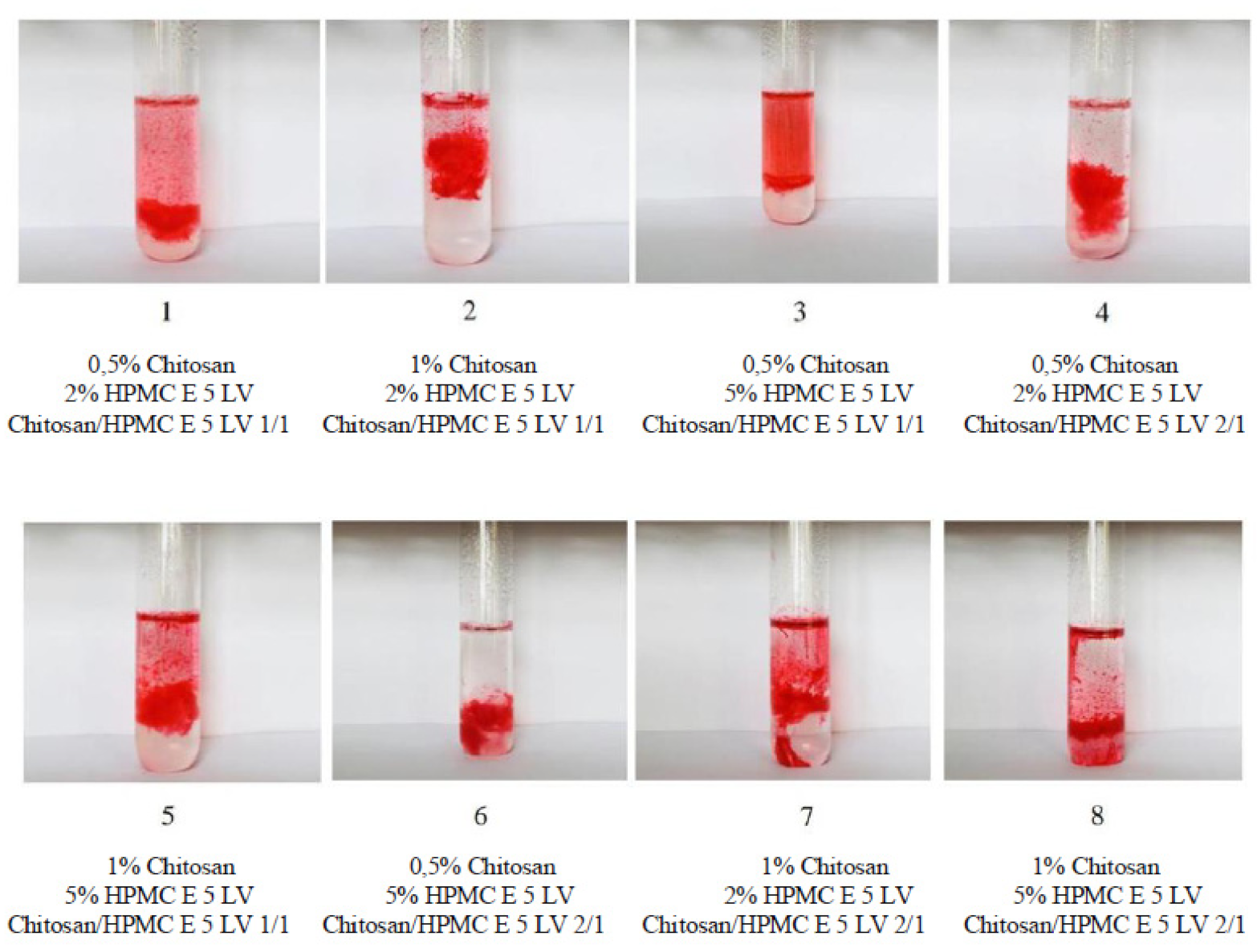
2.3. Determination of Gelling Capacity for Colloidal Chitosan and HPMC E 5 LV Systems

The gelling capacity was determined with a water bath GFL-1012 with a thermostat using previously prepared samples from the colloidal solutions and simulated tear fluid (STF) with a pH of 7.4.
The gelling capacity for the formulated systems was evaluated by adapting the method described by Sheshala et al. (2019) [13]. In test tubes containing 5 mL STF (pH 7.4 at 35 °C), 1 mL of the sample was added. Then, they were placed on the water bath GFL-1012 with a thermostat. The gelling capacity was determined by visual inspection of the degree of rigidity of the gels formed as well as their stability over time. To facilitate visual observation of the gel, a drop of Congo red dye was added to each tube.

2.4. Surface Property Analysis for Ophthalmic Systems with In Situ Gelling Based on Chitosan and HPMC E 5 LV

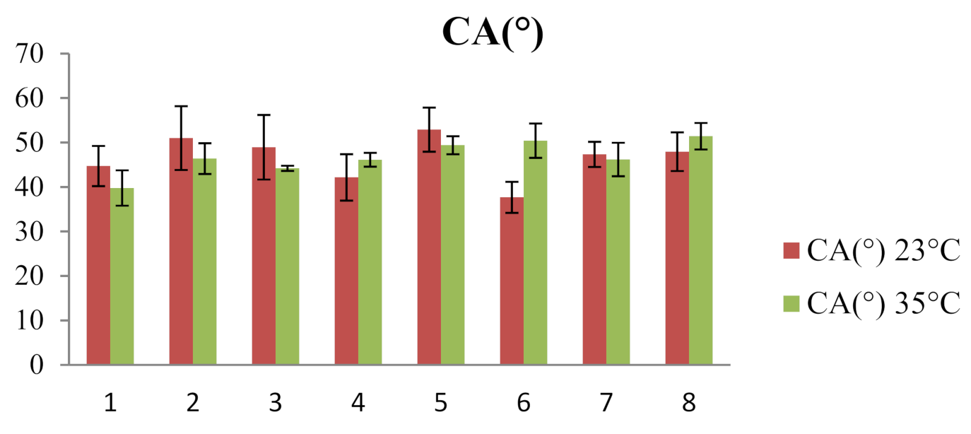
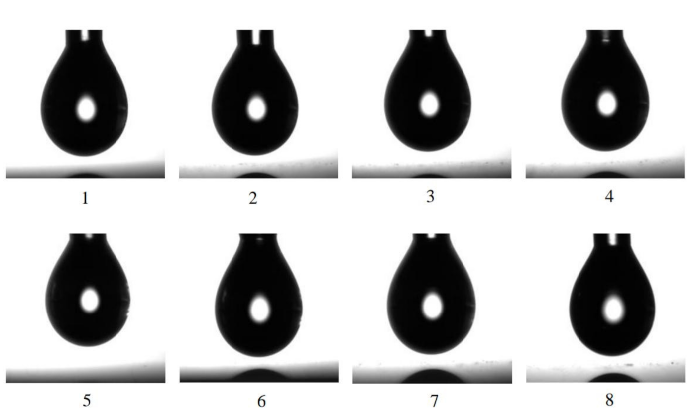
2.4.1. Determination of Contact Angle Values

The contact angle of the in situ gelling systems was determined at 23 °C (storage temperature) and 35 °C (physiological eye temperature) by the sessile drop technique [29]. Evaluation of the contact angle was performed with the following materials: samples from the previously prepared polymer systems with chitosan and HPMC E 5 LV, glass slides, a Hamilton sampling syringe and the CAM 101 sphygmomanometer-goniometer.
The working technique involved displaying a drop of the sample to be analyzed on the surface of a glass slide and processing its image by the Young–Laplace method. For each sample, the measurements were replicated 5 times.

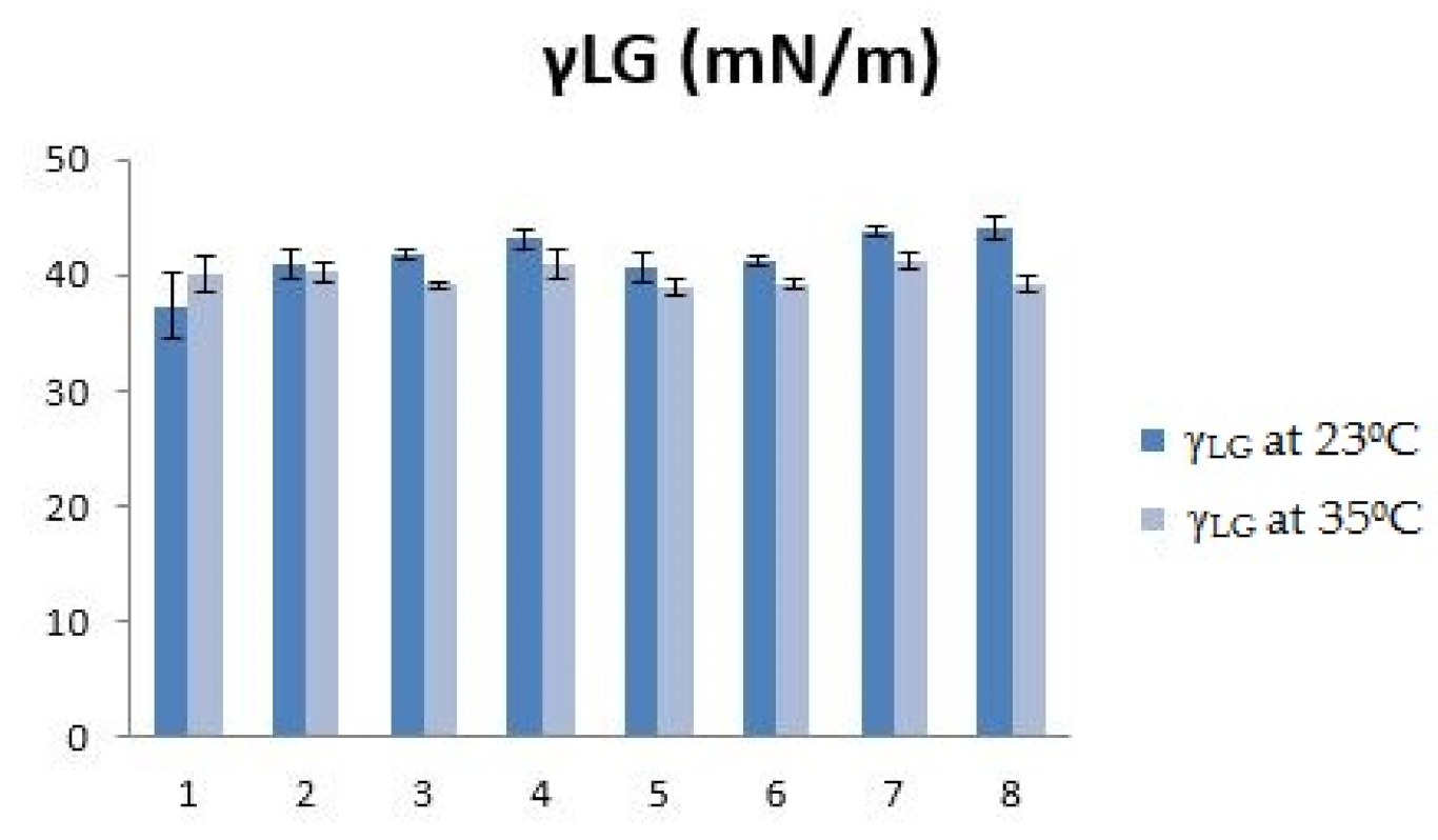
2.4.2. Evaluation of Surface Tension

The determination of the surface tension (ST = γ L G ) as an indicator of the strength of the intermolecular bonds in the fluid system was performed using the “pendant drop method”. The same materials were used to evaluate the surface tension as when determining the contact angle.
The working technique differed from that presented in the section for the contact angle by shooting the image of the drop just before detachment from the needle of the Hamilton syringe. The droplet profile was analyzed based on the Young–Laplace equation, determining the surface tension at the air–droplet interface. For each sample, 5 measurements were performed.

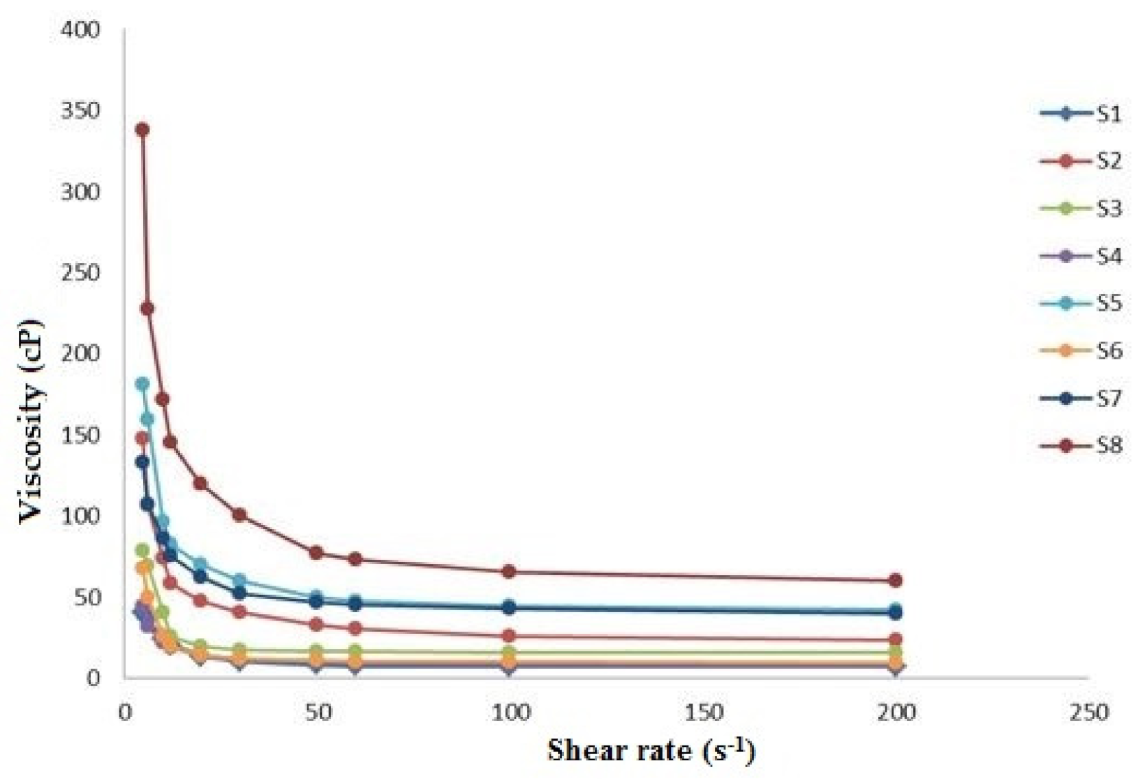
2.5. Rheological Studies on Colloidal Systems with In Situ Gelling Based on Chitosan and HPMC E 5 LV

Rheological studies for the previously prepared chitosan and HPMC E 5 LV formulations were performed at 35 °C using a multi-visc rotary viscometer, to which a thermostat (ThermoHaake P5 Ultrathermostat) was attached to ensure a constant working temperature.
The viscosity values, as a parameter to be optimized for the prepared formulations, were considered to be those obtained at a shear rate of 20 rpm, corresponding to the ocular physiological conditions [30,31].

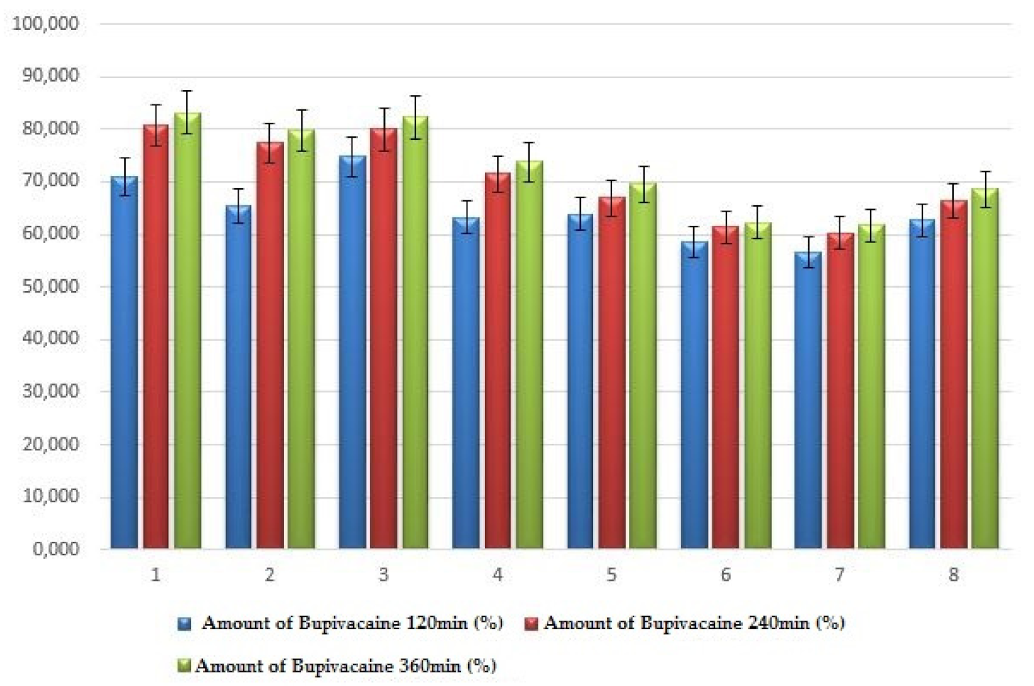
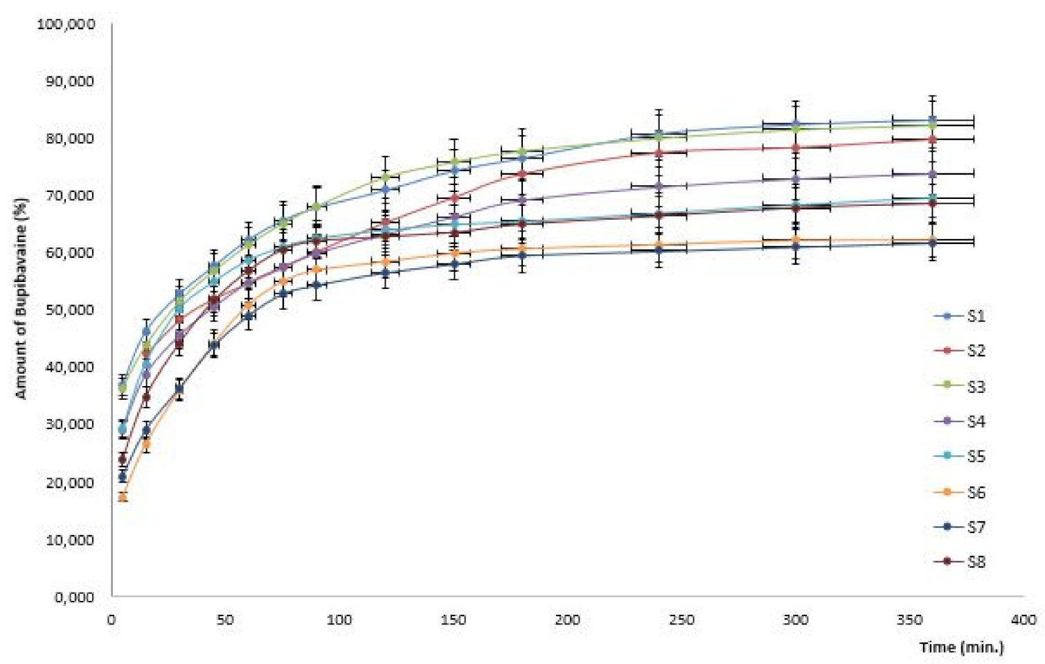
2.6. In Vitro Drug Release Kinetics Studies from Colloidal Systems with Chitosan and HPMC E 5 LV

In vitro drug release studies from chitosan- and HPMC E 5 LV-based in situ colloidal gelling systems were performed using a Hanson Vision® G2 Classic 6TM Dissolution Tester.
For these determinations, 5 mL of each colloidal system with the previously prepared chitosan and HPMC E 5 LV were used, and Visking dialysis bags (from Sigma-Aldrich, Saint Louis, MO, USA) made of cellulose were used as a semipermeable membrane (type: 20/32 inches, thickness: 0.020 mm).
The experiments were performed at 35°C, and the rotational speed of the blades was set at 50 rpm. As a release medium, a phosphate buffer solution with a pH of 7.4 (500 mL volume) was used. At predetermined time intervals over a period of 6 h, volumes of 5 mL of the release medium were taken and replaced with equivalent volumes of the phosphate buffer maintained at 35 °C.
The concentration of bupivacaine hydrochloride in each sample was determined spectrophotometrically by recording the absorbance at 263 nm wavelengths. Using the calibration curve ( A 1 % 1 cm = 14.051 ), the cumulative percentage of the drug substance released after each time interval was calculated.
Data from in vitro release studies on the colloidal systems used were evaluated using the mathematical models of Higuchi (Equation (1)) and Korsmeyer-Peppas (Equation (2)) to evaluate the release mechanism of the active pharmaceutical ingredient bupivacaine hydrochloride:
M t M = k H t 1 2
M t M = k K P t n
where M t is the amount of the substance released over time, M is the total amount of the drug substance in the system under analysis, M t M is the fraction of the drug substance released at each time point, k H is the Higuchi constant, k K P is the Korsmeyer-Peppas kinetic constant and n is the release exponent.

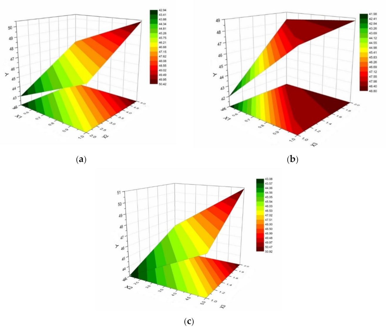
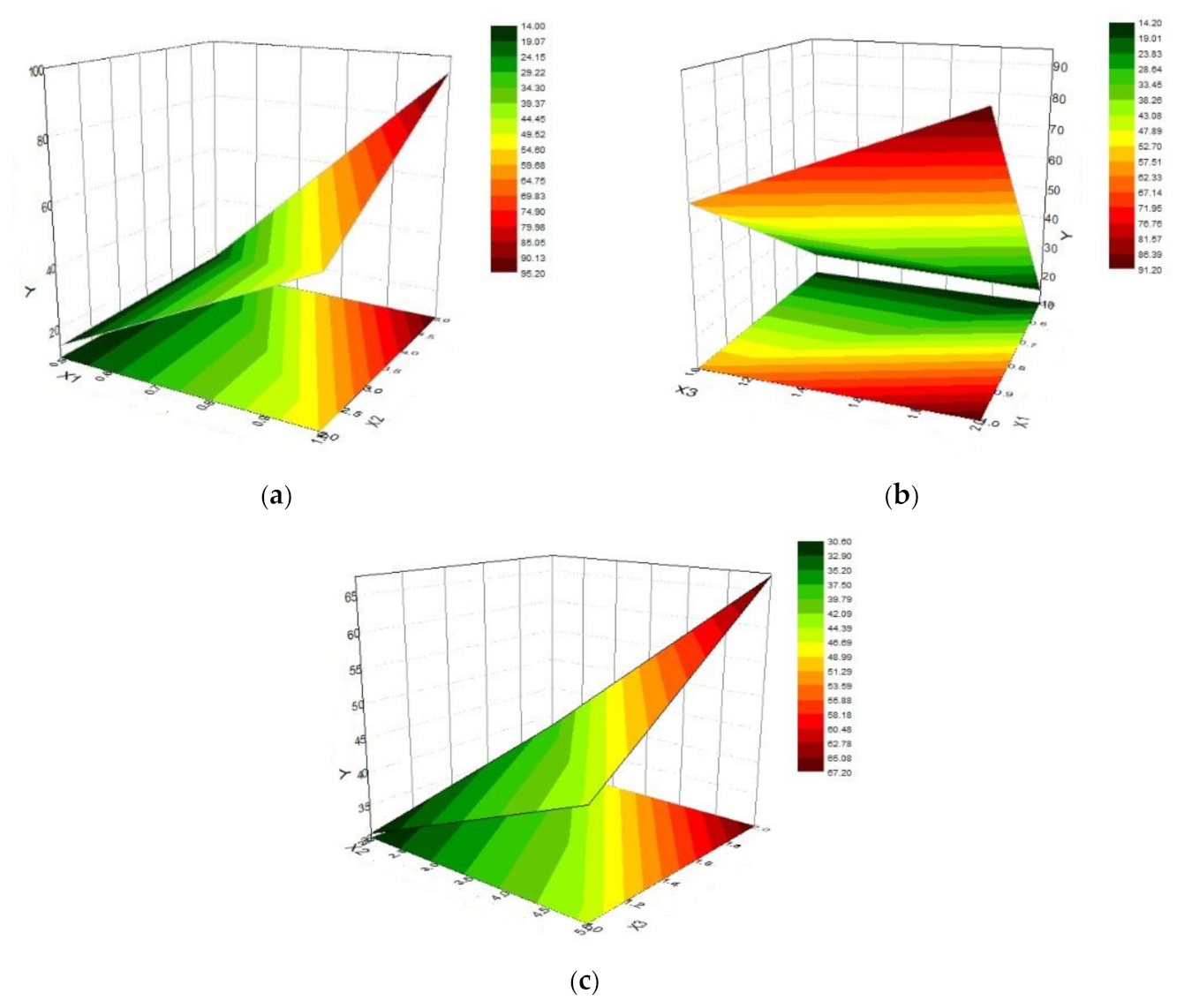
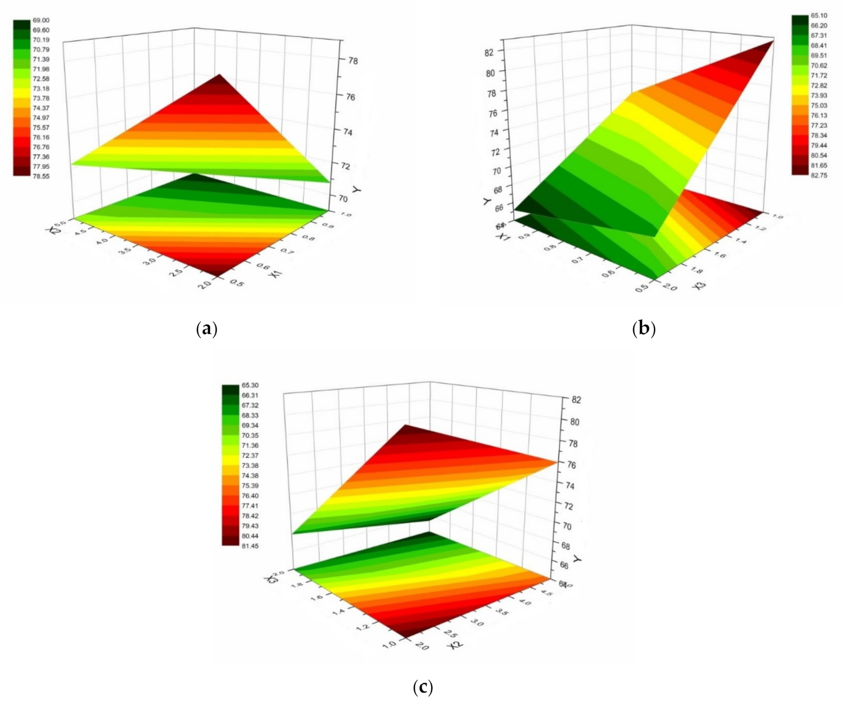
2.7. Optimization of Colloidal Systems with In Situ Gelling Based on Chitosan and HPMC E 5 LV Using Response Surface Analysis

As was previously mentioned, response surface methodology is a statistical method suitable for multi-factorial experiments, providing a relationship between different parameters for optimal operating conditions.
The response of the system is represented graphically in three dimensions, with highlighting of the interactions between the formulation factors [32].
The 3D representation of the responses of the system designed in this study was made using OriginPro 8.5.1 software (OriginLab®).

3. Results

Polymer mixtures of chitosan and HPMC E 5 LV were prepared by varying the concentration and ratio of the polymers in order to increase the ocular bioavailability of bupivacaine hydrochloride.
The design of the experimental matrix for colloidal systems in this study included a factorial plan with three factors (X1, X2 and X3) and two levels of variation (lower and upper) as shown in Table 2, and the three independent variables were as follows:
  • X1: chitosan concentration (0.5% or 1%);
  • X2: HPMC E 5 LV concentration (2% or 5%);
  • X3: (chitosan / HPMC ratio of 1/1 or 2/1).
For the in situ gelling systems obtained, the following parameters were identified and evaluated: a contact angle of 35 °C, viscosity at a shear rate of 20 rpm and cumulative percentage of bupivacaine hydrochloride released in vitro.
Clear, colorless, homogeneous colloidal solutions free of suspended particles were obtained. Increasing the concentration of HPMC E 5 LV from 2% to 5% did not affect the transparency of the formulations. A pH of the samples between 5 and 5.5 ensured the solubility of the components and their stability within the limits of ocular tolerability.
The gelling capacity was coded as follows: (−) not gelled; (+) gelled after a few minutes and quickly despaired; (++) gelled quickly and remained undispersed for several hours; and (+++) gelled instantly and remained undispersed for a long time [33]. The results of the gelling capacity assessment for the designed systems are shown in Table 3.
Sample 2 (chitosan 1%, HPMC E 5 LV 2%) and sample 5 (chitosan 1%, HPMC E 5 LV 5%), both with a polymer ratio of 1/1, gelled instantly in a few seconds and remained undispersed for more than 6 h. In addition, sample 7 (chitosan 1%, HPMC E 5 LV 2%) and sample 8 (chitosan 1%, HPMC E 5 LV 5%), with a polymer ratio of 2/1, had immediate gelation.
Visual analysis of the formulations in which Congo red was added (Figure 1) showed the presence of gelling on contact with the simulated tear fluid. In the case of samples 2, 5, 7 and 8, gelation occurred rapidly in a few seconds, resulting in a gel with high stability. For the other samples, gelation did not occur as quickly, but after gel formation, all samples had high stability. This phenomenon was most likely because they had the highest concentration of chitosan of all the formulations analyzed. Chitosan is a pH-sensitive polymer, with the changing pH of the formulations upon contact with tear fluid in the simulated electrostatic repulsion occurring between the polymer chains to promote cross-linking. Not only was the degree of viscosity of the gel important, but the speed with which the gel was formed was also important. The faster the system gelled, the more it stayed in contact with the mucosa and the harder it was to remove with the tear fluid secreted by the eye system as a method of defending the body against external factors.
The contact angle values for the polymer preparations were intended to evaluate their ability to display on the ocular surface. Table 4 presents the experimental data obtained after analysis of the droplets from the samples taken, captured with the help of the CAM 101 tensiometer-goniometer at 23 °C and at 35 °C.
The recorded quantitative parameters were the average of the two values of the contact angle to the left and to the right CA (M), the volume of the drop (V) (μL) and the interfacial tension ( γ S L ) (mN/m) at 23 °C and 35 °C.
The wettability of the polymeric solutions of chitosan and HPMC E 5 LV analyzed at 23 °C proved to be satisfactory for all 8 samples, an aspect highlighted by the values of the average contact angle (CA (M) (°)) between 37.67° ± 3.49 and 52.89° ± 4.96, respectively.
It was noted that formulations 2 and 5, containing 1% chitosan and different concentrations of HPMC E 5 LV, had the highest CA (°) values, while formulations 4 and 6, incorporating concentrations of only 0.5% chitosan and different concentrations of HPMC E 5 LV and maintaining the same ratio between polymers, had the lowest CA (°) values in the entire series of samples analyzed.
The characteristic CA (°) data for the analyzed in situ gelling colloidal systems showed lower values compared with those obtained for the polymer solutions at 23 °C for most of the samples in the analyzed series, as can be seen in Figure 2, and the p value obtained through the ANOVA test was 0.6999. As an exception, samples 4 (0.5% chitosan, 2% HPMC E 5 LV), 6 (0.5% chitosan, 5% HPMC E 5 LV) and 8 (1% chitosan, 5% HPMC E 5 LV), all having a ratio between the polymers of 2/1, showed higher values for CA (°) at 35 °C. In system 7, although the chitosan/HPMC E 5 LV ratio was 2/1, a lower value of the contact angle was found in the physiological conditions (35 °C) than in the non-physiological ones (23 °C). For this colloidal system formulation with in situ gelling, the concentration of the chitosan biopolymer was at its maximum level (1%).
As in the previous analysis, the dependence of the CA (°) values on the concentrations of the polymers in the systems was maintained such that the samples with lower chitosan concentrations showed lower CA (°) values. Samples 6 and 8, with 5% HPMC E 5 LV and a polymer ratio of 2/1, had the highest CA (°) values in the whole series analyzed at 35 °C, namely 50.41° ± 3.87 and 51.42° ± 2.98, respectively.
For all eight analyzed samples, the optimal values of the surface tension γ L G between 37.32 ± 2.79 mN/m and 43.99 ± 1.05 mN/m were obtained, corresponding to volumes for the drops between 9.23 ± 0.34 μL and 10.82 ± 0.43 μL, respectively. Additionally, a direct proportional relationship between the volume of the sample droplets to be analyzed and the value of the surface tension γ L G was noticed.
The values obtained for the determination of γ L G for polymeric systems with in situ gelling based on chitosan and HPMC E 5 LV at 35 °C were between 38.93 ± 0.64 mN/m and 41.35 ± 0.71 mN/m (Table 5). It was observed that, for the systems evaluated at 35 °C, most γ L G values were lower than those resulting from the analysis at 23 °C (Figure 3), and the p value obtained by the ANOVA test was 0.0666.
Figure 4 shows images of droplets belonging to each test sample, taken at 35 °C before detachment from the needle of the Hamilton syringe, under the action of gravitational force.
The low values of γ L G suggest obtaining preparations with an adequate degree of wettability, as well as a good surface display capacity and corneal adhesion.
An important role in reducing the γ L G values for the studied colloidal systems was the presence of HPMC E 5 LV, with the increase of the concentration of this polymer determining the decrease of the surface tension values. The low values of γ L G determined the watering of the surface and the display and ensuring prolonged contact of the drug substance at the ocular level.
At the same time, the γ L G values close to those of the tears determined a minimal disturbing effect on the tear film, increasing the degree of comfort and compliance of the patient.
It is concluded that by corroborating the results obtained from the experiments evaluating the gelling capacity and surface properties, namely the contact angle and the surface tension, that system 2, system 5 and system 7 had the properties and characteristics of an appropriate quality.
The viscosity values for the colloidal systems with in situ gelling of chitosan and HPMC E 5 LV corresponding to a shear rate of 20 rpm are shown in Table 6.
Figure 5 shows the evolution of the viscosity under the influence of the shear rate. It was observed that there was a decrease in the viscosity of the colloidal systems with an increasing shear rate for all eight systems analyzed. By applying the ANOVA test, we obtained a p value of less than 0.05.
Carrying out experiments at 35 °C had dual roles: to simulate the physiological ocular temperature and to favor the initiation of the gelling process, with an impact on the rheological behavior of the formulations.
The polymer with a major influence on the viscosity of the designed systems was HPMC E 5 LV. Cellulose derivatives of the HPMC type are often used as agents for increasing the viscosity of ophthalmic preparations.
As can be seen from Table 6, the highest viscosity values recorded at 20 rpm were observed in colloidal systems 5 (70.36 cP), 7 (62.32 cP) and 8 (119.87 cP), and they also had high concentrations of polymers in the composition.
Table 7 shows the cumulative percentages of bupivacaine hydrochloride released after 120 min, 240 min and 360 min, along with the 3 formulation variables (X1: concentration percentage of chitosan; X2: concentration percentage of HPMC E 5 LV; and X3: polymer ratio for chitosan/HPMC E 5 LV). The colloidal gelling in situ systems released more than 56% of the drug substance after 2 h, and after 6 h, they released between 61.7% (system 7) and 83.1% (system 1) of the drug substance.
The dynamics of bupivacaine hydrochloride release from the formulations under analysis over 6 h is shown in Figure 6. The highest amount of the drug released after 6 h (83.198%) and was recorded in the case of system 1 (0.5% chitosan, 2% HPMC E 5 LV and polymer ratio 1/1). This was followed by system 3 (0.5% chitosan, 5% HPMC E 5 LV and polymer ratio 1/1), in which the amount of bupivacaine hydrochloride released after the same interval of 6 h was 82.291%. The cumulative percentage of the drug released in vitro from the eight colloidal systems is plotted against time in Figure 7.
In vitro drug release studies looked at the release profile of bupivacaine hydrochloride from chitosan- and HPMC E 5 LV-based in situ gelling colloidal systems. The analyzed formulations showed a prolonged release over a time interval of 6 h (Figure 6) with a p value less than 0.05. This result corresponds to the initial formulation hypothesis and may ensure an increase in the efficacy of the drug at the site of action [34]. Colloidal system 1 with 0.5% chitosan, 2% HPMC E 5 LV and a polymer ratio of 1/1 showed the highest cumulative bupivacaine release percentage, exceeding 83%. The system with the lowest percentage of anesthetic released after 6 h (61.77%) was system 7, with 1% chitosan, 2% HPMC E 5 LV and a polymer ratio of 2/1. Basically, the percentage of the drug given decreases in the order of 1 > 3 > 2 > 4 > 5 > 8 > 6 > 7.
Figure 7 indicates that the colloidal systems shown had similar kinetic profiles. The p value after using the ANOVA test on the data obtained was 0.0179.
In Table 8, the recorded values for the determination coefficient R2 and the release exponent n are shown, which were determined using the mathematical models mentioned above, together with the fraction of bupivacaine hydrochloride M t M released at each time point t.
Table 9 summarizes all the responses of the colloidal systems that followed optimization, summarizing the data obtained for the parameters to be optimized Y1–Y3. Y1 represents the values obtained after measuring the contact angle (CA) at 35 °C, Y2 defines the viscosity of the systems at 35 °C, and Y3 represents the percentage of bupivacaine hydrochloride released from the colloidal systems with chitosan and HPMC E 5 LV after 6 h. The three independent variables were X1 (chitosan concentration), X2 (HPMC E 5 LV concentration) and X3 (chitosan/HPMC E 5 LV polymer ratio).
Regarding the optimization of colloidal systems with gelling in situ, from the point of view of the contact angle (CA (°)), as indicator of hydrophilicity, wetting and mucoadhesivity which was considered the parameter (response variable), it was intended that its value at 35 °C, the ocular physiological temperature, to fall within the range of hydrophilicity, or more precisely, to have a value lower than 90° (Figure 8a–c). Moreover, the values of this quality parameter were in a relatively narrow range: 40–60°. This fact illustrates a uniformity of the formulations, with the chosen composition leading to values of this indicator located around a central optimum. Higher values of the CA (°) parameter were evident for high concentrations of HPMC E 5 LV, as a result of or in relation to the chitosan biopolymer (Figure 8a–c).
The variation of the contact angle values according to X1 (chitosan concentration) and X3 (chitosan/HPMC E 5 LV ratio) (Figure 8b) shows a high “plateau” of values for high concentrations of chitosan (1%) and for a ratio of the two polymers of 2/1 in favor of chitosan, respectively.
The appearance of the response surface of the contact angle parameter according to the variables X2 (HPMC E 5 LV concentration) and X3 (chitosan/HPMC E 5 LV polymer ratio) (Figure 8c) was similar to that in Figure 8a. This supports a similar contribution of the two polymers to the variation of the contact angle parameter. However, high values of CA (°) were obtained for a chitosan/HPMC E 5 LV ratio at a higher level (2/1).
The viscosity response parameter was evaluated at a shear rate of 20 rpm and analyzed from the point of view of the response surfaces, depending on the formulation factors (Figure 9a–c). High viscosity values were recorded for high polymer concentration levels (Figure 9a).
It should be noted that at the higher level of the chitosan concentration (1%), high values of viscosity for the solutions were obtained, even if the level of the polymer HPMC E 5 LV was lower (2%), an example in this respect being system 2 (with a viscosity of 47.6 cP at 20 rpm).
There was a prevalence of the influence of chitosan over the other formulation variables and in the variability of the viscosity of the colloidal systems with in situ gelling being analyzed.
High viscosity values were obtained in system 8, specifically 119.87 cP (colloidal system with 1% chitosan, 5% HPMC E 5 LV and a 2/1 chitosan/HPMC E 5 LV ratio). This is not beneficial for the performance of the system, taking into account that agglomerations of the preparation at the site of action may occur or inadequate display, resulting in either local irritation with acceleration of the washout phenomenon or inadequate availability for absorption of the substance drugs from the polymer matrix.
Regarding the optimization of bupivacaine hydrochloride release, system 1 and system 3, which differed only in the concentration of HPMC E polymer 5 LV, were the formulations that led to the maximum percentages of bupivacaine hydrochloride released after 6 h (83.19% and 82.29%, respectively). However, this observation must also be corroborated with the results of these systems from the point of view of the superficial and rheological properties (Figure 10a–c).
As a result, the proposed range was “increased” to be optimal for the percentage of the drug given after 6 h, meaning a value of at least 70% (Figure 10c).

4. Discussion

In this study, the aim was to formulate ophthalmic systems with in situ gelling in order to increase the ocular bioavailability of bupivacaine hydrochloride by combining chitosan, a pH-sensitive and mucoadhesive polymer, with HPMC E 5 LV, a thermosensitive polymer with the property of increasing the viscosity, with lubricating and mucoadhesive actions.
We set out to design colloidal systems with in situ gelling based on chitosan and HPMC E 5 LV as potential vehicles, which would undergo a phase transition under the action of two existing factors in the tear fluid: a physiological pH of 7.4 and a physiological temperature of 35 °C. An optimal in situ gelation system should be able to undergo phase transition immediately after contact with the ocular surface, ensuring the remainder of the drug substance without causing discomfort to the patient [35].
HPMC E 5 LV is a thermosensitive polymer, but the solutions showed a phase transition at high temperatures. The process takes place gradually with an increasing temperature in four phases, according to Bajwa et al. [36]. In the first phase, a superficial increase of the gelling takes place at temperatures between 10 °C and 40 °C following the cleavage of the intramolecular bonds, increasing the mobility of the polymer chains. At temperatures up to 56 °C, there is a decrease in gelling due to the existence of a rarefied network resulting from the process of separation of the polymeric structures. Reaching 56 °C determines the existence in the solution of an increased number of molecular fractions whose interaction results in a gel with an elastic structure influenced relatively little by the subsequent increase in temperature [36].
The sodium chloride (0.9%) of the compositions of the formulations had an isotonizing role and decreased the transition temperature of HPMC E 5 LV. The addition of sodium chloride influences the temperature at which the onset of gelation occurs, according to Almeida et al. [37]. Chloride ions have a small radius and a high charge density, with the ability to compete for water molecules and reduce the hydrogen bonds between the water and the polymer. As a result, a relatively small number of water molecules remains available for polymer solvation, which favors hydrophobic associations between HPMC chains at low temperatures [38].
By evaluating the contact angle in the optimization study of the ophthalmic systems with in situ gelling based on chitosan and HPMC E 5 LV, the profile of the liquid droplets was investigated, with emphasis on the impact on a hydrophobic surface. The corneal epithelium is known to have a lipophilic character, to which the mucin layer is added on its surface with a protective role, which explains the hydrophobicity of this tissue.
For the colloidal systems of chitosan and HPMC E 5 LV, CA values (°) lower than 90° were obtained, which shows the hydrophilic character and the increased wetting capacity of the analyzed mixtures.
Surface tension is a key parameter in optimizing ophthalmic formulations. The administration of ocular preparations with a surface tension lower than that characteristic of tears can cause the destabilization of the tear film and its dispersion in the form of drops [39].
The surface tension at the air–tear film interface has a physiological value between 40 and 46 mN/m, ensuring the stability and breakup time of the tear film [40]. The results of the γ L G determinations at 23 °C and 35 °C were within the physiological limits.
For the polymeric mixtures of chitosan and HPMC E 5 LV analyzed at the ocular physiological temperature, the γ L G values were lower than those obtained at 23 °C, an aspect that is consistent with the statements of Han et al., according to which γ L G decreases with an increasing temperature [41].
Another important aspect to mention is the influence of γ L G on the volume of the drop. It is known that volumes between 5 and 15 μL have been proven effective in drug release, and the volume of a drop influences the amount of the active pharmaceutical ingredient administered and its maintenance for as long as possible in the eye [42]. The volumes of the droplets in the samples analyzed in this study fell within the aforementioned range, which prevented the onset of the immediate washout phenomenon.
The rate of blinking of a normal, healthy eye varies between 6 and 30 blinks/min, with a reported average of 22 blinks/min when the eye is relaxed [30,31]. Rheological studies follow the influence of the formulation factors and working temperature on the flow behavior of colloidal systems with chitosan and HPMC E 5 LV [43].
Akinosho et al. specified that the methyl and hydroxypropyl groups in the composition of HPMC determine how a colloidal dispersion after gelling can be described as a polymeric network or a gel with different degrees of rigidity, respectively [44]. According to the statements presented by Sandri et al., the increase of the temperature determines the partial dehydration of the polymeric chains of HPMC, favoring the hydrophobic interactions and the cross-linking between them [45].
In the first phase, there is a rapid release, known in the literature as the “burst effect”, followed by a prolonged release for 6 h. This two-stage process corresponds to a rapid initial onset of local anesthetic action of bupivacaine hydrochloride in ocular surgery, followed by a gradual analgesic action maintained for a longer period of time, a beneficial aspect in postoperative pain management [46,47].
The polymer matrix has a significant influence on the percentage and rate of drug release [34]. Thus, an increase in the concentrations of the polymers caused a decrease in the rate of release of the bupivacaine hydrochloride, which is consistent with the statements of Ghosal et al. [48]. A high concentration of polymers causes the formation of a polymer network resistant to the action of the release medium (phosphate buffer solution pH: 7.4), thus reducing the amount of bupivacaine hydrochloride released. Compared with the previously mentioned statements, Chanaj-Kaczmarek et al. stated that the addition of chitosan decreases the permeability of the polymer network [49].
For the approached study, the drug release kinetics were analyzed using the Korsmeyer-Peppas mathematical model, for which the values of the coefficient of determination R2 exceeded the value of 0.98. The values of the yield exponent n within the same model varied between 0.205 and 0.426. In this case, the release mechanism depended on the migration of water in the polymer matrix and the diffusion of the drug substance, a fact also confirmed by Song et al. [50].
For the optimization of the contact angle, a concentration of 1% chitosan and a higher chitosan/HPMC E 5 LV ratio (2/1) were favorable for appropriate hydrophilic or wetting properties, but the HPMC E 5 LV concentration must be at a lower level (2%) to compensate for hydrophobia induced by chitosan. These conditions were met by samples 7 and 8.
The values of the viscosity located in the middle zone of the variation range 13.72–119.87 cP, namely between 60 cP and 70 cP, can be considered the optimal values of the systems. The colloidal systems that correspond to these values are system 5 (1% chitosan, 5% HPMC E 5 LV, 1/1 chitosan/HPMC E 5 LV ratio) and system 7 (1% chitosan, 2% HPMC E 5 LV, 2/1 chitosan/HPMC E 5 LV ratio). So far, only substances soluble at an acidic pH have been analyzed with this type of system, because the natural polymer chitosan is only soluble in an acidic pH.
Corroborating the results of the optimization analysis, the superficial performances of the systems and their rheological profiles, system 5 (1% chitosan, 5% HPMC E 5 LV, polymer ratio 1/1) and system 7 (1% chitosan, 2% HPMC E 5 LV, chitosan/HPMC E 5 LV ratio 2/1) can be considered optimal formulations. However, the higher percentage of bupivacaine hydrochloride released after 6 h was obtained for system 5 (1% chitosan, 5% HPMC E 5 LV, polymer ratio 1/1), meaning a value of 69.55% compared with 61.77% for system 7.

5. Conclusions

This study shows the potential of chitosan in the formulation of colloidal systems with in situ gelling by associating it with a cellulose derivative, namely HPMC sort E 5 LV, in order to increase the ocular residence time of bupivacaine hydrochloride. This study was consolidated upon an experimental plan that aimed to optimize the formulation parameters and their impact on the system responses.
The predominantly hydrophilic behavior of the formulations, expressed by CA (°) values lower than 90°, together with the γLG values located in physiological limits determined a good watering capacity of the corneal surface. Regarding the viscosity parameter evaluated at 20 rpm, high values of it were recorded in systems with a chitosan concentration at a higher level (1%).
In vitro release studies of bupivacaine hydrochloride for the designed systems indicated similar kinetic profiles and a two-stage release of the active substance: an initial rapid release or “burst effect” followed by a gradual release for 6 h. The analysis of the obtained data led to the selection of the Korsmeyer-Peppas model as the mathematical model that characterized the kinetics of the drug release, with the values of the coefficient of determination R2 exceeding 0.98.
The in situ gelling capacities of the designed colloidal systems were mainly influenced by the independent variable X1, represented by the chitosan concentration, such that the systems with a chitosan biopolymer concentration at a higher level (1%) showed a rapid phase transition in a few seconds following contact with the simulated tear fluid.
It can be concluded from the optimization analysis, in terms of the surface properties, rheological profiles and kinetic profiles, that the system with 1% chitosan, 5% HPMC E 5 LV and the chitosan/HPMC E 5 LV 1/1 ratio demonstrated a performance level that can be considered a promising alternative to the prolonged release of bupivacaine hydrochloride to the eye. The developed in situ gelling systems can be further used as delivery platforms to overcome low ocular bioavailability of the drugs.

Author Contributions

Conceptualization, L.P., T.I. and M.V.G.; methodology, L.P. and M.V.G.; experiments, L.P. and T.I.; in vitro drug release experiments, L.P., M.V.G., C.-E.D.-P. and R.P.; writing—original draft preparation, T.I., L.P. and R.P.; writing—review and editing, L.P., R.P. and M.V.G.; supervision, L.P.; funding acquisition, C.-E.D.-P. All authors have read and agreed to the published version of the manuscript.

Funding

This paper was financially supported by the “Carol Davila” University of Medicine and Pharmacy through Contract No. CNFISFDI-2021-0300 and RDI capability consolidation at the institutional level of the multidisciplinary research teams involved in the sustainability of UMFCD priority research directions.

Institutional Review Board Statement

Not applicable.

Informed Consent Statement

Not applicable.

Data Availability Statement

Data are available from the authors upon request.

Conflicts of Interest

The authors declare no conflict of interest.

References

  1. Pai, R.V.; Vavia, P.R. Chitosan oligosaccharide enhances binding of nanostructured lipid carriers to ocular mucins: Effect on ocular disposition. Int. J. Pharm. 2020, 577, 119095. [Google Scholar] [CrossRef]
  2. Thakkar, R.; Komanduri, N.; Dudhipala, N.; Tripathi, S.; Repka, M.A.; Majumdar, S. Development and optimization of hot-melt extruded moxifloxacin hydrochloride inserts, for ocular applications, using the design of experiments. Int. J. Pharm. 2021, 603, 120676. [Google Scholar] [CrossRef] [PubMed]
  3. Tundisi, L.L.; Mostaco, G.B.; Carricondo, P.C.; Petri, D.F.S. Hydroxypropyl methylcellulose: Physicochemical properties and ocular drug delivery formulations. Eur. J. Pharm. Sci. 2021, 159, 105736. [Google Scholar] [CrossRef] [PubMed]
  4. Fan, X.; Torres-Luna, C.; Azadi, M.; Domszy, R.; Hu, N.; Yang, A.; David, A.E. Evaluation of commercial soft contact lenses for ocular drug delivery: A review. Acta Biomater. 2020, 115, 60–74. [Google Scholar] [CrossRef] [PubMed]
  5. Wang, Q.; Zuo, Z.; Cheung, C.K.C.; Leung, S.S.Y. Updates on thermosensitive hydrogel for nasal, ocular and cutaneous delivery. Int. J. Pharm. 2019, 559, 86–101. [Google Scholar] [CrossRef] [PubMed]
  6. Popescu, R.; Ghica, M.V.; Dinu-Pirvu, C.E.; Anuta, V.; Lupuliasa, D.; Popa, L. New Opportunity to Formulate Intranasal Vaccines and Drug Delivery Systems Based on Chitosan. Int. J. Mol. Sci. 2020, 21, 5016. [Google Scholar] [CrossRef]
  7. Irimia, T.; Dinu-Pirvu, C.E.; Ghica, M.V.; Lupuleasa, D.; Muntean, D.L.; Udeanu, D.I.; Popa, L. Chitosan-Based In Situ Gels for Ocular Delivery of Therapeutics: A State-of-the-Art Review. Mar. Drugs 2018, 16, 373. [Google Scholar] [CrossRef] [Green Version]
  8. Sami El-banna, F.; Mahfouz, M.E.; Leporatti, S.; El-Kemary, M.; Hanafy, N.A.N. Chitosan as a Natural Copolymer with Unique Properties for the Development of Hydrogels. Appl. Sci. 2019, 9, 2193. [Google Scholar] [CrossRef] [Green Version]
  9. Chen, C.P.; Hsieh, C.M.; Tsai, T.; Yang, J.C.; Chen, C.T. Optimization and Evaluation of a Chitosan/Hydroxypropyl Methylcellulose Hydrogel Containing Toluidine Blue O for Antimicrobial Photodynamic Inactivation. Int. J. Mol. Sci. 2015, 16, 20859–20872. [Google Scholar] [CrossRef] [Green Version]
  10. Yusufu, M.; Liu, X.; Zheng, T.; Fan, F.; Xu, J.; Luo, Y. Hydroxypropyl methylcellulose 2% for dry eye prevention during phacoemulsification in senile and diabetic patients. Int. Ophthalmol. 2018, 38, 1261–1273. [Google Scholar] [CrossRef]
  11. Chen, Y.A.; Hirnschall, N.; Findl, O. Comparison of corneal wetting properties of viscous eye lubricant and balanced salt solution to maintain optical clarity during cataract surgery. J. Cataract Refract. Surg. 2011, 37, 1806–1808. [Google Scholar] [CrossRef]
  12. El-Sousi, S.; Nacher, A.; Mura, C.; Catalan-Latorre, A.; Merino, V.; Merino-Sanjuan, M.; Diez-Sales, O. Hydroxypropylmethylcellulose films for the ophthalmic delivery of diclofenac sodium. J. Pharm. Pharmacol. 2013, 65, 193–200. [Google Scholar] [CrossRef]
  13. Sheshala, R.; Ming, N.J.; Kok, Y.Y.; Singh, T.R.R.; Dua, K. Formulation and Characterization of pH Induced in situ Gels Containing Sulfacetamide Sodium for Ocular Drug Delivery: A Combination of Carbopol®/HPMC Polymer. Indian J. Pharm. Educ. Res. 2019, 53, 654–662. [Google Scholar] [CrossRef] [Green Version]
  14. Sheil, M.; Chambers, M.; Polkinghorne, A.; Sharpe, B. Topical Application of Lidocaine and Bupivacaine to Disbudding Wounds in Dairy Calves: Safety, Toxicology and Wound Healing. Animals 2021, 11, 869. [Google Scholar] [CrossRef]
  15. Bavli, Y.; Rabie, M.; Fellig, Y.; Nevo, Y.; Barenholz, Y. Liposomal Bupivacaine (Bupigel) Demonstrates Minimal Local Nerve Toxicity in a Rabbit Functional Model. Pharmaceutics 2021, 13, 185. [Google Scholar] [CrossRef]
  16. Irimia, T. Contributions on Formulation and Preliminary Evaluation of Ocular Colloidal Systems of Chitosan and Poloxamer 407 with Bupivacaine Hydrochloride. Farmacia 2019, 67, 702–708. [Google Scholar] [CrossRef]
  17. Minto, B.W.; Zanato, L.; Franco, G.G.; Kawamoto, F.Y.K.; Borsaro, C.P.; Pazzini, J.M.; Carvalho, E.R.; Matsui, A. Topical application of lidocaine or bupivacaine in the healing of surgical wounds in dogs. Acta Circ. Bras. 2020, 35, e202000701. [Google Scholar] [CrossRef]
  18. Goder, D.; Eshkol-Yogev, I.; Matsliah, L.; Lemberger, M.; Harlev, M.; Furer, A.; Zilberman, M.; Egozi, D. In vivo study of the efficacy of bupivacaine-eluting novel soy protein wound dressings in a rat burn model. Burns 2021. [Google Scholar] [CrossRef] [PubMed]
  19. Soliman, M.M.; Macky, T.A.; Samir, M.K. Comparative clinical trial of topical anesthetic agents in cataract surgery: Lidocaine 2% gel, bupivacaine 0.5% drops, and benoxinate 0.4% drops. J. Cataract. Refract. Surg. 2004, 30, 1716–1720. [Google Scholar] [CrossRef]
  20. Kashyap, A.; Varshney, R.; Titiyal, G.S.; Sinha, A.K. Comparison between ropivacaine and bupivacaine in deep topical fornix nerve block anesthesia in patients undergoing cataract surgery by phacoemulsification. Indian J. Ophthalmol. 2018, 66, 1268–1271. [Google Scholar] [PubMed]
  21. Oji, E.; Oji, A. Bupivacaine and lignocaine for ophthalmic surgery. Br. J. Ophthalmol. 1987, 71, 66–68. [Google Scholar] [CrossRef] [Green Version]
  22. Kaewjiaranai, T.; Srisatjaluk, R.L.; Sakdajeyont, W.; Pairuchvej, V.; Wongsirichat, N. The efficiency of topical anesthetics as antimicrobial agents: A review of use in dentistry. J. Dent. Anesth. Pain Med. 2018, 18, 223–233. [Google Scholar] [CrossRef] [PubMed]
  23. Neuwersch, S.; Köstenberger, M.; Sorschag, S.; Ilias, W.; Likar, R. Antimicrobial Activity of Lidocaine, Bupivacaine, Mepivacaine and Ropivacaine on Staphylococcus epidermidis, Staphylococcus aureus and Bacillus subtilis. Open Pain J. 2017, 10, 1–4. [Google Scholar] [CrossRef] [Green Version]
  24. Garg, N.K.; Sharma, G.; Singh, B.; Nirbhavane, P.; Tyagi, R.K.; Shukla, R.; Katare, O.P. Quality by Design (QbD)-enabled development of aceclofenac loaded-nano structured lipid carriers (NLCs): An improved dermatokinetic profile for inflammatory disorder(s). Int. J. Pharm. 2017, 517, 413–431. [Google Scholar] [CrossRef]
  25. Fukuda, I.M.; Pinto, C.F.F.; Moreira, C.d.S.; Saviano, A.M.; Lourenço, F.R. Design of Experiments (DoE) applied to Pharmaceutical and Analytical Quality by Design (QbD). Braz. J. Pharm. Sci. 2018, 54. [Google Scholar] [CrossRef]
  26. Yolmeh, M.; Jafari, S.M. Applications of Response Surface Methodology in the Food Industry Processes. Food Bioprocess Technol. 2017, 10, 413–433. [Google Scholar] [CrossRef]
  27. Kumari, M.; Gupta, S.K. Response surface methodological (RSM) approach for optimizing the removal of trihalomethanes (THMs) and its precursor’s by surfactant modified magnetic nanoadsorbents (sMNP)—An endeavor to diminish probable cancer risk. Sci. Rep. 2019, 9, 18339. [Google Scholar] [CrossRef]
  28. Bajwa, K.; Bishnoi, N.R.; Kirrolia, A.; Gupta, S.; Selvan, S.T. Response surface methodology as a statistical tool for optimization of physio-biochemical cellular components of microalgae Chlorella pyrenoidosa for biodiesel production. Appl. Water Sci. 2019, 9, 1–16. [Google Scholar] [CrossRef] [Green Version]
  29. Prisada, R.M. Perspectives to Describe Surface Properties of Raw Pharmaceutical Materials. A Fractal Approach on the Wetting of Powders. Farmacia 2020, 68, 354–361. [Google Scholar] [CrossRef]
  30. Kim, J.; Kumar, Y.S.; Yoo, J.; Kwon, S. Change of Blink Rate in Viewing Virtual Reality with HMD. Symmetry 2018, 10, 400. [Google Scholar] [CrossRef] [Green Version]
  31. Abusharha, A.A. Changes in blink rate and ocular symptoms during different reading tasks. Clin. Optom. 2017, 9, 133–138. [Google Scholar] [CrossRef] [Green Version]
  32. Saini, S.; Chawla, J.; Kumar, R.; Kaur, I. Response surface methodology (RSM) for optimization of cadmium ions adsorption using C16-6-16 incorporated mesoporous MCM-41. SN Appl. Sci. 2019, 1, 1–10. [Google Scholar] [CrossRef] [Green Version]
  33. Dasankoppa, F.; Kujur, S.; Sholapur, H.N.A.; Jamakandi, V. Design, formulation and evaluation of carboxy methyl tamarind based in situ gelling ophthalmic drug delivery system of dorzolamide hydrochloride. Indian J. Health Sci. 2016, 9, 56. [Google Scholar] [CrossRef]
  34. Ciolacu, D.E.; Nicu, R.; Ciolacu, F. Cellulose-Based Hydrogels as Sustained Drug-Delivery Systems. Materials 2020, 13, 5270. [Google Scholar] [CrossRef] [PubMed]
  35. Kurniawansyah, I.S.; Rusdiana, T.; Sopyan, I.; Ramoko, H.; Wahab, H.A.; Subarnas, A. In situ ophthalmic gel forming systems of poloxamer 407 and hydroxypropyl methyl cellulose mixtures for sustained ocular delivery of chloramphenicole: Optimization study by factorial design. Heliyon 2020, 6, e05365. [Google Scholar] [CrossRef]
  36. Bajwa, G.S.; Sammon, C.; Timmins, P.; Melia, C.D. Molecular and mechanical properties of hydroxypropyl methylcellulose solutions during the sol:gel transition. Polymer 2009, 50, 4571–4576. [Google Scholar] [CrossRef]
  37. Almeida, N.; Rakesh, L.; Zhao, J. Monovalent and divalent salt effects on thermogelation of aqueous hypromellose solutions. Food Hydrocoll. 2014, 36, 323–331. [Google Scholar] [CrossRef]
  38. Joshi, S.C. Sol-Gel Behavior of Hydroxypropyl Methylcellulose (HPMC) in Ionic Media Including Drug Release. Materials 2011, 4, 1861–1905. [Google Scholar] [CrossRef] [Green Version]
  39. Dubashynskaya, N.; Poshina, D.; Raik, S.; Urtti, A.; Skorik, Y.A. Polysaccharides in Ocular Drug Delivery. Pharmaceutics 2019, 12, 22. [Google Scholar] [CrossRef] [Green Version]
  40. Hotujac Grgurević, M.; Juretić, M.; Hafner, A.; Lovrić, J.; Pepić, I. Tear fluid-eye drops compatibility assessment using surface tension. Drug Dev. Ind. Pharm. 2017, 43, 275–282. [Google Scholar] [CrossRef]
  41. Han, K.; Woghiren, O.E.; Priefer, R. Surface tension examination of various liquid oral, nasal, and ophthalmic dosage forms. Chem. Cent. J. 2016, 10, 31. [Google Scholar] [CrossRef] [Green Version]
  42. Kumar, S.; Karki, R.; Meena, M.; Prakash, T.; Rajeswari, T.; Goli, D. Reduction in drop size of ophthalmic topical drop preparations and the impact of treatment. J. Adv. Pharm. Technol. Res. 2011, 2, 192–194. [Google Scholar] [PubMed]
  43. Ghica, M.V.; Hirjau, M.; Lupuleasa, D.; Dinu-Pirvu, C.E. Flow and Thixotropic Parameters for Rheological Characterization of Hydrogels. Molecules 2016, 21, 786. [Google Scholar] [CrossRef]
  44. Akinosho, H.; Hawkins, S.; Wicker, L. Hydroxypropyl methylcellulose substituent analysis and rheological properties. Carbohydr. Polym. 2013, 98, 276–281. [Google Scholar] [CrossRef] [PubMed]
  45. Sandri, G.; Bonferoni, M.C.; Rossi, S.; Ferrari, F.; Mori, M.; del Fante, C.; Perotti, C.; Caramella, C. Thermosensitive eyedrops containing platelet lysate for the treatment of corneal ulcers. Int. J. Pharm. 2012, 426, 1–6. [Google Scholar] [CrossRef] [PubMed]
  46. Ghica, M.V.; Kaya, M.G.A.; Dinu-Pirvu, C.E.; Lupuleasa, D.; Udeanu, D.I. Development, Optimization and In Vitro/In Vivo Characterization of Collagen-Dextran Spongious Wound Dressings Loaded with Flufenamic Acid. Molecules 2017, 22, 1552. [Google Scholar] [CrossRef]
  47. Thitiwichienlert, S.; Saksritawee, K. Postoperative pain control of subtenon bupivacaine injection in adult strabismus surgery: A double-masked randomized trial. Eye South East Asia 2020, 15, 64–67. [Google Scholar] [CrossRef]
  48. Ghosal, K.; Chandra, A.; Rajabalaya, R.; Chakraborty, S.; Nanda, A. Mathematical modeling of drug release profiles for modified hydrophobic HPMC based gels. Pharmazie 2012, 67, 147–155. [Google Scholar] [PubMed]
  49. Chanaj-Kaczmarek, J.; Paczkowska, M.; Osmalek, T.; Kapron, B.; Plech, T.; Szymanowska, D.; Karazniewicz-Lada, M.; Kobus-Cisowska, J.; Cielecka-Piontek, J. Hydrogel Delivery System Containing Calendulae flos Lyophilized Extract with Chitosan as a Supporting Strategy for Wound Healing Applications. Pharmaceutics 2020, 12, 634. [Google Scholar] [CrossRef] [PubMed]
  50. Song, J.; Bi, H.; Xie, X.; Guo, J.; Wang, X.; Liu, D. Preparation and evaluation of sinomenine hydrochloride in situ gel for uveitis treatment. Int. Immunopharmacol. 2013, 17, 99–107. [Google Scholar] [CrossRef] [Green Version]
Figure 1. Highlighting of Congo red gelling for the analyzed colloidal systems.
Processes 09 01694 g001
Figure 2. Comparative graphical representation of the contact angle values for the 8 samples analyzed at the two temperatures (23 °C and 35 °C).
Processes 09 01694 g002
Figure 3. Graphical representation of the comparative values of the surface tension γ L G for the eight samples analyzed at the two temperatures (23 °C and 35 °C).
Processes 09 01694 g003
Figure 4. Images of droplets characteristic of each sample, captured at the time of evaluation of γ L G at 35 °C.
Processes 09 01694 g004
Figure 5. Variation of viscosity as a function of the shear rate for the designed systems.
Processes 09 01694 g005
Figure 6. Dynamics of bupivacaine hydrochloride release from colloidal systems with chitosan and HPMC E 5 LV.
Processes 09 01694 g006
Figure 7. Cumulative release profiles for colloidal systems analyzed at 35 °C.
Processes 09 01694 g007
Figure 8. Highlighting in 3D the evolution of the Y (CA (°)) response parameter as a function of the formulation variables (a) X1 (chitosan concentration) and X2 (HPMC E 5 LV concentration); (b) X1 (chitosan concentration) and X3 (chitosan/HPMC E 5 LV ratio); and (c) X2 (HPMC E 5 LV concentration) and X3 (chitosan/HPMC E 5 LV ratio).
Processes 09 01694 g008
Figure 9. Highlighting in 3D the evolution of the Y (viscosity) parameter according to the formulation variables (a) X1 (chitosan concentration) and X2 (HPMC E 5 LV concentration); (b) X1 (chitosan concentration) and X3 (chitosan/HPMC E 5 LV ratio); and (c) X2 (HPMC E 5 LV concentration) and X3 (chitosan/HPMC E 5 LV ratio).
Processes 09 01694 g009
Figure 10. Highlighting in 3D the evolution of the Y parameter (percentage of bupivacaine hydrochloride) according to the formulation variables (a) X1 (chitosan concentration) and X2 (HPMC E 5 LV concentration); (b) X1 (chitosan concentration) and X3 (chitosan/HPMC E 5 LV ratio); and (c) X2 (HPMC E 5 LV concentration) and X3 (chitosan/HPMC E 5 LV ratio).
Processes 09 01694 g010
Table 1. Experimental matrix for chitosan- and HPMC E5 LV-based in situ gelling systems with bupivacaine hydrochloride.
SystemChitosan
X1
%
HPMC
X2
%
Chitosan/
HPMC
X3
(p:p)
System Volume
V (mL)
NaCl
w (g)
Bupivacaine
Hydrochloride
w (g)
NaOH 10%
V (mL)
pH
10.5021:1300.270.07570.495.35
21.0021:1300.270.07600.505.42
30.5051:1300.270.07570.535.38
40.5022:1450.400.11431.055.48
51.0051:1300.270.07640.345.42
60.5052:1450.400.11531.075.47
71.0022:1450.400.11460.875.37
81.0052:1450.400.11680.895.47
Table 2. Factorial plan for in situ gelling systems based on chitosan and HPMC E 5 LV with three factors and two levels of variation.
FactorsParametersLevels of Variation
Lower (−)Upper (+)
X1Chitosan (%)0.5%1%
X2HPMC E 5 LV (%)2%5%
X3Chitosan/HPMC E 5 LV ratio1/12/1
Table 3. Evaluation of gelling capacity for colloidal chitosan and HPMC E 5 LV systems.
System CodeGelling Capacity
1++
2+++
3++
4++
5+++
6++
7+++
8+++
Table 4. Synthesis of experimental data resulting from CA (°) evaluation for the polymer systems, analyzed at 23 °C and 35 °C.
23 °C35 °C
SystemCA(M) (°) V (µL)γSL (mN/m)CA(M) (°) V (µL)γSL (mN/m)
144.70 ± 4.5211.55 ± 2.4430.65 ± 9.8139.77 ± 3.978.74 ± 0.7628.67 ± 2.71
251.00 ± 7.1810.27 ± 1.1835.14 ± 5.8646.39 ± 3.468.39 ± 0.4731.59 ± 7.34
348.94 ± 7.279.46 ± 1.0733.17 ± 3.4044.19 ± 0.588.39 ± 0.3831.29 ± 4.26
442.16 ± 5.239.65 ± 0.3939.55 ± 4.6746.11 ± 1.569.50 ± 0.5530.63 ± 4.23
552.89 ± 4.9611.44 ± 1.2031.43 ± 4.2149.39 ± 2.029.96 ± 0.3636.95 ± 12.87
637.67 ± 3.497.90 ± 1.1528.84 ± 5.1650.41 ± 3.879.46 ± 1.2030.09 ± 3.37
747.33 ± 2.8410.94 ± 1.6331.78 ± 2.9346.18 ± 3.7712.73 ± 1.4429.33 ± 3.30
847.94 ± 4.3510.65 ± 0.7043.49 ± 13.8151.42 ± 2.989.54 ± 0.9029.46 ± 5.96
Table 5. Synthesis of experimental data resulting from the evaluation of the surface tension γ L G for the polymeric systems analyzed at 23 °C and 35 °C.
23 °C35 °C
SystemV (μL) γ L G   ( mN / m ) V (μL) γ L G   ( mN / m )
19.23 ± 0.3437.32 ± 2.798.97 ± 0.3540.15 ± 1.53
210.54 ± 0.5440.83 ± 1.279.42 ± 0.4340.33 ± 0.82
310.32 ± 0.3241.79 ± 0.419.20 ± 0.1939.10 ± 0.26
410.63 ± 0.4043.10 ± 0.909.06 ± 0.3041.01 ± 1.27
510.56 ± 0.2240.68 ± 1.208.86 ± 0.4638.93 ± 0.64
610.21 ± 0.2241.11 ± 0.459.16 ± 0.1639.34 ± 0.43
710.92 ± 0.4843.81 ± 0.509.52 ± 0.1641.35 ± 0.71
810.82 ± 0.4343.99 ± 1.058.86 ± 0.3939.16 ± 0.64
Table 6. The results obtained after determining the viscosity parameter for the analyzed colloidal systems.
SystemX1
Chitosan (%)
X2
HPMC (%)
X3
Chitosan/HPMC Ratio
Viscosity
(cP)
10.52113.72 ± 0.83
21.02147.60 ± 2.74
30.55119.52 ± 1.15
40.52214.28 ± 0.48
51.05170.36 ± 2.99
60.55214.44 ± 0.14
71.02262.32 ± 3.85
81.052119.87 ± 4.02
Table 7. Cumulative percentage of bupivacaine hydrochloride released at predetermined time intervals (120 min, 240 min and 360 min).
SystemX1
Chitosan (%)
X2
HPMC (%)
X3
Chitosan/
HPMC
Ratio
Amount of Bupivacaine 120 min (%)Amount of Bupivacaine 240 min (%)Amount of Bupivacaine 360 min (%)
10.52170.89 ± 4.6880.78 ± 4.9783.19 ± 5.62
21.02165.26 ± 3.9377.38 ± 3.7179.71 ± 4.56
30.551s74.78 ± 4.8679.99 ± 4.5982.29 ± 5.49
40.52263.23 ± 3.1271.47 ± 4.3973.83 ± 4.35
51.05163.94 ± 2.7566.89 ± 3.6469.55 ± 3.77
60.55258.45 ± 1.8761.41 ± 2.7962.18 ± 4.56
71.02256.55 ± 1.6660.30 ± 2.5861.77 ± 2.94
81.05262.75 ± 3.3266.32 ± 3.7368.52 ± 3.23
Table 8. The values of the determination coefficients R2 and the release exponent n, determined by applying the Korsmeyer-Peppas and Higuchi mathematical models.
SystemKorsmeyer-Peppas ModelModel Higuchi
KnR2R2
10.2650.2050.9980.129
20.2390.2100.9900.473
30.2490.2190.9920.264
40.2020.2370.9950.340
50.1960.2650.9910.574
60.0860.4260.9970.972
70.1180.3420.9940.924
80.1400.3380.9970.873
Table 9. Synthesis of the results obtained at the optimization of the formulation of the colloidal systems with in situ gelling, with gels analyzed containing bupivacaine hydrochloride in a polymeric matrix of chitosan and HPMC E 5 LV.
Formulation VariablesResponse Parameters (Optimized)
SystemX1
Chitosan
(%)
X2
HPMC
(%)
X3
Chitosan/
HPMC
(p/p)
Gelling CapacityY1
Contact Angle
CA (35 °C)
(°)
Y2
Viscosity (35 °C)
(cP)
Y3
Hydrochloride
Bupivacaine (%)
10.5021++39.77 ± 3.9713.72 ± 0.8383.19 ± 5.62
21.0021+++46.39 ± 3.4647.60 ± 2.7479.71 ± 4.56
30.5051++44.19 ± 0.5819.52 ± 1.1582.29 ± 5.49
40.5022++46.11 ± 1.5614.28 ± 0.4873.83 ± 4.35
51.0051+++49.39 ± 2.0270.36 ± 2.9969.55 ± 3.77
60.5052++50.41 ± 3.8714.44 ± 0.1462.18 ± 4.56
71.0022+++46.18 ± 3.7762.32 ± 3.8561.77 ± 2.94
81.0052+++51.42 ± 2.98119.87 ± 4.0268.52 ± 3.23
Publisher’s Note: MDPI stays neutral with regard to jurisdictional claims in published maps and institutional affiliations.

