Article

Boosting Electrochemical Performance of Hematite Nanorods via Quenching-Induced Alkaline Earth Metal Ion Doping

1
Guangdong Architectural Design and Research Institue Co., Ltd., No. 97, Liuhua Road, Liwan District, Guangzhou 510010, China
2
Guangdong Provincial Key Laboratory of Atmospheric Environment and Pollution Control, School of Environment and Energy, South China University of Technology, Guangzhou 510006, China
3
Guangdong Energy Group Science and Technology Research Institute Co., Ltd., Guangzhou 511455, China
*
Authors to whom correspondence should be addressed.
Processes 2021, 9(7), 1102; https://doi.org/10.3390/pr9071102
Submission received: 29 May 2021 / Revised: 20 June 2021 / Accepted: 22 June 2021 / Published: 24 June 2021
(This article belongs to the Special Issue Application of Metal-Based Nanoparticles in Electrochemical Systems)

Abstract

:
Ion doping in transition metal oxides is always considered to be one of the most effective methods to obtain high-performance electrochemical supercapacitors because of the introduction of defective surfaces as well as the enhancement of electrical conductivity. Inspired by the smelting process, an ancient method, quenching is introduced for doping metal ions into transition metal oxides with intriguing physicochemical properties. Herein, as a proof of concept, α-Fe2O3 nanorods grown on carbon cloths (α-Fe2O3@CC) heated at 400 °C are rapidly put into different aqueous solutions of alkaline earth metal salts at 4 °C to obtain electrodes doped with different alkaline earth metal ions (M-Fe2O3@CC). Among them, Sr-Fe2O3@CC shows the best electrochemical capacitance, reaching 77.81 mF cm−2 at the current of 0.5 mA cm−2, which is 2.5 times that of α-Fe2O3@CC. The results demonstrate that quenching is a feasible new idea for improving the electrochemical performances of nanostructured materials.

1. Introduction

At present, the main electrode material of the commercial supercapacitor is still carbon material. However, the surface sites of the carbon-based material cannot be fully utilized, which causes the actual specific capacitance of the electric double layer capacitors (EDLC) to be far less than the theoretical value. Therefore, the energy density of commercial supercapacitors based on the EDLC is not ideal [1]. The nanostructured transition metal active materials have more active sites for faraday redox reaction at the electrode/electrolyte interface to enhance charge storage and, thus, exhibit excellent pseudocapacitance as well as energy density [2,3]; these materials include CoOx [4], MnO2 [5,6,7], RuO2 [8,9], etc.
α-Fe2O3 is considered as a good candidate for the electrode material of supercapacitors owing to its multivalent state, negative potential window, and high theoretical capacitance [10]. However, the poor conductivity of α-Fe2O3 (~10−14 S cm−1) limits the actual capacitance [11,12]. Further, the carbon-based materials have high electric conductivity (2–12 S cm−1) [13]. Therefore, in order to solve the above limitations, researchers have proposed different solutions. For α-Fe2O3 carbon composite materials, Wang et al. anchored α-Fe2O3 nanoparticles on nitrogen-containing graphene, using the superior specific surface area and high conductivity of graphene to increase the ion transmission rate, so that the pseudocapacitance of α-Fe2O3 can be fully utilized [14]. Babasaheb R. Sankapal et al. deposited multiwalled carbon nanotubes (MWCNTs) on a stainless steel sheet and then dipped it into a precursor solution to adsorb Fe3+. After annealing, α-Fe2O3/MWCNTs with high conductivity was obtained [15]. To modify the structure, N. Munichandraiah et al. used a porous ball structure to embed α-Fe2O3, increasing the specific surface area of α-Fe2O3 and overcoming the defect of low conductivity [16]. Additionally, Wang et al. successfully prepared V2O5-decorated α-Fe2O3 composite nanotubes by electrospinning, which shows better reaction reversibility and cycle stability [17].
The conductivity of α-Fe2O3 is critical to increase its pseudocapacitance [18]. As we know, doping can enhance the conductivity and increase oxygen vacancies of nanomaterials. Specially, alkaline earth metal ion doping will affect the band structure and carrier migration rate. M.V. Reddy et al. [19] synthesized Mg-doped α-Fe2O3 electrode materials for the anode of lithium-ion batteries. After 50 charge–discharge cycles, the capacity retention rate was higher and the capacity decay was lower than that of the control sample. Chen et al. [20] improved the oxygen reduction reaction (ORR) performance by doping with alkaline earth metals (Mg, Ca, etc.). Guo et al. [21] used Ca-doped α-Fe2O3 to purify peroxymonosulfate (PMS) in wastewater. Ca doping can not only enhance the electron transfer rate from α-Fe2O3 to PMS but also increase the specific surface area of α-Fe2O3. Here, we have developed a new doping method, quenching, which is widely used in steel manufacturing to change the physical properties of iron. Generally, the iron blocks burning at high temperature are quickly immersed in a solution at a low temperature. In this paper, this method results in more surface defects of as-synthesized materials when the quenching solution is deionized water. The disorganization may further lead to doping of alkaline earth metal ions when the solution is the corresponding alkaline earth metal salt solution [22]. The improved electrochemical performance can be obtained without complex processes. α-Fe2O3 is directly grown on carbon cloths (CC) without additional binders and carbon black. During the conversion of FeOOH to α-Fe2O3, the scorching-hot α-Fe2O3 was quickly put into different alkaline earth metal salt solutions. α-Fe2O3@CC was quenched into Mg(NO3)2, Ca(NO3)2, Sr(NO3)2, and Ba(NO3)2 solutions, respectively. Mg2+, Ca2+, Sr2+, and Ba2+ ions can be successfully doped into α-Fe2O3 nanorods. The influence of these four alkaline earth metal ions on the capacitance performance of α-Fe2O3 has also been explored. Among them, Sr-Fe2O3@CC shows the best results, reaching 77.81 mF cm−2 at the current of 0.5 mA cm−2.

2. Materials and Methods

2.1. Materials

FeCl3 and Na2SO4 of analytical grade were purchased from Shanghai Aladdin Bio-Technology Co., Ltd. (Shanghai, China). Sr(NO3)2, Ba(NO3)2, Mg(NO3)2, and Ca(NO3)2 of analytical grade were purchased from Guangzhou Chemical Reagent Factory Co., Ltd. (Guangzhou, China).

2.2. Fabrication of α-Fe2O3@CC

α-Fe2O3@CC was synthesized by traditional hydrothermal method (Figure S2). In short, 0.946 g FeCl3 and 0.497 g NaSO4 were dissolved in 70 mL deionized water under stirring for 30 min at room temperature. The solution was then transferred to 100 mL autoclave liners, and two pieces of 2 × 3 cm2 CC were vertically immersed into the reaction solution and kept sealed at 160 °C for 6 h. After the reaction, the CC coated with a yellow product was washed with deionized water and ethanol for several times and dried at 70 °C for 12 h. The obtained precursor FeOOH was calcined in a muffle furnace at 400 °C for 1 h to obtain α-Fe2O3@CC.

2.3. Fabrication of Sr-Fe2O3@CC

The solution of Sr(NO3)2 was prepared by dissolving 3.704 g of Sr(NO3)2 in 35 mL of deionized water. After stirring at room temperature for 30 min, the solution was transferred to a refrigerator at 4 °C for 6 h.
The precursor was prepared in the same way. After roasting in a muffle furnace at 400 °C for 1 h, the sample was rapidly put into the prepared Sr(NO3)2 solution for quenching, washed three times with deionized water and ethanol, and then dried at 70 °C for 12 h to obtain Sr-Fe2O3@CC.

2.4. Fabrication of Ba-Fe2O3@CC

The solution of Ba(NO3)2 was prepared by dissolving 4.575 g of Ba(NO3)2 in 35 mL of deionized water. After stirring at room temperature for 30 min, the solution was transferred to a refrigerator at 4 °C for 6 h. The precursor and quenching were prepared in the same way to obtain Ba-Fe2O3@CC.

2.5. Fabrication of Mg-Fe2O3@CC

The solution of Mg(NO3)2 was prepared by dissolving 4.487 g of Mg(NO3)2 in 35 mL of deionized water. After stirring at room temperature for 30 min, the solution was transferred to a refrigerator at 4 °C for 6 h. The precursor and quenching were prepared in the same way to obtain Mg-Fe2O3@CC.

2.6. Fabrication of Ca-Fe2O3@CC

The solution of Ca(NO3)2 was prepared by dissolving 4.132 g of Ca(NO3)2 in 35 mL of deionized water. After stirring at room temperature for 30 min, the solution was transferred to a refrigerator at 4 °C for 6 h. The precursor and quenching were prepared in the same way to obtain Ca-Fe2O3@CC.

2.7. Characterization

Sample morphologies were examined by SEM (ZEISS, Germany, 5.0 kV). The latter was equipped with EDS mapping capabilities. XRD patterns were collected on an X-ray diffractometer (X’Pert Powder, Netherlands) equipped with a Cu Kα radiation source (λ1 = 1.54060 Å, λ2 = 1.54442 Å). XRD patterns were collected over the range 2θ = 10–90° at a scanning rate of 5° min−1 and a step size of 0.01°. Raman spectroscopy (Alpha300 R Raman Instruments, Germany) was introduced to measure materials by using an excitation laser of 532 nm. X-ray photoelectron spectroscopy (XPS) measurements were performed on a Thermo Scientific K-Alpha spectrometer (America) equipped with a monochromatic Al Kα X-ray source (1486.6 eV) operating at 100 W. Adventitious hydrocarbon reference (C 1s = 284.8 eV) was used for binding energy scale calibration. Bulk elemental compositions were examined using ICP-OES (Agilent ICP-OES 730, America). O2-TPD analyses were carried out on a TP-5080B (China) multifunctional adsorption apparatus equipped with a TCD detector. A 50 mg catalyst was pre-treated at 300 °C for 1 h in 30 mL min−1 He flow. The gas was switched to 10% O2/He at a flow rate of 30 mL min−1 for 30 min, and the O2-TPD curves were collected from 50 to 700 °C with a linear heating rate of 10 °C min−1 in the flow of He gas at 30 mL min−1.

2.8. Electrochemical Measurements

The electrochemical performance of the M-Fe2O3@CC was characterized using a Biologic electrochemical workstation. In half-cell tests, the electrochemical measurements were performed in a standard three-electrode cell with an aqueous 5 M LiCl solution as the electrolyte, a platinum foil as the counter electrode, and a saturated calomel electrode (SCE) as the reference electrode. The working electrode consists of M-Fe2O3@CC (carbon cloth, 1.0 cm × 1.0 cm, Figure S3).

3. Results

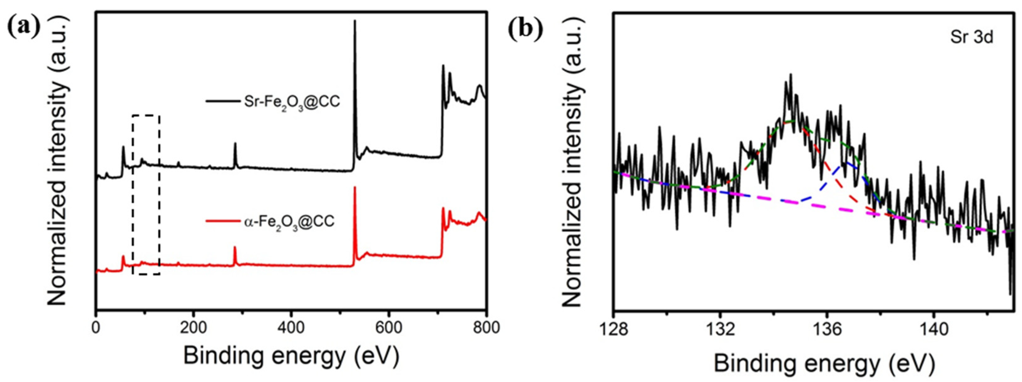
3.1. Characterization

Figure 1 shows the scanning electron microscope (SEM) images of Mg-Fe2O3@CC, Ca-Fe2O3@CC, Sr-Fe2O3@CC, and Ba-Fe2O3@CC. The SEM image of hematite supported on CC is shown in Figure S4. All four samples had similar morphology. SEM images at different magnifications confirm that the quenched α-Fe2O3 had good coverage and was evenly distributed on the surface of CC. It can be seen that the rapid quenching process did not cause the detachment or aggregation of α-Fe2O3 nanorods, and this characteristic still remained with the change of quenching solution, so nanomaterials doped with different alkaline earth metal ions can be obtained. α-Fe2O3 was supported on CC in the form of a one-dimensional nanorod array (Figure 1a), which not only provides a large electrochemically active surface, but also facilitates the rapid transfer of electrons and ions [23]. It is noted that the doping amounts of ions were different after quenching. When the quenching solution concentration was 0.5 mol/L, the doping amounts of Sr2+, Ba2+, Mg2+, and Ca2+ were 0.34 wt.%, 1.30 wt.%, 0.17 wt.%, and 0.9 wt.% respectively (Table 1), which may be related to the radius and electronegativity of ions.
Figure 2a exhibits the X-ray diffraction (XRD) pattern of the FeOOH precursor. The position and relative intensity of the diffraction peak matched with the JCPDS card of FeOOH (#75-1594) [24]. The diffraction peaks in the XRD pattern (Figure 2b) of α-Fe2O3 easily matched the hematite phase (JCPDS#33-0664), 2θ = 24.01°, 33.18°, 35.60°, 40.73°, 49.38°, 54.01°, and 62.32°, corresponding to the crystal planes (012), (104), (110), (113), (024), (116), (214), and (300), respectively [25], which shows that the modification of Sr2+, Ba2+, Mg2+, and Ca2+ did not significantly change the crystal structure of α-Fe2O3. However, it is difficult to detect the different diffraction peaks after quenching attributed to the small amounts of ions doping.
Figure 2c shows the Raman spectra collected for α-Fe2O3@CC, Sr-Fe2O3@CC, Ba-Fe2O3@CC, Mg-Fe2O3@CC, and Ca-Fe2O3@CC [26,27]. It is found that the Raman spectra of all the samples were almost unchanged after quenching treatment, which may be due to the low doping amounts. O2 temperature-programmed desorption (O2-TPD) is used to determine the concentration of oxygen vacancies [28]. The conductivity of the electrode material is related to the property of surface oxygen and the form of the oxygen species adsorbed on the surface. It can be identified by the following transformation reaction: O2 (adsorption)→O2− (adsorption)→O (adsorption)→O2− (adsorption/lattice) [29]. The first peak (~250 °C) is related to the desorption of physically adsorbed oxygen. The second peak (~500 °C) is related to the chemical desorption of O2− (and/or O). The third peak (<600 °C) is related to the desorption of lattice oxygen. It can be seen from Figure 2d that the area of the second peak increased in all samples after quenching in alkaline earth metal solution, which means an increase in oxygen defects. Among them, the area of the second peak of Ba-Fe2O3@CC increased the most, while Sr-Fe2O3@CC increased the least. On the one hand, the increase in oxygen vacancies is conducive to the improvement of electrical conductivity. The capacitance also is affected by doping amount, electrochemical specific surface area (ECSA), and other factors, so comprehensive analysis is needed.
The X-ray photoelectron spectroscopy (XPS) survey spectra for Sr-Fe2O3@CC are shown in Figure 3a, revealing the presence of Sr in Sr-Fe2O3@CC. Furthermore, the Sr 3d XPS spectrum in Figure 3b shows the presence of satellite peaks 134.70 and 136.80 eV for Sr2+ [30,31].
The specific Brunauer–Emmett–Teller (BET) surface area of the sample is slightly differentdue to alkaline earth metal ions doping after quenching (Table 2) [32]. The Nitrogen adsorption/desorption isotherms are shown in Figure S5. Compared with the undoped α−Fe2O3@CC, the specific surface area of Sr-Fe2O3@CC increased from 77.18 m2 g−1 to 79.60 m2 g−1, which may due to the defective surface caused by Sr2+ doping [33]. The specific surface area of the Ba-Fe2O3@CC sample was reduced by 74.65 m2 g−1. The specific surface area of Mg-Fe2O3@CC was almost unchanged due to the low doping amount [34]. The specific surface area of Ca-Fe2O3@CC was greatly reduced by 53.06 m2 g−1, which may be related to the pore destruction of α-Fe2O3 caused by the doping of Ca2+.
The increase in the ECSA enhances the contact area between the electrode and electrolyte and then increases the accumulation of charge [34]. To further explore the factors that affect the increase in capacitance, the ECSA was determined by measuring the electric double layer capacitance (CDL) from the CV curve at various scan rates, shown in Figure 4, and the ECSA value was determined by the slope of ic-v, shown in Table 3. After doping with alkaline earth metal ions, the ECSA of all samples improved, except for Ca-Fe2O3@CC. The doping of Sr2+ ions can induce the change of nanostructured surface of α-Fe2O3, leading to generation of more active sites and the increase in the ECSA. The ECSA of Sr-Fe2O3@CC reached twice that of α-Fe2O3@CC, which can result in a substantial improvement in the capacitance of Sr-Fe2O3@CC. Table 3 shows the ECSA value of Ca-Fe2O3@CC. It can be seen that after Ca2+ doping, the ECSA of α-Fe2O3@CC dropped by 98%. Ca2+ ions can affect the inner surface potential, changing the surface charge due to their adsorption and formation the Stern plane in the EDL. Therefore, Ca2+ may induce particle coagulation, which may destroy the pore structure of the material surface, resulting in a decrease in BET and ECSA [35]. Therefore, the ability of α-Fe2O3 to transport electrons and store charges is reduced.

3.2. Electrochemical Performances of M-Fe2O3@CC Electrode

In order to explore the electrochemical properties of the materials before and after quenching, cyclic voltammetry (CV) and galvanostatic charge–discharge (GCD) tests were carried out in a standard three-electrode system with M-Fe2O3@CC as the working electrode, the platinum mesh as the counter electrode, and an SCE as the reference electrode, tested in 5 M LiCl solution.
Figure 5a shows the CV curves of different electrodes collected at a scan rate of 100 mV s−1. The current density of the Sr-Fe2O3@CC electrode was significantly higher than those of the unquenched α-Fe2O3@CC electrode and the α−Fe2O3@CC electrode doped with Ba2+, Mg2+, and Ca2+, indicating that the Sr2+ modification was the most effective form in the four alkaline earth metal ions [36]. This is because, with the doping of Sr2+ ions, the ECSA increases, which greatly increases the EDLC of Sr−Fe2O3@CC. At the same time, the increase in oxygen vacancies enhances the electron and ion transfer rate between the electrode and electrolyte, which accelerates the rate of redox reactions and greatly increases the pseudocapacitance. The capacitance of the Ca−Fe2O3@CC electrode was significantly smaller than that of the original α−Fe2O3@CC, which proves that the modification of Ca2+ has a side effect on α−Fe2O3@CC. In addition, the shape of the CV curve (Figure 5a) was also different from other electrodes. This may be due to the fact that the surface structure breaks after Ca2+ doping. At the same time, its ability for electron adsorption and charge storage decreased with the reduction in ECSA, resulting in a decrease in EDLC, both of which make the final capacitance reduce massively.
Figure 5b shows the GCD curves of Sr−Fe2O3@CC, Ba−Fe2O3@CC, Mg−Fe2O3@CC, and Ca−Fe2O3@CC collected at a current density of 1 mA cm−2. Sr−Fe2O3@CC exhibited the highest area capacitance among all tested electrodes, reaching 77.81 mF cm−2 at 0.5 mA cm−2. When the current density increased from 0.5 mA cm−2 to 8 mA cm−2 (Figure 5c), the charge–discharge curve of Sr−Fe2O3@CC maintained an equilateral triangle shape, indicating that Sr−Fe2O3@CC has excellent capacitance and faraday reaction reversibility [37]. When tested at high current density (8 mA cm−2), a capacitance of 67.34% was maintained (Figure 5d). The EIS curve (Figure 5e) shows that the Sr−Fe2O3@CC had the lowest equivalent series resistance [38]. For supercapacitors, stability is an important consideration. Figure 5f shows that Ba−Fe2O3@CC, Mg−Fe2O3@CC, and Sr−Fe2O3@CC possessed good cycling stability and had no downward trend (a capacitance of 100% of the initial capacitance maintain) after 800 charge–discharge cycles at the current density of 1 mA cm−2. However, the initial capacity was relatively low, and it needed to be activated for a period of time.

3.3. Formatting of Mathematical Components

The areal capacitance (Cs) in the three-electrode cell was calculated according Equation (1):
C s = I × t s × v
where I is the discharge current, ∆t is the discharge time, s is the electrode area, v is the electrode volume, and ∆v is the potential. The area of the M-Fe2O3@CC fiber is about 1 cm2.
Cyclic voltammetry curves were collected in 1 M NaOH at different scan rates. The electrochemical specific surface areas of Sr-Fe2O3@CC, Ba-Fe2O3@CC, Mg-Fe2O3@CC, and Ca-Fe2O3@CC electrodes were tested under −0.06 V to 0.00 V (relative to SCE). The double-layer current (ic) is equal to the product of the scan rate (v) and the electrochemical double-layer capacitance (CDL), as shown in Equation (2):
i c = v C D L  
The slope value of the ic-v fitted line (Figure 3b,d,f,h,i) is equal to CDL. The ECSA value was obtained through calculation according to Equation (3):
ECSA = C D L C s
where Cs is the specific capacitance, which is 40.00 μF cm−2 for general metal oxides.

4. Conclusions

In summary, we introduced a new method, quenching, to modify the surfaces of nanomaterials for improving electrochemical performance of α−Fe2O3 nanorods. α−Fe2O3@CC was quenched in Sr(NO3)2, Ba(NO3)2, Mg(NO3)2, and Ca(NO3)2 solutions, and Sr2+, Ba2+, Mg2+, and Ca2+ ions were successfully doped into α−Fe2O3 nanorods. The doping amounts of ions varies with the different ionic radius and valence of the ions. The Sr−Fe2O3@CC electrode achieved the best performance, reaching 77.81 mF cm−2 at 0.5 mA cm−2. Due to the doping of Sr2+, the ECSA increases, which greatly increases the EDLC of Sr−Fe2O3@CC. Besides, the increase in oxygen vacancies accelerates the electron and ion transfer rate between the electrode and electrolyte, resulting in the increase in the pseudocapacitance. It is interesting to note that the modification of Ca2+ has the opposite effect. The capacitance of Ca−Fe2O3@CC has decreased, which proves that not all ion modifications can have a positive effect on the capacitance of supercapacitors. The function needs to be selected according to the properties of the ion itself, such as radius, redox, and so on.

Supplementary Materials

The following are available online at https://www.mdpi.com/article/10.3390/pr9071102/s1, Figure S1: Digital photo of (a) the thickness (b) length and width of carbon cloth. (c,d) SEM images of carbon cloth in different magnifications., Figure S2. Schematic of hydrothermal reaction. Figure S3. Schematic of three electrode test. Figure S4. SEM image of hematite supported on CC. Figure S5. Nitrogen adsorption/desorption isotherms for α-Fe2O3@CC, Sr-Fe2O3@CC, Ba-Fe2O3@CC, Mg-Fe2O3@CC and Ca-Fe2O3@CC.

Author Contributions

Conceptualization and methodology, Y.Q. and M.R.; data curation, Q.C. and M.S.; writing—original draft preparation, Q.C. and M.S.; writing—review and editing, M.S. and Y.C.; project administration, Y.Q.; funding acquisition, Y.Q. All authors have read and agreed to the published version of the manuscript.

Funding

This research was funded by National Key Research and Development Program of China (2018YFA0209600), Science and Technology Key Project of Guangdong Province, China (2020B010188002), Guangdong Innovative and Entrepreneurial Research Team Program (2019ZT08L075), Foshan Innovative and Entrepreneurial Research Team Program (2018IT100031), Guangdong Pearl River Talent Program (2019QN01L054), Shenzhen Peacock Plan (KQTD2016053015544057) and Nanshan Pilot Plan (LHTD20170001), Science and Technology Program of Guangzhou, China (202002030153), the Guangdong Science and Technology Program (2017B030314002), National Natural Science Foundation of China (52000076), Innovation and Entrepreneurship Talent Program of Shaoguan City, China Postdoctoral Science Foundation (2020M682714) and the Fundamental Research Funds for the Central Universities (2020ZYGXZR061).

Institutional Review Board Statement

Not applicable.

Informed Consent Statement

Not applicable.

Data Availability Statement

Not applicable.

Conflicts of Interest

The authors declare no conflict of interest.

References

  1. Zhang, L.L.; Zhao, X.S. Carbon−based materials as supercapacitor electrodes. Chem. Soc. Rev. 2009, 38, 2520–2531. [Google Scholar] [CrossRef] [PubMed]
  2. Chen, D.; Zhou, S.; Quan, H.; Zou, R.; Gao, W.; Luo, X.; Guo, L. Tetsubo−like α−Fe2O3/C nanoarrays on carbon cloth as negative electrode for high−performance asymmetric supercapacitors. Chem. Eng. J. 2018, 341, 102–111. [Google Scholar] [CrossRef]
  3. Choudhury, A.; Kim, J.-H.; Yang, K.-S.; Yang, D.-J. Facile synthesis of self−standing binder−free vanadium pentoxide−carbon nanofiber composites for high−performance supercapacitors. Electrochim. Acta 2016, 213, 400–407. [Google Scholar] [CrossRef]
  4. Kandalkar, S.G.; Gunjakar, J.L.; Lokhande, C.D. Preparation of cobalt oxide thin films and its use in supercapacitor application. Appl. Surf. Sci. 2008, 254, 5540–5544. [Google Scholar] [CrossRef]
  5. Huang, H.; Wang, X.; Tervoort, E.; Zeng, G.; Liu, T.; Chen, X.; Sologubenko, A.; Niederberger, M. Nano−Sized Structurally Disordered Metal Oxide Composite Aerogels as High−Power Anodes in Hybrid Supercapacitors. ACS Nano 2018, 12, 2753–2763. [Google Scholar] [CrossRef] [PubMed]
  6. Wei, W.; Cui, X.; Chen, W.; Ivey, D.G. Manganese oxide−based materials as electrochemical supercapacitor electrodes. Chem. Soc. Rev. 2011, 40, 1697–1721. [Google Scholar] [CrossRef] [PubMed]
  7. Jyothibasu, J.P.; Wang, R.-H.; Ong, K.; Ong, J.H.L.; Lee, R.-H. Cellulose/carbon nanotube/MnO2 composite electrodes with high mass loadings for symmetric supercapacitors. Cellulose 2021, 28, 3549–3567. [Google Scholar] [CrossRef]
  8. Xia, H.; Shirley Meng, Y.; Yuan, G.; Cui, C.; Lu, L. A Symmetric RuO2/RuO2 Supercapacitor Operating at 1.6 V by Using a Neutral Aqueous Electrolyte. Electrochem. Solid State Lett. 2012, 15, A60–A63. [Google Scholar] [CrossRef] [Green Version]
  9. Jiang, Q.; Kurra, N.; Alhabeb, M.; Gogotsi, Y.; Alshareef, H.N. All Pseudocapacitive MXene−RuO2Asymmetric Supercapacitors. Adv. Energy Mater. 2018, 8, 1703043. [Google Scholar] [CrossRef]
  10. Yang, Z.; Qiu, A.; Ma, J.; Chen, M. Conducting α−Fe2O3 nanorod/polyaniline/CNT gel framework for high performance anodes towards supercapacitors. Compos. Sci. Technol. 2018, 156, 231–237. [Google Scholar] [CrossRef]
  11. Li, Y.; Xu, J.; Feng, T.; Yao, Q.; Xie, J.; Xia, H. Fe2O3 Nanoneedles on Ultrafine Nickel Nanotube Arrays as Efficient Anode for High−Performance Asymmetric Supercapacitors. Adv. Funct. Mater. 2017, 27, 1606728. [Google Scholar] [CrossRef]
  12. Owusu, K.A.; Qu, L.; Li, J.; Wang, Z.; Zhao, K.; Yang, C.; Hercule, K.M.; Lin, C.; Shi, C.; Wei, Q.; et al. Low−crystalline iron oxide hydroxide nanoparticle anode for high−performance supercapacitors. Nat. Commun. 2017, 8, 14264. [Google Scholar] [CrossRef] [PubMed]
  13. Zhang, X.; Wang, Q.; Xia, X.; He, W.; Huang, X.; Logan, B.E. Addition of conductive particles to improve the performance of activated carbon air−cathodes in microbial fuel cells. Environ. Sci. Water Res. Technol. 2017, 3, 806–810. [Google Scholar] [CrossRef]
  14. Ma, Z.; Huang, X.; Dou, S.; Wu, J.; Wang, S. One−Pot Synthesis of Fe2O3 Nanoparticles on Nitrogen−Doped Graphene as Advanced Supercapacitor Electrode Materials. J. Phys. Chem. C 2014, 118, 17231–17239. [Google Scholar] [CrossRef]
  15. Raut, S.S.; Sankapal, B.R. Comparative studies on MWCNTs, Fe2O3 and Fe2O3/MWCNTs thin films towards supercapacitor application. New J. Chem. 2016, 40, 2619–2627. [Google Scholar] [CrossRef]
  16. Shivakumara, S.; Penki, T.R.; Munichandraiah, N. Synthesis and Characterization of Porous Flowerlike Fe2O3 Nanostructures for Supercapacitor Application. ECS Electrochem. Lett. 2013, 2, A60–A62. [Google Scholar] [CrossRef]
  17. Nie, G.; Lu, X.; Lei, J.; Jiang, Z.; Wang, C. Electrospun V2O5−doped α−Fe2O3composite nanotubes with tunable ferromagnetism for high−performance supercapacitor electrodes. J. Mater. Chem. A 2014, 2, 15495–15501. [Google Scholar] [CrossRef]
  18. Ni, C.; Hou, J.; Li, L.; Li, Y.; Wang, M.; Yin, H.; Tan, W.J.C. The remarkable effect of alkali earth metal ion on the catalytic activity of OMS−2 for benzene oxidation. Chemosphere 2020, 250, 126211. [Google Scholar] [CrossRef]
  19. Reddy, M.V.; Yao, Q.C.; Adams, S. Mg, Cu, Zn doped Fe2O3 as an electrode material for Li−ion batteries. Mater. Lett. 2018, 2, 186–192. [Google Scholar] [CrossRef]
  20. Liu, S.; Li, Z.; Wang, C.; Tao, W.; Huang, M.; Zuo, M.; Yang, Y.; Yang, K.; Zhang, L.; Chen, S.; et al. Turning main−group element magnesium into a highly active electrocatalyst for oxygen reduction reaction. Nat. Commun. 2020, 11, 938. [Google Scholar] [CrossRef] [PubMed] [Green Version]
  21. Guo, S.; Wang, H.; Yang, W.; Fida, H.; You, L.; Zhou, K. Scalable synthesis of Ca−doped α−Fe2O3 with abundant oxygen vacancies for enhanced degradation of organic pollutants through peroxymonosulfate activation. Appl. Catal. B Environ. 2020, 2, 118250. [Google Scholar] [CrossRef]
  22. Su, M.; Pan, Z.; Chong, Y.; Ye, C.; Jin, X.; Wu, Q.; Hu, Z.; Ye, D.; Waterhouse, G.I.N.; Qiu, Y.; et al. Boosting the electrochemical performance of hematite nanorods via quenching−induced metal single atom functionalization. J. Mater. Chem. A 2021, 9, 3492–3499. [Google Scholar] [CrossRef]
  23. Poornajar, M.; Nguyen, N.; Ahn, H.−J.; Büchler, M.; Liu, N.; Kment, S.; Zboril, R.; Yoo, J.; Schmuki, P. Fe2O3 Blocking Layer Produced by Cyclic Voltammetry Leads to Improved Photoelectrochemical Performance of Hematite Nanorods. Surfaces 2019, 2, 131–144. [Google Scholar] [CrossRef] [Green Version]
  24. Bouhjar, F.; Derbali, L.; Marí, B.; Bessaïs, B. Electrodeposited Cr−Doped α−Fe2O3 thin films active for photoelectrochemical water splitting. Int. J. Hydrog. Energy 2020, 45, 11492–11501. [Google Scholar] [CrossRef]
  25. Kumar, P.; Sharma, P.; Shrivastav, R.; Dass, S.; Satsangi, V.R. Electrodeposited zirconium−doped α−Fe2O3 thin film for photoelectrochemical water splitting. Int. J. Hydrog. Energy 2011, 36, 2777–2784. [Google Scholar] [CrossRef]
  26. Shen, S.; Guo, P.; Wheeler, D.A.; Jiang, J.; Lindley, S.A.; Kronawitter, C.X.; Zhang, J.Z.; Guo, L.; Mao, S.S. Physical and photoelectrochemical properties of Zr−doped hematite nanorod arrays. Nanoscale 2013, 5, 9867. [Google Scholar] [CrossRef]
  27. Shen, S.; Jiang, J.; Guo, P.; Kronawitter, C.X.; Mao, S.S.; Guo, L. Effect of Cr doping on the photoelectrochemical performance of hematite nanorod photoanodes. Nano Energy 2012, 1, 732–741. [Google Scholar] [CrossRef]
  28. Wu, Y.; Dong, L.; Li, B. Effect of iron on physicochemical properties: Enhanced catalytic performance for novel Fe2O3 modified CuO/Ti0.5Sn0.5O2 in low temperature CO oxidation. Mol. Catal. 2018, 456, 65–74. [Google Scholar] [CrossRef]
  29. Ayastuy, J.L.; Gurbani, A.; Gutiérrez−Ortiz, M.A. Effect of calcination temperature on catalytic properties of Au/Fe2O3 catalysts in CO−PROX. Int. J. Hydrog. Energy 2016, 41, 19546–19555. [Google Scholar] [CrossRef]
  30. Li, M.; Liu, H.; Zhu, H.; Gao, H.; Zhang, S.; Chen, T. Kinetics and mechanism of Sr(II) adsorption by Al−Fe2O3: Evidence from XPS analysis. J. Mol. Liq. 2017, 2, 364–369. [Google Scholar] [CrossRef]
  31. Komai, S.; Hirano, M.; Ohtsu, N. Spectral analysis of Sr 3d XPS spectrum in Sr−containing hydroxyapatite. Surf. Interface Anal. 2020, 52, 823–828. [Google Scholar] [CrossRef]
  32. Wang, Y.; Wang, Y.; Cao, J.; Kong, F.; Xia, H.; Zhang, J.; Zhu, B.; Wang, S.; Wu, S. Low−temperature H2S sensors based on Ag−doped α−Fe2O3 nanoparticles. Sens. Actuators B Chem. 2008, 131, 183–189. [Google Scholar] [CrossRef]
  33. Banerjee, A.M.; Shirole, A.R.; Pai, M.R.; Tripathi, A.K.; Bharadwaj, S.R.; Das, D.; Sinha, P.K. Catalytic activities of Fe2O3 and chromium doped Fe2O3 for sulfuric acid decomposition reaction in an integrated boiler, preheater, and catalytic decomposer. Appl. Catal. B Environ. 2012, 127, 36–46. [Google Scholar] [CrossRef]
  34. Zhao, X.; Meng, J.; Yan, Z.; Cheng, F.; Chen, J. Nanostructured NiMoO4 as active electrocatalyst for oxygen evolution. Chin. Chem. Lett. 2019, 30, 319–323. [Google Scholar] [CrossRef]
  35. Szymanek, K.; Charmas, R.; Piasecki, W. A study on the mechanism of Ca(2+) adsorption on TiO2 and Fe2O3 with the usage of calcium ion−selective electrode. Chemosphere 2020, 242, 125162. [Google Scholar] [CrossRef] [PubMed]
  36. Liu, D.; Zhao, X.; Su, R.; Hao, Z.; Jia, B.; Li, S.; Dong, L. Highly Porous Graphitic Activated Carbons from Lignite via Microwave Pretreatment and Iron−Catalyzed Graphitization at Low−Temperature for Supercapacitor Electrode Materials. Processes 2019, 7, 300. [Google Scholar] [CrossRef] [Green Version]
  37. Jiang, J.; Liu, X.X.; Han, J.; Hu, K.; Chen, J.S. Self−Supported Sheets−on−Wire CuO@Ni(OH)2/Zn(OH)2 Nanoarrays for High−Performance Flexible Quasi−Solid−State Supercapacitor. Processes 2021, 9, 680. [Google Scholar] [CrossRef]
  38. Zeng, Y.; Han, Y.; Zhao, Y.; Zeng, Y.; Yu, M.; Liu, Y.; Tang, H.; Tong, Y.; Lu, X. Advanced Ti−Doped Fe2O3@PEDOT Core/Shell Anode for High−Energy Asymmetric Supercapacitors. Adv. Energy Mater. 2015, 5, 1402176. [Google Scholar] [CrossRef]
Figure 1. SEM images of (a,b) Sr-Fe2O3@CC, (c,d) Ba-Fe2O3@CC, (e,f) Mg-Fe2O3@CC, and (g,h) Ca-Fe2O3@CC.
Processes 09 01102 g001
Figure 2. (a) XRD patterns for FeOOH. (b) XRD patterns, (c) Raman spectra, and (d) O2-TPD patterns for α-Fe2O3@CC, Sr-Fe2O3@CC, Ba-Fe2O3@CC, Mg-Fe2O3@CC, and Ca-Fe2O3@CC.
Processes 09 01102 g002
Figure 3. (a) XPS survey spectra of α-Fe2O3@CC and Sr-Fe2O3@CC. (b) Sr 3d core-level XPS spectra for Sr-Fe2O3@CC.
Processes 09 01102 g003
Figure 4. CV curves in non-Faradaic region and the ratio of cathodic/anodic current with various scan rates of (a,b) α-Fe2O3@CC, (c,d) Ba-Fe2O3@CC, (e,f) Mg-Fe2O3@CC, (g,h) Sr-Fe2O3@CC, and (i,j) Ca-Fe2O3@CC.
Processes 09 01102 g004
Figure 5. (a) CV curves collected at a scan rate of 100 mV s−1 for Sr-Fe2O3@CC, Ba-Fe2O3@CC, Mg-Fe2O3@CC, α-Fe2O3@CC, and Ca-Fe2O3@CC. (b) GCD curves collected at a current density of 1 mA cm−2 for Sr-Fe2O3@CC, Ba-Fe2O3@CC, Mg-Fe2O3@CC, and Ca-Fe2O3@CC. (c) GCD curves for the Sr-Fe2O3@CC electrode collected at different current densities. (d) Specific areal capacitances and (e) EIS curves for Sr-Fe2O3@CC, Ba-Fe2O3@CC, Mg-Fe2O3@CC, and Ca-Fe2O3@CC at different current densities. (f) Cycling stability for Ba-Fe2O3@CC, Mg-Fe2O3@CC, and Sr-Fe2O3@CC at a current density of 1 mA cm2.
Processes 09 01102 g005
Table 1. The content of Sr2+, Ba2+, Mg2+, and Ca2+ in Sr-Fe2O3@CC, Ba-Fe2O3@CC, Mg-Fe2O3@CC, and Ca-Fe2O3@CC according to the ICP-OES measurement.
SampleM2+
(mg/Kg)
M2+
(wt. %)
Sr-Fe2O3@CC33650.34
Ba-Fe2O3@CC12,9651.30
Mg-Fe2O3@CC16910.17
Ca-Fe2O3@CC90230.90
Table 2. BET surface area for α-Fe2O3@CC, Sr-Fe2O3@CC, Ba-Fe2O3@CC, Mg-Fe2O3@CC, and Ca-Fe2O3@CC.
SampleBET Surface Area
(m2 g−1)
α-Fe2O3@CC77.18
Sr-Fe2O3@CC79.60
Ba-Fe2O3@CC74.65
Mg-Fe2O3@CC77.52
Ca-Fe2O3@CC53.06
Table 3. ECSA for α-Fe2O3@CC, Sr-Fe2O3@CC, Ba-Fe2O3@CC, Mg-Fe2O3@CC, and Ca-Fe2O3@CC.
SampleCDL (mF)ECSA (cm2)
α-Fe2O3@CC0.20505.12
Sr-Fe2O3@CC0.445011.13
Ba-Fe2O3@CC0.28007.00
Mg-Fe2O3@CC0.38009.50
Ca-Fe2O3@CC0.00460.12
Publisher’s Note: MDPI stays neutral with regard to jurisdictional claims in published maps and institutional affiliations.

