Article

UPLC-QToF Nanospray MS and NMR Analysis of Ficus sycomorus Stem Bark and Its Effects on Rabbit

1
Department of Husbandry and Animal Wealth Development, Faculty of Veterinary Medicine, University of Sadat City, Menofia 32897, Egypt
2
Department of Physiology, Faculty of Veterinary Medicine, University of Sadat City, Menofia 32897, Egypt
3
Department of Parasitology, Faculty of Veterinary Medicine, University of Sadat City, Menofia 32897, Egypt
4
Department of Analytical Chemistry, Faculty of Pharmacy, University of Sadat City, Menofia 32897, Egypt
5
Division of Pharmacognosy, Department of Pharmaceutical Biosciences, Uppsala University, Biomedical Centre, Box 574, SE-751 23 Uppsala, Sweden
6
Department of Chemistry, Faculty of Science, Menoufia University, Shebin El-Kom 32512, Egypt
7
Avian and Rabbit Diseases Department, Faculty of Veterinary Medicine, Sadat City University, Menofia 32897, Egypt
8
Research and Development Section, PerNaturam GmbH, 56290 Gödenroth, Germany
9
Department of Pathology, Faculty of Veterinary Medicine, University of Sadat City, Menofia 32897, Egypt
*
Authors to whom correspondence should be addressed.
Processes 2021, 9(7), 1201; https://doi.org/10.3390/pr9071201
Submission received: 26 June 2021 / Revised: 5 July 2021 / Accepted: 8 July 2021 / Published: 12 July 2021

Abstract

:
In the present study, a phytochemical of Ficus sycomorus (Moraceae family) was screened, and the effect of this extract on rabbit performance indices, immunity, and carcass quality measures was determined. Ficus sycomorus samples were collected, air-dried, and extracted with 70% methanol to prepare a solution of 100 mg/mL concentration. The extract was subjected to high-resolution mass spectrometric measurements via ultra-high performance liquid chromatography-quadrupole time-of-flight-nanospray mass spectrometry (UPLC-QToF-MS) and 1H NMR analysis. Forty-eight male rabbits, one-month-old, belonging to the Blanc de Bouscat and New Zealand White breeds were selected and distributed equally in a 2 × 3 factorial trial. The rabbits within each breed received F. sycomorus extract at the dose of 0, 100, and 200 mg/kg for 60 days. Blood samples were collected and serum obtained for the detection of liver enzymes, serum lipids, and proteins. The results of UPLC-QToF-MS and molecular networking analysis revealed the presence of procyanidin B2, procyanidin A1, genistein, eriodyctiol, catechin, luteolin, biochanin A, and chlorogenic acid that might exhibit various pharmaceutical activities. However, the F. sycomorus extract reduced rabbit performance indices and carcass quality measures. In addition, this extract significantly depressed the low-density lipoprotein and triglycerides, which may indicate the antidyslipidemia effect of this extract on rabbits.

1. Introduction

Ficus sycomorus (F. sycomorus) is one of the ancient trees belonging to the family Moraceae. This fruit tree, a native of Africa, is widely distributed in the Mediterranean basin of Egypt. The fruit, known as fig, is used as food and for other medicinal properties. The Moraceae family consists of approximately 40 genera and over 1000 species of flowering plants, most of which are widely distributed in tropical and subtropical regions [1]. The F. sycomorus contains numerous bioactive chemical ingredients, predominantly alkaloids, tannins, flavonoids, and phenolic compounds [2,3]. These bioactive compounds are found in the fruits, leaves, and bark of the tree. Thus, a detailed investigation is required to understand the useful properties of this plant [2,4,5].
The F. sycomorus fruits have been utilized for the treatment of jaundice, fungal infections, and dysentery [6]. Moreover, these fruits are used for controlling cough, liver disease, stomach disorders, diarrhea, skin infection, epilepsy, tuberculosis, lactation disorders, helminthiasis, infertility, and sterility [2,4]. Furthermore, the stem bark milky latex is topically used for treating ringworm [7], ulcers, burns, inflammation, warts, and wound infections. In addition, the boiled F. sycomorus bark is used in the treatment of sore throat, scrofula, respiratory diseases, chest diseases, and infections [8]. The powdered stem bark is used for the treatment of pains, diarrhea, epilepsy, insomnia, and other mental disorders [9,10]. Similarly, it is used for the treatment of animal diarrhea in Nigeria [11].
The aqueous/ethanolic extracts of F. sycomorus leaves have potential therapeutic effects against the methicillin and oxacillin resistant Staphylococcus aureus (MRSA, ATCC43300)-induced keratitis in rabbits. Similarly, significant bacteriostatic activity was reported for the alcoholic extract of F. sycomorus, while a bactericidal activity was achieved with the aqueous extract [12]. Moreover, the stem bark extract of F. sycomorus has many tannins, saponins, flavones, aglycones, reducing sugars, anthraquinone glycosides, flavonoid glycosides, and condensed tannins, which could inhibit either smooth or skeletal muscle contractions [2].
The F. sycomorus extract is a rich source of flavonoids and phenolics [13]. Flavonoids can prevent bacterial growth and suppress its virulence [14]. This antibacterial activity has been reported in the fruit extract of Egyptian Ficus species, including F. sycomorus [15]. The F. sycomorus extract has many medicinal properties, including antitumor [16], antimicrobial [6,17], and anti-inflammatory activities [18]. Moreover, this extract exhibited anticonvulsant [2] and sedative properties [6], antidiabetic activity [19], antidiarrheal activity [20], antioxidant activity [21], and insecticidal and acaricidal activities [22]. The extract also has hepatoprotective activity [12,23,24] and increases sperm cell production in rats [25].
Although many researchers have investigated the various medicinal effects of different F. sycomorus extracts, there is a scarcity of information on the effect of F. sycomorus extracts on growth performance and immunity. Therefore, the current study aimed to investigate the effect of stem bark extract of F. sycomorus on growth performance, hematologic parameters, and carcass quality of growing rabbits.

2. Materials and Methods

2.1. The Plant Materials and Preparation of the Extracts

Stem bark samples of F. sycomorus were collected from the Menofia Governorate, Egypt. The voucher specimens were packaged tightly in plastic bags and then deposited in the investigation laboratory of the University of Sadat City, Egypt. The plant samples were air-dried for ten days under the shade and then uniformly powdered using a Thomas-Willey milling machine with a 60-mesh size. For solvent extraction, 100 g of dried powdered samples were dissolved in 500 mL of 70% methanol for three days. Then, the mixture was filtered under pressure and dried in a rotary evaporator under reduced pressure at 22 °C to obtain the crude extract. This was performed until a constant weight was obtained. The extract of F. sycomorus was stored in a tightly stoppered container of brown glass in the refrigerator at 4 °C until required.

2.2. UPLC-QToF-MS Analysis and Molecular Networking for Screening of Secondary Metabolites

High-resolution mass spectrometric measurements were acquired using an ultra-high performance liquid chromatography (UPLC)-quadrupole time-of-flight (QToF)-nanospray mass spectrometry (MS) (Waters nanoAcquity, QToF Micro, Santa Clara, CA, USA). The UPLC column used was a Waters ACQUITY UPLC M-Class Peptide BEH C18 column (1.7 μm, 130 Å, 75 μm × 150 mm), using solvents A (0.1% in formic acid (FA) in water) and B (0.1% FA in acetonitrile (AcN)) over 75 min with a flow rate of 0.3 µL/min.

2.3. NMR Analysis

Proton nuclear magnetic resonance (1H NMR) spectra were recorded at 298 K on a Bruker 600 MHz (TCI CRPHe TR-1H and 19F/13C/15N 5 mm-EZ CryoProbe) spectrometer. Chemical shifts were referenced to the solvent peak for (CD3)2SO at δH 2.50 ppm.

2.4. Experimental Evaluation of the Effect of F. sycomorus on Rabbit

All experimental procedures of this study have been approved by the Animal Ethics Committee of the University of Sadat City, approval number 20/2020. Forty-eight male rabbits of two different breeds, Blanc de Bouscat (BDB) and New Zealand White (NZW) (24 rabbits from each breed), were selected from a local government farm in Sharqia Governorate, Egypt. The rabbits, which were recently weaned, were on average 30 ± 5 days old with a bodyweight (BW) of 0.730 ± 0.8 kg and 0.650 ± 0.06 kg for BDB and NZW breeds, respectively. The male rabbits were enrolled into a 2 × 3 trial (2 breeds × 3 doses of F. sycomorus stem bark extract). Rabbits within each breed were randomly allocated into 3 groups of 8 animals each. The study was conducted between November 2019 to May 2020, and during this period, the animals were housed in individual cages of galvanized steel, slatted floors, and self-serve waterspout at 25 ± 2 °C and 45 ± 5% humidity under 12 h light/dark cycle (lights on at 6:00 a.m., lights off at 6:00 p.m.). Water was provided ad libitum, while the ration fed to the rabbits comprised of 18% crude protein Table 1. Before the experiment, the animals were acclimatized under controlled standard conditions for 2 weeks. During the acclimatization period, rabbits were subjected to anthelmintic and antiprotozoal therapy via fenbendazole (4 mL/100 kg BW) and toltrazuril 5% (25 mg/kg BW). Fecal samples were examined after 3 days of induction of the anthelmintic and the antiprotozoal therapy to confirm free cases.
Starting from the third week, the F. sycomorus crude extract was weighed and dissolved in distilled water to form a 100 mg/mL solution. Within each rabbit breed, the 1st, 2nd, and 3rd rabbit groups were subjected to oral dosing with F. sycomorus extracts 0, 100, and 200 mg/kg BW [26], respectively, for the next 60 days. Feed intake and body weight were estimated, and daily body gain, feed conversion, and feed efficiency were calculated.
Blood samples were collected between 9:00 and 11:00 a.m. on day 60 post the extract dosing. The samples were drawn from marginal ear vein and divided into three parts; the first one was centrifuged at 3000 rpm to obtain the serum, the second part was heparinized for the estimation of alanine aminotransferase (ALT), aspartate aminotransferase (AST), and alkaline phosphatase (ALP) using a spectrophotometer (Fisher Scientific UK Ltd, Loughborough, UK) and available commercial kits (Spectra CO., 6th October City, Egypt), and the third part was treated with EDTA and was used for estimating WBCs, lymphocytes, RBCs, and hemoglobin using automated hematology analyzers [27]. Serum samples were kept in deep freeze at −20 °C until further investigations. Serum proteins and lipids were estimated using suitable reagents and a spectrophotometer. Serum globulin was estimated by subtracting the serum albumin from total serum protein.
The rabbits were weighed before and after the feed, after a withholding period of 6 h. At the end of the experiment, the animals were humanely slaughtered, and the weights of the hot carcass, kidney, heart, liver, and abdominal fat pad were assessed. In addition, the dressing percentage was calculated.

2.5. Histopathological Examination

Immediately after the slaughter, the tissue samples were taken from the liver and fixed in 10% formalin for 72 h. The samples were trimmed, washed, dehydrated, embedded in paraffin wax, and sectioned with a microtome of 3 μm thickness. Finally, the samples were stained with hematoxylin and eosin and examined under a Leica DMLB microscope (Leica, Wetzlar, Germany). The images were taken via the Leica EC3 digital camera.

2.6. Statistical Analysis

Data of RBCs and WBCs were logarithmically transformed, in addition to the percentage data of lymphocytes and dressing percentage that were Arcsine transformed to obtain normally distributed values. Data on growth performance, hemogram, serum biochemicals, organ weights, and carcass quality were statistically analyzed using the General Linear Models (GLM) procedures of SAS software (SAS User’s Guide: Statistics, Version 8.1 Edition, 2000, SAS Inst. Inc., Cary, NC, USA). Covariate final body weight was used together with final body weight to prevent the inflation of results from the initial body weight. A Tukey post hoc test was conducted as a means separation test. The level of statistical significance was set at p < 0.05.

3. Results

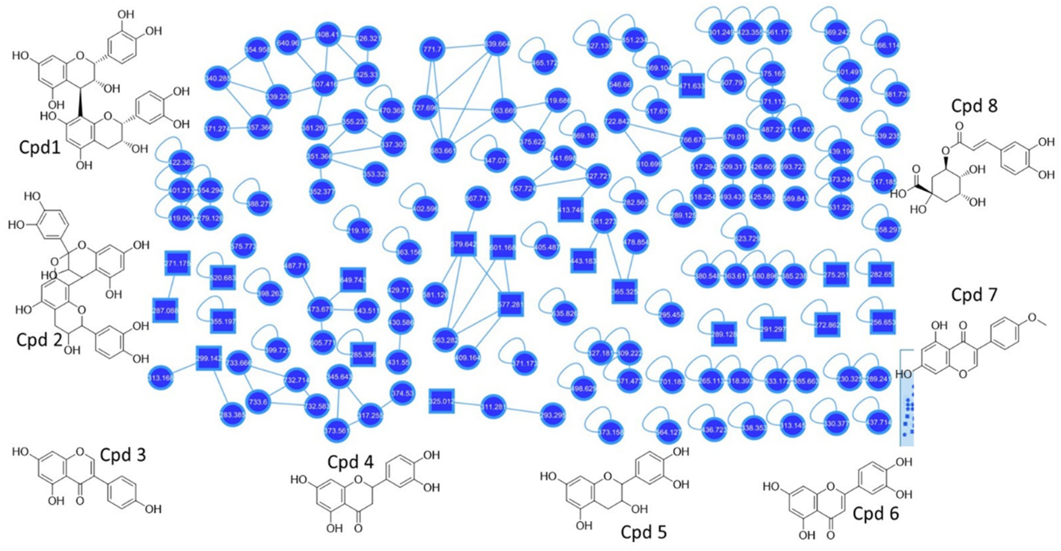
3.1. UPLC-QToF-MS and Molecular Networking Analysis

We investigated the molecular networking of F. sycomorus in this study. The major compounds present in the methanolic extract of F. sycomorus stem bark are listed in Table 2, and their molecular networking is illustrated in Figure 1, Supplementary Figures S1 and S2. The investigation revealed eight major metabolites, which were further confirmed by matching their fragmentation with the database of previously isolated compounds. Cpd1 with m/z 579.642 [C30H27O12]+ was annotated as procyanidin B2, while Cpd2 with m/z 577.281 [C30H25O12]+ was annotated as procyanidin A1. Furthermore, Cpd3 with m/z 271.175 [C15H11O5]+ was reported to be genistein. Cpd4 with the molecular formula [C15H13O6]+ and m/z 289.128 was identified as dihydro-tetrahydroxy flavanone, also known as eriodyctiol. Cpd5 with m/z 291.297 [C15H15O6]+ was elucidated as catechin. Cpd6 with m/z 287.088 and molecular formula [C15H11O6]+ was annotated as tetrahydroxy flavanone, also known as luteolin. Cpd7 with m/z 285.326 [C16H13O5]+ was reported as biochanin A. Finally, Cpd8 with m/z 355.197 [C16H19O9]+ was annotated as chlorogenic acid.

3.2. NMR Analysis

Although NMR did not provide any conclusive information, the combination of MS and NMR data helped in determining the compounds. Terminal CH3 groups at a chemical shift of 0.9 ppm and the long chain of CH2 groups at 1.2 ppm indicated the presence of fatty acids. The 1,4 di substitution aromatic ring in Cpd3 and Cpd7 were observed at a chemical shift of 6.2–6.8 ppm and 8–8.5 ppm, respectively. Moreover, the AB spectrum showed chemical shifts of 5.5–6.2 ppm, as observed in Cpd1, 3, 4, 5, 6, and 7.

3.3. Growth Performance

The effect of rabbit breed on growth performance parameters is given in Supplementary Table S1. Superior growth performance was observed in the BDB breed compared to the NZW breed. A significant increase in the body weight (2063.72 gm), average daily gain (14.63 gm), feed efficiency (0.21), and depressed feed conversion (4.83) was noted in the BDB breed compared to the NZW breed (p < 0.00). However, the stem bark extract of F. sycomorus significantly affected the growth performance of the rabbits, irrespective of the breed. The 200 mg/kg dose significantly depressed average daily gain (12.66 gm) and feed efficiency (0.18) with a high feed conversion ratio (5.66) compared to the doses of 0 and 100 mg/kg (p < 0.001). This dose depressed the final (1953.57 gm), as well as the covariate final body weight (1889.64 gm), compared to the other two doses. However, this decrease was not significant (p < 0.50; p < 0.37, respectively). Moreover, the performance parameters with doses of 0, 100 mg/kg did not differ from each other. The effect of interaction between rabbit breed and extract dose on the performance indices was not significant, except in the bodyweight. The F. sycomorus extract improved the bodyweight growth performance with a dose of 100 mg/kg; however, an opposite trend was observed in rabbits dosed with 200 mg/kg. This effect was evident in the two different rabbit breeds. The highest bodyweight was attained in the BDB breed dosed with 100 mg/kg of F. sycomorus extract, and the lowest in the NZW breed dosed with 200 mg/kg of the extract (p < 0.00).

3.4. Serum Biochemical Analysis and Hemogram

The results revealed no significant difference between the WBCs, lymphocyte, and granulocyte values in the different rabbit breeds or F. sycomorus doses (Supplementary Table S2). However, the WBCs and lymphocyte percentage values tended to decrease in the rabbits with both 100 mg/kg (9.48 ± 0.03 × 10³/mL; 0.48 ± 0.04%) and 200 mg/kg (9.63 ± 0.03 × 10³/mL; 0.47 ± 0.04%) doses of the F. sycomorus extract compared to the control (9.81 ± 0.03 × 10³/mL; 0.52 ± 0.03%). Nevertheless, this decrease was not significant. Moreover, the granulocyte percentage increased in the F. sycomorus-treated groups compared to the control group; however, this increase was not statistically significant. The RBCs, mean corpuscular volume (MCV), and mean corpuscular hemoglobin concentration (MCHC) values did not significantly differ between the rabbit breeds and the different doses of the extract. In contrast, hemoglobin and mean corpuscular hemoglobin (MCH) increased significantly in the BDB breed (13.03 ± 0.22 g/dL; 23.07 ± 0.31 pg) than the NZW breed (11.68 ± 0.21 g/dl; 21.93 ± 0.28 pg) (p < 0.05). In addition, no significant differences were observed in the serum proteins between the rabbit breeds and extract doses (Supplementary Table S2).
The BDB breed had lowest cholesterol and triglycerides values (39.43 ± 3.18 mg/dl; 120.22 ± 6.81 mg/dl) compared to the NZW breed (50.00 ± 3.09 mg/dl; 141.58 ± 6.61 mg/dl). Furthermore, the 100 mg/kg or 200 mg/kg BW F. sycomorus-treated rabbits had lower low density lipoproteins (LDL) (20.75 ± 1.65 mg/dl; 20.32 ± 1.48 mg/dl) and triglycerides (132.03 ± 7.51 mg/dl; 109.17 ± 9.14 mg/dl) compared with the control. This hypolipidemic effect was confirmed by the breed dose interaction, as the dose of 200 mg/kg BW resulted in the lowest level of triglycerides in each rabbit breed (p < 0.05). The BDB breed resulted in higher AST (12.96 U/L) compared with the NZW breed (11.51 U/L). In contrast, the BDB rabbits depressed ALP (33.84 IU/L) compared to NZW rabbits (39.18 IU/L) (p < 0.03). The F. sycomorus extract significantly increased the ALT values compared with the control, while it depressed the ALP values (p < 0.04). However, these parameters remained within normal levels (Supplementary Table S3).

3.5. Internal Organs and Carcass Quality

The BDB rabbits recorded higher liver (77.57) and abdominal fat pad (51.85 g) weights compared to the NZW rabbits (66.91 g; 40.93 g) (p < 0.00; p < 0.02). Moreover, an increase in the F. sycomorus extract dose resulted in a decrease in the kidney, heart, and abdominal fat pad weights. The rabbits receiving 200 mg/kg dose of the extract recorded the lowest kidney (13.36 g), heart (6.13 g), and abdominal fat pad weights (39.44 g) compared to the control group, which recorded the highest kidney (16.36 g), heart (7.11 g), and abdominal fat pad weights (53.63 g). Furthermore, the rabbits receiving 100 mg/kg dose remained intermediate compared to the 200 mg/kg dose and control groups. Moreover, the 200 mg/kg dose decreased the liver weight (66.16 g) compared with the other doses; however, this decrease was not statistically significant (Supplementary Table S4).
The BDB breed significantly attained higher slaughter body weight (2109.33 g), fasten body weight (1936.09 g), and hot carcass weight (1120.55 g). However, it depressed the dressing percentage (57.93%) compared with the NZW breed. Additionally, the results showed that with the increase in the F. sycomorus extract dose, the fasten body weight or hot carcass weight decreased. The highest fasten body weight (1902.10 g) and hot carcass weight (1116.27 g) were attained in the control group, while the lowest values (1748.00 g; 1027.31 g, respectively) were attained by rabbits receiving 200 mg/kg dose of the extract. The rabbits receiving 100 mg/kg dose of the F. sycomorus extract attained intermediated values for fasten body weight (1823.90 g) and hot carcass weight (1057.44 g) compared to the other two groups. The lowest fasten body weight was attained by the NZW rabbits receiving a 200 mg/kg dose of F. sycomorus extract (1564.00 g) followed by a 100 mg/kg dose (1702.00 g). The highest value was attained by the BDB rabbits receiving a 100 mg/kg dose of the extract (1945.80 g), which did not differ from the other doses. The highest dressing percentage was recorded in the NZW rabbits receiving 200 mg/kg dose of the extract (61.43%), while the lowest value was attained by the BDB rabbits receiving the same dose (56.88%) (p < 0.00) (Supplementary Table S4).

3.6. Histopathological Findings

The methanolic extract of the F. sycomorus stem bark severely affected the liver tissue (Figure 2A–F). This outcome was observed in the liver tissues of rabbits dosed with 100 mg/kg BW. This dose increased the fat globules in the blood stem in the central vein and induced the coagulative necrosis and proliferation of Kupffer cells proliferation in the hepatic tissues. The trend was confirmed in the rabbits receiving the 200 mg/kg BW dose, as it maximized the previously mentioned effects in the hepatic tissues.

4. Discussion

Ficus sycomorus is one of the widely spread trees in Egypt. Different extracts of this tree have been studied by various researchers. The UPLC-QToF-MS and molecular networking analysis revealed the presence of procyanidin B2, procyanidin A1, genistein, eriodyctiol, catechin, luteolin, biochanin A, and chlorogenic acid. These metabolites exhibit various pharmaceutical activities; for example, procyanidin B2 is a strong inhibitor of the NF-κB signal pathway, which modulates the NF-κB transcriptional activation at different levels. Procyanidin B2 is also an inhibitor of inflammation activation. It decreases the cytoplasmic pool of NLRP3 and pro-IL-1β, inhibiting inflammation assembly, the subsequent activation of caspase-1, and the maturation of IL-1β, thereby decreasing the levels of extracellular IL-1β secreted in LPS-stimulated human macrophages [35,36]. Furthermore, genistein was reported to decrease the viability of the macrophages. It inhibited the immune response of the macrophages by decreasing phagocytosis and reducing the inflammatory cytokine production, a process that protects the host during infection [37]. Eriodictyol inhibited the induction of macrophage NO release in response to lipopolysaccharide LPS, as this inhibition occurs in a dose-related manner. The NO production was reduced to 31.8 μM by treatment with 20 μM eriodictyol [38].
Catechin is one of the major metabolites in F. sycomorus extract, which can modulate the cellular and humoral immune system in experimental models of immunity. Catechin was also found to decrease cyclophosphamide-induced myelosuppression. It can be used as an immunomodulatory agent [39]. Luteolin was found to reduce the production of IL-1β and TNF-α in a dose-dependent manner. At a concentration of 50 μM, luteolin reduced IL-1β and TNF-α production by 88% and 94%, respectively. The effects of luteolin on the production of IL-1β and TNF-α remained significant after correcting for the possible effects of luteolin on cell proliferation [40]. Furthermore, biochanin A has been reported to interfere with H5N1 replication and/or H5N1-induced pro-inflammatory gene expression in lung epithelial cells and macrophages. Deciphering the complex pharmacological actions exerted by flavonoids in the context of H5N1 infection may result in the identification and/or design of optimized compounds for the treatment of H5N1 disease in humans [41], and it has also been reported to induce AMPK/ULK1/mTOR-mediated autophagy and METs, which enhanced the defense against Salmonella infection in vitro and in vivo [42]. Finally, chlorogenic acid has multifunctional properties as a nutraceutical and food additive. As a nutraceutical, chlorogenic acid has antioxidant, anti-inflammatory, anti-obesity, antidyslipidemia, antidiabetic, and antihypertensive properties, which can help in the prevention and treatment of metabolic syndrome and associated disorders [43].
The potential therapeutic use of F. sycomorus in rabbits in treating methicillin-resistant Staphylococcus aureus (MRSA) was described [12]. In this study, we investigated the effect of the methanolic extract of F. sycomorus stem bark on rabbit performance and their hematologic and carcass quality measures to assess its safety. The results showed that the stem bark extract of F. sycomorus significantly depressed most of the performance indices, such as average daily gain, feed efficiency, and conversion, in rabbits. These results are in agreement with other studies [44,45]. In contrast, no difference was recorded in the body weight, testicular size, and scrotal diameter of albino rats orally dosed with 0, 200, 400, 600 mg/kg of crude stem bark aqueous extract of F. sycomorus for 30 days [9]. Besides, a decrease in body weight was reported during the first week in rats orally dosed with methanolic extract of F. sycomorus stem bark [26]. This decrease was followed by an increase in weight; however, this increase was not significant compared to the control over time. Another study showed that oral dosing of the Wistar rats with aqueous root extract of F. sycomorus at a dose of 320, 640, and 1280 mg/kg for 2, 4, and 6 weeks produced a significant reduction in body weight [45]. In addition, a study revealed that the F. sycomorus leaves could hinder weight gain in rats fed on a high-fat diet along with strong hypolipomic and anti-obesity effects [44].
The BDB rabbit breed demonstrated superior performance indices compared with the NZW breed. In addition, the BDB breed improved the carcass quality measures and internal organs weight compared to the NZW breed, with the exception of the dressing percentage. The difference between the two rabbit breeds could be attributed to their genetic differences [46].
The methanolic extract of F. sycomorus stem bark slightly decreased the WBC count and lymphocyte percentages. This could be due to the presence of genistein, eriodictyol, and catechin flavonoids. Genistein has been shown to significantly inhibit macrophage phagocytosis activity and inflammatory cytokine production [37]. Moreover, eriodictyol has been shown to inhibit the production of NO from macrophages [38]. Catechin can also modulate the cellular or humoral immune functions in animals [39]. Our results are supported by a study where a significant immune-boosting effect was observed for the fruit extract of F. sycomorus in Wistar rats dosed with 400 mg/kg for 21 days or 800 mg/kg for 10 days [47]. Conversely, our extract had no effect on the RBC counts and hemoglobin level. A similar result has been reported, where no hematological and biochemical alterations were observed in rabbits injected intraperitoneally with a single or combined dose of extracts of F. Sycomorus stem bark and Nigella sativa [48].
Prolonged administration of F. sycomorus stem bark extract for 60 days depressed the kidney, heart, and abdominal fat pad weights, indicating the possible toxicity of prolonged administration of this extract on the rabbit body. This could be due to the general decrease in performance indices in rabbits dosed with F. sycomorus extract. In contrast, a study with Wistar rats dosed with 400 mg/kg of fruit extract of F. sycomorus for 21 days and/or 800 mg/kg for 10 days showed no toxicity on the liver, kidney, and blood parameters [47]. In addition, another study reported no significant alteration in liver and kidney weights in rabbits dosed with aqueous root extract of F. sycomorus for 6 weeks with 320, 640, and 1280 mg/kg [45]. The decrease in the abdominal fat pad of the rabbits treated with the extract could be due to the presence of chlorogenic acid in the extract, which has anti-obesity and antidyslipidemia properties [43]. This hypolipidemic nature of the F. sycomorus extracts was also noted in the serum lipid profile. It has been shown that the supplementation of a high-fat diet with F. sycomorus leaves depressed serum high density lipoprotein (HDL), L-histidinol LHL, cholesterol, and triglycerides in rats [44]. Furthermore, a significant reduction in the HDL, LDL, total cholesterol, and triglycerides was detected in Sprague Dawley rats fed with a high-fat diet along with either Ficus carica fruit or leaf ethanolic extract at a dose of 400 mg/kg BW [49]. These extracts could ameliorate dyslipidemia due to supplementation with a high-fat diet. A positive alteration in lipid metabolism and storage in rats has been demonstrated in response to Ficus exasperata leaf extract feeding [50] The Ficus exasperata leaf extract significantly depressed the serum LDL and triglycerides while it increased the serum HDL. It has been shown that certain drugs and herbs could enhance the excretion of natural steroids and acids [51]. Our findings could be due to the enhancement of the excretion of such compounds from the blood due to the F. sycomorus extract’s effect. A significant increase was seen in cholesterol and triglycerides in the liver tissue of the rats fed on Ficus exasperata leaf extract; however, their serum levels were decreased [50]. This could be due to the generation of these compounds within the hepatic tissues to compensate for their shortage in the serum. This may also explain the presence of fat droplets in the central vein of the hepatic tissues in our histopathological findings.
Liver enzymes varied according to the F. sycomorus extract dose. Generally, the extract activated the ALT and depressed the ALP enzymes. Nevertheless, the results remained within normal values. The adverse effect on the liver of the rabbits receiving different doses of the extract may be due to the hepatotoxicity of the extract on the liver tissues. The toxicity of the extract was confirmed via the histopathological finding, which ensured the presence of coagulative necrosis with Kupffer cell proliferation in the hepatic tissue and is dose-dependent. Histopathological changes have been shown in the liver tissue of rats fed on a high-fat diet along with 2.5% F. sycomorus leaves [44]. The changes include the activation of the Kupffer cells along with karyolysis of many hepatocyte nuclei, as well as the elongation and congestion of the central vein. In contrast, Garba et al. reported that the intraperitoneal injection of the aqueous extract of F. sycomorus root bark could be used as a hepatoprotective agent against carbon tetrachloride toxicity in adult albino rats [23]. In addition, the extract protected the liver tissue from degenerative change, fibroplasia, and cirrhosis caused by the effect of carbon tetrachloride toxicity. Samia et al. revealed significant hepatoprotective activities of F. sycomorus leaf and wood extracts at a dose of 400 mg/kg against hepatocarcinogenesis in rats induced by N-nitrosodiethylamine and carbon tetrachloride, while moderate and no effect was reported for stem bark and fruit extracts [8].

5. Conclusions

The UPLC-QToF-MS and molecular networking analysis of the methanolic extract of F. sycomorus stem bark revealed the presence of 8 major flavonoids, including procyanidin B2, procyanidin A1, genistein, eriodyctiol, catechin, luteolin, biochanin A, and chlorogenic acid. Moreover, the BDB rabbit breed had superior performance indices compared to the NZW breed. The methanolic extract of F. sycomorus depressed rabbit performance indices, with a marked decrease in LDL and triglycerides. This indicates the antidyslipidemia nature of the methanolic extract of F. sycomorus stem bark on the rabbits.

Supplementary Materials

The following are available online at https://www.mdpi.com/article/10.3390/pr9071201/s1, Table S1: title, Effects of F. sycomorus extract levels on growth performance of rabbit, Table S2: Effects of F. sycomorus extract level on leukogram and erythrogram parameters of rabbits, Table S3: Effects of F. sycomorus extract level on the serum proteins, serum lipids and liver enzymes of rabbits, Table S4: Effects of F. sycomorus extract level on the internal organ weight of rabbits, Table S5: Effects of F. sycomorus extract level on the carcass quality measures of rabbits.

Author Contributions

Conceptualization, A.D., A.E., A.A.S., and A.A.; methodology, A.D., S.I.F., A.E., N.O., N.S., M.A.H., H.R.E.-S., A.A.S., and A.A.; software, A.D., S.I.F., A.E., N.O., N.S., M.A.H., H.R.E.-S., A.A.S., and A.A.; validation, A.D., A.E., A.A.S., and A.A.; formal analysis, A.D., A.A.S., and A.A.; investigation, A.D., S.I.F., A.E., N.O., N.S., M.A.H., H.R.E.-S., and A.A.; resources, A.D. and A.A.; data curation, A.D., A.E., and A.A.; writing—original draft preparation, A.D. and A.A. writing—review and editing, A.D., S.I.F., A.E., N.O., N.S., M.A.H., and A.A.; visualization, A.D. and A.E.; supervision, A.D., A.A.S., and A.E.; project administration and funding acquisition, A.D. and A.A.S. All authors have read and agreed to the published version of the manuscript.

Funding

This research was funded by the University of Sadat City in project no 13 titled “Using of eco-friendly phytochemical tannins to improve production and parasitic infections control in rabbit.”

Institutional Review Board Statement

The study was conducted according to the guidelines of the Declaration of Helsinki, and approved by Animal Ethics Committee of the University of Sadat City, Egypt (VUSC-20/2020 and date of approval 5 January 2020).

Informed Consent Statement

Not applicable.

Data Availability Statement

Not applicable.

Acknowledgments

The authors acknowledge the continuous encouragement and support of Shaden Moawia, Vice President of Postgraduate Studies and Research Affairs, Sadat City University, to fulfill this research work.

Conflicts of Interest

The authors declare no conflict of interest.

References

  1. Van Noort, S.; Gardiner, A.J.; Tolley, K.A. New records of Ficus (Moraceae) species emphasize the conservation significance of inselbergs in Mozambique. S. Afr. J. Bot. 2007, 73, 642–649. [Google Scholar] [CrossRef] [Green Version]
  2. Sandabe, U.; Onyeyili, P.; Chibuzo, G. Phytochemical screening, and effect of aqueous extract of Ficus sycomorus L. (Moraceae) stembark on muscular activity in laboratory animals. J. Ethnopharmacol. 2006, 104, 283–285. [Google Scholar] [CrossRef]
  3. Zaku, S.G.; Abdulrahaman, F.A.; Onyeyili, P.A.; Aguzue, O.C.; Thomas, S.A. Phytochemical constituents, and effects of aqueous root-bark extract of Ficus sycomorus L. (Moracaea) on muscular relaxation, anaesthetic and sleeping time on laboratory animals. Afr. J. Biotechnol. 2009, 8, 6004–6006. [Google Scholar]
  4. Arnold, H.-J.; Gulumian, M. Pharmacopoeia of traditional medicine in Venda. J. Ethnopharmacol. 1984, 12, 35–74. [Google Scholar] [CrossRef]
  5. Konai, N.; Raidandi, D.; Pizzi, A.; Meva’a, L. Characterization of Ficus sycomorus tannin using ATR-FT MIR, MALDI-TOF MS and 13C NMR methods. Eur. J. Wood Wood Prod. 2017, 75, 807–815. [Google Scholar] [CrossRef]
  6. Hassan, S.W.; Muhammad, L.M.; Umar, B.Y.; Bilbis, R.A.; Saidu, L.S.; Saidu, Y. Effects of anthraquinone glycosides and aqueous ethanol extracts of Ficus sycomorus L. (Moraceae) on rat liver and kidney functions. Asian J. Biochem. 2007, 2, 136–141. [Google Scholar] [CrossRef] [Green Version]
  7. Al-Matani, S.K.; Al-Wahaibi, R.N.S.; Hossain, M.A. Total flavonoids content and antimicrobial activity of crude extract from leaves of Ficus sycomorus native to Sultanate of Oman. Karbala Int. J. Mod. Sci. 2015, 1, 166–171. [Google Scholar] [CrossRef] [Green Version]
  8. El-Sayyad, S.M.; Makboul, M.A.; Ali, R.M.; El-Amir, J.O.; Farag, S.F. Hepatoprotective activity of Ficus sycomorus L. against N-nitrosodiethylamine and CCL4 induced hepatocarcinogenesis in experimental rats. Res. Rev. J. Pharmacogn. Phytochem. 2015, 3, 1–5. [Google Scholar]
  9. Igbokwe, N.A.; Igbokwe, I.; Sandabe, U. Effect of prolonged oral administration of aqueous Ficus sycomorus stem-bark Extract on testicular size of growing albino rat. Int. J. Morphol. 2010, 28, 1315–1322. [Google Scholar] [CrossRef] [Green Version]
  10. Okpara, J.O.; Ochai, C.Z.; Shwarpshakka, S.Y.; Kaigoma, G.J.; Salihu, A. Phytochemical, and antibacterial activities of Ficus sycomorus (linneaus) leaves and unriped fruits extracts. IJASR 2017, 2, 43–56. [Google Scholar]
  11. Offiah, N.V.; Makama, S.; Elisha, I.L.; Makoshi, M.S.; Gotep, J.G.; Dawurung, C.J.; Oladipo, O.O.; Lohlum, A.S.; Shamaki, D. Ethnobotanical survey of medicinal plants used in the treatment of animal diarrhoea in Plateau State, Nigeria. BMC Vet. Res. 2011, 7, 36. [Google Scholar] [CrossRef] [PubMed] [Green Version]
  12. Sayed, W.F.; Salem, W.M.; Haridy, M.A.; Hassan, N.H. Efficacy of Caltropis procera and Ficus sycomorus extracts in treating MRSA (methicillin-resistant Staphylococcus aureus)-keratitis in rabbit. EXCLI J. 2015, 14, 747–757. [Google Scholar] [PubMed]
  13. Nenaah, G. Antimicrobial activity of Calotropis procera Ait. (Asclepiadaceae) and isolation of four flavonoid glycosides as the active constituents. World J. Microbiol. Biotechnol. 2013, 29, 1255–1262. [Google Scholar] [CrossRef] [PubMed]
  14. Oh, I.; Yang, W.Y.; Chung, S.C.; Kim, T.Y.; Oh, K.B.; Shin, J. In vitro sortase A inhibitory and antimicrobial activity of flavonoids isolated from the roots of Sophora flavescens. Arch. Pharmacal. Res. 2011, 34, 217–222. [Google Scholar] [CrossRef]
  15. Arunachalam, K.; Parimelazhagan, T. Anti-inflammatory, wound healing and in-vivo antioxidant properties of the leaves of Ficus amplissima Smith. J. Ethnopharmacol. 2013, 145, 139–145. [Google Scholar] [CrossRef]
  16. Salem, M.Z.; Salem, A.Z.M.; Camacho, L.M.; Hayssam, A.M. Antimicrobial activities and phytochemical composition of extracts of Ficus species: An overview. Afr. J. Microbiol. Res. 2013, 7, 4207–4219. [Google Scholar]
  17. Mousa, O.; Vuorela, P.; Kiviranta, J.; Wahab, S.A.; Hiltunen, R.; Vuorela, H. Bioactivity of certain Egyptian Ficus species. J. Ethnopharmacol. 1994, 41, 71–76. [Google Scholar] [CrossRef]
  18. Ibrahim, G.; Yaro, A.H.; Abdurahman, M. Analgesic and anti-inflammatory activities of Ficus sycomorus (mor acea e). Niger J. Nat. Prod. Med. 2006, 10, 66–68. [Google Scholar]
  19. Njagi, J.M.; Piero, M.N.; Ngeranwa, J.J.N.; Njagi, E.N.M.; Kibiti, C.M.; Njue, W.M.; Maina, D.; Gathumbi, P.K. Assessment of antidiabetic potential of Ficus sycomorus on alloxan-induced diabetic mice. Int. J. Diabetes Res. 2012, 1, 47–51. [Google Scholar]
  20. Ahmadu, A.; Grougnet, R.; Magiatis, P.; Skaltsounis, A.L.; Tillequin, F. New peltogynoids from Ficus sycomorus L. Planta Med. 2007, 73, 385. [Google Scholar] [CrossRef]
  21. El-Sayed, M.; Mahmoud, M.A.; Abel-Khalik, H.; El-Toumy, S.; El-Wakil, E.; Ahmed, M. Bio-guided isolation, and structure elucidation of antioxidant compounds from the leaves of Ficus sycomorus. Pharmacologyonline 2010, 3, 317–332. [Google Scholar]
  22. Romeh, A. Phytochemicals from Ficus sycomorus L. leaves act as insecticides and acaricides. Afr. J. Agric. Res. 2013, 8, 3571–3579. [Google Scholar] [CrossRef] [Green Version]
  23. Garba, S.; Prasad, J.; Sandabe, U. Hepatoprotective effect of the aqueous root bark extract of Ficus sycomorus (Linn) on carbon tetrachloride induced hepatotoxicity in rats. J. Biol. Sci. 2007, 7, 276–281. [Google Scholar] [CrossRef] [Green Version]
  24. El-Sayed, M.M.; Abdel-Hadi, A.M.; Sabra, A.N.; Mahmoud, M.A.; El-Wakil, E.A.; Ghareeb, M.A. Effect of Ficus sycomorus and Azadirachta indica extracts on liver state of mice infected with Schistosoma mansoni. J. Egypt Soc. Parasitol. 2011, 41, 77–88. [Google Scholar] [PubMed]
  25. Igbokwe, N.A.; Sandabe, U.K.; Sanni, S.; Wampana, B.; Wiam, I.M.; Igbokwe, I.O. Aqueous stem-bark extract of Ficus sycomorus increases sperm production and pH of sperm microenvironment in growing albino rat. Anim. Reprod. 2009, 6, 509. [Google Scholar]
  26. Foyet, H.S.; Tchinda Deffo, S.; Koagne Yewo, P.; Antioch, I.; Zingue, S.; Asongalem, E.A.; Kamtchouing, P.; Ciobica, A. Ficus sycomorus extract reversed behavioral impairment and brain oxidative stress induced by unpredictable chronic mild stress in rats. BMC Complement. Altern. Med. 2017, 17, 502. [Google Scholar]
  27. Post, J.; Rebel, J.M.; ter Huurne, A.A. Automated blood cell count: A sensitive and reliable method to study corticosterone-related stress in broilers. Poult. Sci. 2003, 82, 591–595. [Google Scholar] [CrossRef]
  28. Figueroa-Pérez, M.G.; Pérez-Ramírez, I.F.; Paredes-López, O.; Mondragón-Jacobo, C.; Reynoso-Camacho, R. Phytochemical composition and in vitro analysis of nopal (O. Ficus-indica) cladodes at different stages of maturity. Int. J. Food Prop. 2018, 21, 1728–1742. [Google Scholar] [CrossRef] [Green Version]
  29. Wu, L.-Q.; Lei, C.; Gao, L.-X.; Liao, H.-B.; Li, J.-Y.; Li, J.; Hou, A.J. Isoprenylated flavonoids with PTP1B inhibition from Ficus tikoua. Nat. Prod. Commun. 2015, 10, 2105–2107. [Google Scholar] [CrossRef] [PubMed] [Green Version]
  30. Zhang, X.P.; Lv, H.; Li, Z.; Jiang, K.; Lee, M.-R. Identification, and characterization of major chemical compounds in the ethyl acetate extract from Ficus pandurata Hance aerial roots by HPLC-Q-TOF MS. J. Chin. Mass Spectrom. Soc. 2015, 36, 310–320. [Google Scholar]
  31. Soltana, H.; De Rosso, M.; Lazreg, H.; Vedova, A.D.; Hammami, M.; Flamini, R. LC-QTOF characterization of non-anthocyanic flavonoids in four Tunisian fig varieties. J. Mass Spectrom. 2018, 53, 817–823. [Google Scholar] [CrossRef]
  32. Suliman, S.; Yagi, S.; Elbashir, A.A.; Mohammed, A.; Hussein, A.; Ak, G.; Zengin, G.; Orlando, G.; Ferrante, C. Phenolic profile, enzyme inhibition and antioxidant activities and bioinformatics analysis of leaf and stem bark of Ficus sycomorus L. Process. Biochem. 2021, 101, 169–178. [Google Scholar] [CrossRef]
  33. Yunus, S.; Zolkefle, N.K.Z.; Abas, A.H.J.F. Metabolite identification in different fractions of Ficus auriculata Loureiro fruit using the 1H-NMR metabolomics approach and UHPLC-MS/MS. S. Afr. J. Bot. 2021, 138, 348–363. [Google Scholar] [CrossRef]
  34. Zingue, S.; Michel, T.; Nde, C.B.M.; Njuh, A.N.; Cisilotto, J.; Ndinteh, D.T.; Clyne, C.; Fernandez, X.; Creczynski-Pasa, T.B.; Njamen, D. Estrogen-like, and tissue-selective effects of 7-methoxycoumarin from Ficus umbellata (Moraceae): An in vitro and in vivo study. BMC Complement. Altern. Med. 2017, 17, 1–13. [Google Scholar] [CrossRef] [PubMed] [Green Version]
  35. Martinez-Micaelo, N.; González-Abuín, N.; Pinent, M.; Ardévol, A.; Blay, M. Procyanidin B2 inhibits inflammasome-mediated IL-1β production in lipopolysaccharide-stimulated macrophages. Mol. Nutr. Food Res. 2015, 59, 262–269. [Google Scholar] [CrossRef]
  36. He, J.; Sun, M.; Tian, S. Procyanidin B2 prevents lupus nephritis development in mice by inhibiting NLRP3 inflammasome activation. Innate Immun. 2018, 24, 307–315. [Google Scholar] [CrossRef] [PubMed]
  37. Cui, S.; Wienhoefer, N.; Bilitewski, U. Genistein induces morphology change and G2/M cell cycle arrest by inducing p38 MAPK activation in macrophages. Int. Immunopharmacol. 2014, 18, 142–150. [Google Scholar] [CrossRef]
  38. Mokdad-Bzeouich, I.; Maatouk, M.; Mustapha, N.; Mokdad-Bzeouich, I.; Chaaban, H.; Abed, B.; Iaonnou, I.; Ghedira, K.; Ghoul, M.; Ghedira, L.C. Investigation of immunomodulatory and anti-inflammatory effects of eriodictyol through its cellular antioxidant activity. Cell Stress Chaperones 2016, 21, 773–781. [Google Scholar] [CrossRef] [Green Version]
  39. Ganeshpurkar, A.; Saluja, A.K. Protective effect of catechin on humoral and cell mediated immunity in rat model. Int. Immunopharmacol. 2018, 54, 261–266. [Google Scholar] [CrossRef] [PubMed]
  40. Sternberg, Z.; Chadha, K.; Lieberman, A.; Drake, A.; Hojnacki, D.; Weinstock-Guttman, B.; Munschauer, F. Immunomodulatory responses of peripheral blood mononuclear cells from multiple sclerosis patients upon in vitro incubation with the flavonoid luteolin: Additive effects of IFN-β. J. Neuroinflammation 2009, 6, 28. [Google Scholar] [CrossRef] [Green Version]
  41. Sithisarn, P.; Michaelis, M.; Schubert-Zsilavecz, M.; Cinatl, J., Jr. Differential antiviral and anti-inflammatory mechanisms of the flavonoids biochanin A and baicalein in H5N1 influenza A virus-infected cells. Antivir. Res. 2013, 97, 41–48. [Google Scholar] [CrossRef] [PubMed]
  42. Zhao, X.; Tang, X.; Guo, N.; An, Y.; Chen, X.; Shi, C.; Wang, C.; Li, Y.; Li, S.; Xu, H.; et al. Biochanin a enhances the defense against Salmonella enterica infection through AMPK/ULK1/mTOR-mediated autophagy and extracellular traps and reversing SPI-1-dependent macrophage (MΦ) M2 polarization. Front. Cell Infect. Microbiol. 2018, 8, 318. [Google Scholar] [CrossRef] [Green Version]
  43. Santana-Gálvez, J.; Cisneros-Zevallos, L.; Jacobo-Velázquez, D.A. Chlorogenic acid: Recent advances on its dual role as a food additive and a nutraceutical against metabolic syndrome. Molecules 2017, 22, 358. [Google Scholar] [CrossRef] [Green Version]
  44. El-Hashash, S.A. Effect of Ficus sycomorus L. Leaves on high fat diet-fed rats: Possible mechanisms behind the prevention of obesity and its related disorders. IOSR J. Environ. Sci. Toxicol. Food Technol. 2014, 8, 7–16. [Google Scholar] [CrossRef]
  45. Garb, S.; Prasad, J.; Sandabe, U.K. Histomorphological effect of the aqueous root -bark extract of Ficus Sycomorus (Linn) on the liver kidney of Albino rats. Int. J. Pharmacol. 2006, 2, 628–632. [Google Scholar]
  46. Paci, G.; Cecchi, F.; Preziuso, G.; Ciampolini, R.; D’Agata, M. Carcass traits and meat quality of two different rabbit genotypes. Ital. J. Anim. Sci. 2012, 11, 249–252. [Google Scholar] [CrossRef] [Green Version]
  47. Braide, W.; Dokubo, K.O.; Adeleye, S.A.; Uzoh, C.V.; Akobundu, C. Phytochemical properties, toxicological screening, and antibacterial qualities of various parts extracts of Ficus sycomorus. JMPHTR 2018, 6, 1–8. [Google Scholar]
  48. Sandabe, U.K.; Abdurrahaman, F.; Goniri, B.; Baba, U. The combined effects of aqueous extract of Ficus sycomorus l. (Moraceae) Stem bark and Nigella sativa l. (Ranunculaceae) seeds on hematological and biochemical parameters in rabbits. Anim. Res. Int. 2007, 4, 753–757. [Google Scholar] [CrossRef] [Green Version]
  49. Sukowati, Y.K.; Johan, A.; Murwani, R. Ethanol extracts of Ficus carica fruit and leaf normalize high serum lipid profile, TNF-α, and mda due to high fat diet in sprague dawley rat. Curr. Res. Nutr. Food Sci. 2019, 7, 772–782. [Google Scholar] [CrossRef]
  50. Oyewole, O.; Adanlawo, I.; Arise, R. Serum and tissue lipid profile in wistar rats administered leaf extract of Ficus exasperata. Ann. Biol. Res. 2013, 4, 288–291. [Google Scholar]
  51. Nurminen, M.-L.; Korpela, R.; Vapaatalo, H. Dietary factors in the pathogenesis and treatment of hypertension. Ann. Med. 1998, 30, 143–150. [Google Scholar] [CrossRef] [PubMed]
Figure 1. Global molecular networking prescribing the metabolites identified with databases. Circle nodes are unknown, and metabolites square nodes are identified metabolites.
Processes 09 01201 g001
Figure 2. Liver, Rabbit. (A) control group: showing normal histological architectures. CV, central veins. (B,C) 100 mg Ficus sycomorus extract-treated group: (B) shows the aggregation of fat globules in the bloodstream of the central vein (arrows). (C) shows coagulative necrosis of some hepatocytes (arrowhead) and proliferation of Kupffer cells (arrow). (DF) 200 mg Ficus sycomorus extract-treated group: (D) shows the extensive aggregation of fat globules in the bloodstream of the central vein (arrow). (E) shows marked coagulative necrosis of hepatocytes (arrowhead) and extensive proliferation of Kupffer cells (arrow). (E) shows mitosis of hepatocytes (arrowheads) that indicate compensatory regeneration. HE stain, (A,B,D) × 20; (C,E,F) × 40.
Processes 09 01201 g002
Table 1. Gross composition (%) of concentrate diet for the growing rabbits.
IngredientsComposition (%)
Maize40.00
Soybean meal10.00
Dried alfa alfa20.00
Rice bran10.00
White bran14.00
Fish meal (72% CP)2.00
Bone meal2.00
Limestone1.00
Premix (Growers)0.50
Salt (NaCl)0.50
Total100.00
Calculated analysis
Metabolizable energy (MJ/kg)10.91
Crude protein (%)16.73
Crude fiber (%)9.20
Table 2. Identified metabolites from the F. sycomorus methanol extract.
CompoundMSMFReference
Procyanidin B2 (1)579.642[C30H27O12]+[5,28]
Procyanidin A1 (2)577.281[C30H25O12]+[5,28]
Genistein (3)271.175[C15H11O5]+[29]
Dihydro-tetrahydroxy flavanone (4) Eriodictyol289.128[C15H13O6]+[30,31]
Catechin (5)291.297[C15H15O6]+[32]
Tetrahydroxy flavanone (6) Luteolin287.088[C15H11O6]+[33]
Biochanin_A (7)285.356[C16H13O5]+[34]
Chlorogenic acid (8)355.197[C16H19O9]+[33]
Publisher’s Note: MDPI stays neutral with regard to jurisdictional claims in published maps and institutional affiliations.

