Article

Lipids Fraction from Caralluma europaea (Guss.): MicroTOF and HPLC Analyses and Exploration of Its Antioxidant, Cytotoxic, Anti-Inflammatory, and Wound Healing Effects

by
Fatima Ez-Zahra Amrati
1,*,
Meryem Slighoua
1,
Ibrahim Mssillou
2,
Mohamed Chebaibi
3,
Renata Galvão de Azevedo
4,
Smahane Boukhira
1,
Karina Moslova
5,
Omkulthom Al Kamaly
6,
Asmaa Saleh
6,
André Correa de Oliveira
7,
Alice de Freitas Gomes
4,7,
Gemilson Soares Pontes
4,7 and
Dalila Bousta
1
1
Laboratory of Biotechnology, Health, Agrofood and Environment (LBEAS), Faculty of Sciences Dhar El Mehraz, Sidi Mohamed Ben Abdellah University, Fez 30000, Morocco
2
Laboratory of Natural Substances, Pharmacology, Environment, Modeling, Health & Quality of Life (SNAMOPEQ), Faculty of Sciences Dhar El Mahraz, Sidi Mohamed Ben Abdellah University, Fez 30000, Morocco
3
Biomedical and Translational Research Laboratory, Faculty of Medicine and Pharmacy of the Fez, University of Sidi Mohamed Ben Abdellah, BP 1893, Km 22, Road of Sidi Harazem, Fez 30000, Morocco
4
Laboratory of Virology, National Institute of Amazonian Research (INPA), Av. André Araújo 2.936, Petrópolis, Manaus 69067-375, AM, Brazil
5
Department of Chemistry, Faculty of Science, University of Helsinki, 00100 Helsinki, Finland
6
Department of Pharmaceutical Sciences, College of Pharmacy, Princess Nourah bint Abdulrahman University, P.O. Box 84428, Riyadh 11671, Saudi Arabia
7
Post-Graduate Program in Hematology, School of Health Sciences, University of the State of Amazonas, Av. Djalma Batista 3578, Manaus 69050-010, AM, Brazil
*
Author to whom correspondence should be addressed.
Separations 2023, 10(3), 172; https://doi.org/10.3390/separations10030172
Submission received: 24 January 2023 / Revised: 28 February 2023 / Accepted: 1 March 2023 / Published: 3 March 2023

Abstract

:
Caralluma europaea is a medicinal plant used in Morocco to cure a variety of illnesses. This study was conducted to determine the chemical composition, the antioxidant, antiproliferative, anti-inflammatory, and wound healing activities of C. europaea lipids. The chemical composition of C. europaea was analyzed using time-of-flight mass spectrometry and high-performance liquid chromatography. The antioxidant potential was determined using the 2,2-di-phenyl-1-picryl hydrazyl (DPPH), and ferric reducing antioxidant power (FRAP) tests. The antiproliferative effect was evaluated by MTT assay against HL60, K562, Huh-7 cancer cells, and normal Vero cells. The anti-inflammatory potential was conducted against carrageenan-induced paw edema. The wound healing effect was evaluated against skin burns for 21 days. The identified phytochemical compounds were docked for their effect on nicotinamide adenine dinucleotide phosphate oxidase, caspase-3, lipoxygenase, glycogen synthase kinase-3-β, and protein casein kinase-1. The results showed the presence of some lipids, such as linoleic acid and vitamin D3. The DPPH (IC50 = 0.018 mg/mL) and FRAP (EC50 = 0.084 mg/mL) of C. europaea lipids showed an important antioxidant effect. For the anti-inflammatory test, an inhibition of 83.50% was recorded after 6 h of treatment. Our extract showed the greatest wound retraction on the 21st day (98.20%). C. europaea lipids showed a remarkable antitumoral effect against the K562 cell line (IC50 = 37.30 µg/mL), with no effect on Vero cells (IC50 > 100 µg/mL). Lignoceric acid was the most active molecule against caspase-3 (−6.453 kcal/mol). The findings indicate the growing evidence of C. europaea as a potential treatment for several diseases.

Graphical Abstract

1. Introduction

Under normal physiological conditions, free radicals are constantly produced by our body to control the transduction of many signaling pathways such as tumor cell apoptosis, immune cell activation, and cell differentiation [1]. This production is controlled by endogenous enzymes present naturally in the body. Increased levels of free radicals can lead to oxidative stress, which is the origin of the promotion and progression of several illnesses, such as inflammation and cancer [2]. Furthermore, inflammation is one of the essential phases of the wound healing process and is considered an early innate immune response to tissue damage [3]. On the other hand, advances in medical science have not eliminated cancer as one of the leading causes of related death in the world [4].
Throughout history, medicinal plants have been used as a remedy for the treatment of various illnesses. Nowadays, these plants and their by-products still occupy an important place as safe and effective agents for the treatment of many diseases [5]. Consequently, to reduce the risks associated with excessive free radicals, scientists are interested in studying natural antioxidants. In this context, several compounds have been studied for their pharmacological effects including vitamins, polyphenols, saponins, and lipids [6].
Lipids are primary plant metabolites; these compounds are mainly involved in the basic vital functions of the plant and provide various pharmacological effects. Modern chemistry showed the involvement of primary plant metabolites, e.g., lipids, in fundamental biological processes such as cell division, respiration, and reproduction [7]. Moreover, plant lipids have been reported in several studies to possess beneficial health effects [8,9]. In addition, many fatty acids from plants have been reported as excellent antioxidants for the treatment of cancer and inflammation [10,11,12].
Medicinal plants’ potential to cure illnesses, such as inflammation, skin burn, and cancer, inspired researchers to study their pharmacological effects [13]. Caralluma europaea (Guss.) (Apocynaceae) is one of such plants used in popular Moroccan phytomedicine to treat several illnesses including inflammation, hepatotoxicity, and cancer [14,15,16]. Generally, this plant is grown in some Mediterranean countries such as Libya, Egypt, Italy, Algeria, Spain, and Tunisia [17]. The chemical composition of C. europaea is widely studied and numerous studies have been reported on the phytochemical characteristics of different extracts and essential oil of this plant [18,19].
Until now, there have been no studies on C. europaea lipids. The current study aims to determine the chemical composition of C. europaea lipids, and to determine its antioxidant, cytotoxic, anti-inflammatory, and wound healing properties. In order to understand the chemical properties of the identified compounds, in silico study was conducted with the main compounds on NADPH oxidase, caspase-3, lipoxygenase, glycogen synthase kinase-3β, and protein casein kinase-1.

2. Material and Methods

2.1. Plant Material

The aerial parts of C. europaea were harvested in April 2021, around the Middle Atlas Mountains of Morocco (30°40′48″ N 9°28′58″ W). The identification of the plant was carried out by a botanist (Amina Bari), and a reference specimen has been stored in the herbarium of the biology department (USMBA, Fez, Morocco), under voucher number “18I4C001”. The plant was washed, cut, and dried in an oven (40 °C), then ground into a fine powder using an electric grinder (Figure 1). This plant was chosen based on the findings of our botanical study conducted in the Fez-Meknes region [14].

2.2. Preparation of Lipids Extract

One hundred grams of C. europaea powder was mixed with a solution of 200 mL of methanol and 100 mL of chloroform. To this mixture, 100 mL chloroform was then added and after blending, 100 mL of distilled water was added. The obtained solution was filtered using Whatman filter paper. After separation and clarification of the filtrate, the chloroform layer was recorded and the methanol layer was aspirated out. The chloroform layer contains C. europaea lipids [20]. The yield of C. europaea lipids was 21.997%.

2.3. Animal Material

Adult Wistar rats aged 2 months of both sexes were obtained from the animal house of the faculty of sciences Dhar El-Mahraz (USMBA, Fez, Morocco). They were kept under controlled laboratory conditions, with a day/night photoperiod of 12 h and a temperature of 23 ± 2 °C. Animals were allowed free access to water and food. All animal experiments were carried out in conformity with the ethical guidelines for the use and experimentation of laboratory animals [21].

2.4. Ointment Preparation

The ointment was prepared following the method described by Mssillou et al. [22]. One gram of the lipids extract was melted in nine grams of Vaseline®. In a beaker put in a water bath at 50 °C, the lipids extract was added to Vaseline® and continuously stirred until homogeneous. The ointment was stored at 4 °C in airtight containers.

2.5. Chemical Analysis of Lipids Compounds

2.5.1. Solvents and Reagents

Cholesterol, lauric acid, stearic acid, palmitic acid, myristic acid, ascorbic acid, trichloroacetic acid, acetic acid, acetonitrile, methanol, chloroform, HL60 (ATCC® CCL 240TM), K562 (ATCC® CCL 243TM), Vero cell line, RPMI medium, sterile PBS, and MTT (3-(4,5-dimethyl thiazol-2-yl)-2, 5-diphenyl tetrazolium bromide) were purchased from Sigma Aldrich (Hamburg, Germany).

2.5.2. Micro-TOF Analysis

Time-of-flight mass spectrometry (ESI-TOF MS; microTOF, Bruker Daltonics, Bremen, Germany), was used for the chemical screening of C. europaea lipid extract. The settings for the negative ionization mode were kept equal for all measurements. The pulse frequency was 10 × 1.1 Hz, the capillary voltage was 5000 V, the pressure of the nebulizer gas was 0.7 bar, and the temperature and the flow rate of the drying gas were 250 °C and 6 L min−1, respectively. All solutions were injected by a syringe pump (KDScientific, Holliston, MA, USA) using a rate of 240 mm3/h. The instrument was calibrated before each analysis with a sodium formate solution. Data processing was conducted with Bruker Daltonics Data Analysis Version 3.3 software [23].

2.5.3. HPLCMSD Analysis

The chemical characterization of C. europaea lipids fraction was assessed using high-performance liquid chromatography coupled with the single quadrupole MS detector, conforming to the procedure previously described by Seal, with some adjustments [24]. The HPLC system (Agilent Technologies; 6120; Helsinki, Finland) was equipped with a quaternary pump (G7111A) coupled with an MS detector (MS1 + TIC, MS1 − TIC). Under the same conditions, a comparison with standard spectra of myristic acid, lauric acid, stearic acid, cholesterol, and palmitic acid was used to identify lipids in the C. europaea extract. Lipids fraction and standards were filtered through a membrane filter (13 mm syringe filter, 0.2 µm PTFE membrane). Then, 5 µL of C. europaea lipids was injected over a C18 ZORBAX Eclipse Plus (4.6 × 150 mm) column at a flow rate of 0.7 mL/min with the temperature adjusted to 30 °C. The MS was done with electrospray ionization (ESI); the mobile phase was composed of acetic acid 0.1% (A) and acetonitrile (B), with a total running time of 65 mn.

2.6. Antioxidant Effect

2.6.1. Free Radical-Scavenging Capacity (DPPH)

DPPH solution was obtained by mixing 4 mg of DPPH with 100 mL of methanol. An amount of 20 µL of the C. europaea lipids in different concentrations were combined with the DPPH solution (60 µM). After 2 h of incubation, the optical density was determined at 517 nm [25]. Ascorbic acid served as a reference. The inhibition percentage of DPPH was evaluated as follows:
I P ( % ) = A 0 A A 0 100
IP (%): Inhibition percentage of DPPH radicals;
A0: Absorbance of DPPH without lipid fraction;
A: Absorbance of DPPH with lipid fraction.

2.6.2. Ferric Reducing Antioxidant Power (FRAP)

The ferric reducing antioxidant power of C. europaea extract was evaluated as described by Oyaizu [26] with some changes. Firstly, 2.5 mL of the phosphate buffer solution and 2.5 mL of potassium ferricyanide (1%) were combined with 1 mL of C. europaea extract. After the incubation of the obtained solution (20 min, 50 °C), 2.5 mL of trichloroacetic acid (10%) was added. The obtained solution was centrifuged (10 min, 3000 rpm). Finally, 2.5 mL of the supernatant, 0.5 mL of FeCl3 (0.1%), and 2.5 mL of distilled water were mixed. Optical density was performed at 700 nm using a spectrophotometer. The reference utilized was ascorbic acid.

2.7. Cytotoxic Effect

C. europaea lipids extract was evaluated for its cytotoxicity using the MTT assay (INPA, Manaus, Brazil). HL60 (ATCC® CCL 240TM), K562 (ATCC® CCL 243TM), Huh-7, and Vero (kidney cells isolated from an African green monkey) cell lines (2 × 10⁴) were added into a 96-well microplate containing 0.2 mL of RPMI medium per well, for 24 h, at 37 °C and 5% of CO2. Next, the tested cell lines were treated with different doses of C. europaea extract diluted in DMSO 0.05% before being incubated for 24, 48, and 72 h. Negative and positive controls were sterile PBS and DMSO 100%, respectively. Each well’s medium was taken out and 10 µL of diluted MTT was added. After 4 h of incubation, the MTT was removed and 50 µL of MTT solubilization buffer was added to each well; then the mixture was incubated (10 mn, 37 °C). The optical density of the tested extract was determined at 570 nm. The relative cell survival was evaluated with the following formula:
R e l a t i v e   c e l l   s u r v i v a l = O p t i c a l   d e n s i t y   o f   t r e a t e d   c e l l s O p t i c a l   d e n s i t y   o f   u n t r e a t e d   c e l l s 100

2.8. Anti-Inflammatory Activity

The anti-inflammatory activity of C. europaea lipids was assessed using carrageenan-induced paw edema. The animals were divided into three groups (n = 5), then treated as follows:
Group 1: Vaseline®, (Negative control);
Group 2: Diclofenac 1%, (Positive control);
Group 3: Lipids fraction of C. europaea.
Ninety minutes after the topical application of C. europaea lipid extract, 0.1 mL of carrageenan (0.5%) was injected into the right-hand paws of the animals. The basal paw size was measured before the injection of carrageenan, and after 3 h, 4 h, 5 h, and 6 h of treatment [27]. The inhibition of edema (%) was calculated according to the following formula:
%   i n h i b i t i o n = ( S t S 0 )   c o n t r o l ( S t S 0 )   t r e a t e d ( S t S 0 )   c o n t r o l   100
St: Paw diameter after the injection of carrageenan.
S0: Basal paw diameter before the injection of carrageenan.

2.9. Wound Healing Activity

Male Wistar rats were divided into three groups. Pentobarbital (50 mg/kg) was injected intraperitoneally to anesthetize the animals, and after shaving their dorsal areas, burns were applied using a burn set with a heated aluminum rod (100 °C, 1.5 cm). The treatment began 24 h after inducing burns. Ointments were applied daily for 21 days to the entire wound area. The burned area of all rats was photographed using a digital camera and a ruler as a scale. At the end of the study, skin burn images of each day were analyzed using ImageJ software to calculate the wound contraction percentage. Madecassol® 1% served as positive control [22].
Ointments were applied daily for 21 days, over the entire surface of the wound.
Fifteen rats were divided into three groups (n = 5), and treated as follows:
Group 1: Vaseline® (Negative control).
Group 2: Madecassol® 1% (Positive control).
Group 3: Lipids formulation of C. europaea (10%).
The following formula was used to calculate wounds contraction:
W C   ( % ) = ( W S 0 W S S D ) W S 0 × 100
WC (%): Percentage of wound contraction (%),
WS0: Wound size on day of induction,
WSSD: Wound size on a specific day.

2.10. Molecular Docking

In this molecular docking study, we studied the various effects of all lipid compounds revealed in C. europaea extract, including their antioxidant effect (NADPH oxidase), anticancer effect (caspase-3), anti-inflammatory effect (lipooxygenase), and wound-healing effect (GSK-3, and CK1).
All lipids identified in C. europaea by the MicroTOF method were uploaded in SDF format from the PubChem database. Afterward, they were prepared using the LigPrep tool in the Maestro Schrödinger Software V. 11.5. After the ionization states at pH 7.0 ± 2.0, each ligand could produce a maximum of 32 stereoisomers. Using the protein data bank, the 3D crystal structure of NADPH oxidase, caspase-3, lipoxygenase, casein kinase-1 (CK1), and glycogen synthase kinase-3 (GSK3-β) were downloaded in PDB format, with the following PDB IDs: 4EY7, 3GJQ, 6V99, 6GZD, and 1Q5K, respectively. The structure was prepared and refined using the Protein Preparation Wizard of Schrödinger-Maestro version 11.5. The OPLS3 force field was used to minimize the structure. The receptor grid was set and the volumetric spacing was 20 × 20 × 20. SP flexible ligand docking was performed in the Glide of Schrödinger-Maestro v11.5. The most energy-efficient positions were used to determine the glide score of each identified molecule. The ligand’s best-docked position with the lowest glide score value was noted for each ligand [13].

2.11. Statistical Analysis

All statistical analyses of the obtained results were done through GraphPad Prism (GraphPad 5 software, La Jolla), using one-way ANOVA followed by Dunnett’s post hoc test. Results were expressed as mean ± SEM. Values are considered significant at * p < 0.05, ** p < 0.01, *** p < 0.001, and **** p < 0.0001.

3. Results

3.1. Chemical Analysis

3.1.1. MicroTOF Analysis

In the present research, microTOF analysis provides screening information on the lipid composition of C. europaea fraction. The microTOF analysis revealed the presence of palmitic acid, myristic acid, lignoceric acid, linoleic acid, behenic acid, arachidic acid, stearic acid, and vitamin D3 (Figure 2; Table 1).
The results of the identified molecules in C. europaea extract by MicroTOF analysis are summarized in Table 1.

3.1.2. HPLCMSD Analysis

Compared with the standard retention time, the HPLCMSD analysis of lipids extracted from C. europaea confirmed the presence of three potentially lipidic compounds: stearic acid, palmitic acid, and myristic acid (Figure 3; Table 2).
The obtained results of the chemical analysis by HPLCMSD of C. europaea lipids are summarized in Table 2.

3.2. Antioxidant Activity

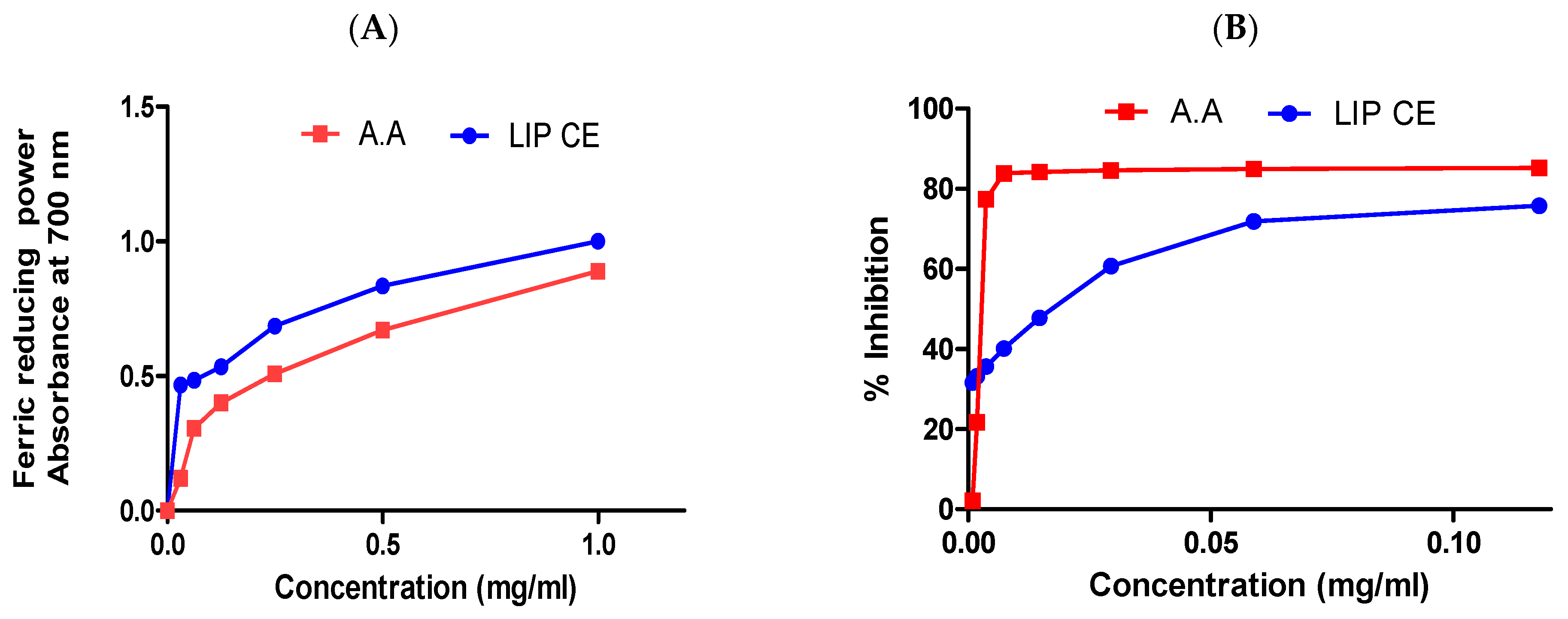
The antioxidant effect of lipids extracted from C. europaea was evaluated by FRAP and DPPH tests. Using the FRAP assay, the tested extract showed an important antioxidant effect when compared with ascorbic acid, with an EC50 of 0.084 and 0.254 mg/ mL, respectively. DPPH test showed an IC50 of 0.018 and 0.003 mg/mL for the lipids fraction and ascorbic acid, respectively (Figure 4).

3.3. Cytotoxic Effect

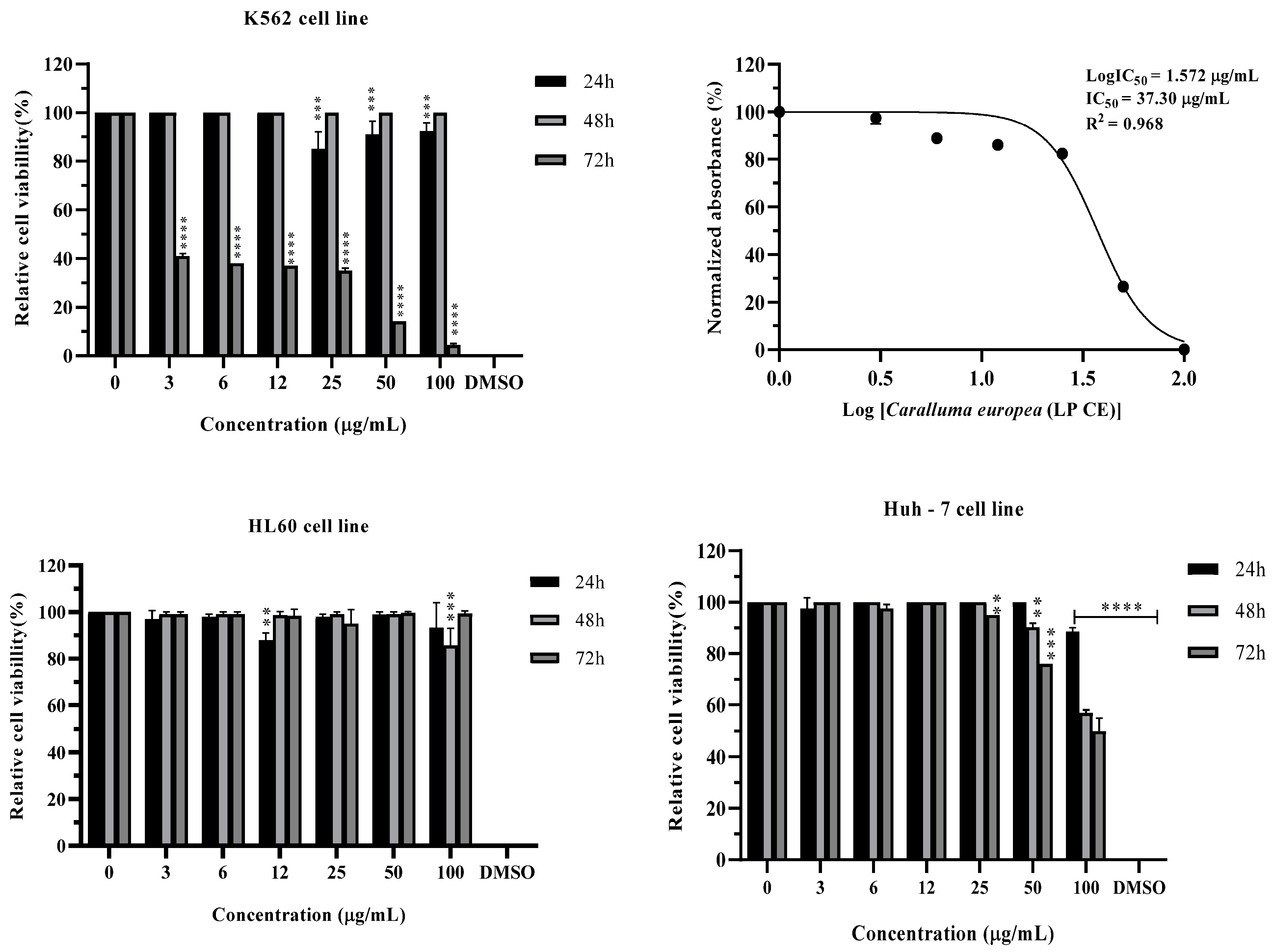
C. europaea extract was evaluated for its cytotoxic effect on three cancer cell lines, K562, HL60, and Huh-7, and on the normal Vero cell line. As shown in Figure 5 and Figure 6, and Table 3, C. europaea lipids extract was able to inhibit cell survival of K562 cells (IC50 = 37.30 µg/mL). No cytotoxicity was observed on HL60, Huh-7, and Vero cells (IC50 > 100 µg/mL).
To test the possibility of having a selective cytotoxic effect of C. europaea extract on cancerous cells, but not on normal cells, we have tested the effect of our extract on normal Vero cells.
Table 3 represents the results of half-maximal inhibitory concentration (IC50) of C. europaea extract towards K562, HL60, Huh-7, and Vero cells by using the MTT test.

3.4. Anti-Inflammatory Activity

In comparison with the positive control (Diclofenac®), the topical application of C. europaea lipid induced important anti-inflammatory activity. The treatment of rats with C. europaea lipids inhibited paw edema, which reached 83.33% after 6 h of the treatment. The obtained data did not show significant statistical results compared to Diclofenac® (10 mg/Kg) (Table 4). Inhibition at 22.22% was observed in the group treated with C. europaea lipids fraction, after 3 h of the carrageenan injection.

3.5. Wound Healing Activity

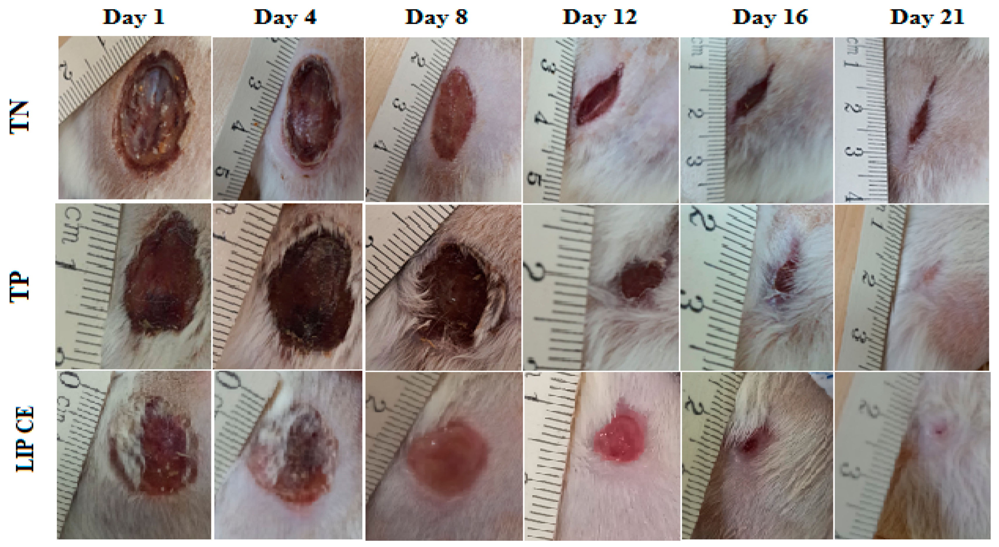
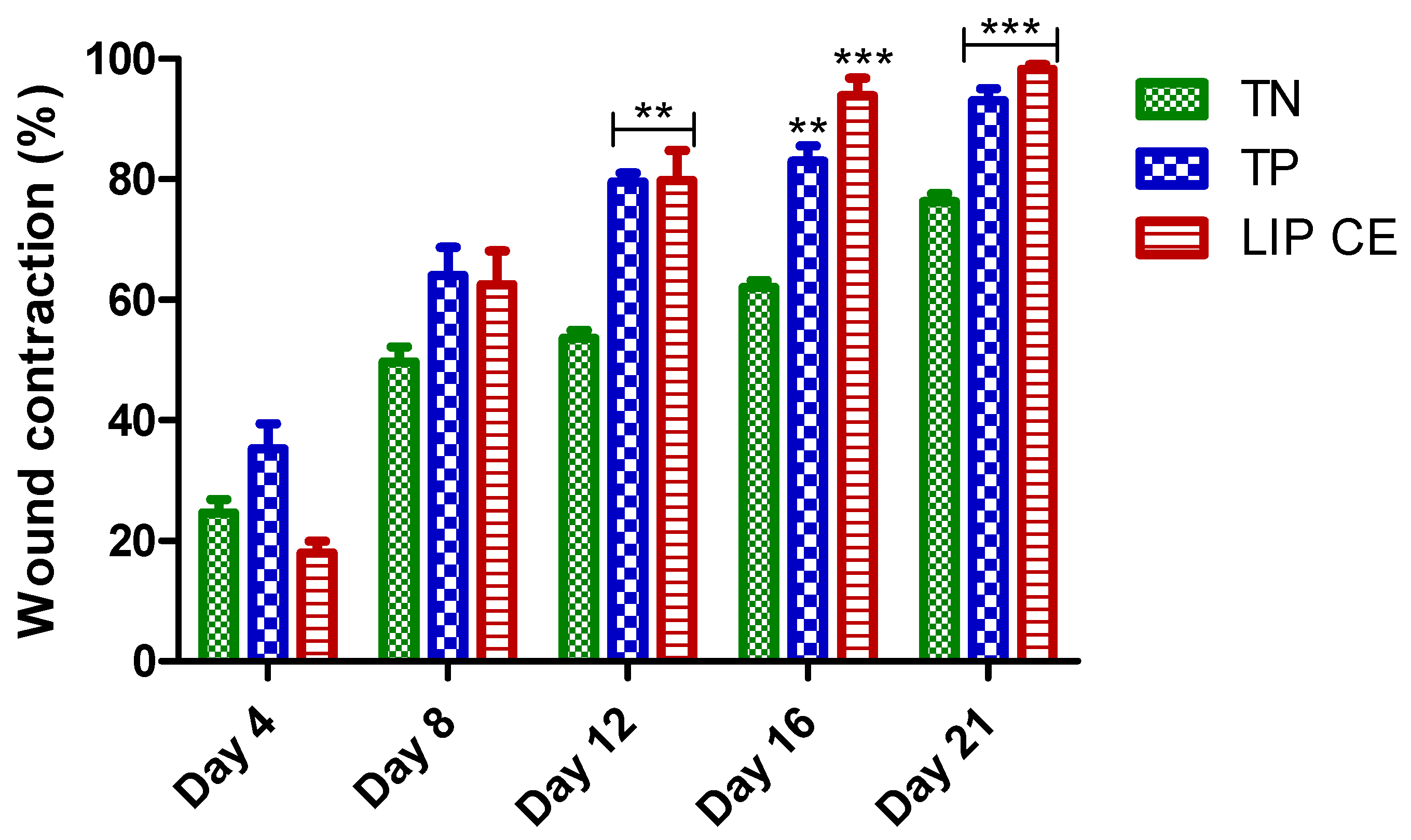
In comparison with the groups of control animals, the topical application of the lipid ointment derived from C. europaea accelerated the healing of the burns. The images in Figure 7 showed the burn healing process for the control group animals, as well as the group treated with the C. europaea lipid ointment. C. europaea ointment significantly reduced wound contraction from the first to the last day. After the 21st day of the test, topical application of C. europaea ointment led to wound closure. However, the wounds in the positive control group (Madecassol®), and the negative control group (Vaseline®) did not completely close (Figure 8).
The results of wound contractions during the 4th, 8th, 12th, 16th, and 21st days are shown in Figure 8. The animals treated with the C. europaea lipid showed the highest percentage of wound contraction on the 4th (17.96%), 8th (62.53%), 12th (79.79%), 16th (93.88%), and 21st day (98.20%).

3.6. Molecular Docking Study

Generally, the in silico study revealed that arachidic acid, lignoceric acid, and vitamin D3 were the most active molecules. In anticancer activity, lignoceric acid and arachidic acid were the most active molecules against caspase-3, with a bond energy of −6.453 and −5.652 kcal/mol, respectively. For antioxidant activity, arachidic acid was the most active molecule against NADPH oxidase with a glide score of −3.479 kcal/mol.
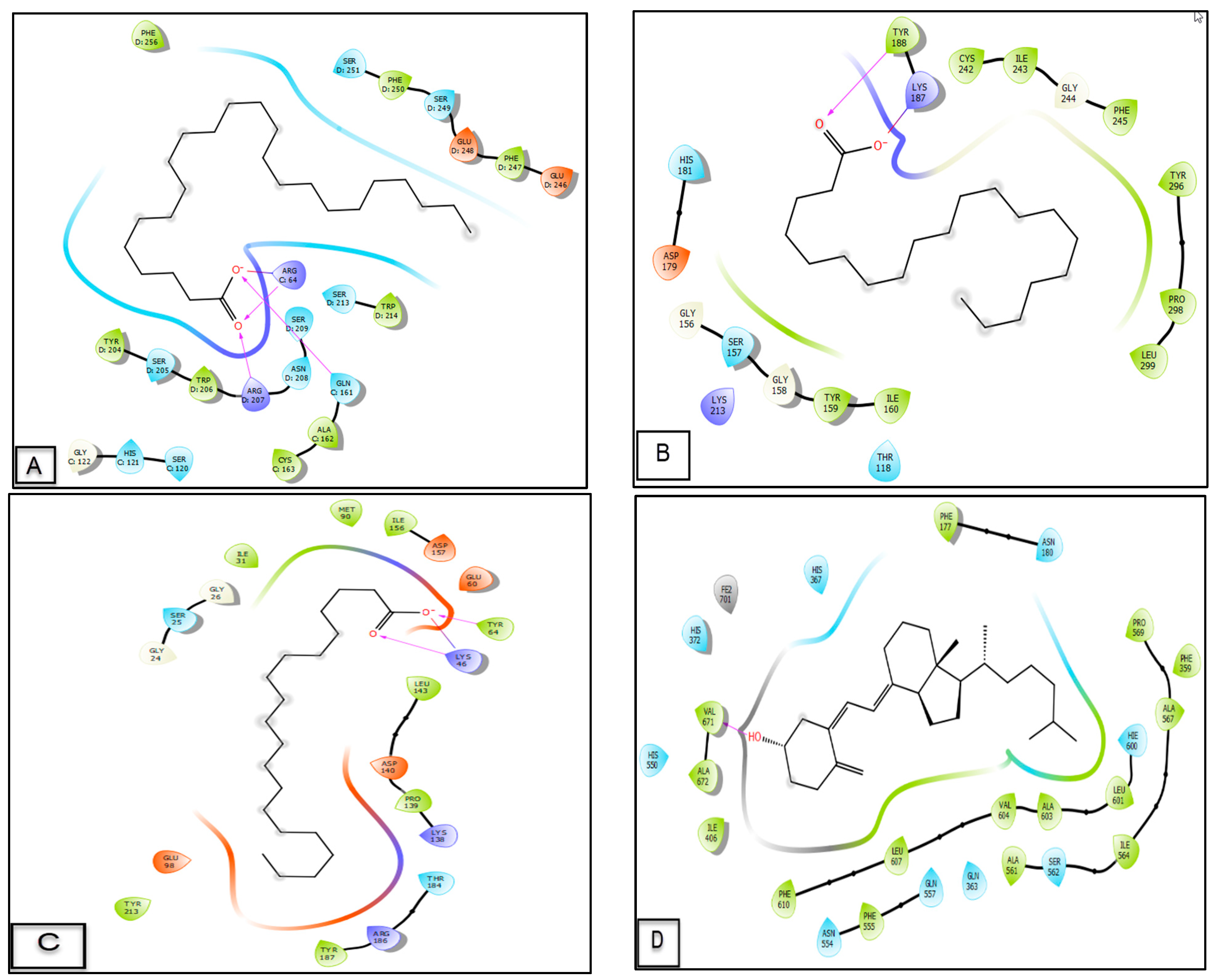
Two-dimensional and three-dimensional viewers of C. europaea lipid compounds docked in the caspase-3 active sites demonstrated that lignoceric acid established three hydrogen bonds with ARG C64, ARG D207, and GLN C161 residues, and one salt bridge with ARG C64 residue. When arachidic acid was docked in the NADPH oxidase active sites, it established one hydrogen bond with residue TYR 188 and a slat bridge with residue LYS 187.
Moreover, in silico evaluation of the anti-inflammatory effect of C. europaea lipid compounds showed that vitamin D3 and arachidic acid were the most energetic molecules against lipoxygenase with a glide score of −4.909 and −4.542 kcal/mol (Table 5). Two-dimensional and three-dimensional viewers of vitamin D3 docked in the active site of lipoxygenase presented the formation of one hydrogen bond with residue VAL 671 (Figure 9 and Figure 10).
Regarding healing activity, arachidic acid and vitamin D3 were the most active molecules against CK1 and GSK3-β, respectively, with a glide energy of −2.853 and −4.538 kcal/mol, respectively. Two-dimensional and three-dimensional viewers of arachidic acid docked in the active site of CK1 revealed the formation of two hydrogen bonds with residues LYS 46 and TYR 64, and one salt bridge with residue LYS 46.
Caspase-3 (PDB: 3GJQ), NADPH oxidase (PDB: 2CDU), casein kinase-1 (CK1) (PDB: 6GZD), glycogen synthase kinase-3 (GSK3-β) (PDB: 1Q5K), and lipoxygenase (PDB: 3V99).
Figure 9 and Figure 10 shows the number and types of possible bonds between the ligands and the active sites.

4. Discussion

For many centuries, people have been actively looking for effective natural remedies extracted from plants to treat various illnesses [28]. Traditional medicine has motivated researchers worldwide for many years because of its few negative effects and beneficial impact on health. The World Health Organization stated that different drugs are obtained from many medicinal plants [29]. In the present work, C. europaea lipids extract was evaluated for its chemical composition, antioxidant effect, and as a treatment of inflammation, skin injury, and cancer. The interaction of C. europaea lipidic compounds with the active sites of NADPH oxidase, CK1, GSK3-β, lipoxygenase, and caspase-3 was also assessed using a molecular docking study.
Chemical screening of C. europaea extract by MicroTOF revealed the presence of some lipids, 7 fatty acids in particular comprised of 6 saturated (myristic acid, stearic acid, etc.) and 1 polyunsaturated (linoleic acid) fatty acids, as well as vitamin D3 (Figure 2; Table 1). Unfortunately, no studies have so far reported the presence of such compounds in the extracts of this species. However, some other Caralluma species have been studied for their fatty acid composition. The study conducted by Augustus et Seiler revealed the presence of seven fatty acids in Caralluma attenuata Wight., including lauric, myristic, palmitic, stearic, oleic, linoleic, and arachidic acid, with concentrations ranging between 29 and 366 g/kg [30]. Furthermore, another study reported the presence of palmitic acid in one of the Caralluma species, Caralluma retrospiciens (Ehrenb) [31]. Oleic acid has been reported in the aqueous extract of Caralluma dalzielii N.E. Brown [32]. In this sense, fatty acids have been proven for their biological properties and for being efficient for many pharmacological activities [33,34].
The lipid extract showed a very interesting DPPH radical scavenging power. This antioxidant effect may be associated with its phytochemical composition. Previous studies have shown that myristic acid and vitamin D3 have strong antioxidant capacities [35,36]. NADPH oxidase enzymes have crucial functions as they regulate enzymatic sources of ROS. Oxidative stress may be successfully reduced by inhibiting NADPH oxidases [37]. This antioxidant potential may also be due to the effect of arachidic acid on NADPH oxidase.
Our findings demonstrated an important cytotoxicity of C. europaea lipids on K652 tumor cells without affecting the normal Vero cells (Figure 5 and Figure 6); the observed effect could be attributed to the apoptotic activity of fatty acids on tumor cells [38]. Caspase-3 inhibits free radical production and is required for the efficient execution of apoptosis [39]. Regarding the activation of caspase-3, lignoceric acid and arachidic acid showed strong activity against the active site of caspase-3; these results may explain the cytotoxic effect obtained for the lipid extract of C. europaea against the K652 cell line.
The lipids extract of C. europaea presented an important anti-inflammatory effect (Table 4); our findings supported earlier research which showed that α-linoleic acid suppresses the production of the inflammatory genes of iNOS, COX-2, and TNF-α through the inhibition of NF-κB and MAPKs in activated macrophages [40]. Previous studies demonstrated that stearic acid has a powerful anti-inflammatory effect, and it is generally linked to liver functions, including lipoprotein and cholesterol metabolism. Additionally, stearic acid can suppress inflammatory cell accumulation in the liver by inhibiting NF-κB activity [38]. Lipoxygenases are oxidative enzymes, which produce pro-inflammatory mediators (leukotrienes), involved in the inflammatory reaction [41]. The molecular docking showed that vitamin D3 has an important effect on lipoxygenase, which may further explain the anti-inflammatory effect of the lipid extract.
The skin is considered the largest organ of the human body and plays crucial roles with aesthetic effect, therefore the management of skin wounds takes an important place in medical science [42,43]. The wounds expose the internal structure of the skin directly to the external environment which can cause severe infections. During the inflammatory phase, the wound releases reactive oxygen species to promote cell proliferation, apoptosis, and homeostasis [44,45].
The C. europaea lipid extract demonstrated a stronger wound healing effect (Figure 7); early studies showed that Lucilia sericata fatty acids accelerate wound healing characterized by faster healing time, due to their related high angiogenic properties [28]. Fatty acids are considered to be useful compounds for promoting wound healing. Arachidonic acid is metabolized by cyclooxygenase and lipoxygenase, and its metabolites act as mediators for a number of processes, including angiogenesis, cellular growth, and the production of extracellular matrix during the healing process. Such fatty acids are continuously metabolized to create intracellular messengers, which in turn regulate a variety of biological processes, including the proliferation of endothelial cells and angiogenesis [28]. Concerning the wound healing effect, arachidic acid and vitamin D3 are the most active molecules against CK1 and GSK3-β, which works with the healing effect of the lipid extract.

5. Conclusions

Caralluma europaea lipid extract has demonstrated antioxidant and cytotoxic effects against K562 cancer cells without affecting the survival of the normal cell line (Vero), this extract may have a selective anti-survival effect against leukemia. Topical application of C. europaea lipids showed anti-inflammatory and wound-healing activities in rats, which proved its importance as an alternative agent to fight skin burns and inflammatory diseases. The molecular docking study revealed that C. europaea compounds might exert the antioxidant effect by NADPH oxidase inhibition; enhance wound healing via CK1 and GSK3-β inhibition; exert an anti-inflammatory effect via lipoxygenase inhibition; and induce apoptosis via caspase-3 activation. Further studies are required for the optimization and validation of this extract and its related lipid composition for therapeutic treatments.

Author Contributions

F.E.-Z.A.: investigation, conceptualization, data curation, software, writing original draft; M.S.: investigation; I.M.: Investigation, writing-review and editing; M.C.: Investigation; R.G.d.A.: investigation, data curation; S.B.: Investigation; K.M.: Investigation, data curation; O.A.K. and A.S.: Resources, formal analysis, methodology; A.C.d.O. and A.d.F.G.: investigation; G.S.P.: resources, formal analysis, methodology, writing-review and editing; D.B.: writing-review and editing, supervision. All authors have read and agreed to the published version of the manuscript.

Funding

This study was funded by Princess Nourah bint Abdulrahman University Researchers Supporting Project number (PNURSP2023R141), Princess Nourah bint Abdulrahman University, Riyadh, Saudi Arabia, and was financed in part by FAPEAM (CT&I ÁREAS PRIORITÁRIAS #01.02.016301.03422/2021-03).

Institutional Review Board Statement

The institutional ethical committee of care and use of the animals at the laboratory of Biotechnology, Health, Agrofood, and Environment of Sidi Mohamed Ben Abdallah University, (Morocco) reviewed and approved the present studies (LBEAS 16/12 April 2022).

Data Availability Statement

Not applicable.

Acknowledgments

The authors extend their appreciation to Princess Nourah bint Abdulrahman University Researchers Supporting Project number (PNURSP2023R141), Princess Nourah bint Abdulrahman University, Riyadh, Saudi Arabia.

Conflicts of Interest

The authors declare no conflict of interest.

Abbreviations

C. europaea/C.E: Caralluma europaea; CK1: casein kinase 1; COX: cyclooxygenase; DMEM: Dulbecco’s Modified Eagle Medium; DMSO: dimethylsulfoxide; DPPH: 2,2-di-phenyl-1-picryl hydrazyl; EC50: half maximal effective concentration; FRAP: ferric reducing antioxidant power; GSK-3: glycogen synthase kinase-3; HPLCMSD: high performance liquid chromatography/mass selective detector; IC50: 50% inhibitory concentration; LYS: lysine; MS: mass spectrometry; PBS: phosphate-buffered saline; PRO: proline; RPMI: Roswell Park Memorial Institute; TIC: total ion chromatogram; TOF: time of flight; Tyr: tyrosine.

References

  1. Gómez, X.; Sanon, S.; Zambrano, K.; Asquel, S.; Bassantes, M.; Morales, J.E.; Otáñez, G.; Pomaquero, C.; Villarroel, S.; Zurita, A.; et al. Key points for the development of antioxidant cocktails to prevent cellular stress and damage caused by reactive oxygen species (ROS) during manned space missions. npj Microgravity 2021, 7, 35. [Google Scholar] [CrossRef]
  2. Sies, H.; Berndt, C.; Jones, D.P. Oxidative Stress. Annu. Rev. Biochem. 2017, 86, 715–748. [Google Scholar] [CrossRef] [PubMed]
  3. Jiang, F.; Ding, Y.; Tian, Y.; Yang, R.; Quan, M.; Tong, Z.; Zhang, X.; Luo, D.; Chi, Z.; Liu, C. Hydrolyzed low-molecular-weight polysaccharide from Enteromorpha prolifera exhibits high anti-inflammatory activity and promotes wound healing. Biomater. Adv. 2022, 133, 112637. [Google Scholar] [CrossRef] [PubMed]
  4. Manikandan, R.; Anjali, R.; Beulaja, M.; Prabhu, N.M.; Koodalingam, A.; Saiprasad, G.; Chitra, P.; Arumugam, M. Synthesis, characterization, anti-proliferative and wound healing activities of silver nanoparticles synthesized from Caulerpa scalpelliformis. Process Biochem. 2019, 79, 135–141. [Google Scholar] [CrossRef]
  5. Said Nasser Al-Owamri, F.; Saleh Abdullah Al Sibay, L.; Hemadri Reddy, S.; Althaf Hussain, S.; Subba Reddy Gangireddygari, V. Phytochemical, Antioxidant, hair growth and wound healing property of Juniperus excelsa, Olea oleaster and Olea europaea. J. King Saud Univ. Sci 2023, 35, 102446. [Google Scholar] [CrossRef]
  6. Hang, C.; Sun, H.; Zhang, A.; Yan, G.; Lu, S.; Wang, X. Recent Developments in the Field of Antioxidant Activity on Natural Products. Амурский Медицинский Журнал 2015, 2, 44–48. [Google Scholar]
  7. Builders, P. Herbal Medicine; IntechOpen: London, UK, 2019; 316p. [Google Scholar]
  8. Khalili Tilami, S.; Kouřimská, L. Assessment of the Nutritional Quality of Plant Lipids Using Atherogenicity and Thrombogenicity Indices. Nutrients 2022, 14, 3795. [Google Scholar] [CrossRef]
  9. Trela-Makowej, A.; Kruk, J.; Jemioła-Rzemińska, M.; Szymańska, R. Acylserotonins—A new class of plant lipids with antioxidant activity and potential pharmacological applications. Biochim. Biophys. Acta (BBA) Mol. Cell Biol. Lipids 2021, 1866, 159044. [Google Scholar] [CrossRef]
  10. Toshkova-Yotova, T.; Georgieva, A.; Iliev, I.; Alexandrov, S.; Ivanova, A.; Pilarski, P.; Toshkova, R. Antitumor and antimicrobial activity of fatty acids from green microalga Coelastrella sp. BGV. S. Afr. J. Bot. 2022, 151, 394–402. [Google Scholar] [CrossRef]
  11. Aydar, E.F.; Mertdinç, Z.; Demircan, E.; Koca Çetinkaya, S.; Özçelik, B. Kidney bean (Phaseolus vulgaris L.) milk substitute as a novel plant-based drink: Fatty acid profile, antioxidant activity, in-vitro phenolic bio-accessibility and sensory characteristics. Innov. Food Sci. Emerg. Technol. 2023, 83, 103254. [Google Scholar] [CrossRef]
  12. Deghima, A.; Righi, N.; Daoud, I.; Ansorena, D.; Astiasarán, I.; Bedjou, F. Fatty acid composition, acute toxicity and anti-inflammatory activity of the n-hexane extract from Ranunculus macrophyllus Desf. roots. S. Afr. J. Bot. 2022, 148, 315–325. [Google Scholar] [CrossRef]
  13. Amrati, F.E.-Z.; Elmadbouh, O.H.M.; Chebaibi, M.; Soufi, B.; Conte, R.; Slighoua, M.; Saleh, A.; Al Kamaly, O.; Drioiche, A.; Zair, T.; et al. Evaluation of the toxicity of Caralluma europaea (C.E) extracts and their effects on apoptosis and chemoresistance in pancreatic cancer cells. J. Biomol. Struct. Dyn. 2022, 1–8. [Google Scholar] [CrossRef] [PubMed]
  14. Amrati, F.E.-Z.; Bourhia, M.; Slighoua, M.; Mohammad Salamatullah, A.; Alzahrani, A.; Ullah, R.; Bari, A.; Bousta, D. Traditional medicinal knowledge of plants used for cancer treatment by communities of mountainous areas of Fez-Meknes-Morocco. Saudi Pharm. J. 2021, 29, 1185–1204. [Google Scholar] [CrossRef] [PubMed]
  15. Amrati, F.E.-Z.; Bourhia, M.; Slighoua, M.; Ibnemoussa, S.; Bari, A.; Ullah, R.; Amaghnouje, A.; Di Cristo, F.; El Mzibri, M.; Calarco, A.; et al. Phytochemical Study on Antioxidant and Antiproliferative Activities of Moroccan Caralluma europaea Extract and Its Bioactive Compound Classes. Evid.-Based Complement. Altern. Med. 2020, 2020, 8409718. [Google Scholar] [CrossRef] [PubMed] [Green Version]
  16. Amrati, F.E.-Z.; Bourhia, M.; Slighoua, M.; Boukhira, S.; Ullah, R.; Ezzeldin, E.; Mostafa, G.A.E.; Grafov, A.; Bousta, D. Protective Effect of Chemically Characterized Polyphenol-Rich Fraction from Apteranthes europaea (Guss.) Murb. subsp. maroccana (Hook.f.) Plowes on Carbon Tetrachloride-Induced Liver Injury in Mice. Appl. Sci. 2021, 11, 554. [Google Scholar] [CrossRef]
  17. Ouassou, H.; Bouhrim, M.; Kharchoufa, L.; Imtara, H.; Daoudi, N.E.; Benoutman, A.; Bencheikh, N.; Ouahhoud, S.; Elbouzidi, A.; Bnouham, M. Caralluma europaea (Guss) N.E.Br.: A review on Ethnomedicinal uses, Phytochemistry, Pharmacological Activities, and Toxicology. J. Ethnopharmacol. 2021, 273, 113769. [Google Scholar] [CrossRef]
  18. Dra, L.A.; Rodrigues, M.J.; da Rosa Neng, N.; Nogueira, J.M.F.; Elamine, Y.; Aghraz, A.; Markouk, M.; Larhsini, M.; Custódio, L. Exploring Caralluma europaea (Guss.) N.E.Br. as a potential source of bioactive molecules: In vitro antioxidant and antidiabetic properties, and phenolic profile of crude extracts and fractions. Ind. Crops Prod. 2019, 139, 111527. [Google Scholar] [CrossRef]
  19. Formisano, C.; Senatore, F.; Della Porta, G.; Scognamiglio, M.; Bruno, M.; Maggio, A.; Rosselli, S.; Zito, P.; Sajeva, M. Headspace Volatile Composition of the Flowers of Caralluma europaea N.E.Br. (Apocynaceae). Molecules 2009, 14, 4597–4613. [Google Scholar] [CrossRef]
  20. Bligh, E.G.; Dyer, W.J. A rapid method of total lipid extraction and purification. Can. J. Biochem. Physiol. 1959, 37, 911–917. [Google Scholar] [CrossRef]
  21. National Research Council Committee. Guide for the Care and Use of Laboratory Animals, The National Academies Collection: Reports Funded by National Institutes of Health, 8th ed.; National Academies Press: Washington, DC, USA, 2011. [Google Scholar]
  22. Mssillou, I.; Agour, A.; Slighoua, M.; Chebaibi, M.; Amrati, F.E.-Z.; Alshawwa, S.Z.; Kamaly, O.A.; El Moussaoui, A.; Lyoussi, B.; Derwich, E. Ointment-Based Combination of Dittrichia viscosa L. and Marrubium vulgare L. Accelerate Burn Wound Healing. Pharmaceuticals 2022, 15, 289. [Google Scholar] [CrossRef]
  23. Jaklová Dytrtová, J.; Moslova, K.; Jakl, M.; Sirén, H.; Riekkola, M.-L. Fluorescein isothiocyanate stability in different solvents. Monatsh Chem. 2021, 152, 1299–1306. [Google Scholar] [CrossRef]
  24. Seal, T. Quantitative HPLC analysis of phenolic acids, flavonoids and ascorbic acid in four different solvent extracts of two wild edible leaves, Sonchus arvensis and Oenanthe linearis of North-Eastern region in India. J. Appl. Pharm. Sci. 2016, 6, 157–166. [Google Scholar] [CrossRef] [Green Version]
  25. Brand-Williams, W.; Cuvelier, M.-E.; Berset, C. Use of a free radical method to evaluate antioxidant activity. LWT Food Sci. Technol. 1995, 28, 25–30. [Google Scholar] [CrossRef]
  26. Oyaizu, M. Studies on products of browning reaction—Antioxidative activities of products of browning reaction prepared from glucosamine. Jpn. J. Nutr. Diet. 1986, 44, 307–315. [Google Scholar] [CrossRef] [Green Version]
  27. Amrati, F.E.-Z.; Bourhia, M.; Saghrouchni, H.; Slighoua, M.; Grafov, A.; Ullah, R.; Ezzeldin, E.; Mostafa, G.A.; Bari, A.; Ibenmoussa, S.; et al. Caralluma europaea (Guss.) N.E.Br.: Anti-Inflammatory, Antifungal, and Antibacterial Activities against Nosocomial Antibiotic-Resistant Microbes of Chemically Characterized Fractions. Molecules 2021, 26, 636. [Google Scholar] [CrossRef]
  28. Zhang, Z.; Wang, S.; Diao, Y.; Zhang, J.; Lv, D. Fatty acid extracts from Lucilia sericata larvae promote murine cutaneous wound healing by angiogenic activity. Lipids Health Dis. 2010, 9, 24. [Google Scholar] [CrossRef] [Green Version]
  29. Aye, M.M.; Aung, H.T.; Sein, M.M.; Armijos, C. A Review on the Phytochemistry, Medicinal Properties and Pharmacological Activities of 15 Selected Myanmar Medicinal Plants. Molecules 2019, 24, 293. [Google Scholar] [CrossRef] [Green Version]
  30. Augustus, G.D.P.S.; Seiler, G.J. Phytochemicals of selected plant species of the Apocynaceae and Asclepiadaceae from Western Ghats, Tamil Nadu, India. Biomass Bioenergy 2011, 35, 3012–3017. [Google Scholar] [CrossRef]
  31. Alqahtani, S.S.; Makeen, H.A.; Menachery, S.J.; Moni, S.S. Documentation of bioactive principles of the flower from Caralluma retrospiciens (Ehrenb) and in vitro antibacterial activity—Part B. Arab. J. Chem. 2020, 13, 7370–7377. [Google Scholar] [CrossRef]
  32. Ugwah-Oguejiofor, C.J.; Amuda, M.B.; Abubakar, K.; Ugwah, O.M.; Ofokansi, M.N.; Mshelia, H.E. An experimental evaluation of anticonvulsant activity of aqueous extract of Caralluma dalzielii N.E. Brown. Phytomed. Plus 2023, 3, 100401. [Google Scholar] [CrossRef]
  33. Zheng, C.J.; Yoo, J.-S.; Lee, T.-G.; Cho, H.-Y.; Kim, Y.-H.; Kim, W.-G. Fatty acid synthesis is a target for antibacterial activity of unsaturated fatty acids. FEBS Lett. 2005, 579, 5157–5162. [Google Scholar] [CrossRef] [PubMed] [Green Version]
  34. Huang, C.B.; Alimova, Y.; Myers, T.M.; Ebersole, J.L. Short- and medium-chain fatty acids exhibit antimicrobial activity for oral microorganisms. Arch. Oral Biol. 2011, 56, 650–654. [Google Scholar] [CrossRef] [PubMed] [Green Version]
  35. Liu, C.; Yuan, C.; Ramaswamy, H.S.; Ren, Y.; Ren, X. Antioxidant capacity and hepatoprotective activity of myristic acid acylated derivative of phloridzin. Heliyon 2019, 5, e01761. [Google Scholar] [CrossRef] [Green Version]
  36. Mokhtari, Z.; Hekmatdoost, A.; Nourian, M. Antioxidant efficacy of vitamin D. J. Parathyr. Dis. 2016, 5, 11–16. [Google Scholar]
  37. Guzik, T.J.; Harrison, D.G. Vascular NADPH oxidases as drug targets for novel antioxidant strategies. Drug Discov. Today 2006, 11, 524–533. [Google Scholar] [CrossRef] [PubMed]
  38. Goradel, N.H.; Eghbal, M.A.; Darabi, M.; Roshangar, L.; Asadi, M.; Zarghami, N.; Nouri, M. Improvement of Liver Cell Therapy in Rats by Dietary Stearic Acid. Iran Biomed. J. 2016, 20, 217–222. [Google Scholar]
  39. Brentnall, M.; Rodriguez-Menocal, L.; De Guevara, R.L.; Cepero, E.; Boise, L.H. Caspase-9, caspase-3 and caspase-7 have distinct roles during intrinsic apoptosis. BMC Cell Biol. 2013, 14, 32. [Google Scholar] [CrossRef] [Green Version]
  40. Sawada, L.A.; Monteiro, V.S.D.C.; Rabelo, G.R.; Dias, G.B.; Da Cunha, M.; do Nascimento, J.L.M.; de Nazareth Tavares Bastos, G. Libidibia ferrea Mature Seeds Promote Antinociceptive Effect by Peripheral and Central Pathway: Possible Involvement of Opioid and Cholinergic Receptors. BioMed Res. Int. 2014, 2014, e508725. [Google Scholar] [CrossRef] [Green Version]
  41. Wisastra, R.; Dekker, F.J. Inflammation, Cancer and Oxidative Lipoxygenase Activity are Intimately Linked. Cancers 2014, 6, 1500–1521. [Google Scholar] [CrossRef] [Green Version]
  42. Naik, S. Wound, heal thyself. Nat. Med. 2018, 24, 1311–1312. [Google Scholar] [CrossRef]
  43. Mssillou, I.; Bakour, M.; Slighoua, M.; Laaroussi, H.; Saghrouchni, H.; Ez-Zahra Amrati, F.; Lyoussi, B.; Derwich, E. Investigation on wound healing effect of Mediterranean medicinal plants and some related phenolic compounds: A review. J. Ethnopharmacol. 2022, 298, 115663. [Google Scholar] [CrossRef] [PubMed]
  44. Chang, R.; Zhao, D.; Zhang, C.; Liu, K.; He, Y.; Guan, F.; Yao, M. Nanocomposite multifunctional hyaluronic acid hydrogel with photothermal antibacterial and antioxidant properties for infected wound healing. Int. J. Biol. Macromol. 2023, 226, 870–884. [Google Scholar] [CrossRef] [PubMed]
  45. Amrati, F.E.-Z.; Chebaibi, M.; de Azevedo, R.G.; Conte, R.; Slighoua, M.; Mssillou, I.; Kiokias, S.; de Freitas Gomes, A.; Pontes, G.S.; Bousta, D. Phenolic Composition, Wound Healing, Antinociceptive, and Anticancer Effects of Caralluma europaea Extracts. Molecules 2023, 28, 1780. [Google Scholar] [CrossRef] [PubMed]
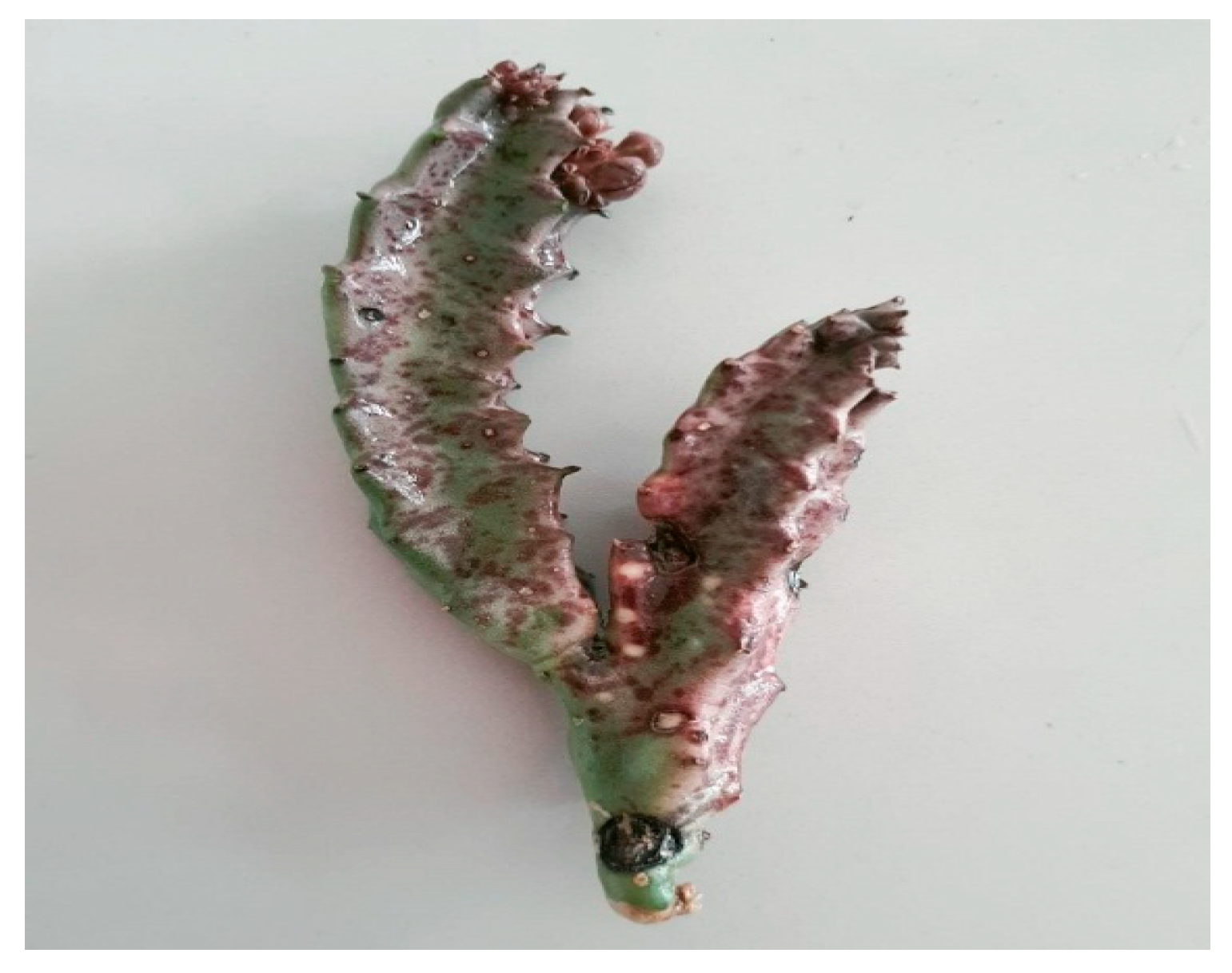
Figure 1. An aerial part of C. europaea.
Separations 10 00172 g001
Figure 2. Micro-TOF profile of lipids extracted from C. europaea.
Separations 10 00172 g002
Figure 3. Total ion chromatogram of the lipids extracted from C. europaea.
Separations 10 00172 g003
Figure 4. Antioxidant potential of C. europaea lipids using FRAP (A) and DPPH (B) assays.
Separations 10 00172 g004
Figure 5. Cytotoxic effect of C. europaea lipids at different concentrations against HL60, Huh-7, and K562 cell lines, after 24, 48, and 72 h. The IC50 for HL60, Huh-7, and K562 cells was evaluated using nonlinear regression (GraphPad Prism v5 software). The cell survival was assessed by the MTT test. ** p < 0.01; *** p < 0.001, **** p < 0.0001.
Separations 10 00172 g005
Figure 6. Cytotoxicity of C. europaea lipid extract at different concentrations against normal Vero cells, after 24, 48, and 72 h. The cell survival was evaluated using the MTT test.
Separations 10 00172 g006
Figure 7. Images of animal wound areas on 1st, 4th, 8th, 12th, 16th, and 21st day of the experiment. CE, Caralluma europaea; TN, Negative control; TP, Positive control; Lip, Lipids.
Separations 10 00172 g007
Figure 8. Wound contraction rate (%) in 4th, 8th, 12th, 16th, and 21st day of treatment. Values are significantly different in comparison with the negative control. **: p < 0.01, ***: p < 0.001.
Separations 10 00172 g008
Figure 9. Two-dimensional representations of ligands interactions with the active sites. (A): Interactions of lignoceric acid with the caspase-3 active sites; (B): Interactions of arachidic acid with the NADPH oxidase active sites; (C): Interactions of arachidic acid with the casein kinase-1 active sites; and (D): Interactions of vitamin D3 with the lipoxygenase active sites.
Separations 10 00172 g009
Figure 10. Three-dimensional representations of ligands interactions with the active sites. (A): Interactions of lignoceric acid with the caspase-3 active sites; (B): Interactions of arachidic acid with the NADPH oxidase active sites; (C): Interactions of arachidic acid with the casein kinase-1 active sites; and (D): Interactions of vitamin D3 with the lipoxygenase active sites.
Separations 10 00172 g010aSeparations 10 00172 g010b
Table 1. Compounds revealed in lipids extract of C. europaea.
PicIdentified CompoundFormulaMWTheoretical [M-H]Found [M-H]Error [ppm]
1Myristic acidC14H28O2228.37227.2017227.204110.765767
2Palmitic acidC16H32O2256.40255.2329255.23559.975
3Linoleic acidC18H32O2280.45279.2330279.23538.402
4Stearic acidC18H36O2284.48283.2643283.26616.517
5Arachidic acidC20H40O2312.53311.2956311.2248−227.115
6Behenic acidC22H44O2340.58339.3269339.32998.977
7Lignoceric acidC24H48O2368.63367.3582367.36169.380
8Cholecalciferol (Vitamin D3)C27H44O384.65383.3319383.356163.026
Table 2. Compounds identified by HPLCMSD in lipids extract of C. europaea.
PeakLipidic CompoundRT (min)Formula
1Stearic acid52.192C18H36O2
2Palmitic acid52.914C16H32O2
3Myristic acid54.186C14H28O2
Table 3. Cytotoxicity of C. europaea lipids.
IC50 (µg/mL)
Human chronic myelogenous leukemia
(K562 cell line)
Human hepatocellular carcinoma
(Huh-7 cell line)
Human acute promyelocytic leukemia
(HL60 cell line)
Normal cell line
(Vero cells)
37.30 ***->100>100
*** Activity observed only during the 72 h of treatment.
Table 4. Anti-inflammatory effect of lipid extract on carrageenan-induced paw edema in Wistar rats after 3, 4, 5, and 6 h of the injection.
Treatment GroupInitial Diameter (cm)Edema Diameter after the Injection of Carrageenan (cm)/Inhibition of Edema (%)
3 h4 h5 h6 h
Vaseline2.370 ± 0.0492.670 ± 0.0372.870 ± 0.0372.838 ± 0.0662.570 ± 0.020
Diclofenac®
(1%)
2.226 ± 0.0352.424 ± 0.037 **
34%
2.358 ± 0.034 *
73.60%
2.302 ± 0.028
83.76%
2.256 ± 0.030
85%
Lipids CE
(10%)
2.417 ± 0.0442.650 ± 0.050 *
22.33%
2.567 ± 0.067
70%
2.500 ± 0.057
82.27%
2.450 ± 0.050
83.50%
Results are statistically different from the negative control: * p < 0.05; ** p < 0.01.
Table 5. Docking results with lipid compounds of C. europaea in the active site of caspase-3, NADPH oxidase, lipoxygenase, CK1, and GSK3-β.
MoleculesGlide G Score (Kcal/mol)
3GJQ2CDU6GZD1Q5K3V99
Arachidic acid−5.652−3.479−2.853−2.968−4.542
Behenic acid−6.334−2.929−2.204−1.817−4.396
Lignoceric acid−6.453−1.641-−1.876−2.177
Linoleic acid−4.141−2.478−1.26−1.207−1.992
Myristic acid−3.759--0.042−0.753
Palmitic acid−3.642--−0.46−1.346
Stearic acid−3.7−0.753−0.636−0.269−0.637
Vitamin D3−4.279--−4.538−4.909
Disclaimer/Publisher’s Note: The statements, opinions and data contained in all publications are solely those of the individual author(s) and contributor(s) and not of MDPI and/or the editor(s). MDPI and/or the editor(s) disclaim responsibility for any injury to people or property resulting from any ideas, methods, instructions or products referred to in the content.