Article

The Molecular Identification and Comprehensive Analysis of Klebsiella pneumoniae Isolated from Industrial Wastewater

Kaifeng Key Laboratory of Food Composition and Quality Assessment, School of Environmental Engineering, Yellow River Conservancy Technical Institute, Kaifeng 475004, China
*
Author to whom correspondence should be addressed.
Separations 2024, 11(4), 121; https://doi.org/10.3390/separations11040121
Submission received: 18 March 2024 / Revised: 7 April 2024 / Accepted: 15 April 2024 / Published: 17 April 2024
(This article belongs to the Topic Sustainable Technologies for Water Purification)

Abstract

:
Industrial wastewater typically contains many organic and inorganic pollutants and is also contaminated by various microorganisms. Microbial species in industrial wastewater have not been extensively investigated. In this experiment, a Klebsiella pneumoniae strain was isolated for the first time from industrial wastewater containing a high concentration of sulfate and phosphate. Mass spectrometry, genetic analysis, and biochemical identification were conducted to understand the genetic and biochemical characteristics of this Klebsiella pneumoniae strain recovered from industrial wastewater. Growth experiments revealed that it exhibited an excellent growth rate in nutrient broth. Further analyses showed that the strain was sensitive to most antibiotics but resistant to chloramphenicol and nitrofurantoin. It also exhibited significant resistance to piperacillin/tazobactam and cefotaxime/clavulanic acid. Resistance gene experiments indicated the presence of gyrA, OqxB, and ParC genes associated with antibiotic resistance in the isolated Klebsiella pneumoniae strain. Proteomics uncovered the following three proteins related to drug resistance: the multi-drug resistant outer membrane protein MdtQ, the multi-drug resistant secretion protein, and the modulator of drug activity B, which are coexistent in Klebsiella pneumoniae. Proteomics and bioinformatics analyses further analyzed the protein composition and functional enrichment of Klebsiella pneumoniae. The isolation of Klebsiella pneumoniae from a high concentration in sulfate and phosphate industrial wastewater provides a new direction for further research on the characteristics and drug resistance traits of industrial wastewater microorganisms and the potential risks they may pose when released into the environment.

1. Introduction

Industrial wastewater has long been recognized as a significant source of environmental pollution. It contains a plethora of toxic, carcinogenic, teratogenic, and mutagenic chemicals. In addition to these chemical compounds, industrial wastewater, either in its untreated state or during the treatment process, also harbors a substantial microorganism load, some of which have pathogenic potential. Recent studies have revealed that antibiotic resistance is prevalent among indicator bacteria and pathogenic strains collected from various industrial and sewage discharge points along the Pydibhimavaram to the Tuni coastal area on the eastern coast of India. Among the detected microorganisms, Klebsiella pneumoniae is one species exhibiting antibiotic resistance [1]. In addition, Escherichia coli co-harboring conjugative plasmids with colistin- and carbapenem resistance genes have a high prevalence and have been identified from different functional areas of an urban wastewater treatment plant in China [2]. Moreover, the mobile colistin resistance (mcr) genes mcr-8 and mcr-9 were the first reported in Ireland, with the mcr-8-positive Klebsiella pneumoniae displaying resistance to colistin [3]. The global spread of the mcr gene highlights that the addition of colistin to veterinary drugs affects human health. Studies have shown that mcr-1 to mcr-9 variants are exclusively identified in Klebsiella pneumoniae, while the mcr-10.1 variant has been found in Klebsiella pneumoniae subspecies quasi-pneumoniae, K. quasipneumoniae, and Klebsiella variicola [4]. Novel characteristics of β-lactamases, as well as their multidrug resistance mechanisms, have been identified in Escherichia coli isolated from hospital wastewater and sewage treatment plants [5]. Human habitation and living often affect antibiotic resistance issues in aquatic environments. Studies have found that the Laru River in Malaysia is contaminated with organic and inorganic pollutants, which is possible because these pollutants are potentially not adequately treated [6]. Antibiotics in wastewater contribute to the production of antibiotic-resistance microorganisms through selection pressure and horizontal gene transfer of resistance genes. Carbapenem-resistant Enterobacteriaceae (CRE) threatens human and animal agriculture worldwide, particularly opportunistic pathogens such as carbapenemase-producing Enterobacteriaceae species. Recent studies have isolated carbapenem-resistant Klebsiella pneumoniae bacteria from urban wastewater treatment plants, receiving water bodies, and wastewater and processed water from poultry and swine slaughterhouses [7]. In this study, large amounts of ammonium sulfate (sodium) were produced from the process of reacting ammonium dihydrogen phosphate (sodium) with ferrous sulfate to produce iron phosphate, and a Klebsiella pneumoniae strain was isolated from the wastewater where some magnesium, manganese, and iron ions, etc., existed.

2. Materials and Methods

2.1. Isolation and Culture of Bacteria

An aliquot of 500 mL of industrial wastewater was collected from Henan Baili New Energy Materials Co., LTD., Qinyang, China. The main components of industrial wastewater were sulfate ions (SO2, 50,000 ppm), ammonia nitrogen (NH3-N, 11,000 ppm), sodium (Na, 5500 ppm), magnesium (Mg, 2100 ppm), phosphorus (P, 1200 ppm), manganese (Mn, 220 ppm), iron (Fe, 120 ppm), and calcium (Ca, 60 ppm). The wastewater in the wastewater reaction tank was collected with a sterile centrifuge tube and quickly sealed. The liquid in the centrifuge tube was mixed on a super-clean laboratory workbench, and the centrifuge tube was opened. The liquid in the centrifuge tube was diluted in a double ratio with ultra-pure water.
A bacteria inoculation ring was dipped in industrial wastewater, streaked on nutrient agar (hopebiol), and placed in a 37 °C incubator for bacterial colony growth. The nutrient broth (hopebiol, HB0108) was used as a liquid medium, and the bacterial cultures were incubated for 14 h at 240 rpm per minute.

2.2. Genome and Plasmid DNA Extraction

DNA was extracted by the Genome and Plasmid Extraction Kit (Tiangen). The 16S rDNA gene was amplified by polymerase chain reaction (PCR) amplification. The universal primer sequence was as follows: 27F: 5′-AGAGTGTGATCCTGGCTGCTCAG-3′, 1492R: 5′-GGTTACCTTGTTACGACTT-3′. PCR conditions: 98 °C for 5 min, then 98 °C for 15 s, 55 °C for 25 s, 72 °C for 20 s, 40 cycles, then 72 °C for 5 min. Moreover, 1% agarose gel was made by Gelred, and the size of PCR products was determined by electrophoresis.

2.3. Phylogenetic Tree Generation

The phylogenetic tree was constructed by the neighbor-joining method with a maximum nucleotide substitution rate of 0.5. The sequences were obtained from the website of the National Center for Biotechnology Information (NCBI). The GenBank entry numbers for the sequences used in the study are shown in parentheses (Figure 1E). B3 was indicated as the novel isolated sequence in this study.

2.4. Matrix-Assisted Laser Desorption/Ionization Time-of-Flight Mass Spectrometry (MALDI-TOF MS) Identification

For the identification of bacterial strains, MALDI-TOF MS was employed using the MEIHUA M-Discover 100 Excellence system (MEIHUA, Lhasa City, China). The samples were processed with a microbial sample preparation reagent for mass spectrometry analysis (MEIHUA, model 2 × 50). The procedure involved transferring an appropriate amount of bacterial cells onto designated spots on a mass spectrometry sample target plate, ensuring even distribution. Next, 1 μL of lysis solution M1 was pipetted onto the bacteria, covering it completely, and dried at room temperature. Finally, 1 μL of the matrix solution was added to the same position, followed by drying at room temperature before analysis.

2.5. Biochemical Analysis and Identification of Microorganisms

The MEIHUA Microbial Identification and Antimicrobial Susceptibility Analysis System (MEIHUA) was utilized for the biochemical identification and antibiotic susceptibility testing of Klebsiella pneumoniae. The biochemical analysis procedure was as follows: Initially, a single bacterial colony was selected for pure culture. The bacterial suspension was diluted in a sterile saline solution to obtain a 0.5 McFarland standard turbidity. Moreover, 100 μL of the aforementioned bacterial suspension was added to the designated wells with a pipette for biochemical identification. Some wells required the addition of sterile mineral oil. After sealing with adhesive tape, the biochemical identification plate was incubated at 37 °C for 24 h before being put into the instrument for analysis.

2.6. Antibiotic Resistance Analysis

The procedure for testing antibiotic resistance in Klebsiella pneumoniae was as follows: 50 μL of the 0.5 McFarland standard bacterial suspension was added to the M-H broth culture medium. Then, a drop of the antibiotic susceptibility indicator solution was thoroughly mixed. Using a pipette, add 100 μL of the mixture to each antibiotic susceptibility testing plate well. After sealing with adhesive tape, the plate was incubated at 37 °C for 24 h before being put into the instrument for analysis.

2.7. Bacterial Growth Curve Analysis

The bacterial growth curve testing for Klebsiella pneumoniae was as follows: 200 μL of the bacterial suspension was added to a 96-well plate with a pipette. The OD600 absorbance values were measured using a microplate reader (Tecan, Sunrise). The bacterial suspension was then diluted to an OD of 1 and used in the different inoculation experiments. The experimental groups were as follows: Group 1: Control with no bacteria in the culture medium. Group 2: 999 μL of culture medium + 1 μL of bacterial suspension. Group 3: 990 μL of culture medium + 10 μL of bacterial suspension. Group 4: 980 μL of culture medium + 20 μL of bacterial suspension. Group 5: 960 μL of culture medium + 40 μL of bacterial suspension. Group 6: 940 μL of culture medium + 60 μL of bacterial suspension. Group 7: 920 μL of culture medium + 80 μL of bacterial suspension. Group 8: 900 μL of culture medium + 100 μL of bacterial suspension. Subsequently, the prepared diluted culture medium was added to each 48-well deep well plate with 1 mL. The 48-well plates were then placed into a high-throughput microbial growth detection system (JIELING, MicroScreen-HT) with incubation conditions set at 37 °C at 600 rpm/min. The OD values were recorded every hour for a total of 90 h, and a growth curve was generated based on the obtained OD values.

2.8. Antimicrobial Resistance Gene Detection

For antimicrobial resistance gene detection, primers were synthesized for the following genes: AAC (3)-II, cmlA, CTX-M-1, gyrA, gyrB, blaKPC, NDM-1, oqxA, oqxB, OXA, parC, qepA, qnrA, qnrB, qnrC, qnrD, qnrS, and Sul2 (References [8,9,10,11,12]). The resistance genes were amplified by a PCR instrument (BIORAD, C1000 Touch, Hercules, CA, USA), followed by agarose gel electrophoresis using a nucleic acid electrophoresis system (BIORAD, PowerPac Basic). Gel imaging (BIORAD, GelDoc Go) was performed to determine the length of the amplified amplicons. The amplification products with the correctly sized amplicons were sequenced (Tsingke Bio) and compared with the NCBI database for identification.

2.9. Proteomic Analysis

This bacterium grows in a broth culture medium that does not have antibiotics. Sample preparation contains the processes of protein extraction, denaturation, reduction, alkylation, tryptic digestion, and peptide cleanup. The total protein of Klebsiella pneumoniae was extracted, and then the appropriate protein was added to 50 µL of lysis buffer and heated at 95 °C for 10 min. After cooling to room temperature, the samples were added to trypsin digestion buffer and incubated at 37 °C for 2 h with shaking. Then, a termination buffer was added to the sample to terminate the enzymolysis reaction. The peptide was desalted using an iST cartridge in the kit and then eluted with an elution buffer. The eluted peptide was vacuum-drained and stored at −80 °C. The polypeptide samples were dissolved in solvent A and analyzed by a Q-Exactive Plus mass spectrometer in the EASY-nanoLC 1200 system (Thermo Fisher Scientific, Waltham, MA, USA). The mass spectrometer operated in data-dependent acquisition (DDA) mode, automatically switching between MS and MS/MS acquisition. The full survey scan MS spectra were m/z 350–1800; the mass resolution was 70,000 in the Orbitrap. The automatic gain control (AGC) target was 3 × 10⁶, and the maximum injection time was 50 ms. A series of mass spectra were analyzed by PEAKS Studio version 10.6 (Bioinformatics Solutions Inc., Waterloo, ON, Canada). The database used was Klebsiella pneumoniae subsp. pneumoniae (strain HS11286) (version 2023, 5728 entries).

3. Results

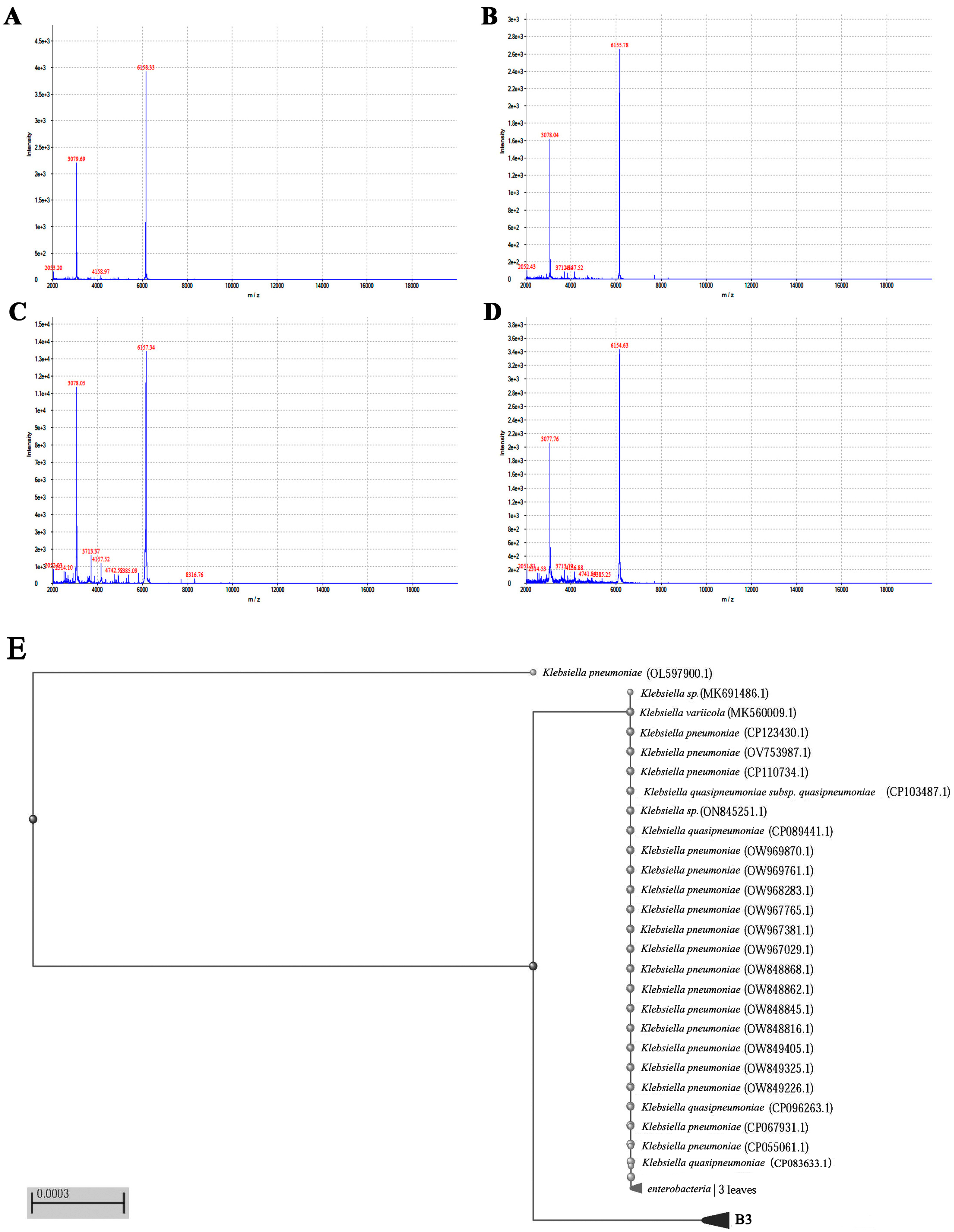
3.1. MALDI-TOF MS Enables the Rapid Identification of Klebsiella Pneumoniae

Using MALDI-TOF MS analysis and the corresponding software (MicroCtrl 1.0), the detected ion peaks were plotted with intensity as the vertical axis and mass-to-charge ratio (m/z) as the horizontal axis. The obtained protein fingerprint map showed a stable baseline, clear protein peaks, and satisfactory results. The main ion peak was observed at m/z values around 6156, with a secondary peak at m/z of approximately 3078. The bacterial protein fingerprints are presented in Figure 1A–D. Comparing them with an in-house database, the isolated strain was identified as Klebsiella pneumoniae.

3.2. Molecular Identification of Klebsiella pneumoniae

The expected size of Klebsiella pneumoniae’s 16S rDNA gene amplification product is approximately 1500 bp. PCR-based identification demonstrated excellent specificity, with only one specific DNA fragment observed. Based on the results of the 16S rDNA sequencing and the comparison with the NCBI database, the isolate was identified as Klebsiella pneumoniae. Additionally, molecular phylogenetic analysis was performed on this strain of Klebsiella pneumoniae isolated from high-concentration metal ions and sulfate compounds in industrial wastewater, revealing a high degree of homology with Klebsiella pneumoniae strains in the genetic database (Figure 1E).

3.3. Biochemical Identification of Klebsiella pneumoniae

The biochemical identification experiment revealed positive reactions for LDC, URE, GLUf, ESC, VP, MANf, INOf, SORf, RHAf, SACf, MELf, ARAf, CELf, CIT, and ONPG. Negative reactions were observed for ADH, ODC, C, H2S, GEL, TDA, IND, AMYf, and LACf (see Table 1).

3.4. Antibiotic Resistance Analysis of Klebsiella pneumoniae

The antibiotic susceptibility experiment demonstrated that the Klebsiella pneumoniae strain newly isolated from industrial wastewater exhibited sensitivity to most antibiotics tested. However, it showed resistance to CHL and FD and intermediate resistance to AMS and PB. Additionally, the MIC values for CTX/C (≤1/4) and CAZ/C (≤1/4) indicated that this Klebsiella pneumoniae strain was still highly sensitive to this combination of antibiotics (see Table 2).

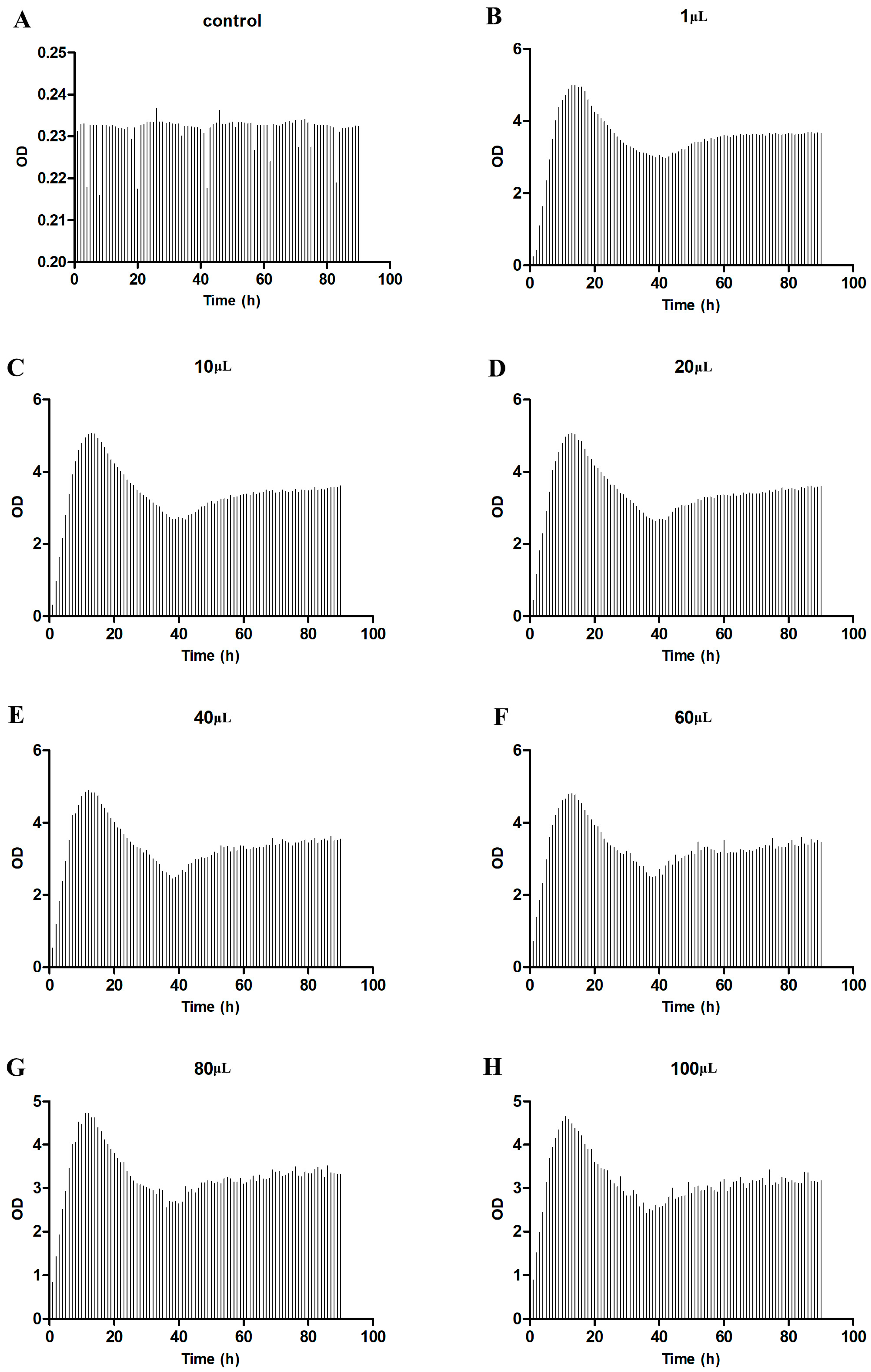
3.5. Growth Experiment of Klebsiella Pneumoniae

After 90 h of culture, the growth characteristics of the isolated Klebsiella pneumoniae strain were evaluated at different dilutions. As shown in Figure 2, the OD value of the bacterial suspension reached its peak at around 11–13 h of culture, followed by a decrease at approximately 40 h. Subsequently, the OD value gradually increased and remained relatively stable until the end of the culture period. However, there were variations in the time it took for different inoculum sizes of Klebsiella pneumoniae to reach their peak. Smaller inoculum sizes peaked after a long time of culture, but they had higher OD values than larger inoculum sizes. The higher OD value indicates a higher bacterial population. Table 3 demonstrates that the OD value of the 10 μL inoculation amount was the highest, and the OD value of the 100 μL inoculation amount was the lowest.

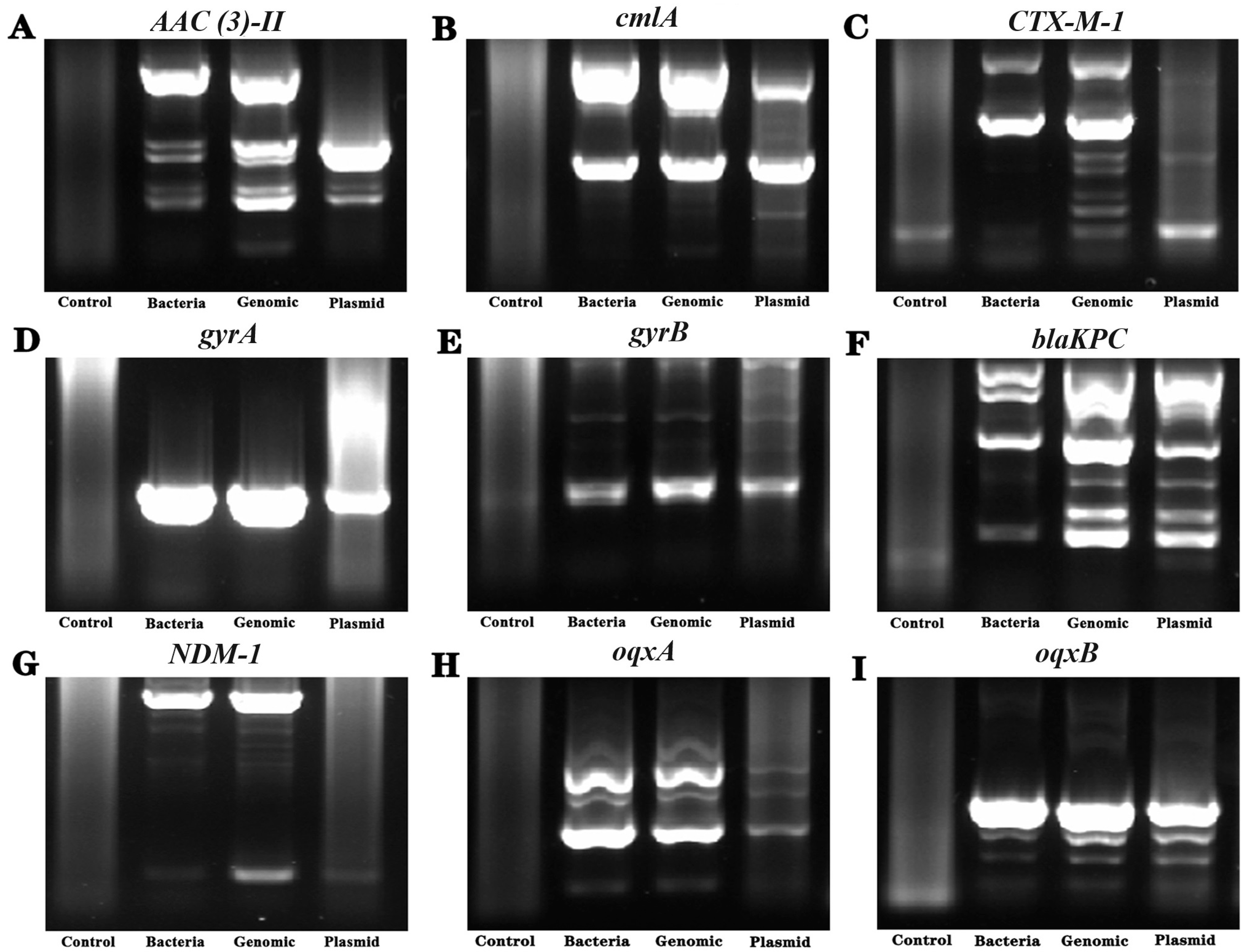
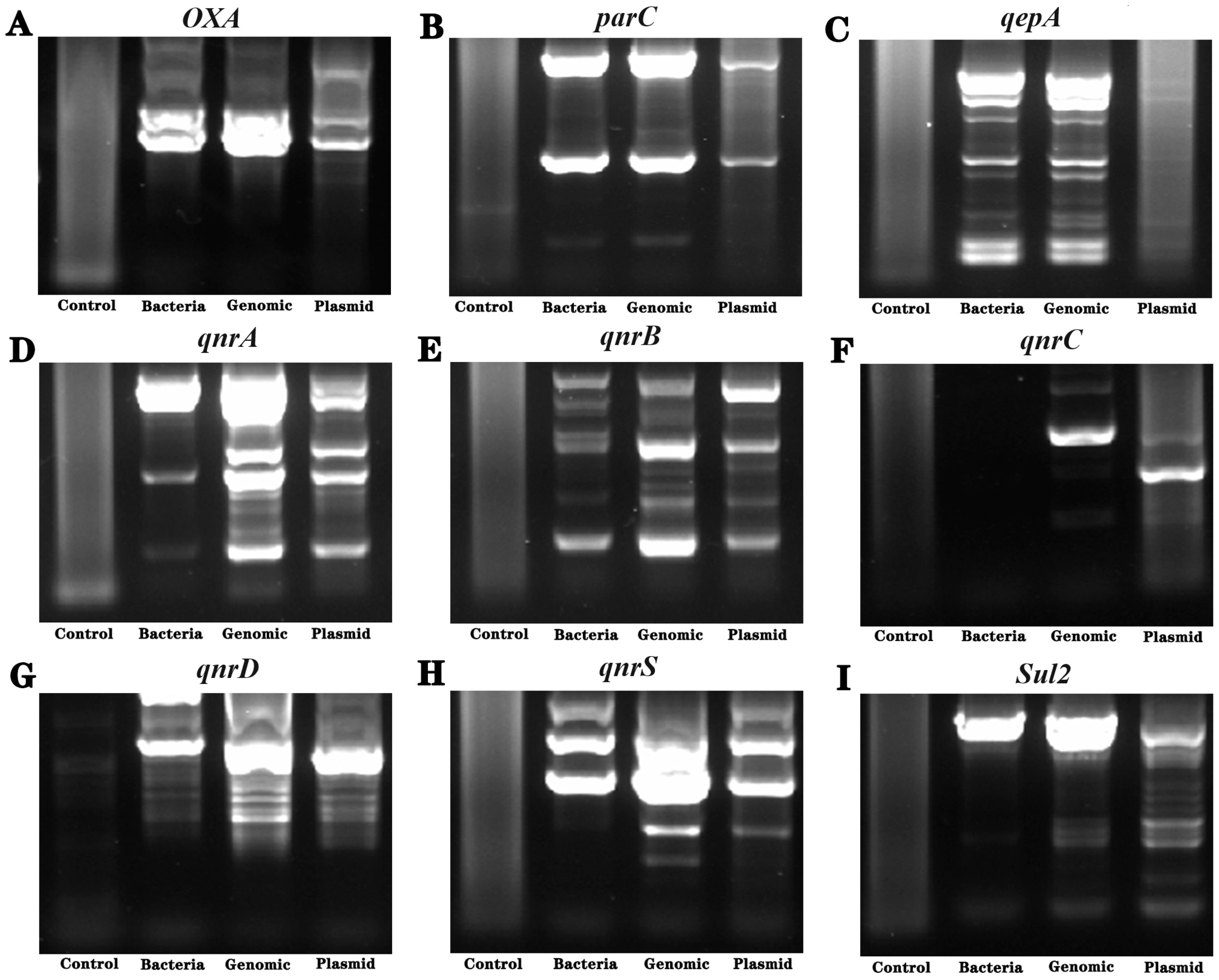
3.6. Detection of Drug Resistance-Related Genes

Specific primers [8,9,10,11,12] were used for gene amplification using the bacterial bodies, extracted bacterial genomes, and plasmid as templates. The length of the amplified product was determined by agarose gel electrophoresis, and the specific bands detected were recovered and sequenced. The results showed that there were drug-resistance genes gyrA, OqxB, and ParC in Klebsiella pneumoniae (Figure 3 and Figure 4).

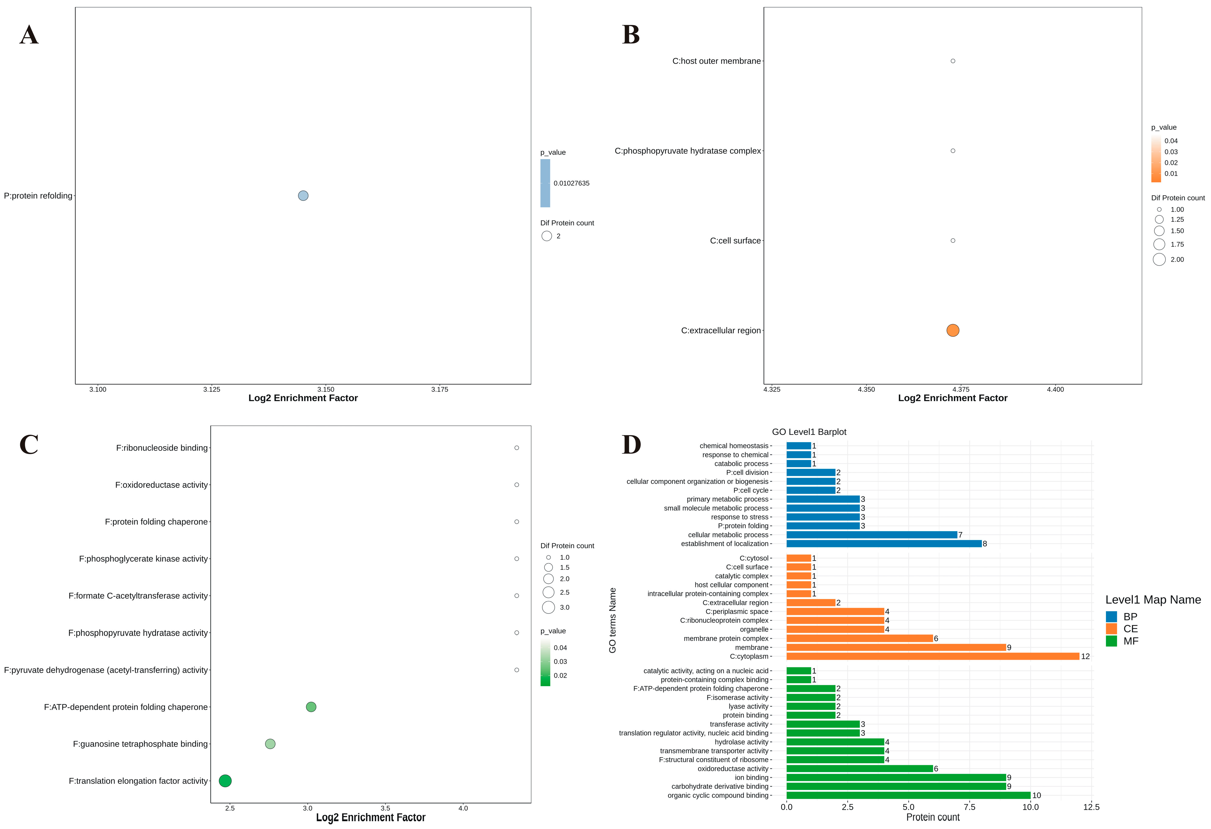
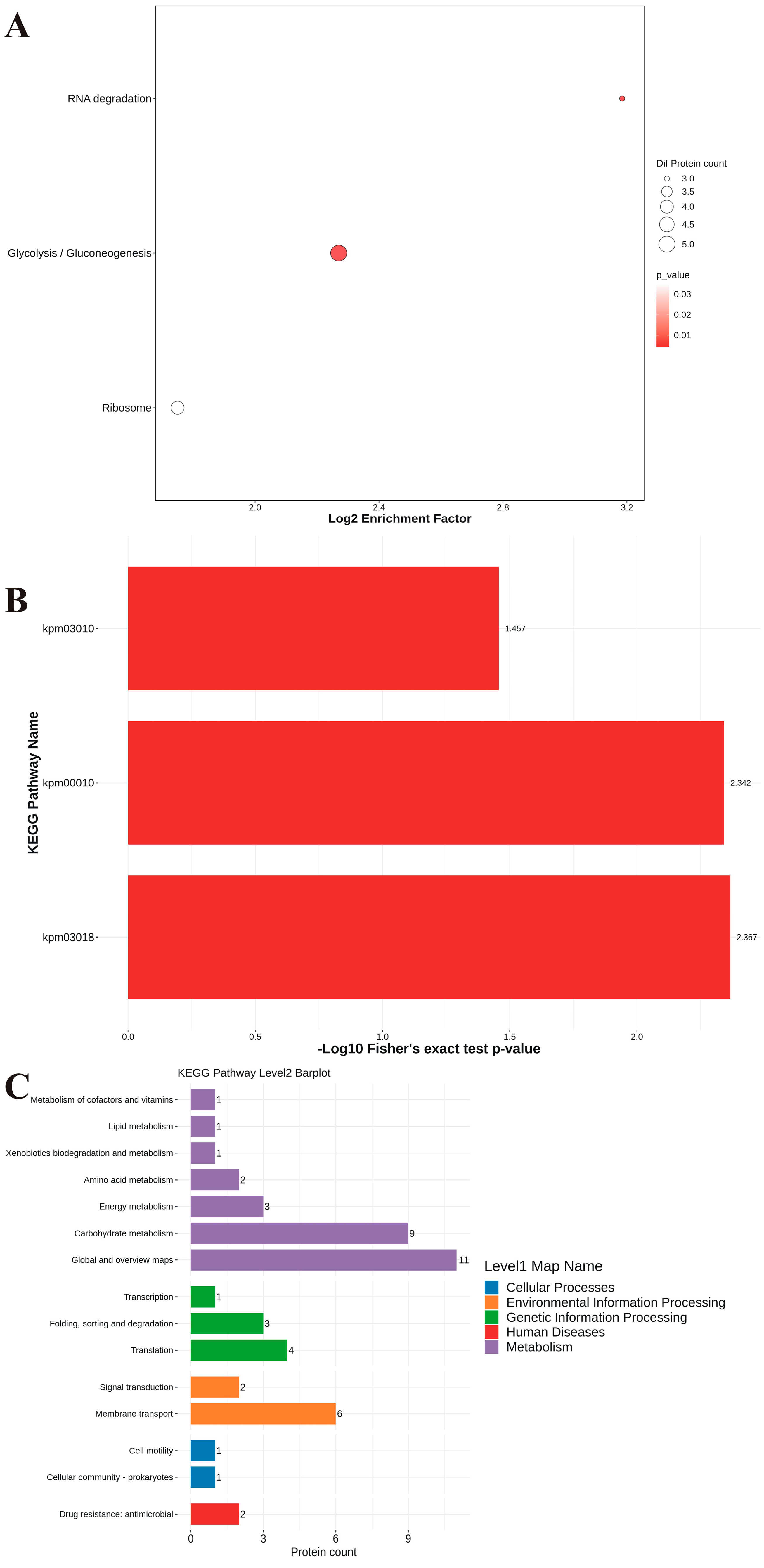
3.7. Proteomic Analysis

Through proteomic analysis, more than 1800 proteins from the isolated Klebsiella pneumoniae were identified. After the screening, three proteins associated with drug resistance were found in Table 4. We selected the top 50 proteins with the most abundant quantities for further analysis. GO enrichment demonstrated that the biological process aspect is mainly related to the cellular metabolic process and establishment of localization; cellular component focus on membrane protein complex, membrane, and cytoplasm; oxidoreductase activity; molecular function mainly included ion binding, carbohydrate derivative binding, and organic cyclic compound binding. Kyoto Encyclopedia of Genes and Genomes (KEGG) pathway enrichment analysis showed that level 1 metabolism was still the main component, and level 2 represented the classification of metabolic and functional pathways (Figure 5). To further analyze the intracellular and extracellular functions of proteins with high enrichment abundance, we selected some high abundance proteins for further KEGG analysis that show cellular processes, environmental information processing, genetic information processing, human diseases, and metabolism related to level 1 map names. Level 2 barplot displays carbohydrate metabolism, translation, and membrane transport as the primary functional pathways; others include cofactors, vitamins, lipid metabolism, etc. (Figure 6).

4. Discussion

Environmental and health hazards caused by industrial wastewater pollution pose a significant global challenge. However, the current environmental management of industrial wastewater mostly focuses on reducing the concentration of chemical pollutants, toxic compound recovery, and wastewater purification. There is still relatively limited research on microorganisms’ environmental adaptability and drug resistance during the treatment processes of different industries.
This study demonstrates that Klebsiella pneumoniae can grow even in the presence of high concentrations of sulfate and phosphate compounds. The bacteria’s protein fingerprint can be quickly identified using protein mass spectrometry, which can help us quickly identify bacteria. This is a very important technical tool in medicine, animal husbandry, and environmental science. From the growth of this strain of Klebsiella pneumoniae, it can be seen that it has a good proliferation density in a short period of time. Although the growth density of the bacteria decreases with time, it will remain at a certain high growth density, which provides us with meaningful thinking. That is, strains that can grow highly efficiently in environmental pollution substances are more likely to become widely promoted and applied in environmental governance. The bacterium exhibited rapid growth and demonstrated adaptation to high concentrations of metallic ions while exhibiting some level of antibiotic tolerance. The emergence and spread of Klebsiella pneumonia isolates that produce broad-spectrum lactamases pose a major threat to public health [13]. The discharge of hospital wastewater is one of the main sources of drug-resistant bacteria. Studies have shown a high frequency of gram-negative bacilli producing β-lactamase, including those in different geographical regions of Colombia, even in wastewater treatment plants [14]. Antibiotic-resistant bacteria originating from hospital wastewater end up in municipal wastewater treatment plants, serving as significant reservoirs for spreading antibiotic-resistance genes. Additionally, industrial wastewater, whether directly discharged or post-treatment, can also act as an important source for disseminating antibiotic-resistance genes. Moreover, concerning findings indicate that, through urban wastewater treatment plants, carbapenemase-producing Klebsiella pneumoniae strains carrying antimicrobial resistance genes can be traced from hospitals to surface waters [15]. Previous research has demonstrated the isolation from urban wastewater treatment plants of Escherichia coli and Klebsiella pneumoniae strains carrying the CTX-M-15 enzyme [16]. Bacteria with carbapenem-resistant Klebsiella spp. genetic characteristics have been found in municipal and slaughterhouse wastewater [7]. Unlike the antimicrobial resistance observed in traditional healthcare-associated sources, the Klebsiella pneumoniae strains identified in this study may possess the ability to adsorb or degrade metal salts. Previous studies have demonstrated significant phenol degradation properties by Pseudomonas aeruginosa and various Klebsiella strains in sludge [17]. Ciprofloxacin and levofloxacin are fluoroquinolone antibiotics that are widely used in hospitals and frequently present in wastewater treatment plants and marine environments. It has been demonstrated that Klebsiella pneumoniae strains obtained from industrial wastewater exhibit biodegradation capabilities toward quinolone drugs [18]. Additionally, newly isolated Klebsiella pneumoniae strains could produce self-secreted biosurfactants that enhance the solubility and bioavailability of xylene, facilitating the biodegradation of hydrophobic and toxic gases [19]. Furthermore, Klebsiella pneumoniae can produce a novel type of bioflocculant that can be applied in cyanobacterial sedimentation and urban wastewater treatment [20]. Through conjugation experiments, the in situ transfer of the multidrug-resistant plasmid RP4 carrying antibiotic resistance genes from the laboratory Escherichia coli strain C600 to indigenous active sludge bacteria has been demonstrated previously, indicating the ability of plasmids carrying resistance genes to be horizontally transferred to other microorganisms in natural wastewater environments [21]. Several genes and the respective proteins involved in antibiotic resistance were identified by gene amplification and proteomics identification, including the multi-drug resistance outer membrane protein MdtQ, the multi-drug resistance secretion protein, and the modulator of drug activity B. The resistance genes and proteins were co-expressed in Klebsiella pneumoniae, providing new insights into the presence and dynamics of resistance elements in bacteria. Moreover, Klebsiella pneumoniae exhibits other functions, such as the biodegradation of 2-methyl quinoline, and strains have been isolated from wastewater with tolerance to arsenic and copper [22,23,24]. Additionally, certain Klebsiella pneumoniae strains demonstrate efficient biodegradation of thermally treated high-density polyethylene, biodegradation of different aromatic compounds, and production of bioflocculants [25,26,27]. Recent studies have shown that bioinformatics has been applied to the study of Klebsiella pneumonia. For example, the isolated strain KPTA-2108 was obtained using whole genome sequencing technology, and further analysis was executed through bioinformatics methods, especially showing that the length of KPTA-2108 carried four plasmids [28]. The mechanisms of drug resistance in the new mutant Klebsiella pneumonia were studied by whole genome sequencing and bioinformatics analysis [29]. Currently, some bioinformatics analytical tools and databases have been applied that predict that it belongs to a cytosolic protein; it was included in the beta-lactamase family and has structural similarity to the FmtA protein of Staphylococcus aureus [30]. However, our research on Klebsiella pneumoniae through the proteomic method is still rare. Proteomic analysis showed more than 1800 proteins were found in the isolated Klebsiella pneumoniae in industrial wastewater. GO enrichment demonstrated that biological processes, cellular components, and molecular function mainly included cellular metabolic processes, membrane protein complex ion binding, organic cyclic compound binding, etc. KEGG pathway enrichment was executed by selecting the most abundant quantity of proteins. The analysis showed cellular processes, environmental information processing, genetic information processing, human diseases, and metabolism relate to a significant functional classification.
This study has demonstrated the rapid growth and inherent drug resistance of a Klebsiella pneumoniae strain isolated from wastewater with high sulfate and phosphate ion concentrations. This study used protein mass spectrometry technology for rapid identification of bacteria and further confirmed Klebsiella pneumoniae through gene sequence analysis of bacterial 16S DNA. Klebsiella pneumoniae has a good growth density, even though it decreases over time, but it still maintains a high density during the experiment period. This bacterium represents a potential drug-resistant pathogenic microorganism that can enter the natural environment. Proteomics and bioinformatics analysis have further enriched the connection between the function and adaptability of this bacterium, allowing us to gain a deeper understanding of its natural properties. These findings provide valuable insights for increasing the sensitivity and accuracy of microorganism detection in industrial wastewater and preventing the proliferation of drug-resistant microorganisms in the environment.

Author Contributions

Methodology, K.Y., C.L. and W.W.; editing, H.W. and J.G.; conceptualization and review, H.W. All authors have read and agreed to the published version of the manuscript.

Funding

This work was supported by the Science and Technology Development Plan of Henan Province (No. 242102310336), the Key Research and Development Special Project of Henan Province (No. 231111320200), the Education and Teaching Reform and Research Project of the National Food Industry Vocational Education and Teaching Guidance Committee in 2023 (SHK2023027), and the Education and Teaching Reform Project of the National Biotechnology Vocational Education and Teaching Guidance Committee in 2023 (XM202341).

Data Availability Statement

Data are contained within the article.

Conflicts of Interest

The authors declare no conflicts of interest.

References

  1. Behera, S.; Tanuku, N.R.S.; Moturi, S.R.K.; Gudapati, G.; Tadi, S.R.; Modali, S. Anthropogenic impact and antibiotic resistance among the indicator and pathogenic bacteria from several industrial and sewage discharge points along the coast from Pydibhimavaram to Tuni, East Coast of India. Environ. Monit. Assess. 2023, 195, 546. [Google Scholar] [CrossRef] [PubMed]
  2. Wang, D.; Zou, H.Y.; Zhao, L.; Li, Q.; Meng, M.; Li, X.W.; Berglund, B. High prevalence of Escherichia coli co-harboring conjugative plasmids with colistin- and carbapenem resistance genes in a wastewater treatment plant in China. Int. J. Hyg. Environ. Health 2023, 250, 114159. [Google Scholar] [CrossRef] [PubMed]
  3. Cahill, N.; Hooban, B.; Fitzhenry, K.; Joyce, A.; O’Connor, L.; Miliotis, G.; McDonagh, F.; Burke, L.; Chueiri, A.; Farrell, M.L.; et al. First reported detection of the mobile colistin resistance genes, mcr-8 and mcr-9, in the Irish environment. Sci. Total Environ. 2023, 876, 162649. [Google Scholar] [CrossRef] [PubMed]
  4. Liu, M.C.; Jian, Z.; Liu, W.; Li, J.; Pei, N. One Health Analysis of mcr-Carrying Plasmids and Emergence of mcr-10.1 in Three Species of Klebsiella Recovered from Humans in China. Microbiol. Spectr. 2022, 10, e0230622. [Google Scholar] [CrossRef] [PubMed]
  5. Mutuku, C.; Melegh, S.; Kovacs, K.; Urban, P.; Virag, E.; Heninger, R.; Herczeg, R.; Sonnevend, A.; Gyenesei, A.; Fekete, C.; et al. Characterization of beta-Lactamases and Multidrug Resistance Mechanisms in Enterobacterales from Hospital Effluents and Wastewater Treatment Plant. Antibiotics 2022, 11, 776. [Google Scholar] [CrossRef] [PubMed]
  6. Bong, C.W.; Low, K.Y.; Chai, L.C.; Lee, C.W. Prevalence and Diversity of Antibiotic Resistant Escherichia coli From Anthropogenic-Impacted Larut River. Front. Public Health 2022, 10, 794513. [Google Scholar] [CrossRef] [PubMed]
  7. Savin, M.; Bierbaum, G.; Mutters, N.T.; Schmithausen, R.M.; Kreyenschmidt, J.; Garcia-Menino, I.; Schmoger, S.; Kasbohrer, A.; Hammerl, J.A. Genetic Characterization of Carbapenem-Resistant Klebsiella spp. from Municipal and Slaughterhouse Wastewater. Antibiotics 2022, 11, 435. [Google Scholar] [CrossRef] [PubMed]
  8. Wang, H.F.; Du, L.Y.; Luo, J.; He, H.X. Isolation, identification and characterization of Morganella morganii from Naja naja atra in Beijing, China. Cell Mol. Biol. 2017, 63, 52–58. [Google Scholar] [CrossRef] [PubMed]
  9. Yigit, H.; Queenan, A.M.; Anderson, G.J.; Domenech-Sanchez, A.; Biddle, J.W.; Steward, C.D.; Alberti, S.; Bush, K.; Tenover, F.C. Novel carbapenem-hydrolyzing beta-lactamase, KPC-1, from a carbapenem-resistant strain of Klebsiella pneumoniae. Antimicrob. Agents Chemother. 2001, 45, 1151–1161. [Google Scholar] [CrossRef]
  10. Yu, Y.; Ji, S.; Chen, Y.; Zhou, W.; Wei, Z.; Li, L.; Ma, Y. Resistance of strains producing extended-spectrum beta-lactamases and genotype distribution in China. J. Infect. 2007, 54, 53–57. [Google Scholar] [CrossRef]
  11. Kim, H.B.; Wang, M.; Park, C.H.; Kim, E.C.; Jacoby, G.A.; Hooper, D.C. oqxAB encoding a multidrug efflux pump in human clinical isolates of Enterobacteriaceae. Antimicrob. Agents Chemother. 2009, 53, 3582–3584. [Google Scholar] [CrossRef] [PubMed]
  12. Liu, J.H.; Deng, Y.T.; Zeng, Z.L.; Gao, J.H.; Chen, L.; Arakawa, Y.; Chen, Z.L. Coprevalence of plasmid-mediated quinolone resistance determinants QepA, Qnr, and AAC(6′)-Ib-cr among 16S rRNA methylase RmtB-producing Escherichia coli isolates from pigs. Antimicrob. Agents Chemother. 2008, 52, 2992–2993. [Google Scholar] [CrossRef] [PubMed]
  13. Obasi, A.; Nwachukwu, S.; Ugoji, E.; Kohler, C.; Gohler, A.; Balau, V.; Pfeifer, Y.; Steinmetz, I. Extended-Spectrum -Lactamase-Producing Klebsiella pneumoniae from Pharmaceutical Wastewaters in South-Western Nigeria. Microb. Drug Resist. 2017, 23, 1013–1018. [Google Scholar] [CrossRef] [PubMed]
  14. Aristizabal-Hoyos, A.M.; Rodriguez, E.A.; Torres-Palma, R.A.; Jimenez, J.N. Concern levels of beta-lactamase-producing Gram-negative bacilli in hospital wastewater: Hotspot of antimicrobial resistance in Latin-America. Diagn. Microbiol. Infect. Dis. 2023, 105, 115819. [Google Scholar] [CrossRef] [PubMed]
  15. Loudermilk, E.M.; Kotay, S.M.; Barry, K.E.; Parikh, H.I.; Colosi, L.M.; Mathers, A.J. Tracking Klebsiella pneumoniae carbapenemase gene as an indicator of antimicrobial resistance dissemination from a hospital to surface water via a municipal wastewater treatment plant. Water Res. 2022, 213, 118151. [Google Scholar] [CrossRef] [PubMed]
  16. Dolejska, M.; Frolkova, P.; Florek, M.; Jamborova, I.; Purgertova, M.; Kutilova, I.; Cizek, A.; Guenther, S.; Literak, I. CTX-M-15-producing Escherichia coli clone B2-O25b-ST131 and Klebsiella spp. isolates in municipal wastewater treatment plant effluents. J. Antimicrob. Chemother. 2011, 66, 2784–2790. [Google Scholar] [CrossRef]
  17. Mahgoub, S.A.; Qattan, S.Y.A.; Salem, S.S.; Abdelbasit, H.M.; Raafat, M.; Ashkan, M.F.; Al-Quwaie, D.A.; Motwali, E.A.; Alqahtani, F.S.; Abd El-Fattah, H.I. Characterization and Biodegradation of Phenol by Pseudomonas aeruginosa and Klebsiella variicola Strains Isolated from Sewage Sludge and Their Effect on Soybean Seeds Germination. Molecules 2023, 28, 1203. [Google Scholar] [CrossRef]
  18. Shaker, R.A.E.; Nagy, Y.I.; Adly, M.E.; Khattab, R.A.; Ragab, Y.M. Acinetobacter baumannii, Klebsiella pneumoniae and Elizabethkingia miricola isolated from wastewater have biodegradable activity against fluoroquinolone. World J. Microbiol. Biotechnol. 2022, 38, 187. [Google Scholar] [CrossRef]
  19. Wang, Y.; Wan, S.; Yu, W.; Yuan, D.; Sun, L. Newly isolated Enterobacter cloacae sp. HN01 and Klebsiella pneumoniae sp. HN02 collaborate with self-secreted biosurfactant to improve solubility and bioavailability for the biodegradation of hydrophobic and toxic gaseous para-xylene. Chemosphere 2022, 304, 135328. [Google Scholar] [CrossRef]
  20. Nie, M.; Yin, X.; Jia, J.; Wang, Y.; Liu, S.; Shen, Q.; Li, P.; Wang, Z. Production of a novel bioflocculant MNXY1 by Klebsiella pneumoniae strain NY1 and application in precipitation of cyanobacteria and municipal wastewater treatment. J. Appl. Microbiol. 2011, 111, 547–558. [Google Scholar] [CrossRef]
  21. Soda, S.; Otsuki, H.; Inoue, D.; Tsutsui, H.; Sei, K.; Ike, M. Transfer of antibiotic multiresistant plasmid RP4 from Escherichia coli to activated sludge bacteria. J. Biosci. Bioeng. 2008, 106, 292–296. [Google Scholar] [CrossRef] [PubMed]
  22. Wang, L.; Li, Y.; Duan, J. Biodegradation of 2-methylquinoline by Klebsiella pneumoniae TJ-A isolated from acclimated activated sludge. J. Environ. Sci. Health A Tox Hazard. Subst. Environ. Eng. 2014, 49, 27–38. [Google Scholar] [CrossRef] [PubMed]
  23. Abbas, S.Z.; Riaz, M.; Ramzan, N.; Zahid, M.T.; Shakoori, F.R.; Rafatullah, M. Isolation and characterization of arsenic resistant bacteria from wastewater. Braz. J. Microbiol. 2014, 45, 1309–1315. [Google Scholar] [CrossRef] [PubMed]
  24. Zulfiqar, S.; Shakoori, A.R. Molecular characterization, metal uptake and copper induced transcriptional activation of efflux determinants in copper resistant isolates of Klebsiella pneumoniae. Gene 2012, 510, 32–38. [Google Scholar] [CrossRef] [PubMed]
  25. Ma, L.; Liang, J.; Wang, S.; Yang, B.; Chen, M.; Liu, Y. Production of a bioflocculant from Klebsiella sp. OS-1 using brewery wastewater as a source. Environ. Technol. 2019, 40, 44–52. [Google Scholar] [CrossRef]
  26. Awasthi, S.; Srivastava, P.; Singh, P.; Tiwary, D.; Mishra, P.K. Biodegradation of thermally treated high-density polyethylene (HDPE) by Klebsiella pneumoniae CH001. 3 Biotech 2017, 7, 332. [Google Scholar] [CrossRef]
  27. Rajkumari, J.; Paikhomba Singha, L.; Pandey, P. Genomic insights of aromatic hydrocarbon degrading Klebsiella pneumoniae AWD5 with plant growth promoting attributes: A paradigm of soil isolate with elements of biodegradation. 3 Biotech 2018, 8, 118. [Google Scholar] [CrossRef] [PubMed]
  28. Liu, Q.; Guo, Z.; Zhu, G.; Li, N.; Bai, G.; Jiang, M. Genomic Characteristics and Phylogenetic Analyses of a Multiple Drug-Resistant Klebsiella pneumoniae Harboring Plasmid-Mediated MCR-1 Isolated from Tai’an City, China. Pathogens 2023, 12, 221. [Google Scholar] [CrossRef]
  29. Yu, M.; Wei, Q.; Song, W.; Yuan, J. Phenotypic and Genetic Analysis of KPC-49, a KPC-2 Variant Conferring Resistance to Ceftazidime-Avibactam and Maintaining Resistance to Imipenem and Meropenem. Infect. Drug Resist. 2023, 16, 2477–2485. [Google Scholar] [CrossRef]
  30. Singh, V.; Dhankhar, P.; Dalal, V.; Tomar, S.; Kumar, P. In-silico functional and structural annotation of hypothetical protein from Klebsiella pneumonia: A potential drug target. J. Mol. Graph. Model. 2022, 116, 108262. [Google Scholar] [CrossRef]
Figure 1. MALDI-TOF MS identification of the isolated Klebsiella pneumoniae strain at different growth times and phylogenetic tree analysis. (AD) represents the MALDI-TOF MS identification results of colony samples cultured for 12, 16, 20, and 24 h. (E) represents the phylogenetic tree based on 16S rDNA gene sequences.
Separations 11 00121 g001
Figure 2. Growth curve of the isolated Klebsiella pneumoniae strain. After 90 h of culture, the growth characteristics of Klebsiella pneumoniae at different inoculum sizes were evaluated. (A) represents the control without inoculation (blank control), while (BH) represents the growth of Klebsiella pneumoniae with different inoculum sizes.
Separations 11 00121 g002
Figure 3. Analysis of drug resistance genes in Klebsiella pneumoniae isolated from industrial wastewater. The bacterial genomic and plasmid DNA were used as templates for amplification with specific primers, and then the electrophoresis bands were sequenced. (AI) represent the nucleic acid gel images of drug-resistance genes.
Separations 11 00121 g003
Figure 4. Analysis of drug resistance genes in Klebsiella pneumoniae isolated from industrial wastewater. The bacterial genome and plasmids were used as templates for amplification with specific primers, and then the electrophoresis bands were sequenced. (AI) represent the nucleic acid gel images of drug-resistance genes.
Separations 11 00121 g004
Figure 5. GO classification of proteomics data for Klebsiella pneumoniae. (A), BP dotplot; (B), CE dotplot; (C), dotplot; (D), the x-axis represents protein count, and the y-axis represents GO term name.
Separations 11 00121 g005
Figure 6. KEGG pathways of proteomics data for Klebsiella pneumoniae. (A), KEGG dot plot; (B), KEGG fisher barplot; (C), level 1 represents the classification of environmental information processing, genetic information processing, and metabolism; and level 2 represents the classification of metabolic and functional pathways.
Separations 11 00121 g006
Table 1. Biochemical identification of Klebsiella pneumoniae.
NameAbbreviationResults
Amino acid controlCNegative
Arginine dihydrolaseADHNegative
UreaseUREPositive
Lysine decarboxylaseLDCPositive
Aescin hydrolysisESCPositive
Ornithine decarboxylaseODCNegative
Rhamnose fermentationRHAfPositive
Melibiose fermentationMELfPositive
GalactosidaseONPGPositive
Glucose fermentationGLUfPositive
Hydrogen sulfide productionH2SNegative
Utilization of citrateCITPositive
Gelatin hydrolysisGELNegative
Lactose fermentationLACfNegative
Tryptophan deaminaseTDANegative
Cellulose disaccharide fermentationCELfPositive
Production of indoleINDNegative
Arabinose fermentationARAfPositive
VP ExperimentVPPositive
Bitter almond glycoside fermentationAMYfNegative
Mannitol fermentationMANfPositive
Inositol fermentationINOfPositive
Sucrose fermentationSACfPositive
Sorbitol fermentationSORfPositive
Table 2. Antibiotic sensitivity assays of Klebsiella pneumoniae.
Drug NameAbbreviationMIC Value
Piperacillin/tazobactamP/T=4/4
TobramycinTOB≤1
FurantoinFD≥128
CefazolinCFZ≤2
CeftazidimeCAZ≤0.5
AmpicillinAMP>32
MoxifloxacinMXF≤0.5
MinocyclineMIN≤4
GentamicinGEN≤1
MeropeneMMRP≤0.06
Cefoperazone/sulbactamCPS≤16/8
Ceftazidime/clavulanic acidCAZ/C≤1/4
ImipenemIPM≤0.25
CefuroximeCXM≤8
Ticarcillin/clavulanic acidTIM≤16/2
CefoxitinFOX≤8
LevofloxacinLEV=0.12
Polymyxin BPB≤1
CefepimeFEP≤0.12
Cefotaxime/clavulanic acidCTX/C≤1/4
Amoxicillin/clavulanateAMC≤8/4
AztreonamATM≤0.25
ErtapenemETP≤0.5
AmikacinAMK≤4
Compound xinnuominSXT≤2/38
CefotaximeCTX≤0.12
CiprofloxacinCIP≤0.015
ChloramphenicolCHL≥32
TigecyclineTGC≤0.25
Ampicillin/sulbactamAMS=16/8
Table 3. The growth peak point of Klebsiella pneumoniae under different concentrations.
Test GroupTime (h)Peak Point (OD)
Klebsiella pneumoniae-1 μL135
Klebsiella pneumoniae-10 μL135.08
Klebsiella pneumoniae-20 μL135.07
Klebsiella pneumoniae-40 μL124.9
Klebsiella pneumoniae-60 μL134.82
Klebsiella pneumoniae-80 μL114.73
Klebsiella pneumoniae-100 μL114.66
Table 4. Proteomics screen out proteins related to drug resistance.
ItemProteomics Analysis
1Multidrug-resistant outer membrane protein (MdtQ)
2Multidrug-resistant secretion protein
3Modulator of drug activity B
Disclaimer/Publisher’s Note: The statements, opinions and data contained in all publications are solely those of the individual author(s) and contributor(s) and not of MDPI and/or the editor(s). MDPI and/or the editor(s) disclaim responsibility for any injury to people or property resulting from any ideas, methods, instructions or products referred to in the content.

