Article

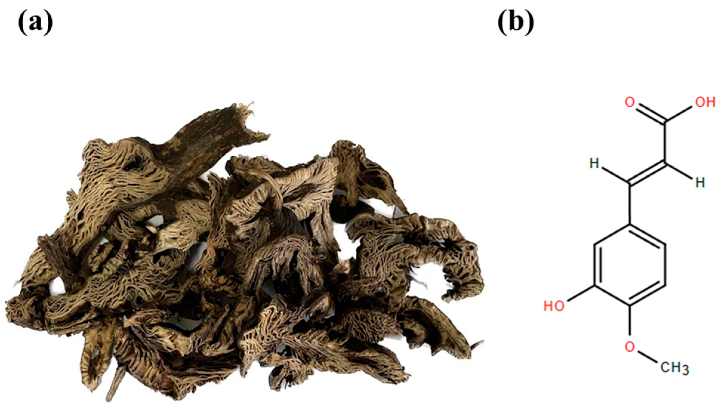
Phytochemical Identification and Anti-Oxidative Stress Effects Study of Cimicifugae Rhizoma Extract and Its Major Component Isoferulic Acid

1
School of Chinese Materia Medica, Beijing University of Chinese Medicine, Beijing 100029, China
2
College of Traditional Chinese Medicine, Xinjiang Medical University, Urumqi 830017, China
*
Authors to whom correspondence should be addressed.
Separations 2024, 11(6), 175; https://doi.org/10.3390/separations11060175
Submission received: 26 April 2024 / Revised: 27 May 2024 / Accepted: 30 May 2024 / Published: 3 June 2024
(This article belongs to the Section Analysis of Natural Products and Pharmaceuticals)

Abstract

:
Background and Objectives: Cimicifugae Rhizoma, also known as ‘Sheng ma’ in Madeiran, is a widely used Chinese herbal medicine that has several pharmacological qualities, one of which is its antioxidant activity. Isoferulic acid, a prominent phenolic compound found in Cimicifugae Rhizoma, has potent antioxidant properties. This study was aimed to comprehensively analyze the components in Cimicifugae Rhizoma and rat plasma to evaluate the in vitro antioxidant and anti-inflammatory properties of Cimicifugae Rhizoma extract and Isoferulic acid as potential candidates for developing herbal formulations targeting podocyte injury in diabetic nephropathy for further clinical utilization. Materials and Methods: UPLC/Q-TOF-MS and HPLC were utilized as analytical tools to identify components of Cimicifugae Rhizoma extract or rat plasma after administrating it. MPC5 cells were cultured with H2O2 and high glucose and subjected to oxidative stress injury. The CXCL12/CXCR4 system plays a crucial role at certain stages of multiple kidney diseases’ injury. Apoptosis-related and target CXCL12/CXCR4/mTOR/Caspase-3 and Cask protein levels were assessed, and the levels of inflammatory-related factors, motility, morphology, ROS level, and apoptosis in podocytes were tested. Results: A total of 82 and 39 components were identified in the Cimicifugae Rhizoma extract and plasma, and Isoferulic acid content was determined as 6.52 mg/g in the Cimicifugae Rhizoma extract. The Cimicifugae Rhizoma extract (1 μg/mL) and Isoferulic acid (10, 25, 50 μM) considerably decreased high glucose and oxidative-stress-mediated toxicity, impaired mobility and adhesion and apoptotic changes in MPC5 cells, and reversed inflammation response. Moreover, the Cimicifugae Rhizoma extract and Isoferulic acid down-regulated Cask, mTOR, and Caspase-3, while significantly blocking the overactivation of CXCL12/CXCR4 in podocytes stimulated by oxidative stress and high glucose. Conclusions: These results indicate that the renal protective mechanism of the Cimicifugae Rhizoma extract and Isoferulic acid on simulating H2O2-induced podocyte injury involves mainly the of CXCL12/CXCR4 pathways and the inactivation of oxidative-stress-mediated apoptotic pathways after comprehensive qualitative and quantitative research by UPLC/Q-TOF-MS and HPLC. These findings provide an important efficacy and ingredient basis for further study on the clinical utilities of Cimicifugae Rhizoma and Isoferulic acid on podocyte and kidney impairment.

1. Introduction

Cimicifugae Rhizoma (Figure 1a) is the dry rhizome of Cimicifuga heracleifolia Kom., Cimicifuga dahurica (Turcz.) Maxim., or Cimicifuga foetida L. of Ranunculaceae. Cimicifugae Rhizoma. Cimicifugae Rhizoma shows great antioxidant capacity and contains triterpenoid saponins, phenolic acids, chromogenic ketones, alkaloids and so on, with caffeic acid, ferulic acid, isoferulic acid, and ascophyllin as the main components [1]. And the antioxidant effect may come from the phenolic acids that Cimicifugae Rhizoma contains. The amount of phenolic acid in Cimicifugae Rhizoma is strongly linked to its antioxidant effect [2]. A previous study identified the most important beneficial components are caffeic acid, ferulic acid, and Isoferulic acid, which work by metal-chelating and radical-scavenging to reduce oxidative damage [3]. Isoferulic acid (Figure 1b), which is the representative phenolic acid component of Cimicifugae Rhizoma and the specified quality control target component of Cimicifugae Rhizoma, according to the 2020 Edition of the Chinese Pharmacopoeia, can exert antioxidant, anti-inflammatory, and anti-apoptosis activities and a potential role in regulating blood glucose [4,5,6]. Isoferulic acid has been found to have a mixed-inhibition effect on intestinal α-glucosidase by kinetic study, which also showed that Isoferulic acid would block intestinal maltase [7]. Isoferulic acid may help stop the glycation and oxidation of bovine serum albumin, which is linked to diabetes complications [8]. This is shown by lowering the levels of fructosamine, carboxymethyllysine, and amyloid cross-β structure, as well as the protein carbonyl content and stopping thiol group modification in bovine serum albumin [9]. Methylglyoxa causes failure, ROS production, and death in INS-1 cells. Isoferulic acid pretreatment decreases these effects by increasing GLO1 activity and the mitochondrial survival pathway [10]. Administering streptozotocin and Isoferulic acid (5.0 mg/kg) three times in one day intravenously to diabetic rats results in reduced levels of glucose in the blood plasma and alterations in the quantities of GLUT4 and PEPCK proteins. These findings indicate that Isoferulic acid may have potential benefits in the treatment of diabetes [11]. In the subsequent investigation of Isoferulic acid, it was determined that the decline in plasma glucose levels in streptozotocin -diabetic rats might perhaps be attributed to the activation of opioid mu-receptors, leading to an augmented release of beta-endorphins and alterations in glucose utilization or hepatic glucose production [12]. Due to its potential anti-inflammatory and anti-glycemic properties, Isoferulic acid might potentially be used for the treatment of diabetic nephropathy. Diabetic nephropathy is the primary factor leading to end-stage renal disease globally and is a prevalent vascular complication of both type 1 and type 2 diabetes mellitus. Elevated levels of TNF-α and IL6 are strongly associated with oxidative stress and inflammation, resulting in the development of diabetic nephropathy [13]. To enhance diabetic nephropathy, it is possible to inhibit the cell signaling pathways responsible for the production of ROS [14]. Elevated levels of ROS activate cellular pathways that trigger apoptosis and necrosis [15]. Exogenous H2O2 triggers a signaling cascade by mimicking the endogenous ROS signaling pathway, leading to ROS accumulation, which inhibits cell viability and triggers mitochondria-dependent apoptosis. Meanwhile, high glucose induction could lead MPC5 podocytes to the situation of diabetic nephropathy.
Cimicifugae Rhizoma has also been found to have good anti-inflammatory and antioxidant activity in previous studies. Phenolic acid compounds such as Cimidahuside E and Cimidahuside D can inhibit the production of PGE2 in LPS-stimulated RAW 264.7 macrophages, down-regulate the expression of COX-2, and then exert anti-inflammatory activity [16]. Caffeic-acid-containing cinnamic acid derivatives (such as Isoferulic acid) with potential anti-inflammatory activity were screened by UPLC/Q-TOF-MS-integrated NF-κB luciferase reporter gene detection system. Caffeic acid significantly inhibited the activation of NF-κB in TNF-α-induced injury of BEAS-2B cells in a dose-dependent manner [17]. The ethanol extract of Cimicifugae Rhizoma can effectively alleviate OVA ovalbumin-induced inflammatory and oxidative stress injury by up-regulating Nrf2/HO-1/NQO1 signaling and down-regulating NF-κB phosphorylation [18]. The previous studies focused on the quality markers and components of Cimicifugae Rhizoma from original plants [19,20]. But our study aimed to comprehensively identify the components absorbed into the plasma after administration of Cimicifugae Rhizoma and screen out the component with the best antioxidant effect on podocytes impaired by oxidative stress, so as to provide the potential evidence of the renal protection of Cimicifugae Rhizoma.
Isoferulic acid (6.52 mg/g) was screened for the strongest antioxidant activity from 82 and 39 components identified by the UPLC/Q-TOF-MS of the Cimicifugae Rhizoma extract and plasma after administration of Cimicifugae Rhizoma extract combined with MTT assay. The objective of this research was to determine the protective effects of Cimicifugae Rhizoma and Isoferulic acid on oxidative-stress-damaged podocytes. This was carried out by examining the influence of Cimicifugae Rhizoma extract and Isoferulic acid on MPC5 cells that were subjected to high levels of glucose and H2O2. Researchers discovered that it operates by specifically targeting the CXCL12/CXCR4 pathway and its association with Caspase-3 and mTOR, hence alleviating cell death and oxidative stress. Consequently, the Cimicifugae Rhizoma extract and Isoferulic acid should be useful for devising a prospective component for the treatment.

2. Materials and Methods

2.1. Reagents and Chemicals

The conditionally immortalized mouse podocyte cell line (MCP5) was purchased from the BeNa Culture Collection (BNCC342021, Beijing, China). Cell line identification was performed by the same company. Isoferulic acid was obtained from China National Institute for Food and Drug Control (Lot: 111698-201904, purity = 99.3%, Beijing, China). Ferulic acid (Lot: B20007, purity ≥ 98%), Methyl ferulate (Lot: B30655, purity ≥ 98%), Caffeic acid (Lot: B20660, purity ≥ 98%), Paeonol (Lot: B20266, purity ≥ 98%), Ethyl caffeate (Lot: B20661, purity ≥ 98%), Methyl caffeate (Lot: B25588, purity ≥ 98%), Sinapic acid (Lot: B30104, purity ≥ 98%), 4-Hydroxyphenylacetic acid (Lot: B30104, purity ≥ 98%), Cimifugin (Lot: B21156, purity ≥ 98%), Cimigenol (Lot: B23157, purity ≥ 98%), and Acetylcimigenol 3-O-alpha-L-arabinopyranside (Lot: B21504, purity ≥ 98%) were purchased from Shanghai yuanye Bio-Technology Co., Ltd. (Shanghai, China). Cimicifugae Rhizoma-prepared herbal medicine was purchased from Beijing TaiRenKang Pharmaceutical Co. (Beijing, China) (Lot: 22081501): Roswell Park Memorial Institute 1640 (RPMI 1640). The 0.05% trypsin and penicillin/streptomycin solutions were Gibco products. Fetal bovine serum (FBS), Hydrogen peroxide (H2O2), and DMSO were products of Sigma-Aldrich (St. Louis, MO, USA). Phosphate-buffered saline (PBS) and methyl thiazolyl tetrazolium (MTT) were purchased from Beijing Solarbio Science & Technology Co., Ltd. (Beijing, China). Anti-beta Actin antibody (ab8227), Anti-SDF1 antibody (ab155090), Anti-CXCR4 antibody (ab181020), Anti-mTOR antibody (ab134903), Anti-Caspase-3 antibody (ab184787), and Anti-CASK antibody (ab3383) were obtained from Abcam (Cambridge, UK). Mouse IL-6 ELISA kit (SEA079Mu), CXCR4 ELISA kit (SEA940Mu), CXCL12 ELISA kit (SEA122Mu), and TNF-α ELISA kit (SCA133Mu) were products from Cloud-Clone Corp. (Wuhan, China), while IL-1β ELISA kit (E-EL-R0012c) was from Elabscience Biotechnology Co., Ltd. (Wuhan, China). Annexin V/FITC apoptosis assay kit, DCFH-DA assay kitand Actin-Tracker Red-594 were purchased from Beyotime Biotechnology (Shanghai, China).

2.2. Extract Samples Preparation and Components Identification

2.2.1. HPLC Parameters and Cimicifugae Rhizoma Extract Preparation

Cimicifugae Rhizoma extracts were prepared into 5 mg/mL methanol solution, filtered by a 0.22 μm filter member, and analyzed by HPLC (LC-20AT HPLC with SPD-20A UV-VIS detector, Shimadzu, Kyoto, Japan). A C18 (4.6 × 250 mm, 5 μm) column (Agilent Eclipse XDB-C18) was used to perform compounds’ separations. The mobile phase consisted of water containing acetonitrile (A) and 0.1%phosphoric acid (B). The gradient program was as follows: 0~5 min, 5% A, 5~20 min, 5% → 20% A, 20~60 min, 20% → 40% A. Injection volume was 10 μL, and the flow rate was 0.8 mL/min and under 320 nm.
The crude powder of Cimicifugae Rhizoma was taken and soaked in 50 times the amount of 70% ethanol for about 12 h. It was extracted twice by ultrasonication (37 °C, 200 W, 40 kHz) for 60 min and dried to the extract. Isoferulic acid was prepared with methanol and stored in cold storage at 4 °C for later use.

2.2.2. UPLC/Q-TOF-MS Parameters and Sample Preparations

The Cimicifugae Rhizoma extract sample was prepared by dissolving 50 mg in 1 mL of menthol via ultrasonication at 200 W and 40 kHz for 30 min and centrifugation at 12,000 r/min for 10 min at 4 °C. The solution was diluted tenfold, and the supernatant was centrifuged at a low temperature and high speed (12,000 r/min, 10 min). The resultant supernatant was the final sample to do the analysis. The standards of Isoferulic acid, ferulic acid, methyl ferulate, caffeic acid, paeonol, ethyl caffeate, methyl caffeate, sinapic acid, and 4-hydroxyphenylacetic acid were weighed precisely and prepared into 1 mg/mL single standard solution with methanol. Fifty μL of each single standard solution was added into a 50 mL measuring flask and, then, made up with methanol solution to form a 1 μg/mL mixed standard solution. The solution was diluted into 10 ng/mL, and the supernatant was centrifuged at a low temperature and high speed (12,000 r/min, 10 min).
In this study, 3 SD rats (SiPeiFu (Beijing) Biotechnology Co., Ltd. (Beijing, China)) were administered the Cimicifugae Rhizoma extract at a dose of 0.253 g/kg for the calculations of body areas of humans and rats (based on the maximum dosage of 10 g for human use as stipulated in the 2020 edition Chinese Pharmacopoeia). Rats were fasted for 24 h before drug administration. Blank blood samples were obtained before drug administration. Plasma was taken from the orbital plexus at 0.5 h, 1 h, 2 h, 4 h, and 6 h after administration and before administration as blank comparison. The blood samples were centrifuged at 4000 r/min for 10 min, and the plasma samples (supernatants) were kept at −80 °C prior to analysis. For plasma processing methods, refer to the previous study [21].
The experiments were approved by the Laboratory Animal Ethics Subcommittee, Academic Committee, Beijing University of Chinese Medicine (BUCM) under the Animal Ethics Licence No. BUCM- 202350804- 2115.
SYNAPT G2-Si Mass Spectrometer (Waters Corp., Milford, MA, USA) and I-Class Ultra High-Performance Liquid Chromatography (Waters Corp., Milford, MA, USA) were used. Column: ACQUITY UPLCTM T3 C18 column (100 × 2.1 mm, 1.7 μm); mobile phase: 0.1% formic acid in water (A) − 0.1% formic acid in acetonitrile solution (B); column temperature: 35 °C; sample holding temperature in the instrument: 10 °C; flow rate: 0.25 mL.min−1; injection volume: 3 μL. In both the positive and negative mode, elution was set as the gradient program (0–3 min, 0% B; 3–70 min, 0–100% B; 70–76 min, 100% B; 76–80 min, 100–0% B).
The MS parameters in the positive mode were as follows: mass scan range, 50 Da to 1500 Da; capillary voltage, 3000 V; lock spray capillary, 3000 V; desolvation temperature, 500 °C; gas flow rate, 500 L/Hr; cone gas flow rate, 50 L/Hr. Most MS parameters in the negative mode were same as those in the positive mode except for gas flow rate (800 L/Hr), capillary voltage (2000 V).
The identification was performed using the UNIFI Scientific Information System with the commercial Traditional Chinese Medicine database, online databases (e.g., Massbank), relative data from reports or articles, and results obtained from mixed standard solutions (Table A3) in this study.

2.3. Cell Cultures and Treatment

MPC5 cells were resuspended and inoculated in RPMI 1640 containing 10 U/mL interferon-gamma and 10% FBS and cultured at 33 °C with 5% CO2 for proliferation and passaging. The cells were transferred to RPMI 1640 without IFN-γ and allowed to differentiate for 7–12 days at 37 °C with 5% CO2.
Fully differentiated podocytes were divided into the low glucose group (LG), high glucose with H2O2-Induced group (HG + H2O2), and drug group. The cells in LG were treated with 5.6 mM glucose medium as the control group. HG + H2O2 cells were treated with 30 mM glucose medium and 150 μM H2O2. The drug group cells were treated with 10, 25, 50 μM Isoferulic acid or 1 μg/mL Cimicifugae Rhizoma extract under the same condition of HG + H2O2 or LG, respectively. The whole treatment time was 24 h.

2.4. MTT Assay on Cell Viability

The MTT assay was applied to assess the influence of Isoferulic acid and Cimicifugae Rhizoma extract on HG + H2O2 MPC5 cells. Harvested cells (6 × 10³ cells/well) were seeded in 96-well plates, followed by incubation for 18 h. Thereafter, the cells were subjected to 24 h incubation after treatment as described in Section 2.3. Then, the cells were exposed to 100 μL of MTT (0.5 mg/mL) for 4 h, followed by replacement of the medium with 150 μL DMSO to solubilize the resultant formazan crystals. The absorbance of each well was read at 490 nm in a microplate reader (Thermo Fisher Scientific, Waltham, MA, USA).

2.5. Transwell Test for Podocyte Migration

MPC5 cells were seeded in 6-well plates at 5 × 10⁵ cells/well and treated as described in Section 2.3 after 18 h incubation. Treated cells were washed by PBS and collected after digestion by trypsin. Then, 2 × 10⁴ cells/well podocytes with 200 μL RPMI 1640 from different groups were seeded onto Transwell culture inserts (pore size 5 μm, Costar Corporation, Richmond, VA, USA), while the inserts were flowed into 750 μL RPMI 1640 with 10% FBS. After 24 h incubation, the inserts were emptied of medium and washed by PBS twice. Migrated podocytes were fixed with 4% paraformaldehyde for 15 min and stained with 0.1% crystal violet solution. The number of migrated cells was counted randomly using a phase-contrast microscope with a ×20 objective (BH2-RFCA; Olympus, Tokyo, Japan).

2.6. Podocyte Adhesion Ability Test

Firstly, 2 × 10⁴ cells/well treated podocytes with 100 μL RPMI 1640 were seeded into pre-coated 96-well plates and incubated for 2 h. Cell staining solution (10 μL) was added and incubated for another 2 h. Absorbance was measured at 450 nm. The adhesion ratio was calculated by dividing OD of HG or drug group podocytes minus OD of blank group by OD of LG podocytes minus OD of blank group.

2.7. Determination and of ROS Levels

MPC5 cells were seeded in 6-well plates at 5 × 10⁵ cells/well and treated as described in Section 2.3after 18 h incubation. Treated cells were washed by PBS and collected after being digested by trypsin. Intracellular ROS levels were assessed with the DCFH-DA method. Briefly, the cells were seeded and incubated as mentioned earlier. Finally, incubation was carried out in darkness for 10–20 min with DCFH-DA at 37 °C. This was followed by rinsing and re-suspension with PBS or RPMI 1640. And they were analyzed using flow cytometry (BD Biosciences, Franklin Lakes, NJ, USA) which was used to quantitate the relative FITC-A Mean values.

2.8. 4′,6-Diamidino-2-phenylindole (DAPI) Staining and Flow Cytometry Apoptosis

MPC5 cells were seeded in 24-well plates at 5 × 10⁴ cells/well and treated as described in Section 2.3 after 18 h incubation. Thereafter, the medium was removed, and the cells were fixed with 4% paraformaldehyde solution at room temperature for 10 min. Subsequently, the cells were washed twice with PBS, and 0.3 mL of DAPI reagent was added, followed by reacting with the stain in the dark for 3 min. Finally, the changes in the shape of the nuclei were observed under a fluorescence microscope (in the center fields while at ×100 magnification).
MPC5 cells were seeded in 6-well plates at 5 × 10⁵ cells/well and treated as described in Section 2.3 after 18 h incubation. Treated cells were washed by PBS and collected after digested by trypsin.
Annexin FITC-Annexin V/PI assay distinguished between the apoptotic and viable cells. After treatment as described in Section 2.3, the cells were incubated with the fluorescent probe, followed by using a flow cytometer to determine apoptotic rates.

2.9. Enzyme Linked Immunosorbent Assay (ELISA) Assay

The ELISA kits were used to detect the levels of inflammatory cytokines, including TNF-α, IL-6, and IL-1β. CXCR4 and CXCL12 levels of podocytes were preliminarily determined by ELISA kits at the same time.

2.10. Observation of Actin-Tracker Red-594 Phalloidin Staining Podocyte Cytoskeleton

The fully developed and specialized MPC5 cells were placed in 12-well plates with a concentration of 5 × 10⁴ cells per well. Each group received the respective intervention medium for 24 h, with a total volume of 600 μL supplied to each well. The intervention medium was aspirated and rinsed three times with 300 μL of PBS for 3 min each time. Following the washing process, a solution of 300 μL of 4% paraformaldehyde was applied and allowed to undergo fixation at room temperature for 20 min. Rinse three times with 300 microliters of PBS for 3 min each time. Following the washing step, a solution containing 0.5% TritonX100 diluted with PBS was applied. A volume of 0.5 mL was used. The membrane was then pierced at room temperature for 30 min. It is important to note that in this case, membrane penetration was not necessary since the target protein was already a membrane protein. The Actin-Tracker Red-59 dilution (1:50) was introduced and allowed to incubate at a temperature of 37 °C for a duration of 50 min. Subsequently, it was rinsed twice with 300 μL of PBS for 3 min each time. After applying 300 μL of DAPI staining solution for 1 min, the sample was rinsed twice with 300 μL of PBS for 3 min each time. A single droplet of a solution that inhibits fluorescence was applied and introduced again.

2.11. Western Blot Analysis

Following the incubation of cells with different treatments, the cells were collected and PBS-rinsed. The protein concentration was quantified by the Bradford Protein Quantification Kit. Protein samples were mixed with 5× loading buffer and were heated for 5 min for protein denaturation, followed by an ice bath for 5 min. The samples were loaded onto 10% SDS-PAGE and transferred onto polyvinylidene difluoride membranes at 350 mA for 2 h. The membranes were incubated overnight with the following primary antibodies: Cask, CXCL-12, CXCR4, mTOR, Caspase-3 (1:1000). Then, the immunoblots were incubated with corresponding secondary antibodies and developed using Western LightningTM Chemiluminescence Reagent (PerkinElmer, Waltham, MA, USA) and analyzed with LabworksTM 4.0 digital quantification software (UVP, Upland, CA, USA).

2.12. Statistical Analysis

Results are presented as mean ± S.D. Multiple-group comparison was performed with one-way ANOVA. All statistical analyses were carried out with GraphPad Prism 8.0 device. A statistically significant difference was assumed at p < 0.05.

3. Results

3.1. Identification of the Components in Cimicifugae Rhizoma Extract by UPLC-MS/MS and Quantification of Isoferulic Acid by HPLC

General ion flow diagrams (Figure 2a–d) were collected in UPLC-MS/MS positive and negative ion modes, and the UNIFI scientific information system was used for data analysis (Cimicifugae Rhizoma (Cimicifuga heracleifolia Kom., Cimicifuga dahurica (Turcz.) or Cimicifuga foetida L.)). Rapid identification was assisted by UNIFI platform or Masslynx 4.1. The secondary fragment ion peaks were compared with the data in the references and database (https://massbank.us/ (accessed on 27 February 2024)) or the test results of the mixed standard solution.
As shown in Table 1 and Table S1, 82 and 39 components were identified in the medication and plasma, respectively. Most components in the Cimicifugae Rhizoma extract are filed under triterpene and triterpenoid saponins (46) and phenolic acid (30), respectively. In the plasma, most components are still filed under triterpene and triterpenoid saponins (26) and phenolic acid (12), respectively.

3.1.1. Identification of Phenolic Acids in Cimicifugae Rhizoma Extract

The fragment of phenolic acids was characterized by neutral losses of 15.02 Da (–CH3), 45.01 Da (–COOH), and 29.06 Da (–C2H5). Based on the same fragment ions m/z 193 as ferulic acid, components 33 and 34 were characterized as Shomaside C and Methyl-ferulic acid. A fragment response at m/z 177 was expected to occur when a methoxy group was present on the benzene ring together, which could be found in Shomaside C. The same fragment ions m/z 178 as caffeic acid could also be observed in Ethyl caffeate (Figure 2e). A fragment response at m/z 135 was captured in caffeic acid and its derivatives and Isoferulic acid. To distinguish Ferulic acid and Isoferulic acid, the fragment ions m/z 133 could be seen in Ferulic acid and its derivatives (Table A3).

3.1.2. Identification of Triterpene in Cimicifugae Rhizoma Extract

Triterpene and triterpenoid saponins were very common in the Cimicifugae Rhizoma extract. A lot of triterpene and triterpenoid saponins were liable to form [M + H]+ ion and [M + Na]+ ion in positive mode. The main cleavage pathways of triterpenoid saponins were prone to lose 18.01 Da (H2O), 17.01 Da (OH), and 132.05 Da (arabinose). Components 21–24 were the specific triterpenoid saponin constituents of Cimicifuga dahurica (Turcz.) Maxim. The following were examples of the conventional fragmentation pathways for triterpenoid saponins. For example, m/z 451 ([M + H − H2O − Ara]+ and [M + H − 2H2O − Ara]+) was detected in Cimidahuside E and Cimidahuside F (Figure 2f).

3.1.3. Identification of Isoferulic Acid Content in Cimicifugae Rhizoma Extract by HPLC

This HPLC methodological result was sufficient to meet the standard which was shown in Appendix A. In the HPLC analysis, both standard Isoferulic acid and the Cimicifugae Rhizoma extract exhibited the same retention time (<0.1 min). This matching retention time indicates the same chemical component. The Isoferulic acid concentration in the Cimicifugae Rhizoma extract was calculated to be 6.52 mg/g (n = 3, calculated by standard curve from 1.56 μg/mL to 50.00 μg/mL). This quantification was confirmed through the HPLC analyses, as depicted in Figure 2g,h.

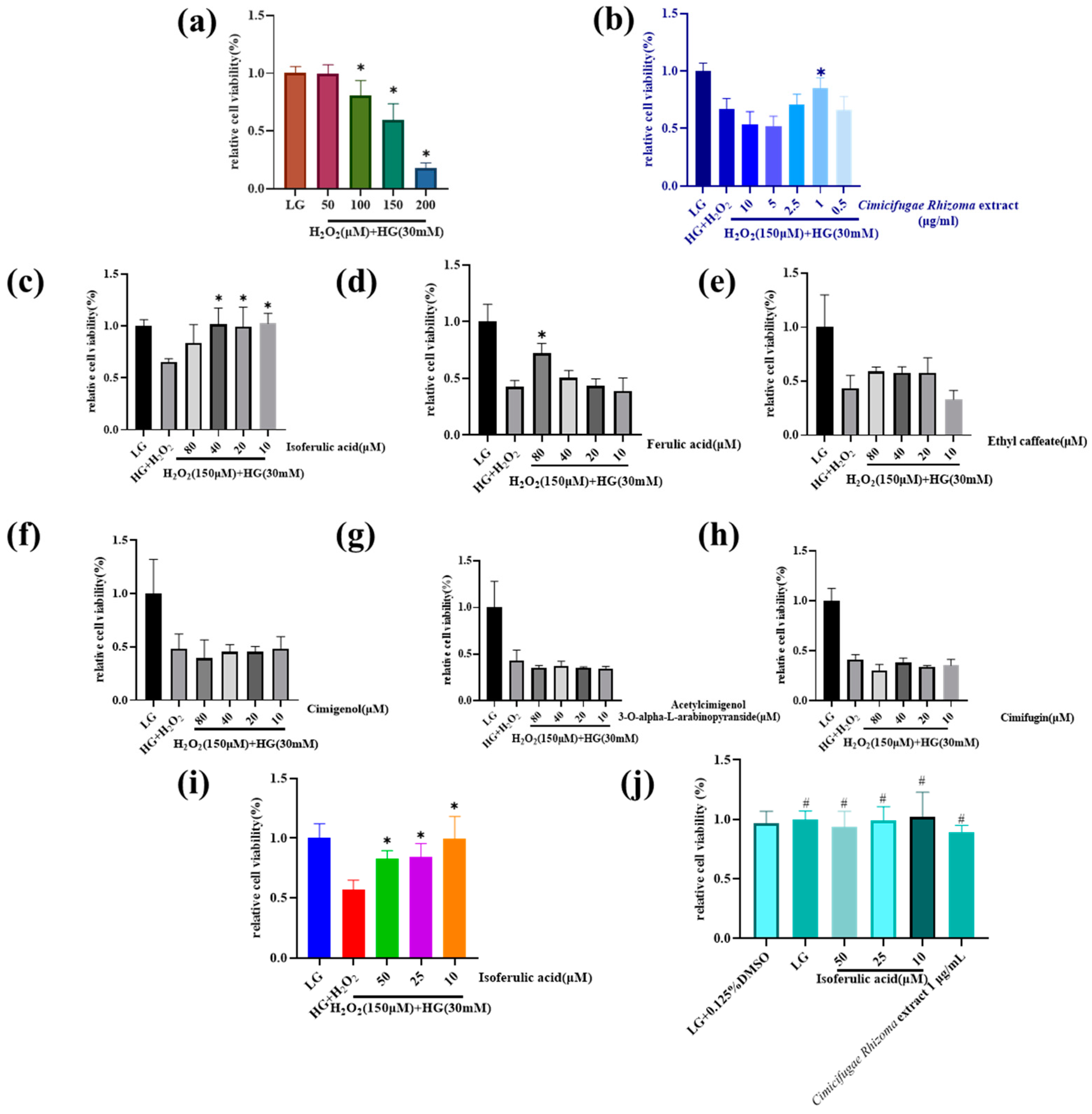
3.2. Cimicifugae Rhizoma Extract and Isoferulic Acid Inhibit H2O2 + HG Cytotoxicity in MPC5 Cells

To optimize the dose of H2O2 in the HG environment, the effects of 50~200 μM H2O2-inducion were tested. And the result (Figure 3a) showed MPC5 cells’ viability reduced significantly and stably at the 150 μM H2O2 level. Under the condition of H2O2 (150 μM) + HG (30 mM), the Cimicifugae Rhizoma extract was able to significantly increase cell survival at a dose of 1 μg/mL (Figure 3b). We performed MTT screening of three representative phenolic acid constituents and triterpenoid constituents of the Cimicifugae Rhizoma extract (Figure 3c–h) and found that two phenolic acid constituents, ferulic acid and Isoferulic acid, exhibited antioxidant properties. And Isoferulic acid had the strongest antioxidant property at 10–40 μM, which might be the main antioxidant component of the Cimicifugae Rhizoma extract.
The repeated experiment showed that 10, 25, and 50 μM of Isoferulic acid recovered the podocytes viability, which could protect most podocytes from H2O2 + HG injury (Figure 3i). On the other hand, the solution DMSO itself, 1 μg/mL Cimicifugae Rhizoma extract, or the different levels of Isoferulic acid could not result in the abnormal proliferation of MPC5 cells in the LG environment (Figure 3j). The above two results meant that Isoferulic acid was moderating H2O2 + HG injury and protecting the podocytes.

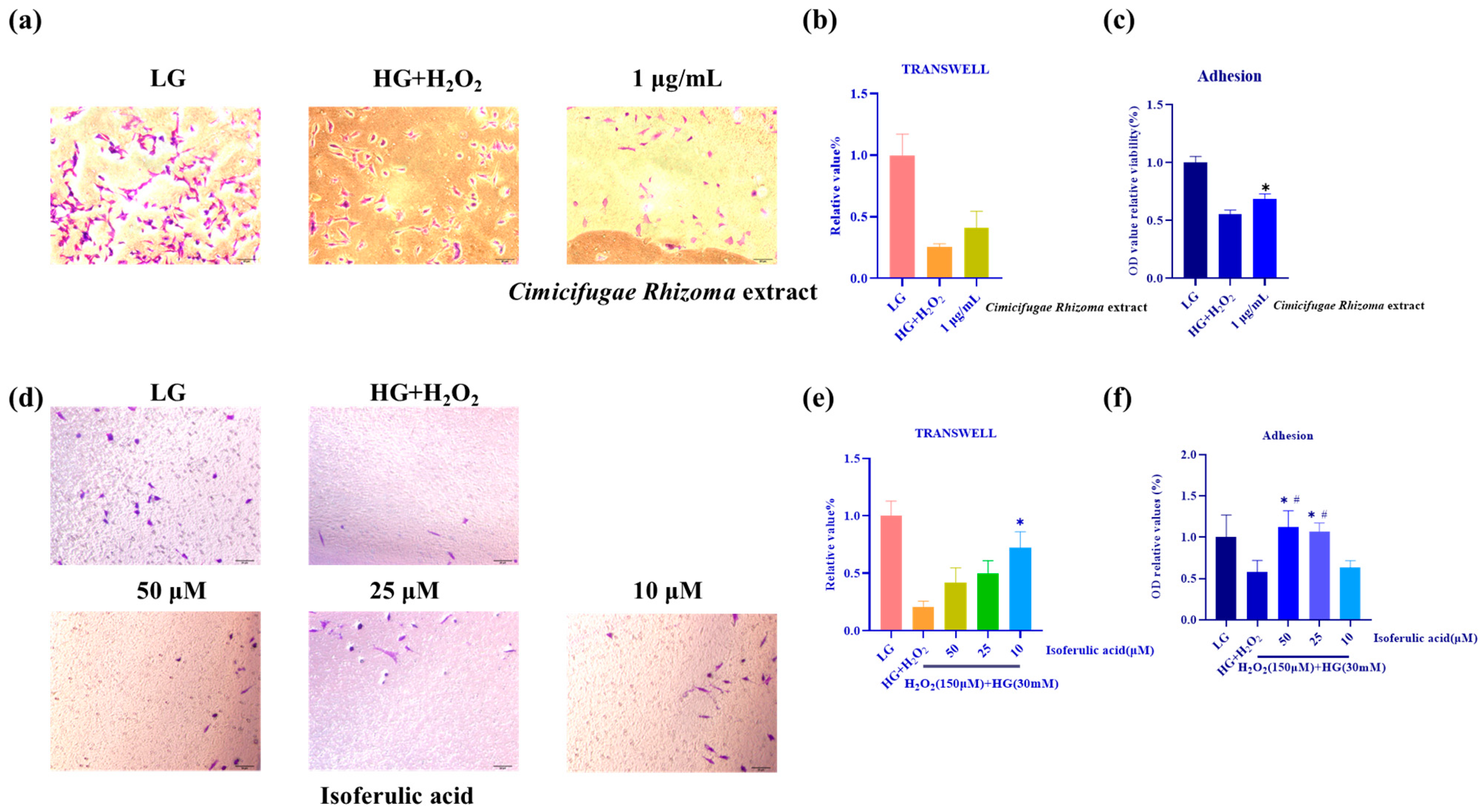
3.3. Isoferulic Acid Shows Potential in Improving MPC5 Cells’ Impaired Mobility and Adhesion

The Cimicifugae Rhizoma extract could increase the migration of podocytes, but there was no statistical difference from the H2O2 + HG group (Figure 4a,b). The Cimicifugae Rhizoma extract could restore damaged podocyte adhesion (Figure 4c). Isoferulic acid treatment improved MPC5 and regained the migration (Figure 4d,e) and cell adhesion (Figure 4f) of conditionally immortalized podocytes cultured in high-glucose and H2O2 media in comparison with normal-glucose media.

3.4. Isoferulic Acid Inhibits H2O2 + HG-Mediated Cell Apoptosis and Oxidative Stress

3.4.1. Isoferulic Acid Inhibits H2O2 + HG -Mediated Cell Apoptosis

The changes in the nucleus of MPC5, as well as apoptosis, were primarily assessed through the DAPI staining. Based on the results (Figure 5a), the nucleus of cells in the LG and Cimicifugae Rhizoma extract (1 μg/mL) and Isoferulic acid (10–50 μM) group were uniformly maintained. Stronger fluorescence intensity and blue blot with chromosome fragmentation and chromatin condensation could be observed in the HG + H2O2 group. The Cimicifugae Rhizoma extract (1 μg/mL) and Isoferulic acid (10–50 μM) reduced the above nuclear morphologies and apoptotic changes.
The results in Figure 5b,c showed that the Cimicifugae Rhizoma extract (1 μg/mL) had a favorable inhibitory effect on podocyte apoptosis induced by high-glucose H2O2. The number of viable cells was significantly increased (79.13 ± 2.47% to 89.03 ± 0.96%), the amount of early apoptotic cells was significantly reduced (13.20 ± 2.51% to 7.36 ± 0.21%), and the number of late apoptotic versus early necrotic cells was also significantly reduced (from 7.20 ± 0.36% to 3.13 ± 0.60%), returning to the same level as the LG control (p > 0.05).
As shown in Figure 5d,e, the number of viable cells in the HG + H2O2-injured group was significantly reduced (94.53 ± 1.00% versus 70.99 ± 2.74%), while the amount of early apoptotic cells and late apoptotic and early necrotic cells was significantly increased (4.38 ± 0.56% versus 22.13 ± 3.08%; 0.87 ± 0.39% versus 6.50 ± 1.76%) compared to the LG group. Isoferulic acid (50, 25, 10 μM) exhibited anti-apoptotic capability by reducing the rate of apoptosis: the number of viable cells rebounded significantly (90.68 ± 1.57%, 89.23 ± 3.90%, 87.13 ± 5.93%), and the number of early apoptotic cells decreased significantly (6.70 ± 0.96%, 7.53 ± 2.31%, 8.97 ± 3.49%). In the meantime, the number of late apoptotic and early necrotic cells decreased (2.45 ± 0.80%, 2.93 ± 1.45%, 3.70 ± 2.39% respectively). The induction of HG + H2O2 could produce cytotoxicity to MPC5 cells that further leads to cell apoptosis. The results showed that Isoferulic acid, especially at 50 μM, was able to inhibit the apoptosis of podocytes.

3.4.2. Isoferulic Acid Inhibits H2O2 + HG -Mediated Oxidative Stress and Inflammatory Response in MPC5 Cells

Oxidative stress which can affect triggering of the apoptosis signal is indicated by enhanced ROS levels. In order to further explore whether Isoferulic acid protects MPC5 cells through antioxidant activity, after the podocytes were exposed to HG + H2O2 in the absence or presence of Isoferulic acid (10–50 µM), the relative ROS levels were quantified by flow cytometry. Results in Figure 6 indicate that intracellular ROS which was induced by HG + H2O2 were suppressed by Isoferulic acid. These results suggest that the anti-apoptotic protective effect of Isoferulic acid on MPC5 cells may be involved in oxidative-stress-mediated apoptosis.

3.5. Effects of Cimicifugae Rhizoma Extract and Isoferulic Acid on the Morphology of the Damaged Podocyte Cytoskeleton

The results depicted in Figure 7 demonstrated that in the LG normal control group, the podocyte exhibited prominent pedicle protrusions with clear edges and neatly arranged actin fiber bundles. However, after subjecting the cells to H2O2-induced injury in a high sugar environment for 24 h, the HG + H2O2 injury group displayed the absence of pedicle protrusions, inward rolling of the podocyte cell edges, reduced cell volume, and fragmented or even vanished actin fiber bundles. Consequently, this led to a significant decrease in fluorescence intensity. Following the intervention of the Cimicifugae Rhizoma extract and Isoferulic acid, the podocyte protrusions were successfully repaired; the size of the podocyte cells remained within the normal range, and the edge of the cells was clearly defined. The actin fiber bundles were less fragmented; the fluorescence intensity significantly increased, and both the podocyte protrusions and actin fiber bundles exhibited a favorable morphology.

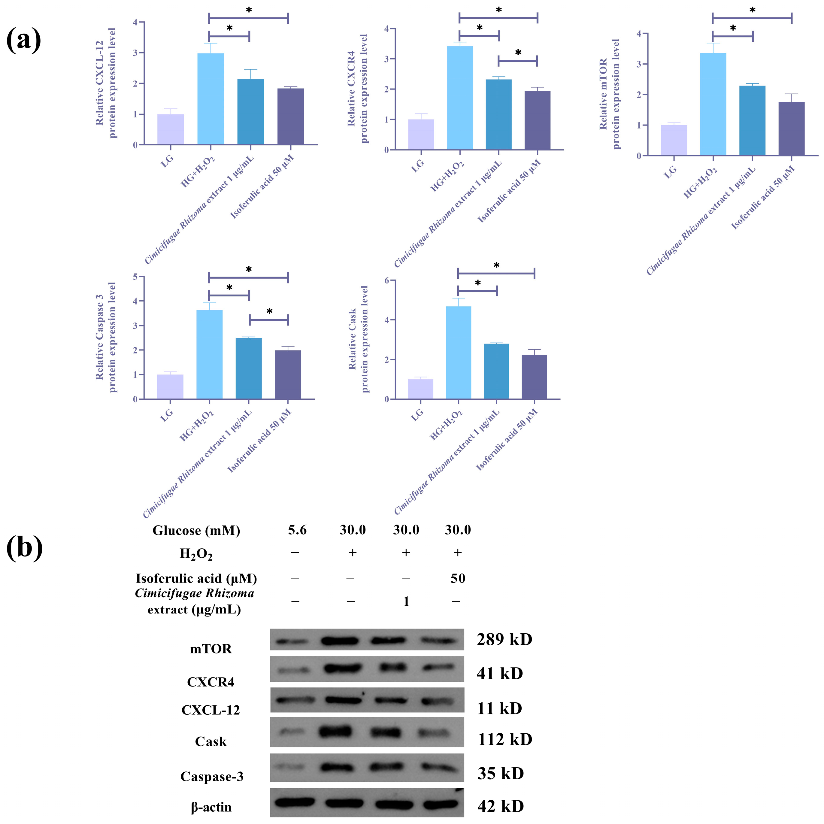
3.6. Cimicifugae Rhizoma Extract and Isoferulic Acid Inhibit H2O2 + HG -Mediated Inflammatory Response and CXCL12/CXCR4 Signaling Pathway in MPC5 Cells

TNF-α, IL-6, and IL-1β belong to the pro-inflammatory cytokine family. Figure 8a–c demonstrated that TNF-α, IL-6, and IL-1β exhibited excessive production in the cell medium treated with high glucose and hydrogen peroxide (HG + H2O2). The ELISA findings demonstrated that the administration of several dosages of Isoferulic acid (10, 25, 50 μM) in combination with the Cimicifugae Rhizoma extract effectively reduced the excessive release of inflammatory factors induced by high-glucose H2O2. Every quantity of Isoferulic acid effectively decreased the aberrant production of IL-6 (p < 0.05). One μg/mL Cimicifugae Rhizoma extract and 50 μM Isoferulic acid reduced the abnormal release of TNFα (p < 0.05). Additionally, the Cimicifugae Rhizoma extract and Isoferulic acid reduced the abnormal release of IL-1β (p < 0.05) and relieved the inflammatory phenomena. Out of all the options, the intervention impact on inflammatory variables was most significant at a concentration of 50 μM Isoferulic acid.
The ELISA findings shown in Figure 8d,e reveal an ample increase in the expression of CXCL12 and CXCR4 in podocytes stimulated by HG + H2O2. The administration of an Cimicifugae Rhizoma extract and Isoferulic acid effectively controlled the levels of CXCL12/CXCR4 in podocytes, indicating that the protective actions of podocytes are linked to the targets CXCL12 and CXCR4. Fifty μM Isoferulic acid could regulate the CXCL12/CXCR4 expression level back to LG group.

3.7. Cimicifugae Rhizoma Extract and Isoferulic Acid Regulates Protein Levels in MPC5 Cells That Were Influenced by HG + H2O2

As we can see in the Figure 9, CXCL12 and CXCR4 were abnormally up-regulated in podocytes induced by HG + H2O2 induction, and the intervention of Isoferulic acid significantly regulated the levels of these target proteins in podocytes, which matched the results of the previous ELISA experiments. Consistent with the results of ELISA, Cimicifugae Rhizoma extract also inhibited the aberrant expression of the CXCL12/CXCR4 pathway in injured podocytes, but 50 μM Isoferulic acid had a stronger inhibitory effect on the CXCR4 target.
The increased activity of Caspase-3 appeared to be detected after 24 h HG + H2O2 incubation, suggestive of the cells’ entry point into the apoptotic-signaling pathway after exposure to HG + H2O2. And 50 μM Isoferulic acid treatment could down-regulate the apoptosis-related protein Caspase-3 in the HG + H2O2 environment.
In the present study, the podocyte injury caused by high-glucose H2O2 resulted in a rise of mTOR in the cells of the model group, and the down-regulation of mTOR protein levels by Isoferulic acid may indicate that mTOR occupies a major role in the apoptotic pathway in the present model of injury. Also, the Cimicifugae Rhizoma extract inhibited apoptosis and podocyte motility and affected the overexpression of the targets mTOR, Caspase-3, and Cask. The inhibitory effects of mTOR and Cask were consistent with that of 50 μM Isoferulic acid, and the inhibitory effect of 50 μM Isoferulic acid was better in regulating the apoptosis-related target Caspase-3 that surged in response to the damage by oxidative stress.

4. Discussion

This study identified 82 and 39 components in a Cimicifugae Rhizoma extract and plasma after Cimicifugae Rhizoma extract administration by UPLC/Q-TOF and screening for the effective antioxidant component Isoferulic acid (6.52 mg/kg). We found the cleavage pattern of triterpene glycoside components and phenolic acid components in the Cimicifugae Rhizoma extract, which led to a rapid and comprehensive analysis of the active components of the Cimicifugae Rhizoma extract. The in vitro part explored the renoprotective role of Cimicifugae Rhizoma extract and Isoferulic acid in mice podocyte cells. Under the condition of H2O2 (150 μM) + HG (30 mM), the cytotoxicity, cell apoptosis, oxidative stress, and inflammatory response of MPC5 cells aggravated immediately. Through MTT assay, flow cytometry apoptosis assay, and ELISA test, the Cimicifugae Rhizoma extract and Isoferulic acid were found to alleviate cytotoxicity, cell apoptosis, ROS level, and pro-inflammatory cytokines and maintain the healthy state of podocyte morphology. Also, the unique features of podocyte cells needed to be explored. The migration and adhesion abilities of MPC5 cells were degraded in the HG + H2O2-treated cell group. Isoferulic acid could reverse the decline in the migration and adhesion abilities of MPC5 cells.
However, the mechanisms and pathways behind the therapeutic efficacies of Cimicifugae Rhizoma extract and Isoferulic acid still need to be further studied. Not many studies have tried to investigate this area. The CXCL12/CXCR4 system is vital in the regulation of stem and progenitor cell trafficking in some processes of kidney injuries. CXCL12/CXCR4 signaling leads to several different intracellular signaling pathways and can culminate in multiple downstream effects like PI3K/Akt/mTOR and β-catenin [24]. Renal CXCR4 expression was observed to be increased in diabetic rats, while CXCL12/CXCR4 antagonistAMD3100 unmasked albuminuria and accelerated tubular epithelial cell death while reducing podocyte VEGF secretion. CXCR4 blockade prevented high-glucose/CXCL12-augmented phosphorylation of the pro-survival kinase. MMP9 was also up-regulated in diabetic nephropathy biopsies, and up-regulated MMP9 could prevent CXCL12/CXCR4 signaling in tubular epithelial cells [25]. On the other hand, DPP4 inhibition contributed to protection of the diabetes-injured kidney through CXCL12-dependent antioxidative and antifibrotic effects and the amelioration of adverse renal hemodynamics [26].
In the present study, we found that Cimicifugae Rhizoma extract and Isoferulic acid reversed the HG + H2O2-induced increase in CXCL12 and CXCR4 levels in MPC5 cells, while indicating a significant activation of CXCL12/CXCR4 system might relate to the diabetic nephropathy injury in mice podocyte cells. Consequentially, Cimicifugae Rhizoma extract and Isoferulic acid down-regulated mTOR, Caspase-3, and Cask. As the potential downstream target, mTOR differentially affects both podocyte apoptosis and autophagy in diabetic nephropathy. Caspase 3 has been shown to recognize and cleave a conserved DXXD common tetrapeptide motif, and the putative recognition site for Caspase 3 is found in the amino acid sequence of RelA/p65 [27]. Partial p65 cleavage-activated fragmentation by caspase 3 interferes with anti-apoptotic transcription conferred by RPS3/NF-κB, leading to programmed cell death [28]. High glucose induces podocyte apoptosis in the apoptotic pathway, and the expression of AMPK in apoptotic cells is inhibited and activates mTOR, whereas the inhibition of mTOR has no effect on the phosphorylation of AMPK, suggesting that mTOR is downstream of these signaling molecules [29]. Also, mTOR promotes the oxidative-stress-induced apoptosis of thylakoid cells in diabetic nephropathy [30]. The roles of mTOR in autophagy include rapamycin’s ability to effectively inhibit the mTOR/P70S6K/4EBP1 signaling pathway and activate pedicellular autophagy, thereby reducing pedicellular apoptosis [31]. Complex interactions between nephrin and adhesion junction proteins mediated by the scaffolding proteins Cask, CD2AP, and ZO-1 may be critical for maintaining the structural integrity and signaling properties of glomerular podocyte [32]. This is the first study linking Cask overexpression in podocytes to oxidative stress and high glucose damage.
The anti-oxidative stress properties of Isoferulic acid and Cimicifugae Rhizoma extract were examined. It was found that Isoferulic acid at the doses of 10, 25, and 50 μM and the Cimicifugae Rhizoma extract at the dose of 1 μg/mL could alleviate the oxidative stress damage induced by H2O2 in the high-glucose environment and could reduce the apoptosis and the level of intracellular ROS, increase the survival rate of the podocytes, maintain the morphology of the podocyte cytoskeleton, alleviate the burst of inflammation factors, and improve the motor ability of the damaged podocytes. And in the connection of apoptosis as well as inflammatory factor inhibition, 50 μM Isoferulic acid exhibited greater efficiency. In the modulation of CXCL12/CXCR4 signaling pathway and other associated proteins, the effectiveness of Isoferulic acid revealed a dose-dependent impact. Cimicifugae Rhizoma extract demonstrated a similar impact on the CXCL12/CXCR4 signaling pathway, although the modulatory effect on CXCR4 and Caspase-3 target proteins was marginally lower than that of 50 μM Isoferulic acid.
The research indicates that Isoferulic acid has a substantial antioxidant impact on H2O2-induced oxidative stress damage in the simulated diabetic nephropathy environment, which may preserve the podocytes and minimize the inflammatory response. The phenolic acid-richness of Cimicifugae Rhizoma also demonstrates effective mitigation of oxidative stress damage.

5. Conclusions

Overall, this study identified 82 components in Cimicifugae Rhizoma extract and 39 components in rat plasma. Cimicifugae Rhizoma extract (1 μg/mL) and Isoferulic acid (10, 25, 50 μM) significantly lowered high glucose and H2O2-mediated toxicity, reduced movement and binding and apoptotic changes in MPC5 podocytes, and reversed oxidative-stress-induced inflammation response. Cimicifugae Rhizoma extract and Isoferulic acid down-regulated Cask, mTOR, and Caspase-3. Moreover, Cimicifugae Rhizoma extract and Isoferulic acid treatment significantly stopped overactivation of CXCL12/CXCR4 in podocytes triggered by oxidative stress and high glucose. These results suggest that the protective mechanism of Cimicifugae Rhizoma extract and Isoferulic acid on MPC5 cells modelling diabetic nephropathy symptoms includes mainly the of CXCL12/CXCR4 pathways and the inhibition of oxidative-stress-mediated apoptotic pathways. Overall, our work found novel mechanisms by which Cimicifugae Rhizoma extract and its bioactive component detected in identification, Isoferulic acid, reduce oxidative stress reactions brought on in podocytes, suggesting their therapeutic potential in diabetic nephropathy. An in vivo experiment on streptozotocin-induced mice to confirm the renal and podocyte protection of Cimicifugae Rhizoma and Isoferulic acid and the pharmacokinetics study of phenolic acid components in Cimicifugae Rhizoma will be conducted in the future.

Supplementary Materials

The following supporting information can be downloaded at: https://www.mdpi.com/article/10.3390/separations11060175/s1, Table S1: The identification results of Cimicifugae Rhizoma extract.

Author Contributions

Conceptualization, J.L.; methodology, J.L.; software, A.C.; validation, J.L., X.Y. and X.D.; formal analysis, J.L.; investigation, H.P.; resources, C.Q.; data curation, J.N.; writing—original draft preparation, J.L.; writing—review and editing, J.N., C.Q. and X.D.; visualization, J.L.; supervision, J.N.; project administration, J.L. All authors have read and agreed to the published version of the manuscript.

Funding

This research received no external funding.

Institutional Review Board Statement

Not applicable.

Informed Consent Statement

Not applicable.

Data Availability Statement

The original contributions presented in the study are included in the article and the Supplementary Material, further inquiries can be directed to the corresponding authors.

Conflicts of Interest

The authors declare no conflicts of interest.

Appendix A

Appendix A.1. Precision Investigation

This HPLC method examinedd both ferulic acid and isoferulic acid in the methodological examination phase, but only the isoferulic acid content determination part was used in the text. The same mixed standard solution was taken in six consecutive injections; the results showed that the RSD values of the peak areas of ferulic acid and Isoferulic acid were 0.22% and 0.20%, respectively, and the RSD values of the peak areas were less than 3%, indicating that the precision of the instrument and the method was good.

Appendix A.2. Repeatability

According to the previous operation, the test solution was prepared into the test solution, six test solutions of the Asclepias extract were prepared in parallel, and 10 μL of the test solution was sucked into the sample for determination. The RSD values of ferulic acid and soferulic acid were 3.52% and 0.72%, respectively, indicating that the reproducibility of the method was good.

Appendix A.3. Stability

An appropriate amount of the Asclepias extract was weighed and prepared as the test solution, and the samples were injected into the sample for determination at the 0th, 2nd, 4th, 8th, 12th and 24th h. The results of ferulic acid and Isoferulic acid were obtained from the samples. The RSD values of the peak area of ferulic acid and Isoferulic acid were 4.78% and 0.29%, respectively, which indicated that the stability of ferulic acid and Isoferulic acid was good within 24 h. The results showed that the stability of ferulic acid and Isoferulic acid was good.

Appendix A.4. Linearity Study

With the precision dilution of the mixed control stockpile, making the original concentration of 2/3, 1/2, 1/4, 1/8, 1/16, 1/32 of the mixed control solution, respectively, 10 μL of precision was pipetted into the sample; the determination of the linear regression equation results showed that the components of the linear relationship were good.

Appendix A.5. Sample Recovery Test

The average recoveries of ferulic acid and Isoferulic acid were 99.35% and 98.89% with RSD values of 2.45% and 1.42%, respectively, which showed good accuracy of the method.

Appendix A.6. Optimization of Extraction Methods by Orthogonal Screening

For further optimization of the Cimicifugae Rhizoma extraction method, the L9(34) orthogonal table recommended the material–liquid ratio (A) (10-fold, 25-fold, 50-fold), the number of extraction times (B) (1, 2, 3 times), and the extraction duration (C) (30 min, 60 min, 120 min). Multi-indicator comprehensive scoring was used to analyze Isoferulic acid content and extraction ratio, and the experiment was evaluated using the rate system: (index value − indicator minimum value)/(index maximum value − indicator minimum value) with a full score of 100 points while Isoferulic acid content and extraction ratio were 50% weighted. Table A1 and Table A2 exhibit the composite score ANOVA and orthogonal test results. The optimal orthogonal test and ANOVA level combination was A3B2C2 with the highest K-value in each group: The crude powder of Cimicifugae Rhizoma was taken and soaked in 50 times amount of 70% ethanol for about 12 h. It was extracted twice by ultrasonication (37 °C, 200 W, 40 kHz) for 60 min and dried to the extract.
Table A1. Analysis of variance.
FactorDegree of FreedomMean SquareFp-Value
A23413.34527.6230.035
B294.8310.7670.566
C252.7280.4270.701
Table A2. Results of orthogonal experiment.
NumberABCD (Blank)Isoferulic Acid Content (mg/g)Isoferulic Acid Extraction Ratio (%)Rate
11013013.40 16.089.45
210212023.06 30.3914.56
31036033.01 37.5819.89
425112033.78 36.0936.97
52526013.08 54.0936.86
62533023.62 28.0625.60
75016025.10 53.0084.16
85023034.75 70.1291.76
950312014.03 59.1564.29
K143.90130.58126.81110.60
K299.43143.18140.91124.32
K3240.21109.78115.82148.62
R196.3133.40 25.09 38.02
Table A3. The identification results of mixed standards.
LabelComponent FormulaMass
(Da)
m/z (Precursor Ion)Mass Error
(ppm)
Observed RT
(min)
AdductsMS/MS Fragment (Product Ion)
1Caffeic acidC9H8O4180.04226179.03510.712.56−H135.0447
2Ferulic acidC10H10O4194.0579193.0511.819.17−H133.0294
3PaeonolC9H10O3166.063165.05612.325.41−H133.0295
44-Hydroxyphenylacetic acidC8H8O3152.0473151.04010.311.46−H107.0500
5Sinapic acidC11H12O5224.0685223.06172.316.53−H149.0242
6Methyl caffeateC10H10O4194.0579193.05070.416.22−H121.0291/93.0340
7Ethyl caffeateC11H12O4208.0736207.06682.422.84−H178.0826/135.0448/133.0294
8Isoferulic acidC10H10O4194.0579193.0505−0.516.89−H178.0286/135.0431/134.0380
9Methyl ferulateC11H12O4208.0736207.06672.023.51−H133.0293

References

  1. Ma, Z.; Liu, M.; Liu, G.; Zhou, Z.; Ren, X.; Sun, L.; Wang, M. A comprehensive quality evaluation of Cimicifugae Rhizoma using UPLC-Q-Orbitrap-MS/MS coupled with multivariate chemometric methods. J. AOAC Int. 2023, 106, 1313–1322. [Google Scholar] [CrossRef]
  2. Niu, X.N.; Qin, R.L.; Zhao, Y.D.; Han, L.; Lu, J.C.; Lv, C.N. Simultaneous determination of 19 constituents in Cimicifugae Rhizoma by HPLC-DAD and screening for antioxidants through DPPH free radical scavenging assay. Biomed. Chromatogr. 2019, 33, e4624. [Google Scholar] [CrossRef] [PubMed]
  3. Li, X.; Lin, J.; Gao, Y.; Han, W.; Chen, D. Antioxidant activity and mechanism of Rhizoma Cimicifugae. Chem. Cent. J. 2012, 6, 140. [Google Scholar] [CrossRef] [PubMed]
  4. Wang, X.; Li, X.; Chen, D. Evaluation of antioxidant activity of isoferulic acid in vitro. Nat. Prod. Commun. 2011, 6, 1285–1288. [Google Scholar] [CrossRef] [PubMed]
  5. Lee, K.H.; Lee, W.J.; Yang, S.J.; Huh, J.W.; Choi, J.; Hong, H.N.; Hwang, O.; Cho, S.W. Inhibitory effects of Cimicifuga heracleifolia extract on glutamate formation and glutamate dehydrogenase activity in cultured islets. Mol. Cells 2004, 17, 509–514. [Google Scholar] [CrossRef] [PubMed]
  6. Wang, F.; Zhao, S.; Li, F.; Zhang, B.; Qu, Y.; Sun, T.; Luo, T.; Li, D. Investigation of antioxidant interactions between Radix Astragali and Cimicifuga foetida and identification of synergistic antioxidant compounds. PLoS ONE 2014, 9, e87221. [Google Scholar] [CrossRef] [PubMed]
  7. Adisakwattana, S.; Chantarasinlapin, P.; Thammarat, H.; Yibchok-Anun, S. A series of cinnamic acid derivatives and their inhibitory activity on intestinal alpha-glucosidase. J. Enzym. Inhib. Med. Chem. 2009, 24, 1194–1200. [Google Scholar] [CrossRef] [PubMed]
  8. Goh, S.Y.; Cooper, M.E. Clinical review: The role of advanced glycation end products in progression and complications of diabetes. J. Clin. Endocrinol. Metab. 2008, 93, 1143–1152. [Google Scholar] [CrossRef] [PubMed]
  9. Meeprom, A.; Sompong, W.; Chan, C.B.; Adisakwattana, S. Isoferulic Acid, a New Anti-Glycation Agent, Inhibits Fructose- and Glucose-Mediated Protein Glycation in Vitro. Molecules 2013, 18, 6439–6454. [Google Scholar] [CrossRef] [PubMed]
  10. Meeprom, A.; Chan, C.B.; Sompong, W.; Adisakwattana, S. Isoferulic acid attenuates methylglyoxal-induced apoptosis in INS-1 rat pancreatic beta-cell through mitochondrial survival pathways and increasing glyoxalase-1 activity. Biomed. Pharmacother. 2018, 101, 777–785. [Google Scholar] [CrossRef]
  11. Liu, I.M.; Hsu, F.L.; Chen, C.F.; Cheng, J.T. Antihyperglycemic action of isoferulic acid in streptozotocin-induced diabetic rats. Br. J. Pharmacol. 2000, 129, 631–636. [Google Scholar] [CrossRef] [PubMed]
  12. Liu, I.M.; Chen, W.C.; Cheng, J.T. Mediation of beta-endorphin by isoferulic acid to lower plasma glucose in streptozotocin-induced diabetic rats. J. Pharmacol. Exp. Ther. 2003, 307, 1196–1204. [Google Scholar] [CrossRef] [PubMed]
  13. Elmarakby, A.A.; Sullivan, J.C. Relationship between Oxidative Stress and Inflammatory Cytokines in Diabetic Nephropathy. Cardiovasc. Ther. 2012, 30, 49–59. [Google Scholar] [CrossRef] [PubMed]
  14. Kashihara, N.; Haruna, Y.; Kondeti, V.K.; Kanwar, Y.S. Oxidative stress in diabetic nephropathy. Curr. Med. Chem. 2010, 17, 4256–4269. [Google Scholar] [CrossRef] [PubMed]
  15. Redza-Dutordoir, M.; Averill-Bates, D.A. Activation of apoptosis signalling pathways by reactive oxygen species. Biochim. Et Biophys. Acta (BBA) Mol. Basis Dis. 2016, 1863, 2977–2992. [Google Scholar] [CrossRef]
  16. Lu, Q.; Zhang, W.Y.; Pan, D.B.; Shi, D.F.; Pang, Q.Q.; Li, H.B.; Yao, X.J.; Yao, Z.H.; Yu, Y.; Yao, X.S. Phenolic acids and their glycosides from the rhizomes of Cimicifuga dahurica. Fitoterapia 2019, 134, 485–492. [Google Scholar] [CrossRef] [PubMed]
  17. Wang, Z.-Y.; Wang, Q.; Han, Y.-Q.; Jiang, M.; Gao, J.; Miao, Y.; Bai, G. Bioactivity-based UPLC/Q-TOF/MS strategy for screening of anti-inflammatory components from Cimicifugae Rhizoma. Chin. Chem. Lett. 2017, 28, 476–481. [Google Scholar] [CrossRef]
  18. Lim, J.O.; Song, K.H.; Lee, I.S.; Lee, S.J.; Kim, W.I.; Pak, S.W.; Shin, I.S.; Kim, T. Cimicifugae Rhizoma Extract Attenuates Oxidative Stress and Airway Inflammation via the Upregulation of Nrf2/HO-1/NQO1 and Downregulation of NF-κB Phosphorylation in Ovalbumin-Induced Asthma. Antioxidants 2021, 10, 1626. [Google Scholar] [CrossRef] [PubMed]
  19. Zhang, Q.; Chen, S.; Wen, J.; Wang, R.; Lu, J.; Muhammad-Biu, A.; Wang, S.; Du, K.; Wei, W.; Tian, X.; et al. A comprehensive strategy integrating metabolomics with DNA barcoding for discovery of combinatorial discriminatory quality markers: A case of Cimicifuga foetida and Cimicifuga dahurica. Arab. J. Chem. 2024, 17, 105613. [Google Scholar] [CrossRef]
  20. Fan, M.; Qin, K.; Ding, F.; Huang, Y.; Wang, X.; Cai, B. Identification and differentiation of major components in three different “Sheng-ma” crude drug species by UPLC/Q-TOF-MS. Acta Pharm. Sin. B 2017, 7, 185–192. [Google Scholar] [CrossRef] [PubMed]
  21. Liu, J.; You, L.; Yang, C.; Sai, N.; Wu, H.; Sun, M.; Cai, M.; Peng, H.; Liang, X.; Yin, X.; et al. Phytochemical identification of Xiaoer Huanglong Granule and pharmacokinetic study in the rat using its seven major bioactive components. J. Sep. Sci. 2022, 45, 2804–2818. [Google Scholar] [CrossRef] [PubMed]
  22. Hu, L.F.; Song, X.J.; Nagai, T.; Yamamoto, M.; Dai, Y.; He, L.L.; Kiyohara, H.; Yao, X.S.; Yao, Z.H. Chemical profile of Cimicifuga heracleifolia Kom. And immunomodulatory effect of its representative bioavailable component, cimigenoside on Poly(I: C)-induced airway inflammation. J. Ethnopharmacol. 2021, 267, 113615. [Google Scholar] [CrossRef] [PubMed]
  23. Dan, C.; Zhou, Y.; Ye, D.; Peng, S.; Ding, L.; Gross, M.L.; Qiu, S.X. Cimicifugadine from Cimicifuga foetida, a new class of triterpene alklaoids with novel reactivity. Org. Lett. 2007, 9, 1813–1816. [Google Scholar] [CrossRef] [PubMed]
  24. Alagpulinsa, D.A.; Cao, J.J.L.; Sobell, D.; Poznansky, M.C. Harnessing CXCL12 signaling to protect and preserve functional beta-cell mass and for cell replacement in type 1 diabetes. Pharmacol. Ther. 2019, 193, 63–74. [Google Scholar] [CrossRef] [PubMed]
  25. Siddiqi, F.S.; Chen, L.H.; Advani, S.L.; Thai, K.; Batchu, S.N.; Alghamdi, T.A.; White, K.E.; Sood, M.M.; Gibson, I.W.; Connelly, K.A.; et al. CXCR4 promotes renal tubular cell survival in male diabetic rats: Implications for ligand inactivation in the human kidney. Endocrinology 2015, 156, 1121–1132. [Google Scholar] [CrossRef] [PubMed]
  26. Takashima, S.; Fujita, H.; Fujishima, H.; Shimizu, T.; Sato, T.; Morii, T.; Tsukiyama, K.; Narita, T.; Takahashi, T.; Drucker, D.J.; et al. Stromal cell-derived factor-1 is upregulated by dipeptidyl peptidase-4 inhibition and has protective roles in progressive diabetic nephropathy. Kidney Int. 2016, 90, 783–796. [Google Scholar] [CrossRef] [PubMed]
  27. Kang, K.H.; Lee, K.H.; Kim, M.Y.; Choi, K.H. Caspase-3-mediated cleavage of the NF-kappa B subunit p65 at the NH2 terminus potentiates naphthoquinone analog-induced apoptosis. J. Biol. Chem. 2001, 276, 24638–24644. [Google Scholar] [CrossRef] [PubMed]
  28. Wier, E.M.; Fu, K.; Hodgson, A.; Sun, X.; Wan, F. Caspase-3 cleaved p65 fragment dampens NF-kappaB-mediated anti-apoptotic transcription by interfering with the p65/RPS3 interaction. FEBS Lett. 2015, 589, 3581–3587. [Google Scholar] [CrossRef] [PubMed]
  29. Eid, A.A.; Ford, B.M.; Bhandary, B.; Cavaglieri, R.D.; Block, K.; Barnes, J.L.; Gorin, Y.; Choudhury, G.G.; Abboud, H.E. Mammalian Target of Rapamycin Regulates Nox4-Mediated Podocyte Depletion in Diabetic Renal Injury. Diabetes 2013, 62, 2935–2947. [Google Scholar] [CrossRef] [PubMed]
  30. Lu, Q.; Zhou, Y.; Hao, M.; Li, C.; Wang, J.; Shu, F.; Du, L.; Zhu, X.; Zhang, Q.; Yin, X. The mTOR promotes oxidative stress-induced apoptosis of mesangial cells in diabetic nephropathy. Mol. Cell. Endocrinol. 2018, 473, 31–43. [Google Scholar] [CrossRef] [PubMed]
  31. Jin, J.; Hu, K.; Ye, M.; Wu, D.; He, Q. Rapamycin Reduces Podocyte Apoptosis and is Involved in Autophagy and mTOR/P70S6K/4EBP1 Signaling. Cell. Physiol. Biochem. 2018, 48, 765–772. [Google Scholar] [CrossRef] [PubMed]
  32. Lehtonen, S.; Lehtonen, E.; Kudlicka, K.; Holthöfer, H.; Farquhar, M.G. Nephrin Forms a Complex with Adherens Junction Proteins and CASK in Podocytes and in Madin-Darby Canine Kidney Cells Expressing Nephrin. Am. J. Pathol. 2004, 165, 923–936. [Google Scholar] [CrossRef] [PubMed]
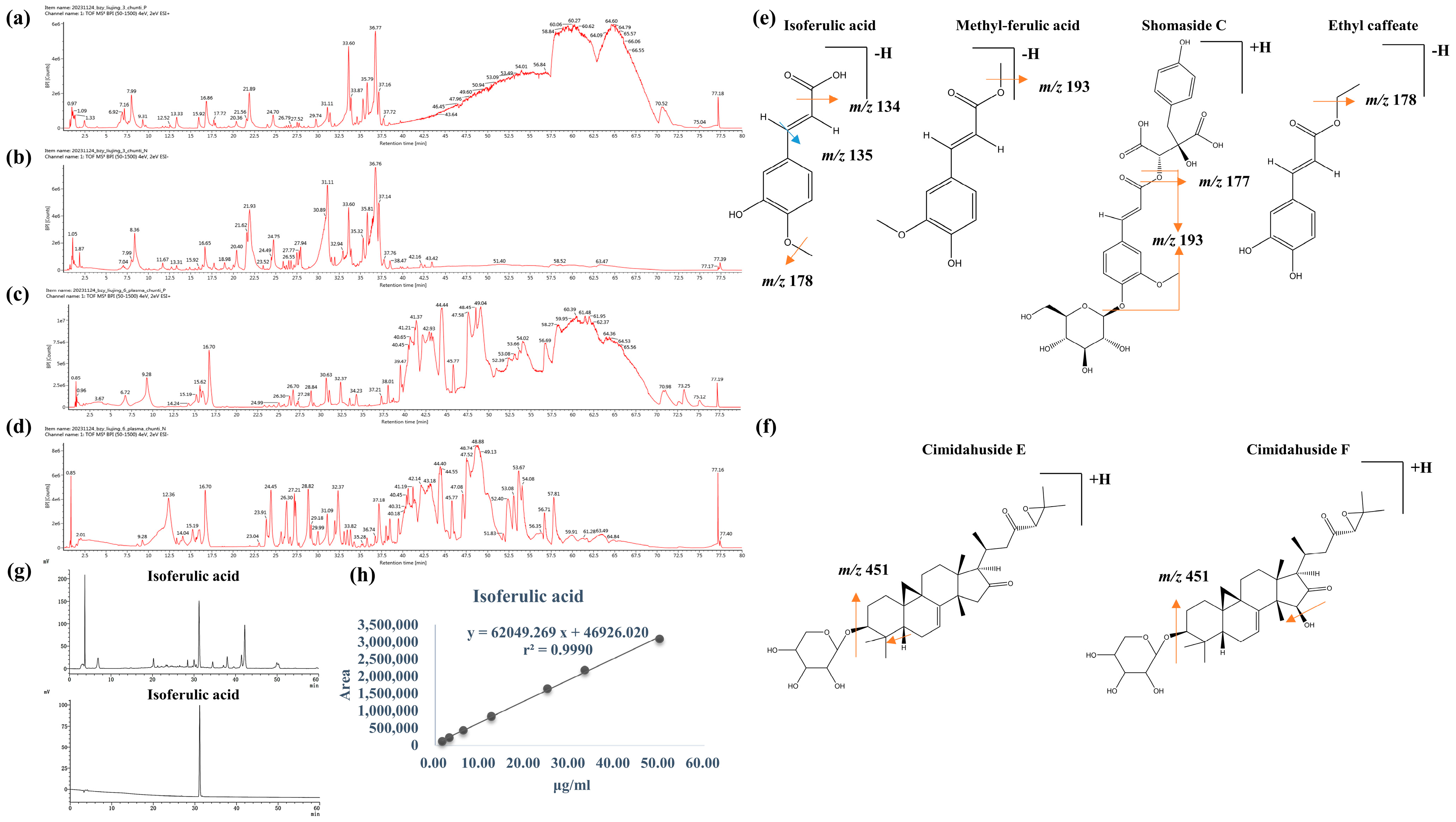
Figure 1. The picture of (a) Cimicifugae Rhizoma and the chemical structure of (b) Isoferulic acid.
Separations 11 00175 g001
Figure 2. The general ion flow diagram of Cimicifugae Rhizoma extract at positive mode (a) and negative mode (b). The general ion flow diagram of Cimicifugae Rhizoma extract at positive mode (c) and negative mode (d) in the plasma. Illustration for the structural elucidation of phenolic acids (e) and triterpenoid saponins (f). HPLC analysis of Isoferulic acid in the Cimicifugae Rhizoma extract (gup) and standard Isoferulic acid (gdown) and standard curve of Isoferulic acid content in Cimicifugae Rhizoma extract (h).
Separations 11 00175 g002
Figure 3. (a) The effect of H2O2 on MPC5 podocytes viability for 24 h was assessed by the MTT assay (n = 6, * p < 0.05, vs. LG group). (b) The protective effect of 1 μg/mL Cimicifugae Rhizoma extract on podocyte (n = 6, * p < 0.05, vs. HG + H2O2 group). Effects of components in the concentration range of 10 μM-80 μM on the H2O2-induced injury MPC5: (c) isoferulic acid; (d) ferulic acid; (e) Ethyl caffeate; (f) cimigenol; (g) Acetylcimigenol 3-O-alpha-L-arabinopyranside; (h) cimifugin (n = 3, * represents p < 0.05 compared to HG + H2O2 group). MTT assays were also performed to evaluate the effects of (i) Isoferulic acid on HG + H2O2-induced injury and (j) solvent versus the drug itself on the survival of MPC5 cells (n = 6, # p > 0.05, vs. LG group, * p < 0.05, vs. HG + H2O2 group).
Separations 11 00175 g003
Figure 4. Cimicifugae Rhizoma extract and Isoferulic acid exerted the mobility and adhesiveness improvement effect: Representative images of podocytes on the upper surface of the membranes in Transwell (×200) of (a) Cimicifugae Rhizoma extract and (d) Isoferulic acid. Migration assay results of podocytes from (b) Cimicifugae Rhizoma extract and (e) Isoferulic acid intervention. And the relative adhesion ratio of (c) Cimicifugae Rhizoma extract and (f) Isoferulic acid. (n = 3) (* p < 0.05, vs. HG + H2O2 group, # p < 0.05 vs. LG group).
Separations 11 00175 g004
Figure 5. Cimicifugae Rhizoma extract (1 μg/mL) and Isoferulic acid (10–50 μM) exerted an anti-apoptotic protective effect. (a) The representative image of DAPI staining by fluorescence microscope (×100). The representative image of Annexin V-FITC/PI double staining by flow cytometry of Cimicifugae Rhizoma extract (b) and Isoferulic acid (d). Results of co-cultured podocytes’ cell apoptosis of Cimicifugae Rhizoma extract (c) and Isoferulic acid (e) were presented as mean  ±  SD (n = 3). (* p < 0.05, vs. HG + H2O2 group. # p >0.05 vs. LG group).
Separations 11 00175 g005
Figure 6. Cimicifugae Rhizoma extract (1 μg/mL) and Isoferulic acid (10–50 μM) exerted the anti-oxidative stress protective effect. ROS level of Cimicifugae Rhizoma extract (a) and Isoferulic acid (c) detected by flow cytometry. Relative ROS levels from different treatment groups of Cimicifugae Rhizoma extract (the mean ratio of HG + H2O2 or treatment group P2 FITC-A value versus LG group P2 FITC-A Mean value) (b) and Isoferulic acid (d). (* p < 0.05, vs. HG + H2O2 group.).
Separations 11 00175 g006
Figure 7. Actin Tracker Red-594 observation of podocyte cytoskeleton morphology in each group (×400).
Separations 11 00175 g007
Figure 8. The effect of Cimicifugae Rhizoma extract and Isoferulic acid on MPC5 cell releasing IL-6 (a), TNFα (b), IL-1β (c), CXCL12 and (d) CXCR4 (e) was assessed. (n = 4) (* p < 0.05, vs. HG + H2O2 group. # p >0.05 vs. LG group).
Separations 11 00175 g008
Figure 9. HG + H2O2-induced podocyte injury and intracellular protein levels in MPC5 cells: (a) The results of the homogenization calculations of intracellular protein levels after 1 μg/mL Cimicifugae Rhizoma extract intervention and compared with 50 μM Isoferulic acid, and the (b) representative images (n = 3) (* p < 0.05 vs. group).
Separations 11 00175 g009
Table 1. The identification results of Cimicifugae Rhizoma extract in the plasma.
LabelComponent FormulaMass
(Da)
m/z (Precursor Ion)Mass Error
(ppm)
Observed RT
(min)
AdductsMS/MS Fragment (Product Ion)Reference
112β-O-Acetyl-Liposide BC37H58O11678.3979 677.39070.036.76−H617.3701 [19]
212β-O-Acetylcimifugoside AC37H56O11676.3823 677.3870−3.727.88+H541.3164/195.1206/181.1043[19]
312β-HydroxystigmasterolxylosideC35H56O10636.3874 635.38010.031.04−H577.3384 [19]
415α-HydroxycimifugosideH2C35H54O11650.3666 649.36092.425.09−H531.2990 [22]
524-O-Acetyl-7,8-didehydroclerosterolxylosideC37H58O11678.3979 677.3901−0.833.58−H [20]
624-O-Acetyl-CresolxylosideC37H60O11680.4136 679.4059−0.537.16−H [20]
77,8-DidehydroclerosterolxylosideC35H54O9618.3768 619.3830−1.631.06+H469.3325/451.3226/433.3109[20]
8Acetylcimigenol 3-O-alpha-L-arabinopyransideC37H58O10662.4030 663.4090−1.937.16+H623.3551 [22]
925-Anhydrocimigenol-xylosideC35H54O8602.3819 603.3875−2.833.68+H, +Na583.3613 [22]
1012β-HydroxycitrinolC30H48O6504.3451 503.3370−1.636.73−H503.3370 [19]
11ArcotoxinC37H56O11676.3823 677.3888−1.126.69+H131.0894
12Cimigenol xylosideC35H56O9620.3924 643.38383.428.02+Na435.3249
13Cimiside AC35H56O10636.3874 635.38081.228.16−H445.8311 [19]
14Cimiside CC43H70O16842.4664 841.45980.831.41−H637.3959/447.3112
15Cimiside H1C35H52O9616.3611 615.35451.136.33−H251.2021 [19]
16Cimiside H2C35H54O10634.3717 633.3642−0.327.75−H501.2875/369.2438[19]
17CimicifugineC35H51NO8613.3615 614.36930.922.45+H446.3062/184.0776[23]
18Cimaroside VC47H76O19944.4981 943.4898−1.068.58−H303.2335 [22]
19Cimaroside IC36H58O11666.3979 665.39100.533.69−H [20]
20Cimiacerin AC35H54O9618.3768 619.3831−1.535.79+H, +Na451.3225 [22]
21Cimidahuside EC35H52O8600.3662 601.3728−1.235.77+H451.3195 [19]
22Cimidahuside FC35H52O9616.3611 617.36870.527.77+H451.3328 [22]
23Cimidahuside GC35H56O9620.3924 621.3972−4.031.51+H453.3379
24Cimidahuside HC35H54O9618.3768 641.3644−2.535.29+Na451.3223/433.3122[22]
25CimicidanolC30H44O5484.3189 485.3271.627.76+H449.3901 [22]
26Isodahurinol/CimigenolC30H48O5488.3502 489.3563−2.431.51+H453.3379 [22]
27Ferulic acidC10H10O4194.0579 193.0495−5.919.52−H standards data
28CimidahurinineC14H20O8316.1158 315.1069−5.38.11−H153.9128/149.2179
29Ethyl caffeateC11H12O4208.0736 207.06640.822.80−H standards data
30Cimicifugic acid EC21H20O10432.1057 431.0983−0.121.93−H165.0557 [22]
31IsocimicifugamideC25H31NO10505.1948 504.1861−2.821.81−H343.9951 [22]
32Cimicifugaside BC33H40O18724.3962 723.3952−3.633.62−H723.3952[16]
33Shomaside CC27H30O15594.3390 617.3677−3.027.73+Na, +H193.9128/177.0620[19]
34Methyl-ferulic acidC11H12O4208.0679 207.0673−6.822.93−H194.1196/193.0924
35Caffeicacid-β-D-glucopyranosylesterC15H18O9342.0951 341.08934.510.54−H
36CimicifugamideC25H31NO10505.1948 504.1860−3.117.76−H [22]
37CimidahurinineC14H20O8316.1158 315.1077−2.79.09−H153.0564
38Isoferulic acidC10H10O4194.0579 193.0505−0.516.89−H standards data
39CimifuginC16H18O6306.2462 305.23792.153.65−H235.9243 [19]
Note: Reference, i.e., relevant research literature and standard data referenced on behalf of the constituents, so as to screen and analyze the constituents of Cimicifugae Rhizoma extract in the plasma.
Disclaimer/Publisher’s Note: The statements, opinions and data contained in all publications are solely those of the individual author(s) and contributor(s) and not of MDPI and/or the editor(s). MDPI and/or the editor(s) disclaim responsibility for any injury to people or property resulting from any ideas, methods, instructions or products referred to in the content.

