Article

A Facile and Efficient Protocol for Phospholipid Enrichment in Synovial Joint Fluid: Monodisperse-Mesoporous SiO2 Microspheres as a New Metal Oxide Affinity Sorbent

by
Serhat Aladağ
1,
İlayda Demirdiş
2,
Burcu Gökçal Kapucu
3,
Emine Koç
4,
Ozan Kaplan
4,
Batuhan Erhan Aktaş
5,
Mustafa Çelebier
1,4,
Ali Tuncel
1,3 and
Feza Korkusuz
1,5,*
1
Department of Bioengineering, Hacettepe University, 06810 Ankara, Türkiye
2
Department of Biology, Hacettepe University, 06810 Ankara, Türkiye
3
Department of Chemical Engineering, Hacettepe University, 06810 Ankara, Türkiye
4
Department of Analytical Chemistry, Faculty of Pharmacy, Hacettepe University, 06230 Ankara, Türkiye
5
Department of Sports Medicine, Faculty of Medicine, Hacettepe University, 06230 Ankara, Türkiye
*
Author to whom correspondence should be addressed.
Separations 2024, 11(9), 262; https://doi.org/10.3390/separations11090262
Submission received: 16 August 2024 / Revised: 27 August 2024 / Accepted: 29 August 2024 / Published: 5 September 2024

Abstract

:
Phospholipids (PLs), essential components of cell membranes, play significant roles in maintaining the structural integrity and functionality of joint tissues. One of the main components of synovial joint fluid (SJF) is PLs. Structures such as PLs that are found in low amounts in biological fluids may need to be selectively enriched to be analyzed. Monodisperse-mesoporous SiO2 microspheres were synthesized by a multi-step hydrolysis condensation method for the selective enrichment and separation of PLs in the SJF. The microspheres were characterized by SEM, XPS, XRD, and BET analyses. SiO2 microspheres had a 161.5 m2/g surface area, 1.1 cm3/g pore volume, and 6.7 nm pore diameter, which were efficient in the enrichment of PLs in the SJF. The extracted PLs with sorbents were analyzed using Q-TOF LC/MS in a gradient elution mode with a C18 column [2.1 × 100 mm, 2.5 μM, Xbridge Waters (Milford, MA, USA)]. An untargeted lipidomic approach was performed, and the phospholipid enrichment was successfully carried out using the proposed solid-phase extraction (SPE) protocol. Recovery of the SPE extraction of PLs using sorbents was compared to the classical liquid–liquid extraction (LLE) procedure for lipid extraction. The results showed that monodisperse-mesoporous SiO2 microspheres were eligible for selective enrichment of PLs in SJF samples. These microspheres can be used to identify PLs changes in articular joint cartilage (AJC) in physiological and pathological conditions including osteoarthritis (OA) research.

1. Introduction

Osteoarthritis (OA) is a common disabling joint disorder associated with symptoms such as joint pain, swelling, and inflammation. The disease and its conditions have socioeconomic impacts on the aging society [1,2]. OA affects the articular cartilage, subchondral bone, synovium, meniscus, periarticular ligaments, and adipose tissue [3]. Recently, OA research has focused not only on phenotypic and genotypic but also on omic studies [4]. Metabolomics is a reliable and promising tool for distinguishing different subtypes of OA [5]. It is a useful tool for examining different phenotypes, as it reveals findings related to environmental, epigenetic, and genetic factors [6]. Recent metabolomic profiling approaches have revealed changes associated with pathways such as amino acid [7,8] and phospholipid (PL) metabolisms involving the conversion of phosphatidylcholine (PC) to lysophosphatidylcholine (LPC) [9,10]. The SJF contains nutrients and resorbs articular cartilage degradation and degeneration markers. An SJF omic examination [11] may provide phenotyping information for the early diagnosis and treatment of OA. Therefore, the SJF is a promising biofluid for OA phenotyping. It contains growth factors and cytokines, as well as lubricin, hyaluronic acid, and PLs, which are necessary for articular joint function [12]. PLs are the main structures of cellular membranes and contribute structural and functional properties [13]. Some PLs are involved in biological processes such as cell proliferation, differentiation, apoptosis and oxidative stress [14]. PL-related metabolites and enzymes play a role in many malignant diseases such as cancer of the breast, endometrium, colon, and kidney, in addition to leukemia, malignant lymphomas, and multiple myeloma [15]. One of the main components of the SJF is PLs. Approximately ~41% of the total lipids of SJF consist of phosphatidylcholines (PCs) [16]. PLs can bind with hyaluronan (HA), which is the main link protein of articular joint cartilage, to form complexes that facilitate boundary lubrication [16]. PLs found on the surface of articular joint cartilage (AJC) are identified as phosphatidylcholine (41%), phosphatidylethanolamine (PE) (27%), and sphingomyelin (SM) (32%) [17]. It has also been reported that PL composition changes in OA and rheumatoid arthritis (RA) conditions [18]. In case of joint inflammation, PLs are degraded and their amounts in the SJF doubles or triples from the normal range [18]. It is known that PLs on the surface of AJC are active in the boundary lubrication process and decrease in OA [19]. The amount of PLs, which is ~0.1 to 0.2 mg/mL in the normal human to SJF, increases around ~0.2–0.3 mg/mL in the case of OA and decreases to ~0.02–0.08 mg/mL after traumatic injury [15,20,21]. Although PLs are important boundary lubricants, they can be quantified by lipidomics [18]. Some PLs are also active in immune modulation during inflammation, cartilage degradation, cell differentiation, apoptosis, and signaling processes [22,23]. Phospholipids consist of a polar head group containing a phosphate moiety and various fatty acids attached to a glycerol backbone. According to their polar head groups, they are divided into subtypes such as phosphatidylcholine, phosphatidylethanolamine, phosphatidylinositol (PI), phosphatidylserine (PS), sphingomyelin, and lysophosphatidylcholine [24,25]. The hydrophilic/polar head groups that are attached to the basic phospholipid structure are shown in red (Figure 1). The structural diversity and complexity of PLs make them difficult to analyze [24,25].
Enrichment of PLs can enhance selectivity for evaluating changes in low abundance structures such as PLs. The liquid–liquid extraction (LLE) and solid-phase extraction (SPE) protocols are the methods used in PLs extraction [26]. LLE in lipidomics uses modified Bligh and Dyer extraction or Folsch methods that target non-polar compounds in the sample fraction unselectively [26]. SPE for lipidomics or phospholipidomics analyses, on the other hand, is an extraction method aimed at selectivity enriching the sample fraction.
Solid materials commonly used for SPE include porous silica or silica modified with the octadecyl, cyanopropyl, aminopropyl, and 2,3-dihydroxypropoxypropyl groups [27]. These materials, however, could be insufficient in terms of selectivity and effectiveness for specific proposes like PL extraction. Metaloxides (e.g., ZrO2, TiO2), which have a higher affinity to PLs, are used for the selective extraction of PLs instead of these materials [28,29].
Phospholipidomics SJF studies are a novel interest for researchers trying to find potential biomarkers of lipid levels to monitor the disease situation [5,12,18,30,31,32,33]. Extraction of the SJF by the LLE method and subsequent selective enrichment of PLs by the SPE method is promising for profiling lipid fractions. By considering traditional SPE methods, we hypothesized that SiO2 microsphere-based metal oxide affinity chromatography could be adapted as a new micro-extraction technique for the extraction of PLs from SJF.
The analysis of the SJF presents unique challenges compared to plasma or tissue samples due to its distinct properties. For plasma samples, there are well-established protocols for sample preparation that are relatively straightforward. Tissue samples can also be prepared effectively using techniques such as freezing and mechanical disruption. Synovial joint fluid, however, exhibits characteristics of both plasma and tissue, being highly viscous and containing a complex mixture of proteins, lipids and other biomolecules. This complexity makes sample preparation particularly challenging, as standard methods are not directly applicable. Our first challenge, therefore, was to develop an effective method for preparing SJF samples for analysis. The second challenge involved the enrichment of phospholipids from this intricate matrix. The diverse composition of SJF complicates the selective extraction and enrichment of phospholipids, necessitating specialized techniques to ensure accurate and reliable analysis. These challenges encouraged us to develop a facile and efficient protocol for phospholipid enrichment in SJF, utilizing monodisperse-mesoporous SiO2 microspheres as a new metal oxide affinity sorbent.

2. Materials and Methods

2.1. Chemicals and Reagents

Methacrylic acid (MAA), ethyleneglycol dimethacrylate (EGDMA), polyvinylalcohol (PVA, 85–87% hydrolyzed), glycidyl methacrylate (GMA), polyvinylpyrrolidone K-30 (PVP K-30, MW: 25,000 Da), ethanol (Et-OH), tetraethyl orthosilicate (TEOS), trifluoroacetic acid (TFA), tetrabutylammonium bromide (TBAI), benzoyl peroxide (BPO), and isopropanol (Iso-PrOH) were obtained from Sigma-Aldrich, Inc (St. Louis, MO, USA). The syntheses were performed using deionized (DI) water with a resistivity of 18 MΩcm, obtained from purification system (Millipore, Direct-Q3 UV, MA, USA). Methanol (Me-OH) and chloroform (TCM) for LLE were HPLC grade and supplied from Sigma-Aldrich, Inc (St. Louis, MO, USA). LC-MS grade methanol, and Iso-PrOH [Sigma-Aldrich, Inc (St. Louis, MO, USA)] were used for the chromatographic analyses.

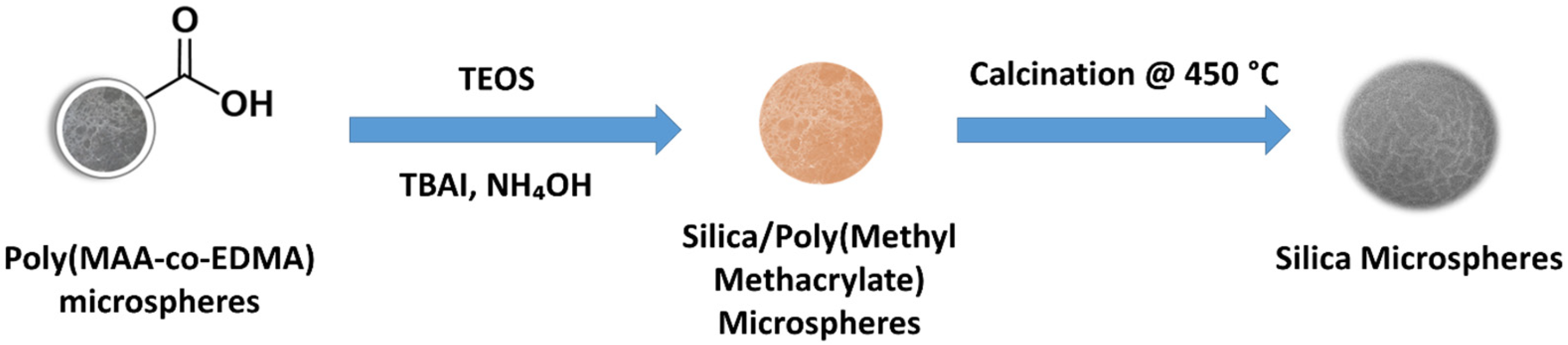
2.2. Preparation of Monodisperse-Mesoporous SiO2 Metal Oxide Affinity Sorbent

To obtain SiO2 microspheres, poly(methacrylic acid-co-ethylene glycol dimethacrylate), poly (MAA-co-EDMA) microspheres were used as the starting material [34]. Briefly, poly (MAA-co-EDMA) microspheres were dispersed in a solution containing Iso-PrOH (50 mL), water (5 mL), tetrabutylammonium iodide (TBAI, 0.25 g), and NH4OH (0.25 mL, % 25 w/w). The dispersion was mixed with a magnetic stirrer to ensure the adsorption of ammonium ions into the microspheres. Tetraethyl orthosilicate (TEOS) was used as a silica precursor and added dropwise to the dispersion medium. Then, the solution was magnetically stirred at room temperature for 24 h. The obtained silica gel/polymer composite microspheres were washed two times with 2-propanol and water by successive centrifugation–decantation and then dried in an oven and calcined at 450 °C for 4 h with a ramp of 2 °C to remove the polymer mold material. Thus, monodisperse-mesoporous SiO2 microspheres were obtained.

2.3. Characterization of SiO2 Microspheres

Synthesized SiO2 microspheres were characterized in terms of X-ray Photoelectron Spectroscopy (XPS) (K-Alpha XPS system, Thermo Fischer Scientific, Waltham, MA, USA), Scanning Electron Microscopy (SEM) (Tescan, Brno, Czech Republic), X-ray diffraction spectroscopy (XRD) (Aeris, Malvern Panalytical, Malvern, UK) with CuKa1 radiation operating at 40 kV. The porous properties were determined according to the Brunauer–Emmett–Teller (BET) model using the nitrogen (N2) adsorption–desorption method using surface area and pore size analyzer (Quantochrome, Nova 2200e, Boynton Beach, FL, USA).

2.4. Synovial Joint Fluid Sample Collection

The SJF was obtained from patients at Hacettepe University Hospital after the approval of Hacettepe University Health Sciences Research Ethics Committee (#: 2023/09-44 date: 19 December 2023, Research no: SBA 23/262). Patients with knee OA (n = 24) who did not improve with medical and physical therapy were referred to intra-articular HA injection treatment according to the European Society for Clinical and Economic Aspects of Osteoporosis, Osteoarthritis and Musculoskeletal Diseases (ESCEO) guidelines were included. Patients who received injections into the knee joint in the last three months and food supplements such as glycosaminoglycan and chondroitin sulfate, which may affect joint metabolism, were not included.
After the suprapatellar knee joint area of the supine patient was cleaned with 10% povidone iodine (Isosol, Merkez Lab AŞ, Istanbul, Türkiye), a 0.7 × 32 mm 22 G sterile, a non-pyrogenic needle (Berika Technology Medikal, Konya, Türkiye) was used for local anesthesia (1.5 mL of 2% Prilocaine Hydrochloride Priloc, VEM İlaç San ve Tic AŞ, Istanbul, Türkiye). Then, 5 min after the injection, the SJF was collected from the joint space by passing through the skin, subcutaneous and joint capsule with a 1.2 × 38 mm 18 G sterile needle attached to a 50 mL syringe. The collected SJF was placed in 2.0 mL micro storage tubes (Sarstedt, Germany) and stored at −20 °C for the transfer to the laboratory (Figure 2).

2.5. Selective Extraction of PLs Using Monodisperse-Mesoporous SiO2 Microspheres

Non-selective LLE of lipids in SJF was performed using a modified Folch method [35]. The SJF samples, stored at −20 °C, were retrieved on the day of the study. They were centrifuged at 6000 rpm for 20 min upon thawing to room temperature. Subsequently, 160 µL of the upper phase of the fluid was transferred to a new microcentrifuge tube. To this, 160 µL of distilled water and 960 µL of cold chloroform/methanol (1:1, v/v) were added and vortexed for a minute (IKA, VG 3, Königswinter, Germany). The vortexed samples were then centrifuged at 10,000 rpm at +4 °C for 20 min using a refrigerated centrifuge (Hettich Universal 320 R, Tuttlingen, Germany). After centrifugation, three distinct phases were observed in the tube: the chloroform phase containing nonpolar lipids concentrated at the bottom, a thin protein layer in the middle and the methanol–water phase containing polar metabolites at the top (Figure 3).
A total of 400 µL of the lower chloroform phase was carefully transferred to a separate tube and evaporated in a vacuum centrifuge (Labconco, CentiVap, Kansas City, MO 7310030, USA). The resulting dried samples were then redissolved in 0.6 mL Iso-PrOH. Non-selectively obtained lipid extracts were vortexed for a minute afterwards and then centrifuged at 10,000 rpm at +4 °C for another 10 min. An adsorption buffer consisting of ACN-DI water (1:1, v/v) and 0.1% TFA was prepared to adsorb lipids to the particles. Then, 10 mg SiO2 microspheres were mixed with 200 µL lipid extracts and 600 µL binding buffer for 30 min on the rotator for each aliquot. After adsorption, the particles were washed with binding buffer. For the desorption of lipid fractions, 500 µL ammonia solution (0.4 M) was prepared as a desorption buffer. The microspheres were treated with desorption buffer for 30 min and the obtained supernatant was pooled and filtered through an ultrafilter (Amicon® Ultra Centrifugal Filter, 3 kDa, Millipore, Darmstadt, Germany). In total, 0.1 mL of the supernatant was taken into a vial and analyzed with the Q-TOF LC/MS system. The non-selectively extracted lipids were also analyzed in identical experimental conditions to compare the results to evaluate the selectivity and recovery capability of the sorbents.

2.6. Q-TOF LC/MS Analysis

Analysis was performed using an Agilent 6530 quadrapole-time-of-flight mass spectrometer. Samples were injected through a gradient elution using a C18 chromatography column [2.1 × 100 mm, 2.5 μM, Xbridge Waters (Milford, MA, USA)] and the column temperature was set to 60 °C autosampler +4 °C. Mobile phase A: 10 mM ammonium acetate containing 0.1% acetic acid B: ACN: Iso-PrOH (7:3, v/v) containing 10 mM ammonium acetate and 0.1% acetic acid. Samples were analyzed in positive scan mode between 100 and 1700 m/z in MS, ESI ionization mode. Mobile phase and QC samples were injected every 6 injections. Samples were injected in a mixed order.

2.7. Data Pre-Processing and Analyses

Lipidomic data evaluation was performed via MzMine ver. 2.53 software using “.mzdata” files taken from the device. Using MzMine, peaks at 0.005 m/z (mass/charge) ratio were grouped with a retention time deviation of 0.01 min. Isotope peaks were matched with molecular ion peaks, and peaks that were not seen in at least 50% of all samples were filtered out. Peaks were identified according to which lipid group they belonged to, based on their MS/MS spectrum match and retention time. The peak areas were normalized using total peak areas.

3. Results

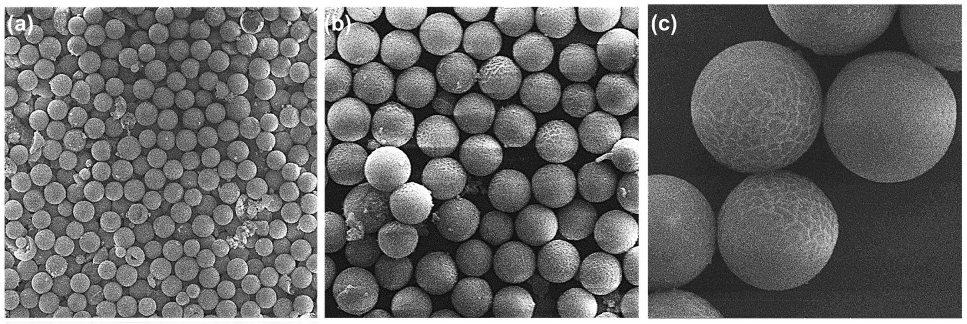
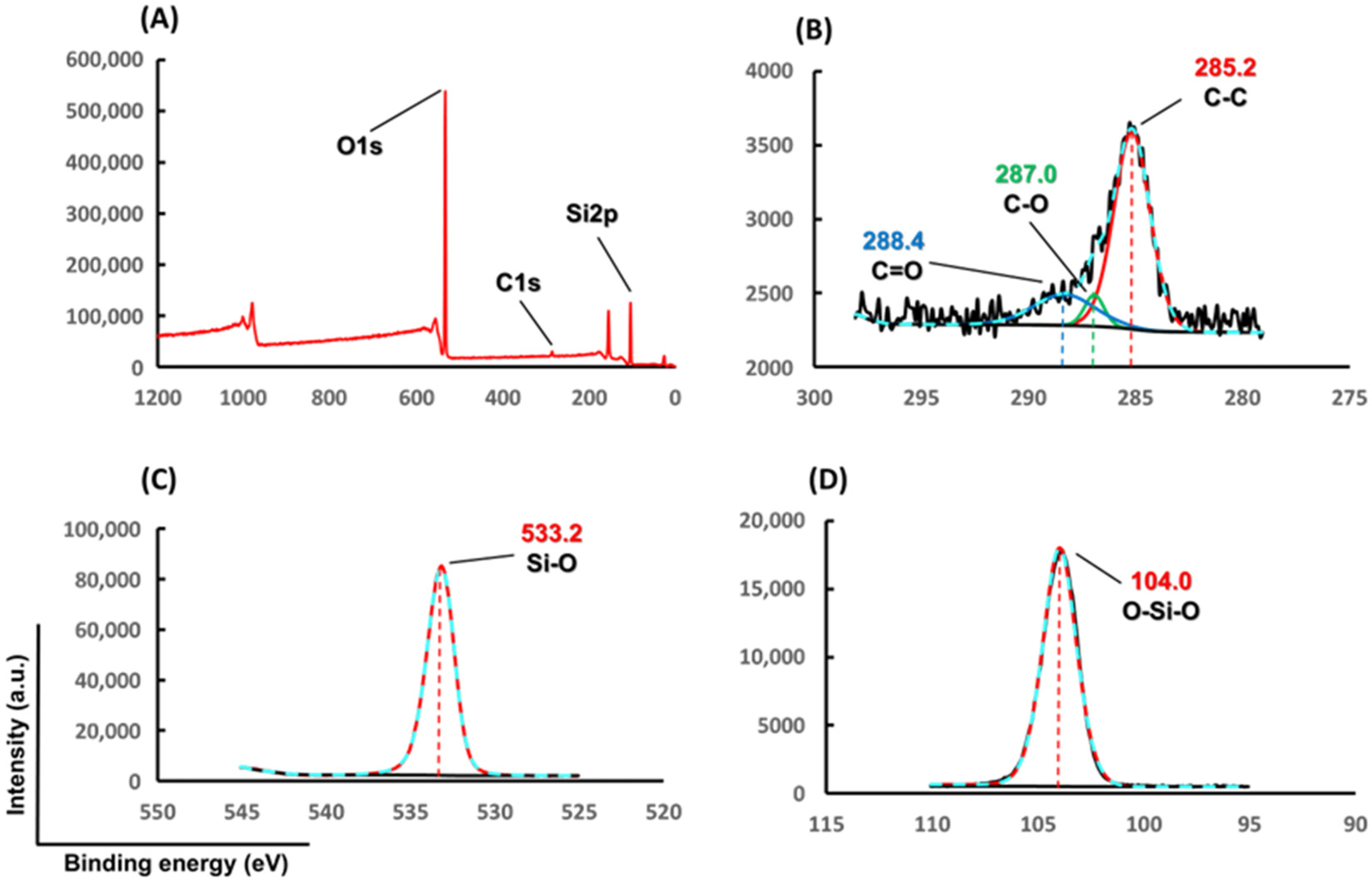
3.1. Characterization of Monodisperse-Mesoporous SiO2 Microspheres

Monodisperse-mesoporous SiO2 microspheres were synthesized by the multi-step hydrolysis condensation method (Figure 4) [36,37]. Poly(methacrylic acid-co-ethylene dimethacrylate) [poly(MAA-co-EDMA)] microspheres synthesized in the monodisperse- mesoporous form, ca. 5.0 μm in size, were used as the starting material in the synthesis of silica microspheres. To obtain poly(MAA-co-EDMA) microspheres, 2 μm sized polyglycidyl methacrylate [poly(GMA)] microspheres obtained by dispersion polymerization were used [38]. Silica gel/polymethacrylate composite microspheres were obtained by a hydrolysis–condensation reaction of the organosilicon precursor (tetraethoxysilane, TEOS) on poly(MAA-co-EDMA) microspheres. For this purpose, TEOS was adsorbed by poly(MAA-co-EDMA) microspheres in an alcohol–water mixture and the adsorbed TEOS molecules were transformed into silica gel form within the porous matrix of polymer microspheres by a hydrolysis and condensation reaction [39]. Next, silica gel/polymethacrylate composite microspheres were calcined at 450 °C in air, for the removal of the polymeric part, and the silica microspheres in monodisperse-mesoporous form were obtained (Figure 4).
The SEM photograph demonstrated the monodisperse character of produced microspheres (Figure 5). The mean particle size was determined as 5.5 μm. SiO2 microspheres were obtained with a mean pore size of 6.7 nm, a mode pore size of 41.1 nm, a pore volume of 1.1 cm3/g and a specific surface area of 161.5 m2/g (Table 1). The pore size distribution in the range of 3–140 nm demonstrated the mesoporous character of the obtained microspheres (Figure 6). The XRD spectrum confirmed the amorphous structure of SiO2 microspheres (Figure 7).
In the survey XPS spectrum, C 1 s, O 1 s and Si 2p bands were observed at the binding energies of 285.2, 287.0, 288.4, 533.2 and 104.0 eV, respectively (Figure 8A). The core level spectra for the C 1 s scan are deconvoluted into three peaks belonging to the C-C, C-O, and C=O groups 285.2, 287.0, and 288.4 eV (Figure 8B) [36,37]. In the core level spectra for the Si 2p scan, a single, sharp deconvoluted peak belonging to the O-Si-O bond was observed at 104.0 eV (Figure 8C) [37]. Similar to the Si 2p scan, the Si-O bond was observed at 533.2 eV in the O 1s scan (Figure 8D) [40].
The characterization demonstrated the monodisperse-mesoporous character of SiO2 microspheres with amorphous character and the presence of organic residual material coming from the template containing C, O species. By considering their enhanced porous properties, including a high pore volume and high surface area and also their suitable mean particle size and very narrow size distribution, for their facile isolation in a solid phase extraction process performed in batch fashion the produced SiO2 microspheres are termed as a material suitable for the separation of various target molecules.

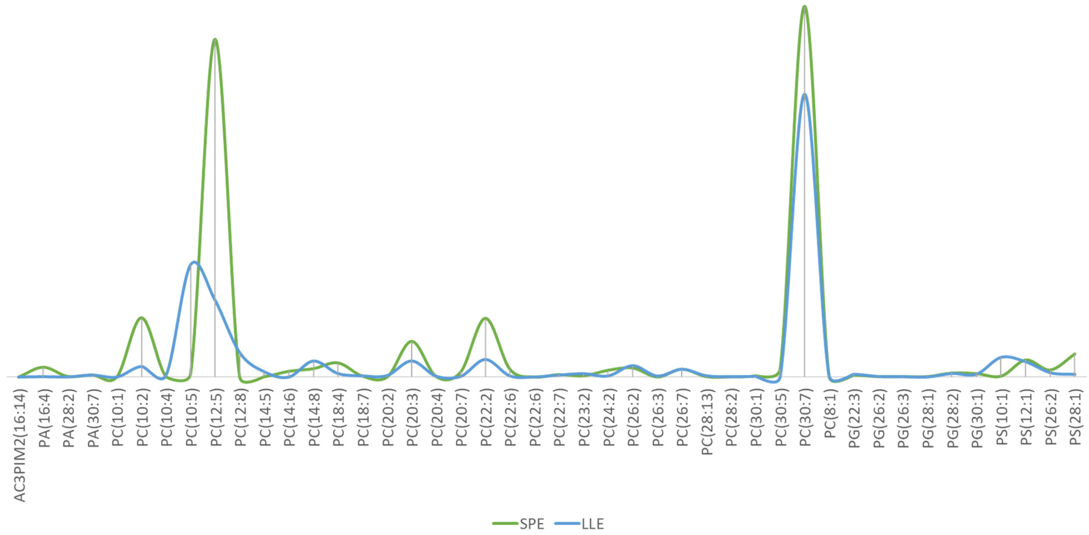
3.2. Q-TOF LC/MS-Based Lipidomics Results

Although the SJF was collected using identical procedures, we pooled the samples in order to prevent variations from the sample itself to just focus on the selective extraction capability of the monodisperse-mesoporous SiO2. Phospholipid enrichment using monodisperse-mesoporous SiO2 microspheres is schematically presented in Figure 9. SiO2 and phosphonic acid groups have possible types of attachment which are monoester, diester, diester with hydrogen bonding to a silanol group, monoester with hydrogen bonding to a surface oxygen atom, diester with Lewis acid/base interaction with a silicon atom, and pure hydrogen bond interactions [41]. In our study, we used SiO2 and phospholipid interactions, especially silanol groups.
Among 7667 peaks, 164 were matched with PLs for LLE extraction of the SJF with a modified Folch method, whereas among 3872 peaks, 141 were matched with PLs for SPE. This situation clearly indicates the capability of SiO2 microspheres to clean up the sample matrix as seen in the chromatograms (Figure 10).
To consider the extraction yield for the novel monodisperse-mesoporous SiO2 microspheres, the peak areas were normalized for both LLE and SPE fractions. The phospholipid enrichment performances of the particles were examined on 44 phospholipid fractions were detected and identified as common across both methods. The comparison of these 44 lipids, based on normalized peak areas and using total peak areas, is visualized in Figure 11. The recovery for the the extracted PLs were at least identical, and for some lipids, it was better than those obtained using LLE (Figure 11).
The phospholipid classes detected in the SJF and their dominance levels after LLE and particle treatment were presented (Figure 12) and listed (Table 2). The sorbents used for phospholipid enrichment in the literature and the detected phospholipid classes are listed in Table 3. In our experimental conditions, PC, PG, PA, and PS were dominantly enriched when the sorbents were used.

4. Discussion

The matrix in which PLs are analyzed can significantly impact the results due to their unique composition. Complex biological matrices, such as SJF, human serum, or tissues, contain various proteins, lipids, and other biomolecules that can interact with sorbents used in PL enrichment. These interactions can affect the extraction efficiency and detection of specific PL classes, requiring tailored extraction procedures and analytical techniques. Table 3 highlights the diversity of sorbents used for PL enrichment across various matrices and analytical techniques. It briefly shows that previous studies have successfully employed monodisperse-mesoporous SiO2 microspheres, HOA–BaTiO3 nanoparticles and Fe3O4/TiO2 nanoparticles to extract PLs from complex biological samples. For instance, monodisperse-mesoporous SiO2 microspheres were utilized with SPE coupled with Q-TOF LC/MS to analyze PLs such as PC, PG, PA, and PS from SJF, as demonstrated in the present work. Similarly, HOA–BaTiO3 nanoparticles and Fe3O4/TiO2 nanoparticles have been used for micro-SPE and SPE, respectively, for analyzing PLs in samples like Escherichia coli and tumor cells. These examples underscore the adaptability and effectiveness of different sorbents in extracting PLs from a wide range of complex biological matrices, highlighting the importance of selecting appropriate sorbents and extraction techniques based on the specific matrix and analytical goals.
When the SEM, XPS, XRD and BET analyses are examined, it is seen that SiO2 microspheres have been synthesized successfully. Our multi-step hydrolysis condensation method and the monodisperse character of our microspheres were in line with previous studies [37,38,39]. The materials used in these studies served as sorbents for SPE. In this current study, we used the sorbent as a microextraction agent and aimed to enrich the SJF in terms of PLs by applying it with the sorbent (Table 3). It has been observed that phospholipidomic studies conducted on the SJF were carried out directly on the sample or on the extract obtained by the LLE method in a targeted manner (Table 4). In our study, SiO2 microspheres were used as a microextraction sorbent to increase selectivity. We were able to produce SiO2 microspheres with a surface area of 161.5 m2/g, pore volume of 1.1 cm3/g, and pore diameter of 6.7 nm, which were efficient in the enrichment of PLs in SJF. The XRD and survey XPS spectra were compatible to previous studies [36,37,40].
The modified Folch method is a non-selective method that extracts all non-polar molecules by dissolving them in a non-polar organic solvent. However, selective identification and determination of PLs in SJF is crucial for diagnosing and monitoring joint diseases. Phospholipids, essential components of cell membranes, play significant roles in maintaining the structural integrity and functionality of joint tissues. By studying PL levels, researchers can gain insights into the underlying mechanisms of joint diseases, such as increased degradation reflecting heightened inflammatory activity or altered synthesis rates pointing to metabolic or enzymatic dysregulations within the joint. This makes the PL concentration in the SJF a promising biomarker for early diagnosis, disease monitoring, and personalized treatment in joint diseases. At this point, traditional LLE methods, such as the modified Folch method, are not sufficient for selectively extracting PLs due to their non-selective nature, which leads to the co-extraction of other non-polar molecules. Therefore, targeted approaches to extract phospholipids from very small volumes of SJF samples are essential. Advanced techniques such SPE, which can selectively isolate PLs based on their chemical properties or mass spectrometry-based methods that allow for high specificity and sensitivity, are needed. These targeted extraction methods not only improve the accuracy of PL quantification but also enhance the reliability of these molecules as biomarkers. Implementing such precise extraction techniques can facilitate more accurate monitoring of joint health, provide deeper insights into the pathophysiology of joint diseases and support the development of more effective, individualized therapeutic strategies.
When the non-selective LLE fraction was injected, the chromatographic peaks observed include all lipids in a higher concentration and therefore contain more peaks with higher intensity as seen in the chromatogram (Figure 10). However, the primary goal of our study was to enrich even very small sample amounts for phospholipids (PLs), which is why we applied sorbent at the milligram scale. Especially in cases such as OA, where PL levels change and need to be characterized, enriching PLs for selective analysis in very low amounts of SJF samples becomes important. The developed SPE technique extracted the PLs selectively and 141 PLs were identified. To determine the extraction efficiency, the normalized peak areas of 44 common peaks given in Table 2 (intersection of the identified peaks for LLE and SPE after LLE) were considered, and the results are given in Figure 10 and Figure 11. Our study presents the capability of the SiO2 microspheres as an efficient sorbent for extracting PLs from SJF. The utilization of this sorbent demonstrates remarkable efficacy in the cleanup of the complex SJF matrix (Figure 10) while achieving selective enrichment of PLs (Figure 11). Importantly, only a minimal quantity of the sorbent is required for this task, showcasing its high efficiency and cost-effectiveness. Considering the critical role of PL determination in the diagnosis of SJF-related diseases, our findings suggest a new approach for disease prognosis and monitoring of treatment efficacy. This innovative methodology holds promise for facilitating early and accurate diagnosis, thus potentially improving patient outcomes and healthcare management in this clinical context. In this case, we think that it would be valuable to use the sorbent developed for studies on the detection of biomarkers in arthritic diseases, such as OA, based on the amount of PL.

5. Limitations

In this study, a notable limitation arises from the comparison between two distinct sample types. The first involves the extraction of SJF using a modified LLE method, in which lipids are non-selectively collected within the nonpolar organic phase. This approach results in the detection of 164 PLs alongside other lipid species, with the concentration of these lipids being dependent on the extraction yield. In contrast, the use of our sorbent for PL extraction offers a more selective approach, where the extraction yield is influenced by the sorbent’s enrichment capacity and the amount used for adsorption and desorption of PLs. Therefore, a normalization process was performed by assessing the peak areas of identical PL peaks observed in both sample types. However, to better demonstrate the efficiency of the sorbent, it is essential to inject samples with equivalent lipid concentrations and evaluate the proportion of identified lipids constituted by PLs versus other lipid types.
Another limitation of this study is the use of pooled samples to minimize variations in matrix composition. While this approach provides consistency, it does not account for individual sample variability. Future studies should adapt this methodology to analyze individual samples from distinct groups, such as healthy and test groups, to determine if the sorbent can efficiently identify diagnostic or prognostic PLs and discern differences between these groups.
The third limitation is the untargeted nature of this study. Untargeted lipidomics approaches have inherent limitations, such as a lack of specificity for particular lipid classes, potential issues with lipid identification accuracy, and the inability to quantify low-abundance lipids effectively. Moreover, untargeted methods may miss certain lipid species due to their reliance on the sensitivity and resolution of the analytical techniques used.

6. Conclusions

In the study, monodisperse-mesoporous SiO2 microspheres, a new metal oxide affinity sorbent, were used for the first time to selectively enrich PLs in SJF. The efficiency of the developed method was shown using Q-TOF LC/MS-based untargeted lipidomics. PLs are critical biomarkers for better understanding OA and proposing personalized therapies. This study showed that small amounts of SJF can be selectively extracted using the proposed SPE technique and the findings on PLs can be compared within groups with different stages of OA. In the near future, personalized therapies will be taken into action and such sorbents developed in the present study can be used in routine clinical applications.

Author Contributions

Conceptualization, S.A., F.K., M.Ç. and A.T.; methodology, S.A., F.K., M.Ç. and A.T.; formal analysis, S.A., İ.D., B.G.K., E.K. and O.K.; investigation, S.A., İ.D., B.G.K., E.K., O.K. and B.E.A.; data curation, S.A., B.G.K., E.K. and O.K.; writing—original draft preparation, S.A. and F.K.; writing—review and editing, S.A., F.K., M.Ç. and A.T.; visualization, S.A. and O.K.; supervision, F.K.; project administration, F.K.; funding acquisition, F.K. All authors have read and agreed to the published version of the manuscript.

Funding

This study was supported by Scientific and Technological Research Council of Türkiye (TUBITAK) under the Grant Number 223S509. This work also was supported by Research Fund of the Hacettepe University under the Project Number: 21455.

Institutional Review Board Statement

The study was conducted in accordance with the Declaration of Helsinki, and approved by the Hacettepe University Health Sciences Research Ethics Committee (Approval no and date: 2023/09-44—19 December 2023, Research no: SBA 23/262).

Informed Consent Statement

Informed consent was obtained from all subjects involved in the study. Written informed consent has been obtained from the patient(s) to publish this paper.

Data Availability Statement

The data used to support the findings of this study are available from the corresponding author [F.K.] upon request.

Acknowledgments

TUBITAK granted this study in relevance to the EU COST CA21110—“Building an open European Network on OsteoArthritis research (NetwOArk)” Action. The authors thank TUBITAK for their support. Part of this study was presented at the Cells & Extracellular Templates (CET)—2nd International Conference in Rome between 8 and 9 June 2023 and the 3rd Regenerative Medicine Orthopaedic Summit (RMOS) in İstanbul between 30 November and 2 December 2023. Feza Korkusuz and Ali Tuncel are active members of the Turkish Academy of Sciences (TUBA).

Conflicts of Interest

This study is based on the PhD Thesis of Serhat Aladağ, MSc, entitled “Development of Monodisperse-Mesoporous SiO2 Microspheres as a New Metal Oxide Affinity Chromatography (MOAC) Sorbent for Phospholipid Enrichment in Synovial Joint Fluid”. Feza Korkusuz MD and Ali Tuncel are active members of the Turkish Academy of Sciences (TUBA). The authors declare that they have no known competing financial interests or personal relationships that could have appeared to influence the work reported in this paper.

References

  1. Kraus, V.B.; Blanco, F.J.; Englund, M.; Karsdal, M.A.; Lohmander, L.S. Call for standardized definitions of osteoarthritis and risk stratification for clinical trials and clinical use. Osteoarthr. Cartil. 2015, 23, 1233–1241. [Google Scholar] [CrossRef]
  2. Martel-Pelletier, J.; Barr, A.J.; Cicuttini, F.M.; Conaghan, P.G.; Cooper, C.; Goldring, M.B.; Goldring, S.R.; Jones, G.; Teichtahl, A.J.; Pelletier, J.P. Osteoarthritis. Nat. Rev. Dis. Primers 2016, 2, 16072. [Google Scholar] [CrossRef]
  3. Goldring, S.R.; Goldring, M.B. Changes in the osteochondral unit during osteoarthritis: Structure, function and cartilage-bone crosstalk. Nat. Rev. Rheumatol. 2016, 12, 632–644. [Google Scholar] [CrossRef] [PubMed]
  4. Deveza, L.A.; Melo, L.; Yamato, T.P.; Mills, K.; Ravi, V.; Hunter, D.J. Knee osteoarthritis phenotypes and their relevance for outcomes: A systematic review. Osteoarthr. Cartil. 2017, 25, 1926–1941. [Google Scholar] [CrossRef]
  5. Rocha, B.; Illiano, A.; Calamia, V.; Pinto, G.; Amoresano, A.; Ruiz-Romero, C.; Blanco, F.J. Targeted phospholipidomic analysis of synovial fluid as a tool for osteoarthritis deep phenotyping. Osteoarthr. Cartil. Open 2021, 3, 100219. [Google Scholar] [CrossRef] [PubMed]
  6. Priori, R.; Scrivo, R.; Brandt, J.; Valerio, M.; Casadei, L.; Valesini, G.; Manetti, C. Metabolomics in rheumatic diseases: The potential of an emerging methodology for improved patient diagnosis, prognosis, and treatment efficacy. Autoimmun. Rev. 2013, 12, 1022–1030. [Google Scholar] [CrossRef]
  7. Zhang, W.; Sun, G.; Likhodii, S.; Liu, M.; Aref-Eshghi, E.; Harper, P.E.; Martin, G.; Furey, A.; Green, R.; Randell, E.; et al. Metabolomic analysis of human plasma reveals that arginine is depleted in knee osteoarthritis patients. Osteoarthr. Cartil. 2016, 24, 827–834. [Google Scholar] [CrossRef]
  8. Zhai, G.; Sun, X.; Randell, E.W.; Liu, M.; Wang, N.; Tolstykh, I.; Rahman, P.; Torner, J.; Lewis, C.E.; Nevitt, M.C.; et al. Phenylalanine Is a Novel Marker for Radiographic Knee Osteoarthritis Progression: The MOST Study. J. Rheumatol. 2021, 48, 123–128. [Google Scholar] [CrossRef]
  9. Zhang, W.; Sun, G.; Aitken, D.; Likhodii, S.; Liu, M.; Martin, G.; Furey, A.; Randell, E.; Rahman, P.; Jones, G.; et al. Lysophosphatidylcholines to phosphatidylcholines ratio predicts advanced knee osteoarthritis. Rheumatology 2016, 55, 1566–1574. [Google Scholar] [CrossRef]
  10. Zhai, G.; Pelletier, J.P.; Liu, M.; Aitken, D.; Randell, E.; Rahman, P.; Jones, G.; Martel-Pelletier, J. Activation of The Phosphatidylcholine to Lysophosphatidylcholine Pathway Is Associated with Osteoarthritis Knee Cartilage Volume Loss Over Time. Sci. Rep. 2019, 9, 9648. [Google Scholar] [CrossRef]
  11. Jaggard, M.K.J.; Boulangé, C.L.; Graça, G.; Vaghela, U.; Akhbari, P.; Bhattacharya, R.; Williams, H.R.T.; Lindon, J.C.; Gupte, C.M. Can metabolic profiling provide a new description of osteoarthritis and enable a personalised medicine approach? Clin. Rheumatol. 2020, 39, 3875–3882. [Google Scholar] [CrossRef]
  12. Kosinska, M.K.; Liebisch, G.; Lochnit, G.; Wilhelm, J.; Klein, H.; Kaesser, U.; Lasczkowski, G.; Rickert, M.; Schmitz, G.; Steinmeyer, J. Sphingolipids in human synovial fluid—A lipidomic study. PLoS ONE 2014, 9, e91769. [Google Scholar] [CrossRef]
  13. Bian, J.; Xue, Y.; Yao, K.; Gu, X.; Yan, C.; Wang, Y. Solid-phase extraction approach for phospholipids profiling by titania-coated silica microspheres prior to reversed-phase liquid chromatography-evaporative light scattering detection and tandem mass spectrometry analysis. Talanta 2014, 123, 233–240. [Google Scholar] [CrossRef] [PubMed]
  14. Bellamy, N.; Campbell, J.; Welch, V.; Gee, T.L.; Bourne, R.; Wells, G.A. Viscosupplementation for the treatment of osteoarthritis of the knee. Cochrane Database Syst. Rev. 2006, 2006, CD005321. [Google Scholar] [CrossRef] [PubMed]
  15. Ludwig, T.E.; McAllister, J.R.; Lun, V.; Wiley, J.P.; Schmidt, T.A. Diminished cartilage-lubricating ability of human osteoarthritic synovial fluid deficient in proteoglycan 4: Restoration through proteoglycan 4 supplementation. Arthritis Rheum. 2012, 64, 3963–3971. [Google Scholar] [CrossRef] [PubMed]
  16. Sun, K.; Shoaib, T.; Rutland, M.W.; Beller, J.; Do, C.; Espinosa-Marzal, R.M. Insight into the assembly of lipid-hyaluronan complexes in osteoarthritic conditions. Biointerphases 2023, 18, 021005. [Google Scholar] [CrossRef]
  17. Sarma, A.V.; Powell, G.L.; LaBerge, M. Phospholipid composition of articular cartilage boundary lubricant. J. Orthop. Res. 2001, 19, 671–676. [Google Scholar] [CrossRef]
  18. Kosinska, M.K.; Liebisch, G.; Lochnit, G.; Wilhelm, J.; Klein, H.; Kaesser, U.; Lasczkowski, G.; Rickert, M.; Schmitz, G.; Steinmeyer, J. A lipidomic study of phospholipid classes and species in human synovial fluid. Arthritis Rheum. 2013, 65, 2323–2333. [Google Scholar] [CrossRef] [PubMed]
  19. Pawlak, Z.; Mrela, A.; Kaczmarek, M.; Cieszko, M.; Urbaniak, W. Natural joints: Boundary lubrication and antiphospholipid syndrome (APS). Biosystems 2019, 177, 44–47. [Google Scholar] [CrossRef]
  20. Mazzucco, D.; Scott, R.; Spector, M. Composition of joint fluid in patients undergoing total knee replacement and revision arthroplasty: Correlation with flow properties. Biomaterials 2004, 25, 4433–4445. [Google Scholar] [CrossRef]
  21. Schmidt, T.A.; Gastelum, N.S.; Nguyen, Q.T.; Schumacher, B.L.; Sah, R.L. Boundary lubrication of articular cartilage: Role of synovial fluid constituents. Arthritis Rheum. 2007, 56, 882–891. [Google Scholar] [CrossRef] [PubMed]
  22. Sabatini, M.; Thomas, M.; Deschamps, C.; Lesur, C.; Rolland, G.; de Nanteuil, G.; Bonnet, J. Effects of ceramide on aggrecanase activity in rabbit articular cartilage. Biochem. Biophys. Res. Commun. 2001, 283, 1105–1110. [Google Scholar] [CrossRef]
  23. Liebisch, G.; Drobnik, W.; Lieser, B.; Schmitz, G. High-throughput quantification of lysophosphatidylcholine by electrospray ionization tandem mass spectrometry. Clin. Chem. 2002, 48, 2217–2224. [Google Scholar] [CrossRef] [PubMed]
  24. Fahy, E.; Subramaniam, S.; Murphy, R.C.; Nishijima, M.; Raetz, C.R.; Shimizu, T.; Spener, F.; van Meer, G.; Wakelam, M.J.; Dennis, E.A. Update of the LIPID MAPS comprehensive classification system for lipids. J. Lipid Res. 2009, 50, S9–S14. [Google Scholar] [CrossRef]
  25. Henry, S.A.; Kohlwein, S.D.; Carman, G.M. Metabolism and regulation of glycerolipids in the yeast Saccharomyces cerevisiae. Genetics 2012, 190, 317–349. [Google Scholar] [CrossRef]
  26. Bligh, E.G.; Dyer, W.J. A rapid method of total lipid extraction and purification. Can. J. Biochem. Physiol. 1959, 37, 911–917. [Google Scholar] [CrossRef] [PubMed]
  27. Ruiz-Gutiérrez, V.; Pérez-Camino, M.C. Update on solid-phase extraction for the analysis of lipid classes and related compounds. J. Chromatogr. A 2000, 885, 321–341. [Google Scholar] [CrossRef]
  28. Nelson, C.A.; Szczech, J.R.; Xu, Q.; Lawrence, M.J.; Jin, S.; Ge, Y. Mesoporous zirconium oxide nanomaterials effectively enrich phosphopeptides for mass spectrometry-based phosphoproteomics. Chem. Commun. 2009, 43, 6607–6609. [Google Scholar] [CrossRef]
  29. Kweon, H.K.; Håkansson, K. Selective zirconium dioxide-based enrichment of phosphorylated peptides for mass spectrometric analysis. Anal. Chem. 2006, 78, 1743–1749. [Google Scholar] [CrossRef]
  30. Khoury, S.; Colas, J.; Breuil, V.; Kosek, E.; Ahmed, A.S.; Svensson, C.I.; Marchand, F.; Deval, E.; Ferreira, T. Identification of Lipid Biomarkers for Chronic Joint Pain Associated with Different Joint Diseases. Biomolecules 2023, 13, 342. [Google Scholar] [CrossRef]
  31. Kosinska, M.K.; Ludwig, T.E.; Liebisch, G.; Zhang, R.; Siebert, H.C.; Wilhelm, J.; Kaesser, U.; Dettmeyer, R.B.; Klein, H.; Ishaque, B.; et al. Articular Joint Lubricants during Osteoarthritis and Rheumatoid Arthritis Display Altered Levels and Molecular Species. PLoS ONE 2015, 10, e0125192. [Google Scholar] [CrossRef] [PubMed]
  32. Koh, J.H.; Yoon, S.J.; Kim, M.; Cho, S.; Lim, J.; Park, Y.; Kim, H.S.; Kwon, S.W.; Kim, W.U. Lipidome profile predictive of disease evolution and activity in rheumatoid arthritis. Exp. Mol. Med. 2022, 54, 143–155. [Google Scholar] [CrossRef] [PubMed]
  33. Van de Vyver, A.; Clockaerts, S.; van de Lest, C.H.A.; Wei, W.; Verhaar, J.; Van Osch, G.J.V.M.; Bastiaansen-Jenniskens, Y.M. Synovial Fluid Fatty Acid Profiles Differ between Osteoarthritis and Healthy Patients. Cartilage 2020, 11, 473–478. [Google Scholar] [CrossRef] [PubMed]
  34. Salimi, K.; Usta, D.D.; Çelikbıçak, Ö.; Pinar, A.; Salih, B.; Tuncel, A. Ti(IV) carrying polydopamine-coated, monodisperse-porous SiO2 microspheres with stable magnetic properties for highly selective enrichment of phosphopeptides. Colloids Surf. B Biointerfaces 2017, 153, 280–290. [Google Scholar] [CrossRef] [PubMed]
  35. Folch, J.M.L.; Lees, M.P.; Stanley, G.H.A. A Simple Method for the Isolation and Purification of Total Lipides from Animal Tissues. J. Biol. Chem. 1957, 226, 497–509. [Google Scholar] [CrossRef]
  36. Tosun, R.B.; Hamaloglu, K.O.; Tuncel, A. Bimetallic Pd-Au nanoparticles supported monodisperse porous silica microspheres as an efficient heterogenous catalyst for fast oxidation of benzyl alcohol. ChemistrySelect 2022, 7, e202201646. [Google Scholar] [CrossRef]
  37. Al-Qurahi, M.A.A.; Demir, M.; Tümer, B.; Gökçal, B.; Tuncel, A. Formic acid dehydrogenation catalyzed by bimetallic nanoalloys supported by monodisperse-porous microspheres: Catalytic and visible light driven photocatalytic hydrogen generation. Int. J. Hydrogen Energy 2024, 51, 111–132. [Google Scholar] [CrossRef]
  38. Elmas, B.; Tuncel, M.; Yalçın, G.; Şenel, S.; Tuncel, A. Synthesis of uniform, fluorescent poly (glycidyl methacrylate) based particles and their characterization by confocal laser scanning microscopy. Colloids Surf. A Physicochem. Eng. Asp. 2005, 269, 125–134. [Google Scholar] [CrossRef]
  39. Gökçal, B.; Kip, Ç.; Tuncel, A. Monodisperse-porous silica microspheres with flexible phenylboronic acid functionalized-polycationic molecular brushes as a sorbent for teamed boronate affinity chromatography in batch and capillary column systems. Colloids Surf. A Physicochem. Eng. Asp. 2023, 676, 132143. [Google Scholar] [CrossRef]
  40. Choi, D.; Lee, C.H.; Lee, H.B.; Lee, M.W.; Jo, S.M. Electropositive Membrane Prepared via a Simple Dipping Process: Exploiting Electrostatic Attraction Using Electrospun SiO2/PVDF Membranes with Electronegative SiO2 Shell. Polymers 2023, 15, 2270. [Google Scholar] [CrossRef]
  41. Weinberger, C.; Heckel, T.; Schnippering, P.; Schmitz, M.; Guo, A.; Keil, W.; Marsmann, H.C.; Schmidt, C.; Tiemann, M.; Wilhelm, R. Straightforward Immobilization of Phosphonic Acids and Phosphoric Acid Esters on Mesoporous Silica and Their Application in an Asymmetric Aldol Reaction. Nanomaterials 2019, 9, 249. [Google Scholar] [CrossRef]
  42. Avalli, A.; Contarini, G. Determination of phospholipids in dairy products by SPE/HPLC/ELSD. J. Chromatogr. A 2005, 1071, 185–190. [Google Scholar] [CrossRef]
  43. Donato, P.; Cacciola, F.; Cichello, F.; Russo, M.; Dugo, P.; Mondello, L. Determination of phospholipids in milk samples by means of hydrophilic interaction liquid chromatography coupled to evaporative light scattering and mass spectrometry detection. J. Chromatogr. A 2011, 1218, 6476–6482. [Google Scholar] [CrossRef] [PubMed]
  44. Calvano, C.D.; Jensen, O.N.; Zambonin, C.G. Selective extraction of phospholipids from dairy products by micro-solid phase extraction based on titanium dioxide microcolumns followed by MALDITOF-MS analysis. Anal. Bioanal. Chem. 2009, 394, 1453–1461. [Google Scholar] [CrossRef]
  45. Kailasa, S.K.; Wu, H.F. Surface modified BaTiO3 nanoparticles as the matrix for phospholipids and as extracting probes for LLME of hydrophobic proteins in Escherichia coli by MALDI-MS. Talanta 2013, 114, 283–290. [Google Scholar] [CrossRef] [PubMed]
  46. Shen, Q.; Cheung, H.Y. TiO2/SiO2 core-shell compositebased sample preparation method for selective extraction of phospholipids from shrimp waste followed by hydrophilic interaction chromatography coupled with quadrupole time-of-flight/mass spectrometry analysis. J. Agric. Food Chem. 2014, 62, 8944–8951. [Google Scholar] [CrossRef] [PubMed]
  47. Ferreiro-Vera, C.; Priego-Capote, F.; Luque de Castro, M.D. Comparison of sample preparation approaches for phospholipids profiling in human serum by liquid chromatography-tandem mass spectrometry. J. Chromatogr. A 2012, 1240, 21–28. [Google Scholar] [CrossRef] [PubMed]
  48. Pegoraro, C.; Silvestri, D.; Ciardelli, G.; Cristallini, C.; Barbani, N. Molecularly imprinted poly(ethylene-co-vinyl alcohol) membranes for the specific recognition of phospholipids. Biosens. Bioelectron. 2008, 24, 748–755. [Google Scholar] [CrossRef]
  49. Ten-Doménech, I.; Martínez-Pérez-Cejuela, H.; Lerma-García, M.J.; Simó-Alfonso, E.F.; Herrero-Martínez, J.M. Molecularly imprinted polymers for selective solid-phase extraction of phospholipids from human milk samples. Microchim. Acta 2017, 184, 3389–3397. [Google Scholar] [CrossRef]
  50. Liu, A.; Zhang, H.; Ding, J.; Kou, W.; Yan, F.; Huang, K.; Chen, H. Enrichment of phospholipids using magnetic Fe3O4/TiO2 nanoparticles for quantitative detection at single cell levels by electrospray ionization mass spectrometry. Talanta 2020, 212, 120769. [Google Scholar] [CrossRef]
  51. Kosinska, M.K.; Mastbergen, S.C.; Liebisch, G. Comparative lipidomic analysis of synovial fluid in human and canine osteoarthritis. Osteoarthr. Cartil. 2016, 24, 1470–1478. [Google Scholar] [CrossRef]
  52. Jónasdóttir, H.S.; Brouwers, H.; Kwekkeboom, J.C. Targeted lipidomics reveals activation of resolution pathways in knee osteoarthritis in humans. Osteoarthr. Cartil. 2017, 25, 1150–1160. [Google Scholar] [CrossRef]
Figure 1. Structures of the major phospholipids [24,25].
Separations 11 00262 g001
Figure 2. Radiographic imaging result and SJF aspiration sample of a 57-year-old patient with grade 4 OA diagnosis.
Separations 11 00262 g002
Figure 3. Representation of metabolite, protein, and lipid phases from top to bottom, respectively, after the LLE protocol.
Separations 11 00262 g003
Figure 4. The schematic representation of the synthesis protocol for monodisperse-mesoporous SiO2 microspheres.
Separations 11 00262 g004
Figure 5. Different scales SEM photographs of monodisperse-mesoporous SiO2 microspheres ((a): 20 µm scale, (b): 10 µm scale, (c): 2 µm scale).
Separations 11 00262 g005
Figure 6. Brunauer–Emmett–Teller (BET) result of monodisperse-mesoporous SiO2 microspheres.
Separations 11 00262 g006
Figure 7. XRD spectra of the monodisperse-mesoporous SiO2 microspheres.
Separations 11 00262 g007
Figure 8. (A) Survey XPS spectrum, (B) core level spectra for C1s scan, (C) core level spectra for O1s scan, and (D) core level spectra for Si 2p scan with monodisperse-mesoporous SiO2 microspheres.
Separations 11 00262 g008
Figure 9. The schematic representation of the phospholipid enrichment using monodisperse-mesoporous SiO2 microspheres.
Separations 11 00262 g009
Figure 10. Overlapped based peak chromatograms (A) Base peak chromatograms of the SPE fraction. (B) Base peak chromatograms of the LLE fraction.
Separations 11 00262 g010
Figure 11. Comparative recovery for PLs using different extraction techniques (SPE: solid-phase extraction, LLE: liquid–liquid extraction).
Separations 11 00262 g011
Figure 12. The enrichment rates of the sorbent for the phospholipid species predominantly found in SJF (PA: Phosphatidic acid, PC: Phosphatidylcholines, PG: Phosphatidylglycerol, PS: Phosphatidylserine).
Separations 11 00262 g012
Table 1. The size and porous properties of SiO2 microspheres.
Specific Surface Area (m2/g)Pore Volume (cc/g)Pore Diameter (nm)Mode Pore Size (nm)
SiO2161.51.16.741.1
Table 2. PLs enriched jointly by LLE and SPE methods.
NoPhospholipidIdentity
1TetraacylglycerophosphoinositoldimannosideAC3PIM2(16:14)
2DialkylglycerophosphatesPA(16:4)
3AlkylacylglycerophosphatesPA(28:2)
4AlkylacylglycerophosphatesPA(30:7)
5DialkylglycerophosphocholinesPC(10:1)
6DialkylglycerophosphocholinesPC(10:2)
7DialkylglycerophosphocholinesPC(10:4)
8DialkylglycerophosphocholinesPC(10:5)
9DialkylglycerophosphocholinesPC(12:5)
10DialkylglycerophosphocholinesPC(12:8)
11DialkylglycerophosphocholinesPC(14:5)
12AlkylacylglycerophosphocholinesPC(14:6)
13DialkylglycerophosphocholinesPC(14:8)
14DialkylglycerophosphocholinesPC(18:4)
15DialkylglycerophosphocholinesPC(18:7)
16AlkylacylglycerophosphocholinesPC(20:2)
17AlkylacylglycerophosphocholinesPC(20:3)
18DiacylglycerophosphocholinesPC(20:4)
19DialkylglycerophosphocholinesPC(20:7)
20DiacylglycerophosphocholinesPC(22:2)
21AlkylacylglycerophosphocholinesPC(22:6)
22DialkylglycerophosphocholinesPC(22:6)
23DialkylglycerophosphocholinesPC(22:7)
24DiacylglycerophosphocholinesPC(23:2)
25AlkylacylglycerophosphocholinesPC(24:2)
26DialkylglycerophosphocholinesPC(26:2)
27AlkylacylglycerophosphocholinesPC(26:3)
28DialkylglycerophosphocholinesPC(26:7)
29AlkylacylglycerophosphocholinesPC(28:13)
30DialkylglycerophosphocholinesPC(28:2)
31DiacylglycerophosphocholinesPC(30:1)
32DialkylglycerophosphocholinesPC(30:5)
33DialkylglycerophosphocholinesPC(30:7)
34DialkylglycerophosphocholinesPC(8:1)
35DialkylglycerophosphoglycerolsPG(22:3)
36AlkylacylglycerophosphoglycerolsPG(26:2)
37DialkylglycerophosphoglycerolsPG(26:3)
38DialkylglycerophosphoglycerolsPG(28:1)
39DialkylglycerophosphoglycerolsPG(28:2)
40AlkylacylglycerophosphoglycerolsPG(30:1)
41DiacylglycerophosphoserinesPS(10:1)
42DiacylglycerophosphoserinesPS(12:1)
43AlkylacylglycerophosphoserinesPS(26:2)
44DiacylglycerophosphoserinesPS(28:1)
Table 3. Analytical methods and sorbents used for PL enrichment across different matrices.
Sorbent UsedExtraction ProcedureCoupled TechniqueMatrixPLs StudiedRef.
Monodisperse-mesoporous SiO2 microspheresSPEQ-TOF LC/MSSynovial joint fluidPC, PG, PA, PSPresent Work
Silica gel base materialSPENP-LC-ELSDDairy productsPC, PE, PI, PS, SM[42]
Silica gel base materialSPEHILIC-ELSDBovine and donkey milkPC, PE, PI, PS, SM[43]
TiO2 beadsMicro-SPEMALDI-TOF-MSDairy productsPC, PE, PI, PS, SM[44]
HOA–BaTiO3 NPsLLMEMALDI-TOFEscherichia coliPS, L-α-PA[45]
Titania-coated silica (TiO2/SiO2) core–shell compositesSPEHILIC-MS/MSShrimp wastePC, PE, PI, PS[46]
Zirconia-bonded silica particlesSPELC-TOF-MSHuman serumLyso-PC, PC, lyso-PE[47]
Poly(ethylene-co-vinyl alcohol) base MIM using PC as templatePermeabilityUVPhospholipid standardsPC[48]
Methacrylate base MIP using PC as templateSPEHILIC-ELSDHuman milkPC, PE, SM[49]
Fe3O4/TiO2 NPsSPEESI-MSTumor CellsPC[50]
Table 4. Overview of PL profiling studies in SJF using mass spectrometry.
YearGroup(s)MatrixExtraction TechniqueMS ModeMS TechnologyDetected PL ClassesRef.
2024OA (24) [Pooled Sample]SJFModified Folsch Method + SPEUntargetedQ-TOF LC/MSPC, PG, PA, PSPresent Work
2013eOA (17)
lOA (13)
RA (18)
Cont (9)
SJFBligh and Dyer MethodTargetedESI-MS/MSPC, LPC, PE, PS, PG, SM[18]
2014eOA (17)
lOA (13)
RA (18)
Cont (9)
SJFBligh and Dyer MethodTargetedESI-MS/MSSM, PA, LPA, PG, LPG[12]
2015OA (48)
RA (20)
Cont (16)
SJFBligh and Dyer MethodTargetedESI-MS/MSPC, LPC, PE, PS, PG, SM[31]
2016Healthy (9)
eOA (17)
lOA (13)
SJFBligh and Dyer MethodTargetedESI-MS/MSPC, LPC, SM, PI[51]
2017OA (11)
RA (12)
SJFSPE ColumnTargetedLC-MS/MSLipid mediators[52]
2021OA (13)
RA (6)
PsA (12)
SynoviumDirectUntargetedMALDI-MSISM, LPC, PA, PC, PE, PI[5]
Disclaimer/Publisher’s Note: The statements, opinions and data contained in all publications are solely those of the individual author(s) and contributor(s) and not of MDPI and/or the editor(s). MDPI and/or the editor(s) disclaim responsibility for any injury to people or property resulting from any ideas, methods, instructions or products referred to in the content.

