Article

Specific and Simultaneous Detection of E. coli O157:H7 and Shiga-like Toxins Using a Label-Free Photonic Immunosensor

by
Ana Fernández
1,
Manuel Hernández
2,
Yolanda Moreno
3,* and
Jorge García-Hernández
2
1
Lumensia Sensors S.L., 46020 Valencia, Spain
2
Centro Avanzado de Microbiología de Alimentos, Biotechnology Department, Universitat Politècnica de València, 46022 Valencia, Spain
3
Instituto de Ingeniería de Agua y del Medioambiente, Universitat Politècnica de València, 46022 Valencia, Spain
*
Author to whom correspondence should be addressed.
Photonics 2024, 11(4), 374; https://doi.org/10.3390/photonics11040374
Submission received: 21 February 2024 / Revised: 3 April 2024 / Accepted: 4 April 2024 / Published: 16 April 2024
(This article belongs to the Special Issue Editorial Board Members’ Collection Series: Photonics Sensors)

Abstract

:
The current study outlines the advancement of an innovative technique for the simultaneous detection of E. coli O157:H7 and its Shiga-like toxins in food samples by utilizing a photonic label-free biosensor coupled with a microfluidic system. This detection method relies on ring resonator transduction that is functionalized with specific bioreceptors against O157:H7 on silicon nitride surfaces capable of binding specifically to the antigen bacterium and its verotoxins. This experiment included the characterization of selected monoclonal and polyclonal antibodies employed as detection probes through ELISA immunoassays exposed to target bacterial antigens. A thorough validation of photonic immunosensor detection was conducted on inoculated minced beef samples using reference standards for E. coli O157:H7 and its verotoxins (VTx1 and VTx2) and compared to gold-standard quantification. The lowest limit-of-detection values of 10 CFU/mL and 1 ppm were achieved for the detection of bacteria and its verotoxins. In this study, the lowest limit of quantification (LoQ) achieved for bacterial quantification was 100 CFU/mL, and, for verotoxins, it was 2 ppm. This study confirmed the effectiveness of a new quality control and food hygiene method, demonstrating the rapid and sensitive detection of E. coli O157:H7 and its verotoxins. This innovative approach has the potential to be applied in food production environments.

1. Introduction

Escherichia coli O157:H7 (E. coli O157:H7) is considered one of the most hazardous types of bacteria, responsible for severe illnesses such as hemorrhagic colitis and hemoytic uremic syndrome, particularly affecting young and immunocompromised individuals [1]. Despite notable advancements in disinfection techniques within the food manufacturing and agricultural sectors, controlling E. coli O157:H7 remains a persistent challenge [2]. In the case of ground beef, the presence of E. coli O157:H7 at one colony-forming unit (CFU)/25 g or above is considered a substantial health risk, given the robust proliferative capacity of E. coli O157:H7 [3]. As a result, the swift and sensitive monitoring of E. coli O157:H7 is crucial to ensuring food and water safety and facilitating prompt and accurate disease diagnosis and treatment [4].
The scientific committees associated with the European Union have established preventative measures and applicable recommendations for avoiding possible food-borne infections caused by strains of verotoxigenic/Shiga toxin-producing/enterohemorrhagic Escherichia coli (VTEC/STEC/EHEC) [5]. According to the Report of the Scientific Committee of the Spanish Agency for Food Safety and Nutrition (AESAN), preventive measures for the contamination of this pathogen should be adopted, covering the entire food chain. These measures include good agricultural practices, biosecurity programs on farms with livestock, good hygiene practices and the inspection of slaughterhouses, and good practices in the processing of vegetables for fresh consumption. Additionally, according to the recommendations established by the European Union, food industry workers must adopt procedures based on the principles of Hazard Analysis and Critical Control Points (HACCP), and consumers must be taught good conservation practices and the cooking of food [6].
In the EU, regulatory microbiological criteria for outbreaks have been established for the absence of specific STEC strains with the highest potential risk of severe disease, while, in other countries, testing for specific STEC strains may be required during outbreak processing as a measure of process performance [5]. Most E. coli O157:H7 strains produce type 2 Shiga toxin, with its subunits binding to the surface of enterocytes. The subunit entering the cells halts protein synthesis by disrupting the function of the large ribosomal subunit [7]. The Shiga toxin can cause symptoms such as stomach pain, diarrhea, inflammation, secretion of intestinal fluids, ulcers, and, in severe cases, conditions like hemorrhagic enteritis and hemolysis, especially in infants and young children, often leading to sepsis or meningitis. Contaminated food is a significant source of infection with verocytotoxigenic E. coli.
The European Commission regulation regarding compliance with the microbiological criteria applicable to food products establishes the absence of E. coli O157:H7 required in 25 g or 25 mL [8]. Several strategies have been employed for the detection of E. coli O157:H7, including colony counting (considered the gold standard) as well as techniques such as enzyme-linked immunosorbent assay (ELISA), polymerase chain reaction (PCR), and quartz crystal microbalance resonators (QCM).
Traditional culture-based methods involve the ability of bacteria to grow and multiply, requiring 24 h of incubation under laboratory conditions [9]. Traditional methods are considered the “gold standards” and consist of culture techniques with specific and selective enrichment media [10], followed by biochemical tests or the counting of microorganisms indicating contamination in the food. These techniques are effective and low cost, but they can take a longer time to obtain results and have low sensitivity and specificity. These methods can be slow and time-consuming in obtaining results, which can delay the diagnosis and treatment of diseases [10]. Additionally, these methods can be expensive and require highly trained personnel to perform the tests.
Nucleic acid-based strategies, particularly those involving polymerase chain reaction (PCR), have shown promise due to their high sensitivity and rapid detection capabilities. The PCR methods used to detect trace amounts of E. coli O157:H7 often involve amplifying the bacteria present in a sample. These approaches are known for being labor-intensive, time-consuming, and expensive. Additionally, they necessitate highly trained personnel, leading to delays in obtaining results and hindering the timely prevention of epidemic outbreaks [11]. On the other hand, methods based on nucleic acids have emerged as powerful tools in the detection of pathogens in food. The advantages of these assays, together with their ease of use and susceptibility to automation, make them very attractive for applications in food, in order to overcome the long enrichment cultivation stage. Polymerase chain reaction (PCR), multiplex PCR, real-time PCR, hybridization, microarrays, and nucleic acid sequence-based amplification offer high sensitivity and specificity, allowing the detection of multiple pathogens in one single sample and providing quick results [12]. However, it is important to highlight that the use of the PCR technique in the detection of pathogens in food has some limitations. One of the main limitations is the need to know the target DNA sequence to design specific primers [13]. This means that a specific set of primers is needed for each pathogen, which can limit the detection capacity of the technique.
Multiplex PCR is an efficient and sensitive technique for the detection of E. coli O157:H7 and its verotoxins in food samples. This technique is a variant of PCR and can detect multiple pathogens in a single reaction [14], reducing sample analysis time and costs. Furthermore, multiplex PCR is capable of detecting pathogens present in low quantities in the sample, making it a highly sensitive technique. However, multiplex PCR also has some limitations in its application. The presence of inhibitors in the sample can affect the sensitivity and specificity of the technique, which can result in false-negative or false-positive results. Furthermore, multiplex PCR requires careful validation to ensure the specificity and sensitivity of the technique for each pathogen and toxin to be detected.
Immunological methods involve obtaining antibody responses, which can be assessed through various techniques, such as an enzyme-linked immunosorbent assay (ELISA). ELISA typically requires approximately 24 h for completion [15,16]. However, these methods demand considerable time, skilled personnel, and costly equipment [17,18]. The ELISA test is a highly sensitive and specific technique. But, to achieve an effective ELISA technique, it is necessary to isolate the antigens of interest and generate specific antibodies by immunizing animals.
An enzyme-linked immunosorbent assay (ELISA) is a highly sensitive and selective method used to detect specific antigens or antibodies in various biological samples [19,20]. The immobilization of the target antigen on a plastic substrate and its subsequent detection using a labeled selective antibody is a crucial feature of ELISA [21]. The numerical values obtained from ELISA measurements are crucial for quantitatively determining the concentration of specific antigens in a sample. The intensity of the signal generated by the labeled antibody (often an enzyme-linked reaction) is directly proportional to the amount of antigen present in the sample.
While ELISA provides relatively rapid results compared to traditional culture techniques, it is essential to note that the sensitivity might differ from more time-consuming methods. The current ELISA used to detect E. coli O157:H7 is commonly based on cELISA. In this case, anti-E. coli O157:H7 antibodies are immobilized on a plate to capture E. coli O157:H7 antigens and subsequently bind the primary antibodies and secondary antibodies linked to an enzyme or directly bind to a conjugated antibody. This method is widely used in laboratories for its speed (3–5 h). It provides a balance between speed and accuracy for routine testing, but it represents a loss of sensitivity [22].
The lateral flow immunoassay is a widely used detection method in the food industry due to its sensitivity and specificity in detecting E. coli O157:H7 in food samples [23]. The simplicity, rapidity, and low cost of the lateral flow immunoassay make it a valuable tool for the detection of pathogens in food, especially in settings where access to laboratories is limited [24]. However, it should be noted that, compared to other pathogen detection methods, the lateral flow immunoassay may have limitations in terms of sensitivity and specificity. Furthermore, this technique can produce false positives or false negatives, which can have serious consequences for food safety [25].
The progress of nanotechnology across diverse domains has led to the development of biosensors as precise diagnostic tools. These biosensors offer a solution to the demand for selective, rapid, and accurate procedures in identifying microorganisms within medical, food, or environmental samples [26]. The use of biosensors in the detection of pathogens and other contaminants in food offers numerous advantages compared to conventional detection methods, such as speed and sensitivity [27]. A notable aspect of biosensors is their high specificity, which means that they can detect the presence of a particular pathogen in a food sample with high precision [28]. Furthermore, biosensors can also be designed to simultaneously detect multiple pathogens or contaminants in food, reducing analysis time and increasing detection accuracy [29].
The implementation of photonic biosensors on silicon photonic-integrated circuits (PICs) indeed offers several advantages, making them a promising technology for various applications, including point-of-care diagnostics [30]. In summary, as determined by LoD and LoQ, sensitivity is a fundamental aspect of biosensor performance [31]. Achieving a high sensitivity enhances the effectiveness of biosensors across various applications where the detection and quantification of specific substances are critical. The sensitivity of waveguide sensors is intricately linked to the extent of overlap between the evanescent field and the sample being analyzed [32,33].
Compared to traditional methods, optical biosensors offer a faster and more effective response to possible food contamination. This is because optical biosensors can detect pathogens in food samples in short periods of time, in some cases within minutes [34]. These devices can detect extremely low levels of pathogens in food samples, allowing for early detection and a rapid response to potential disease outbreaks [34]. Additionally, optical biosensors are also highly specific, meaning that they can distinguish between different species of pathogens. Finally, it is worth noting that optical biosensors are also portable and easy to use, making them ideal for use in field environments [34].
Hence, it is of utmost importance to develop novel techniques for the rapid and sensitive detection of E. coli O157:H7, aiming to prevent potential disease outbreaks resulting from the public consumption of contaminated food. Emerging technologies, including isothermal amplification methods, biosensors, surface-enhanced Raman spectroscopy, paper-based diagnostics, and smartphone-based digital methods, represent innovative approaches in E. coli O157:H7 detection. In the realm of optical biosensors, two primary types are distinguished: the first relies on any potential alteration in the internal optical properties of a biomolecule due to its interaction with the target analyte, encompassing changes in absorption, emission, polarization, or luminescence reduction. The second type employs markers and optical probes. These systems, driven by reagent-mediated detection, utilize changes in the optical response of an intermediate, typically an analyte-sensitive dye molecule, to monitor the concentration of the analyte [35]. These nanobiosensors use electrochemical, fluorescence, colorimetric, and other techniques [36,37,38].
A biosensor falls into the category of bio-affinity sensors when the bio-identifying component is an antigen/antibody, DNAzymes, or DNA from a single-stranded DNA (ssDNA)/RNA sequence immobilized on a solid substrate through linker molecules that specifically interact with the target. Other classifications include enzyme sensors and sensor receptors, which involve ligand binding and biosensors for whole cells, depending on the type of physical probe utilized [39].
Fluorescent biosensor technologies are gaining significance in detecting E. coli O157:H7, offering practical advantages such as rapidity, portability, ease of use, sensitivity, and cost-effectiveness. Fluorescent dyes allow for direct measurement due to their stability compared to natural enzymes. Nevertheless, the challenge of low sensitivity persists, particularly in colorimetric detection methods [40]. The applicability of a detection method is also an important factor to consider. Some methods may be more suitable for detecting E. coli O157:H7 (culture, real-time PCR) or its verotoxins (ELISA, lateral flow immunoassays) or for use on certain food samples, while other methods may have broader applications. Optical and electrochemical biosensors are an example of this due to their limited applicability.
Microfluidic-based biosensors characterized by microchannels for fluidic samples are increasingly gaining relevance because they also facilitate on-chip immunoassays [40]. These systems enable the concurrent in situ execution of various laboratory processes, including detection, sampling, separation, and mixing [40,41].
Biosensors designed for E. coli O157 detection still rely on labor-intensive manual procedures, limiting their practical use in laboratories. While existing optical microfluidic biosensors exhibit accurate sensitivities, their high cost and complex assembly processes remain areas that require improvement [42].
This research aims to develop and optimize a photonic immunosensor for the simultaneous detection of E. coli O157:H7 and its verotoxins. This allows for the early detection and quantification of viable bacteria or their verotoxins, enabling the prediction of pathogen contamination in meat products, fresh vegetables, and prepared dishes. The significance of this technology in overcoming existing limitations in current pathogen detection systems in the food industry is highlighted by its potential applications in predicting and quantifying pathogen contamination.

2. Materials and Methods

2.1. Reagents and Antibodies

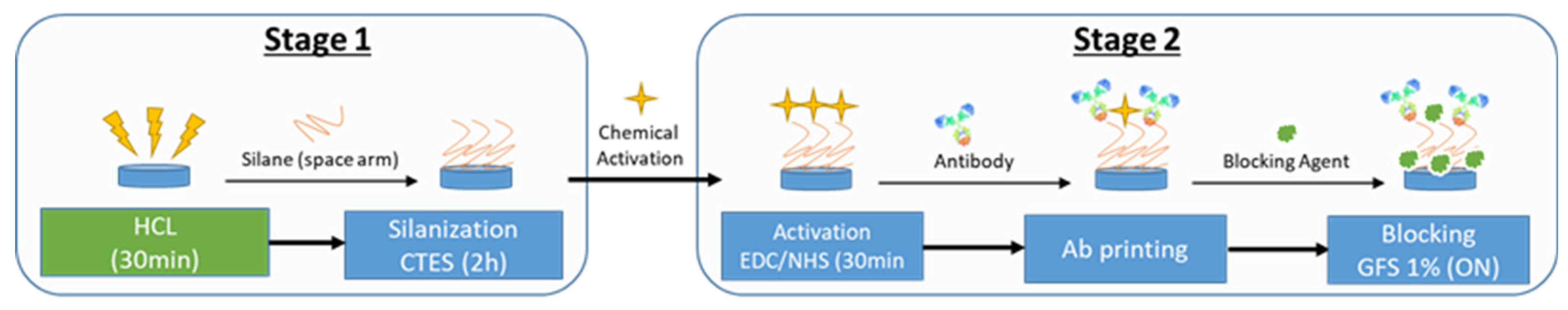
2.1.1. Functionalization

The functionalization reagents comprised CTES (carboxyethylsilanetriol, disodium salt 25% in MilliQ water, ABCR) at 1%, EDC (1-Ethyl-3-(3-dimethylaminopropyl) carbodiimide, Sigma)/NHS (N-hydroxysuccinim) (Merck KGaA, Darmstadt, Germany), and MES (2-(N-morpholino) ethanesulfonic acid (ThermoFisher, Waltham, MA, USA) at 0.1 M.
The antibodies employed in this study included a polyclonal (rabbit) anti-E. coli O157:H7 antibody (BS-1563R, Bioss Antibodies, Woburn, MA, USA) and an anti-E. coli O157:H7 core monoclonal (mouse) antibody (clone E28) (CerTest Biotec, Zaragoza, Spain). The polyclonal and monoclonal antibodies were tested against target antigens derived from E. coli O157:H7 to evaluate the primary antibodies. The specific antigens used were the MT-25STX E. coli O157 VT1 recombinant protein and the MT-25VT2 E. coli O157 VT2 recombinant protein.
A rabbit polyclonal antibody anti-fish obtained from Eurofins Inmunolab (Reinbek, Germany) was also included as a negative control.

2.1.2. Indirect ELISA

The primary polyclonal antibody (Bioss Antibodies, Woburn, MA, USA) and the selected monoclonal anti-E. coli O157:H7 core monoclonal (mouse) antibody (clone E28) (CerTest Biotec, Zaragoza, Spain) were assessed against different concentrations of the inactivated E. coli O157 antigen (native extract) MT-28EC7U.
The indirect enzyme-linked immunosorbent assay (iELISA) reagents comprised commercial TMB substrate (Thermo Scientific, Waltham, MA, USA), H2SO4 solution (for slowing down the reaction), and 0.1 M hydrochloric acid from Scharlab (Barcelona, Spain). The secondary ELISA antibodies included the GARPO polyclonal anti-rabbit IgG (whole molecule) peroxidase antibody produced in goat and the GAMPO polyclonal anti-mouse IgG (whole molecule) peroxidase antibody produced in goat from Abcam (Cambridge, UK).
Each sample was inoculated in duplicate in 0.05 M carbonate buffer with a pH of 9.6 (Merck, Darmstadt, Germany). Furthermore, a phosphate-buffered saline (PBS) solution served as a negative control. In contrast, a standard solution of inactivated E. coli O157 antigen (native extract) MT-28EC7U (CerTest Biotech, Zaragoza, Spain) was employed as a positive control.
A total of seven serial dilutions from 1 mL of the stock of an inactivated E. coli O157 antigen (MT-28EC7U, CerTest Biotech, Spain) were prepared in 0.05 M carbonate buffer with a pH of 9.6 to assess antibody specificity: 1/10, 1/50, 1/80, 1/100, 1/1000, and 1/5000.
Serial dilutions of both MT-25STX E. coli O157 VT1 and MT-25VT2 E. coli O157 VT2 recombinant proteins (CerTest Biotech, Spain) in 0.05 M carbonate buffer with a pH of 9.6 were prepared to assess antibody specificity. The concentrations of the final dilutions of VTx1 were 48.4 ppm, 24.2 ppm, 12.1 ppm, 6.05 ppm, 3.025 ppm, and 1.1909 ppm. And the concentrations of the final dilutions of VTx2 were 38 ppm, 19 ppm, 9.5 ppm, 4.75 ppm, 2.375 ppm, and 1.1875 ppm.
The emitted absorbance was measured at 450 and 650 nm using a Varioskan Flash multimode spectral scanning plate reader (Multilabel Victor 1420 Counter). The positive control was defined as a sample with an OD450 nm value 2.1 times higher than the negative control (P/N 2.1).

2.1.3. Immunosensor Validation

To validate the detection capability of the photonic technique and the conducted detection tests against E. coli O157:H7, a total of five 1/10 serial dilutions from an overnight E. coli O157:H7 CECT 4972 (Spanish Type Culture Collection, Valencia, Spain) culture were prepared to inoculate in TSBm+n broth medium (Triptone Soya Broth + Novobiomiocin supplement) at a specified concentration range of 10 to 106 CFU/mL.
To validate the biosensor detection capability against E. coli O157:H7 verotoxins, serial dilutions of both MT-25STX E. coli O157 VT1 and MT-25VT2 E. coli O157 VT2 recombinant proteins (CerTest Biotech, Spain) were prepared to be inoculated in TSBm+n broth medium (Triptone Soya Broth + Novobiomiocin supplement). The concentrations of the final dilutions of VTx1 ranged from 1 to 50 ppm: 48.4 ppm, 24.2 ppm,12.1 ppm, 6.05 ppm, 3.025 ppm, and 1.1909 ppm. And the concentrations of the final dilutions of VTx2 were 38 ppm, 19 ppm, 9.5 ppm, 4.75 ppm, 2.375 ppm, and 1.1875 ppm.
Additionally, the biosensor response was further evaluated through multiple replicates of artificially contaminated minced beef meat samples obtained from the same batch (25 g of each batch was suspended in 250 mL of TSBm+n). The samples were subsequently inoculated with both different concentrations of E. coli O157 culture ranging from 10 to 106 CFU/mL and dilutions of VTx1 and VTx2 ranging from 1 ppm to 50 ppm. The batches of minced beef samples were collected over three months from a meat processing facility in the Valencia region. These samples had undergone a prior assessment using quantitative polymerase chain reaction (qPCR) and traditional plate counting to verify the presence of the bacteria E. coli O157:H7, confirming negative results. Significantly, these batches were obtained directly from their original packaging and were intended for immediate consumption. They were purchased from a local grocery store and stored at a temperature of 4 °C until employed in the assays.
Concentration tests were conducted, followed by confirmation of the bacteria presence through the separation and concentration of the microorganism using immunomagnetic particles coated with anti-E. coli O157:H7 antibodies (Dynabeads anti-E. coli O157, 71003, Applied Biosystems by Thermo Fisher Scientific Baltics UAB, Vilnius, Lithuania). For each isolated colony which was examined, one drop was dispensed to be confirmed by a latex agglutination test for the identification of the E. coli serogroup 0157:H7 from Oxoid (TSMX9410, RIM latex test for E. coli O157:H7, Oxoid, Termo Fisher-Scientific). These incubation conditions align with established recommendations and standards for bacterial culture and growth [43].
A data-reading setup according to [44] was employed for reading the signal transduction related to the resonance of 100 Photonic-Integrated Circuits (PICs) designed by Lumensia Sensors (Spain). One hundred photonic biosensors were integrated on silicon photonic-integrated circuits (PICs). A two-channel microfluidic system connected to the PICs and a peristaltic pump were connected to the data-reading setup.

2.2. Indirect ELISA

To assess the suitability of the developed biosensor for detecting E. coli O157:H7, it was necessary to characterize the antibodies used. This involved evaluating the binding capacity of both polyclonal and monoclonal antibodies specific to E. coli O157:H7. An iELISA protocol was designed for this purpose, based on established methods from the existing literature [19,20].
This new protocol was based on previous descriptions with modifications [45,46]. The primary polyclonal antibody (Bioss Antibodies, Woburn, MA, USA) and the selected monoclonal anti-E. coli O157:H7 core monoclonal (mouse) antibody (clone E28) (CerTest Biotec, Zaragoza, Spain) were assessed against different concentrations of the inactivated E. coli O157 antigen (native extract) MT-28EC7U.
A 96-well ELISA microplate was loaded with 100 µL/well of the corresponding concentration of the E. coli O157:H7 inactivated antigen (native extract) MT-28EC7U. Additionally, replicates from a batch of minced beef meat spiked with inactivated E. coli O157:H7 (native extract) MT-28EC7U (CerTest Biotech, Zaragoza, Spain), as above described in 2.1, were included to assess antibody specificity. The wells underwent three washes with PBS with 0.05% Tween-20. Subsequently, all the wells were blocked with 1% BSA in PBS by adding 100 µL to each well and were incubated for one hour at room temperature (25 °C).
Following the washes, 100 µL of a PBS solution containing 1 ppm of the selected antibody specific for E. coli O157 was added to all the wells previously inoculated with the antigen. The plates were then incubated at 37 °C for 1 h, with a control group lacking immobilized antibodies. Subsequently, three successive washes were conducted with PBST, and the wells were filled with 100 µL of a goat anti-rabbit–HRP conjugate, followed by another incubation at 37 °C for 1 h. After three additional washes with PBST, the detection reaction was initiated by adding 100 µL of the substrate based on 1,2-diaminobenzene (OPD) (4 mg of 1,2-diaminobenzene and 15 L of H2O2 in 10 mL of citrate buffer, pH 4.5). After a 15 min incubation period, the reaction was halted by adding 50 µL of 2 M sulfuric acid. Finally, the emitted absorbance was measured at 450 and 650 nm using a Varioskan Flash multimode spectral scanning plate reader (Multilabel Victor 1420 Counter). The positive control was defined as a sample with an OD450 nm value 2.1 times higher than the negative control (P/N 2.1).

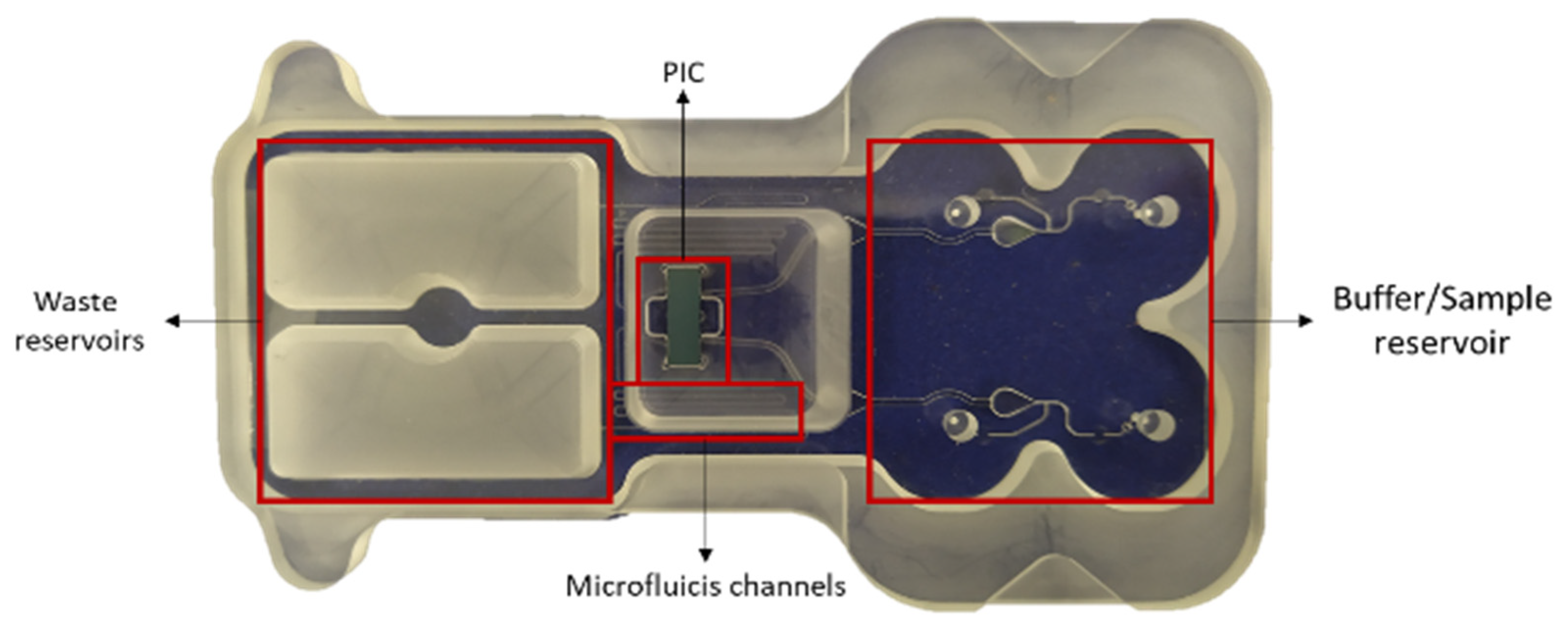
2.3. PIC Fabrication and Functionalization Process

The optical photonic-integrated circuits (PICs) developed for this study underwent fabrication in a controlled clean room environment, specifically within a class 10–100 clean room. The process involved utilizing an electron beam writing technique on a positive resist layer made of polymethylmethacrylate (PMMA) with a thickness of 100 nm [44]. The manufacturing process of the photonic-integrated circuits (PICs) began with preparing a silicon wafer, which served as the substrate for the PICs, and positive photoresist was deposited onto the wafer surface. The circuit patterns were defined on the photoresist layer using lithography techniques (electron beam process or ultraviolet light). This involved exposing the photoresist to light through a photomask containing the desired circuit pattern. After exposure, the photoresist was developed, removing the unexposed areas and leaving behind the desired circuit patterns on the wafer. A layer of chromium was deposited onto the wafer using an evaporation process. After deposition, the remaining photoresist was dissolved or lifted off, leaving the metal patterns on the wafer. The silicon nitride layer on the wafer was etched to define the waveguide structures required for photonic circuits. A layer of silicon oxide was deposited onto the wafer to cover the circuitry.
The detection of E. coli O157:H7 and its verotoxins (VTx1 and VTx2) was achieved through the functionalization of 100 photonic biosensors (PICs). These biosensors were obtained from various batches and wafers, and the functionalization process followed the established procedure outlined by [44].
The process began with the oxidation of the PIC surface, which was achieved by immersing it in 5 mL of a 0.1 M hydrochloric acid solution provided by Scharlab (Barcelona, Spain) for 30 min (Figure 1). The oxidation step was carried out on an orbital shaker plate at 30 rpm. Subsequently, the surface underwent a meticulous rinse with deionized water (DIW) and was dried.
After the oxidation step, the surface underwent silanization with a carboxyethylsilanetriol (CTES) solution for 2 h. Simultaneously, the activation of the carboxylic group within the CTES organosilane on the surface was accomplished by introducing a mixture of carbodiimide and N-hydroxysuccinimide (EDC/NHS), followed by a 30 min incubation at room temperature. The surface was then thoroughly rinsed and dried under a stream of airflow.
The covalent immobilization of anti-E. coli O157:H7 antibodies on the biosensor surface were conducted with the specific anti-E. coli O157:H7 rabbit polyclonal antibody (Bioss Antibodies, Woburn, MA, USA) and the anti-E. coli O157:H7 monoclonal (mouse) antibody (clone E28) (CerTest Biotec, Zaragoza, Spain). This immobilization process was carried out in an oriented manner (Figure 1).
After the covalent immobilization of anti-E. coli O157:H7 antibodies on the biosensor surface, the surfaces were washed with phosphate-buffered saline (PBS), dried, and then blocked. The blocking step involved incubating the surfaces overnight with a solution containing 1% cold water fish skin gelatin (GFS) in PBS. This ensured the effective blocking of the non-specific binding sites on the surface (Figure 1).
Throughout these steps, precise control and alignment were crucial to ensure the accuracy and functionality of the resulting PICs. The completed PICs could then be further processed and integrated into a two-channel microfluidic cartridge (Figure 2).

2.4. Photon Transduction Setup Reader

Photon transduction is the fundamental principle underlying the detection process of this system. Resonant cavities, specifically ring resonators (RRs) fabricated with silicon nitride technology, are employed for this purpose. The analysis of the refractive indices obtained from these ring resonators (RRs) has been proposed for various applications. In these transducers, a change in the refractive index of light occurs when the analyte of interest interacts with the bioreceptor anchored on the surface of the ring. This enables the correlation of the analyte concentration attached to the ring with the observed signal. The optical transduction systems used on biosensors offer unique characteristics conferring several advantages.
Indeed, these biosensors leverage significantly smaller sample volumes by integrating microfluidic and nanophotonic systems, enabling analyses at the nanometric or micrometric scale. Furthermore, the label-free detection capability of these biosensors allows for real-time monitoring, simplifying procedures by reducing the necessary steps and reagents.
The photonic biosensors integrated on silicon photonic-integrated circuits (PICs) offer label-free performance with high detection sensitivities. Notably, they are disposable, making them particularly interesting for point-of-care diagnostics as they do not necessitate specialized personnel [30].
The current detection system is designed to capture the sensor’s transduction signal during sample analysis. It involves the integration of three essential components forming a measurement setup: a photonic-integrated circuit (PIC) designed by Lumensia Sensors (Spain), a two-channel microfluidic system connected to the PIC, and a peristaltic pump. Both microfluidic channels are linked to the microfluidic system attached to the PIC, through which the samples to be detected flow (Figure 2). Four sealed reservoirs are housed per channel, in which the flow direction is carried out in the following order: a reservoir to introduce the washing buffer; another one, below, in which the test sample to be analyzed is introduced; a hole in which the adhered PIC is housed and embedded with the washing buffer first and then the problem sample; and, finally, a waste reservoir which is reached by all the fluids which have passed through the PIC housing. The system ensures that there is no cross-reactivity since both channels are sealed and separated (Figure 2).
A data-reading setup according to [44] was employed for the signal transduction related to resonance. This reading system was based on software and hardware developed by Lumensia Sensors, enabling the translation of the optical signal into resonance measured in picometers (pm).

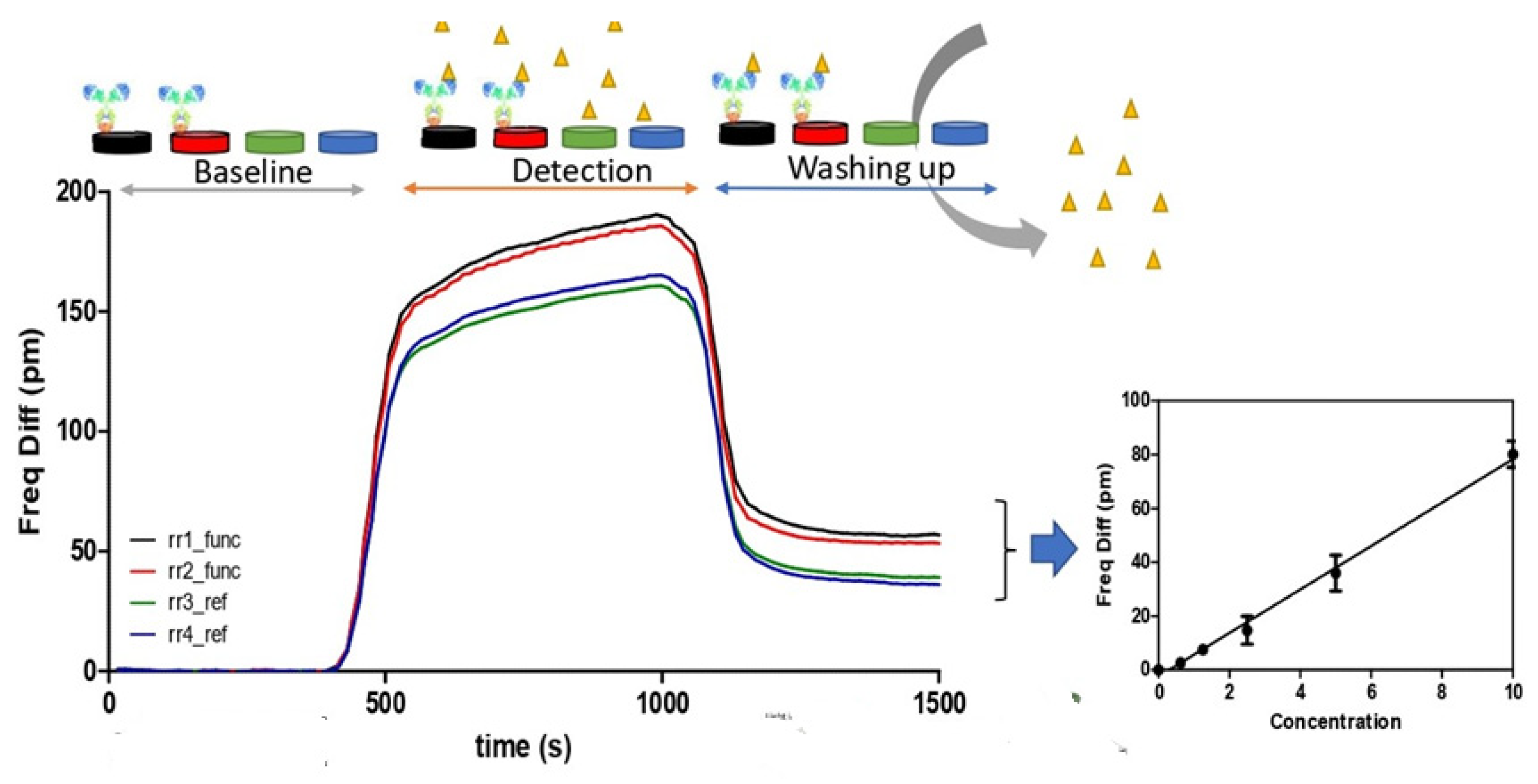
2.5. Immunosensor Validation for E. coli O157:H7 and Shiga-like Toxins

The performance and reliability of the detection technique were evaluated by flowing different minced meat samples spiked with antigens of E. coli O157:H7 over previously functionalized photonic biosensors (PICs). Various experimental tests were conducted to determine this alternative detection system’s optimal sensitivity and limit of detection (LoD). This critical step facilitated a comprehensive assessment of the technique’s effectiveness in detecting and responding to various E. coli O157:H7 antigens, including a strain of the bacterium and its verotoxins VT1 and VT2 in isolation, contributing to its validation and reliability.
Different samples of minced meat from the same batch, inoculated with E. coli O157:H7 CECT 4972 (range of 10 to 106 CFU/mL, as detailed in Section 2.2), were included in the analysis to assess the sensor’s detection efficiency. Moreover, the sensor’s detection efficiency within a food matrix was also stablished by inoculating different dilutions of verotoxin 1 (VTx1) and verotoxin 2 (VTx2) to compare the immunosensor’s response against both types of antigens. Moreover, serial dilutions of verotoxin 1 and verotoxin 2 produced by E. coli O157:H7, only suspended in the pre-enrichment medium, as described in Section 2.1, were used to evaluate the specific immunosensor detection.
Current biosensor trends focus on improving accessibility, robustness, and the overall efficiency of the detection process. Integrating these components creates a comprehensive biosensor system capable of detecting and analyzing analytes with high sensitivity and precision. The combination of optical transduction on a PIC, two microfluidics channels, and a controlled sample flow (peristaltic pump) enhances the performance of the biosensor. The setup is specifically designed to facilitate the controlled flow of samples over the sensor for analysis [44].
The next step in the experimental procedure involved systematically flowing the samples and subjecting them to bacterial sensing using the setup detector optimized for PICs, which was fabricated by Lumensia Sensors [44]. A critical aspect of this step was facilitating the controlled flow of samples by attaching the microfluidic adhesive layer. The coupled microfluidic system, integrated with eight ring resonators arranged within the sensor and distributed across two channels, enhanced the detection capabilities by enabling the concurrent measurement of two samples. The sensor incorporated eight ring resonators, which are optical components which respond to changes in the refractive index. These resonators were distributed across two channels, each containing four resonant rings [44]. The configuration, with multiple resonators and channels, contributed to a high sensitivity in detection. The system could detect concentrations down to the ng/mL scale [44].
Additionally, the immunosensors were securely housed within the previously functionalized PICs obtained according to the procedure outlined in Section 2.4. Tests were conducted using the same batch of sensors that had been manufactured and printed on the same date. The sensors were used to detect samples with the same levels of contamination on the same matrix on different dates, up to six months and one year after their manufacturing and functionalization.
Indeed, the setup for reading resonance data is a crucial component of the biosensor system, allowing for the interpretation of the optical signals generated during the interaction between the biosensor and the target analytes (E. coli O157:H7 bacterium, VTx1, and VTx2). The hardware and software components developed by Lumensia Sensors play a crucial role in translating these optical signals into measurable resonance values, typically expressed in picometers (pm). This reading system is essential for monitoring and quantifying the binding events on the biosensor surface, providing valuable information about the presence and concentration of the target analytes. The integration of this advanced reading system enhances the overall performance and reliability of the biosensor platform [44].
A peristaltic pump was utilized to control the flow rate of the sample dilutions, operating at a rate of 15 µL/min. This precise flow method ensured that the prepared samples reached the immunosensors, where the crucial reaction between the E. coli O157:H7 antigen and the functionalized antibodies specific to that antigen occurred in the E. coli O157 immunodetection assay (Figure 3). A specific flowing protocol consisting of the following steps was implemented. Initially, TSBm+n broth was run for 3 min to establish a reference signal. Following this, the bacterial sample, previously diluted in the same broth, was flowed for 15 min, allowing the immunosensors to interact with the E. coli O157:H7 bacterium and its verotoxins in the sample. Subsequently, a cleaning buffer was employed to prepare the system for subsequent analyses and ensure its cleanliness and readiness. For the next five minutes, a cleaning buffer (TSBm+n) was flowed. This step effectively cleared any residual materials and contaminants from the system.
As a graphical representation of the biosensor’s response to the analyte over time, a sensogram represents the observation of the resonance signal (Figure 3). This provides insights into the biosensor’s sensitivity, specificity, and overall performance in detecting and quantifying E. coli O157:H7. In this sense, the X-axis, indicating time in seconds, and the Y-axis, representing resonance values in picometers (pm), for monitoring and analyzing the biosensor’s response. The positive difference in the resonance values between the rings functionalized with specific antibodies and the reference rings is a reliable indicator of the presence of the target analyte. This differential signal is crucial for detecting and quantifying the concentration of E. coli O157:H7 or its verotoxins in the sample.
The units of measurement, such as colony-forming units per milliliter (CFU/mL) for E. coli O157:H7 or parts per million (ppm) for verotoxins (VTx1 or VTx2), are indicative of the quantity of the analyte present in the sample. A higher concentration of the E. coli O157:H7 antigen results in a more pronounced increase in the resonance values on the sensogram. This tool serves for the quantitative and indirect estimation of the concentration of the target analyte.

2.6. Data Analysis

The evaluation of the method’s sensitivity and specificity involved a double-blind assay, where negative minced beef meat samples were intentionally spiked with E. coli O157:H7 and its verotoxins 1 and 2. The obtained results were then subjected to a statistical analysis to determine their significance [47,48]. Multiple repetitions of the detection and quantification using the biosensor were conducted for each concentration of this study (strains and verotoxins), under identical conditions, utilizing similar chemicals and instrumentation elements. This approach helped ensure the reliability and consistency of the method across different repetitions and concentrations.
The statistical analysis was conducted to assess each variable’s impact using an ANOVA test. Additionally, variations in the frequency of positive samples were determined through a chi-square test at a significance level of 95%. The data analysis was performed using the Systat version 9 software (SPSS Inc., Chicago, IL, USA). Statistically significant differences, as determined by a one-way analysis of variance (ANOVA), were considered when the p-values were less than or equal to 0.05.

3. Results and Discussion

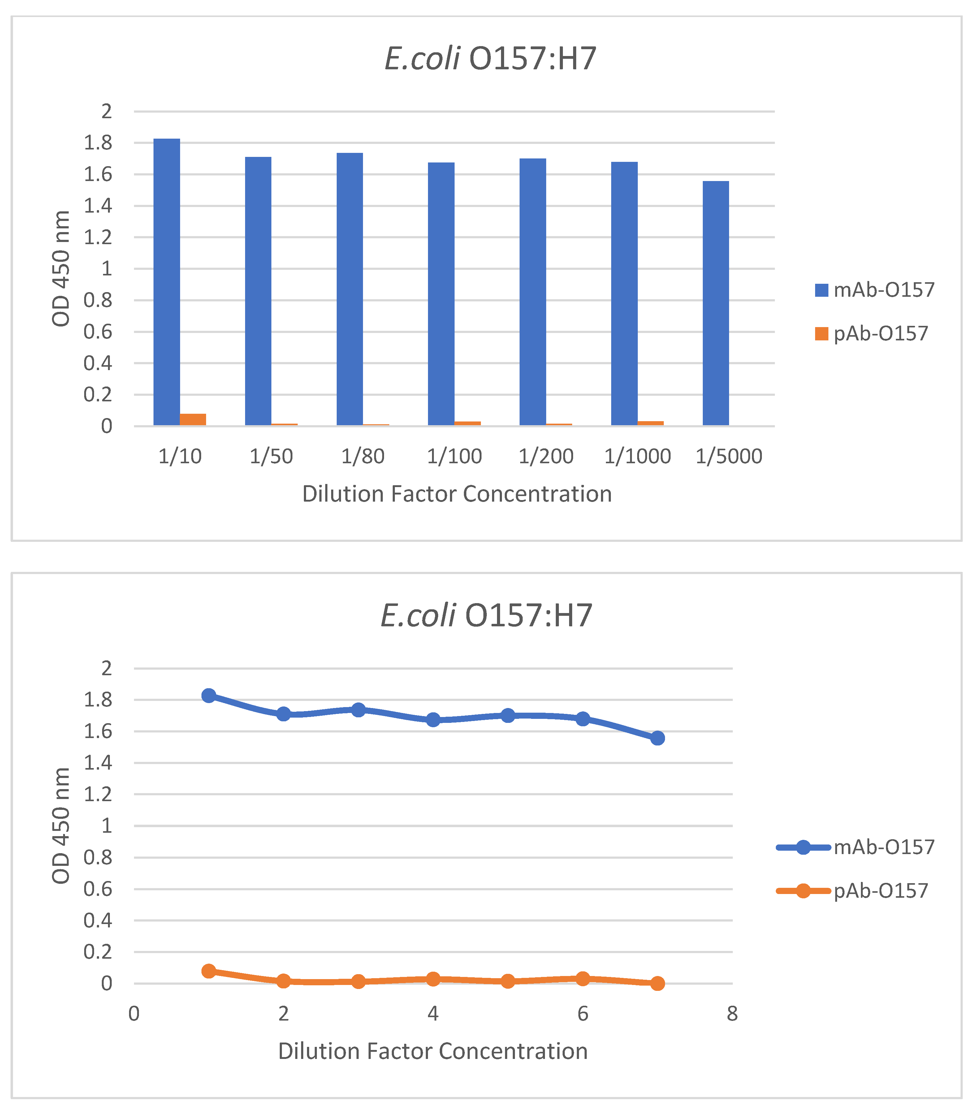
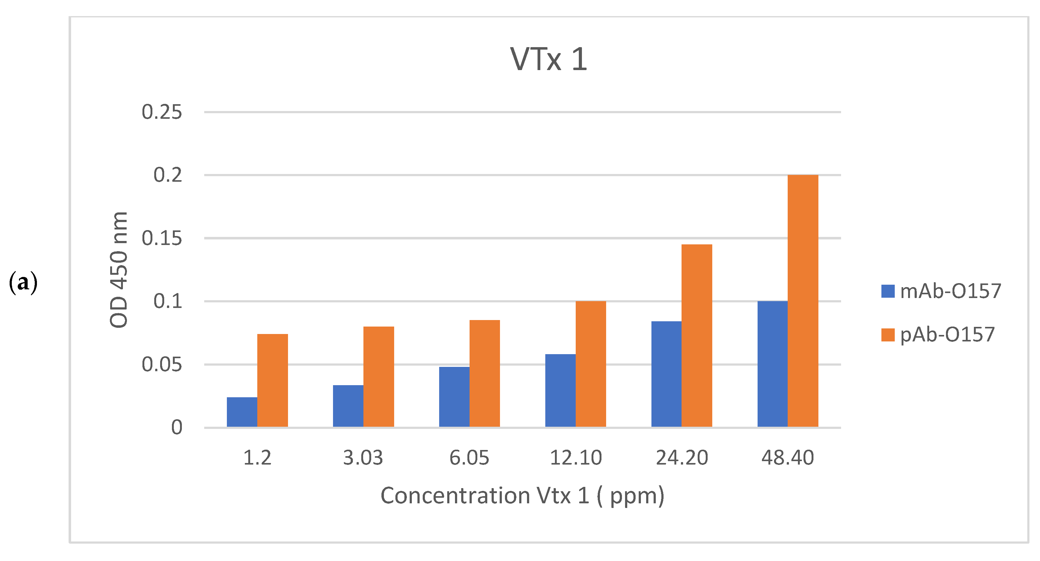
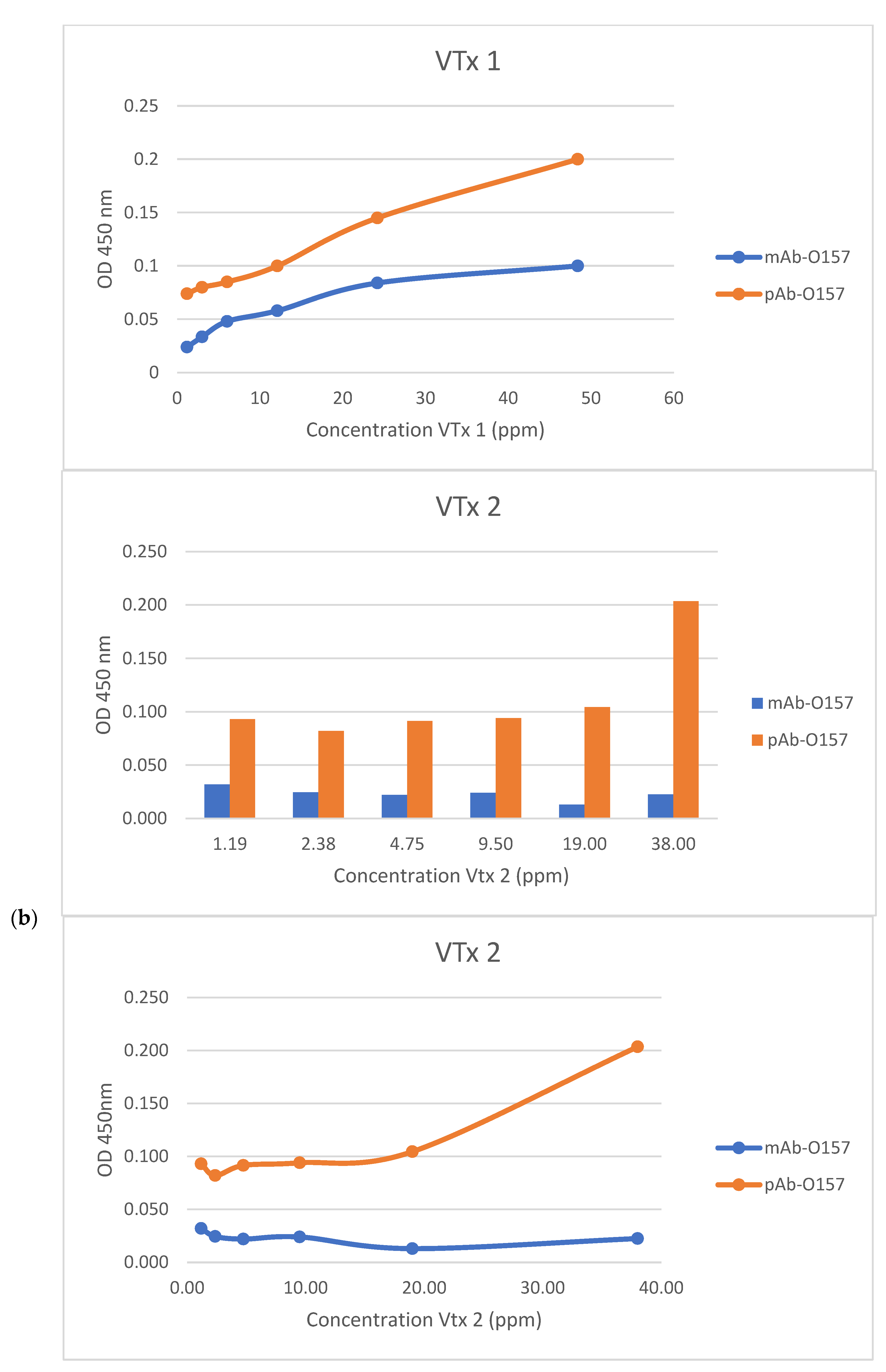
3.1. iELISA and Sensitivity Studies

The evaluation of the absorbance values obtained for each antibody against different E. coli O157:H7 antigens (obtained and described in Section 2.3 Reagents and Antibodies) is a crucial step in characterizing the binding capacity and specificity of the antibodies. The absorbance values obtained through the iELISA method typically indicate the amount of antigen–antibody complexes formed during the assay. Higher absorbance values suggest stronger binding between the antibodies and the target antigens, demonstrating specificity and sensitivity (Figure 4 and Figure 5).
Specificity and sensitivity results are typically generated by plotting the absorbance values against known analyte concentrations. Each analyte (commercial E. coli O157:H7 strain, commercial VTx, and commercial VTx2) were used with known concentrations. For E. coli O157:H7, the sensitivity and specificity in terms of absorbance versus concentration in the dilution factor allowed for quantifying the bacterial concentration in the samples based on the obtained absorbance values. The resultant curves established a comparison between the signal (OD 450 nm) and the actual concentration of E. coli O157:H7 (inactivated E. coli O157 antigen MT-28EC7U, MT-25STX, CerTest Biotec, Zaragoza, Spain) and its verotoxins VTx1 and VTx2 (E. coli O157 VT1 recombinant protein and MT-25VT2, E. coli O157 VT2 recombinant protein, CerTest Biotec, Zaragoza, Spain).
The observed results, in the case of the commercial E. coli O157:H7 strain, show that for each anti-E. coli O157:H7 monoclonal antibody there is a greater affinity for the entire concentration range used, since higher absorbance values are obtained for each range of concentrations. Similarly, for verotoxin 1 (VTx1) and verotoxin 2 (VTx2), the results of specificity and sensibility in terms of absorbance versus ppm (parts per million) provide a means to quantify the concentration of these toxins in the samples based on the absorbance measurements. For both verotoxins, the affinity of the polyclonal antibody is greater across the entire range of concentrations used, since higher absorbance values are obtained for each range of concentrations of each verotoxin.
The observed trend, where higher concentrations of the commercial E. coli O157:H7 strain and its verotoxins (VTx1 and VTx2) result in higher absorbance values, is consistent with expectations in an indirect enzyme-linked immunosorbent assay (iELISA). This behavior indicates that the monoclonal antibody and the polyclonal antibody, selected as probes for the biosensor developed, respond appropriately to varying concentrations of the target E. coli O157:H7 and its verotoxins, respectively. This evaluation of the binding capacity of each antibody and their specificity and sensitivity results are consistent with the previous bibliography, strengthening the reliability of the results [22,49,50,51,52,53,54,55].
Observing optimal results in terms of affinity at both high and low concentrations of E. coli O157:H7 and its verotoxins (VTx1 and VTx2) is a positive finding. This suggests that the selected polyclonal antibody is effective across various concentrations of verotoxins and does not saturate at high concentrations. The high binding efficiency at both low and high concentrations of the entire bacterium E. coli O157:H7 antigen is an important characteristic, indicating the versatility and sensitivity of the selected monoclonal antibody as an immunosensor probe [50].

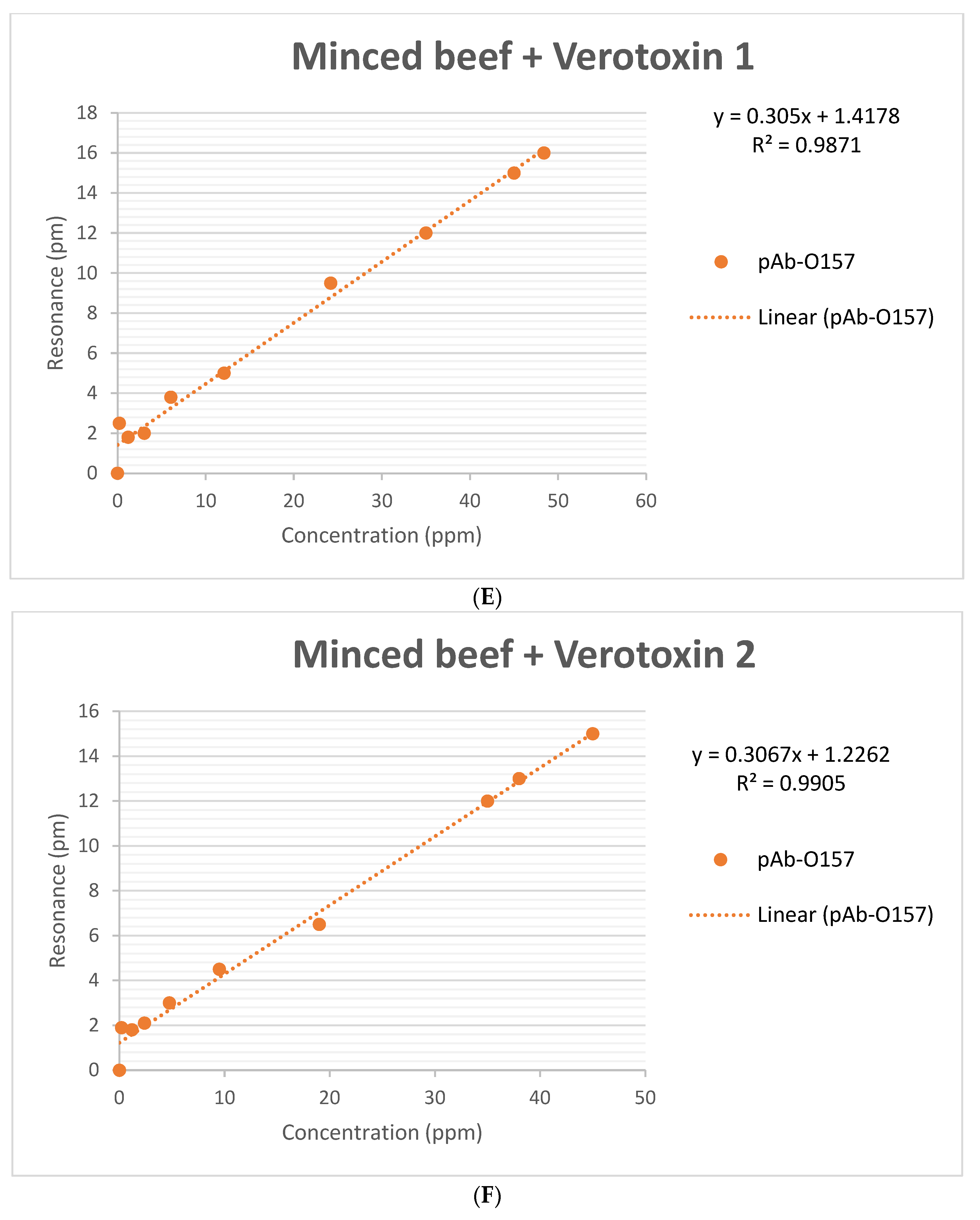
3.2. Immunosensor Specificity and Sensitivity

The detection sensitivity of the immunosensor method for E. coli O157:H7 was evaluated over a concentration range from 10 to 106 CFU/mL. This study aimed to determine the most effective enrichment method for detecting E. coli O157:H7. For this purpose, the assay was conducted using both a pure culture of the bacteria and food samples inoculated with the bacteria. The validation process included several dilutions of Shiga-like toxins VTx1 and VTx2 with a range of concentrations for evaluation, and the assessment involved analyzing the presence or absence of the pathogen. The results of the comparison between the response detection of the immunosensor and quantification through a culture of E. coli O157:H7 (CFU/mL) (Table S1, Supplementary Materials file attached) and quantification through a chemical concentration of verotoxins (ppm) (Table S2, Supplementary Materials file attached) were compared with those obtained following gold standards.
A low p-value suggests that the observed results are unlikely to be due to random chance, strengthening the confidence in the effectiveness of the biosensor for detecting E. coli O157:H7 and its verotoxins. In this sense, the statistical significance obtained (p = 0.0026) further supports the reliability of the detection method (see Section 2.6 Materials and Methods Section and the results reflected in Tables S1 and S2, Supplementary Materials file attached).
The biosensor in development has achieved a 100% observed agreement or relative accuracy with the reference methods. This level of agreement is crucial for validating the biosensor as a reliable alternative for detecting E. coli O157:H7, especially compared to established reference methods (Figure 6).
Sensitivity is a crucial parameter in biosensor performance as it reflects the ability to correctly identify true-positive cases. This case study demonstrates the effectiveness of the biosensor method in detecting samples contaminated with the E. coli O157:H7 antigen while correctly identifying samples without the microorganism or its verotoxins (VTx1 and VTx2) (Tables S1 and S2, Supplementary Materials).
Specificity is another parameter that reflects the ability of the biosensor to accurately identify samples that do not contain the target microorganism or its associated toxins. The obtained predictive value (PPV) and negative predictive value (NPV) of 100% suggest that the biosensor method can distinguish between true negatives and true positives for the presence of E. coli O157:H7 and its verotoxins. These values confirm the accuracy and reliability of the biosensor method in detecting positive samples and correctly identifying negative samples.
The results suggest that either of the two tested methods can effectively detect E. coli O157:H7 and its verotoxins in minced beef samples, whether spiked with the bacterium strain or Shiga-like toxins. Both methods have comparable specificity levels and can accurately identify the presence of E. coli O157:H7 antigens in the sample (Tables S1 and S2, Supplementary Materials).
This method presents reproducibility based on the specific binding of E. coli O157:H7 antigens observed in 97.6% of the contaminated samples. This value suggests a 97.6% probability of obtaining the same detection result when analyzing identical samples under standard reproducibility conditions at different times (Tables S1 and S2, Supplementary Materials). This is a valuable characteristic, as it indicates that the methods can consistently produce accurate results across multiple analyses and under varying conditions.
The generation of calibration curves is crucial in assessing the immunosensor method’s potential for providing a quantitative response. Calibration curves relate the measured parameter—in this case, the resonance in picometers (pm)—to known concentrations of E. coli O157:H7 and its verotoxins (VTx1 and VTx2). This allows for establishing a relationship that can be used to quantify the concentration of the target analyte in unknown samples. This is essential for enumerating E. coli O157:H7 on food safety assessments to confirm the acceptability of a food product [8].
The sensitivity of the biosensor to different concentrations of E. coli O157:H7 and its verotoxins is reinforced by the dependent relationship between the observed concentration of the bacterium or its verotoxin versus the optical signal it generates. As observed in the data presented in Figure 6, the optical signals (resonance notch shifts) are stronger for samples with higher concentrations and weaker for more diluted samples, which is consistent with the biosensing principles. Figure 6, illustrating the micro-ring resonance notch shift in picometers (pm) during the E. coli O157:H7 experiment with different dilution factors and concentrations, provides valuable information about the biosensor’s performance.
In a quantitative analysis, the working interval refers to a range of values for which the test method exhibits adequate precision, trueness, and linearity. By examining the six curves (Figure 6) presented graphically, establishing the working interval for each calibration curve facilitated the estimation of key parameters. A reliable working interval ensures that the method performs accurately across a specified concentration range of 10 to 106 CFU/mL for the entire bacterium E. coli O157:H7 and across a range of 1 to 50 ppm for its verotoxins.
The LoD is a critical parameter that indicates the lowest concentration of the target analyte that the biosensor can reliably detect. A notable obtained result is achieving a 100% detection rate for samples containing as few as 10 CFU/mL of E. coli O157:H7 or 1 ppm of VTx1 or VTx2. This indicates that the biosensor method can reliably detect low concentrations of E. coli O157:H7 and its verotoxins (Figure 6).
The results of the detection of spiked samples with dilution factors up to 10 CFU/mL and concentrations up to 1 ppm/mL indicate that the biosensor has an LoD of 10 CFU/mL for the detection E. coli O157:H7 and an LoD of 1 ppm/mL for the detection of its verotoxins (Figure 6A–C).
To calculate the Limit of Detection (LoD), the formula LoD = 3.3 * s0 was applied. To calculate the Limit of Quantification (LoQ), the formula LoQ = 10 * s0 was used. The Upper Limit of Quantification (ULoQ) was determined based on the specific requirements of the analysis. The threshold spread (s0) for the detection method was estimated by performing at least six determinations of samples at the calculated breakpoint concentration. The CFU/mL of E. coli O157:H7 and the ppm of VTx1 and VTx2 in the enrichment cultures were obtained from Tables S1 and S2, respectively. The unit of measurement used for resonance was picometers (pm), and these data were obtained from a laboratory setup reader after processing the spiked samples, as detailed in Section 2.
The lower limit of quantification (LoQ) represents the lowest concentration of the analyte (E. coli O157:H7 and verotoxins) that can be reliably quantified with acceptable precision and accuracy. In this study, the LoQ was approximately 100 CFU/mL for the bacteria and two ppm for verotoxin 1 and verotoxin 2. This indicates that the biosensor method can reliably quantify concentrations above this threshold. The limit of detection (LoD) is the lowest concentration of the analyte that can be reliably detected, although not necessarily quantified, by the method. In this study, the LoD was positioned at around 10 CFU/mL for pathogen detection and 1 ppm for verotoxins detection. This signifies the method’s sensitivity in detecting even lower concentrations, albeit without precise quantification. The upper limit of quantification (ULoQ) represents the highest concentration of the analyte that can be reliably quantified without introducing significant measurement errors. The ULoQ was observed to be approximately 106 CFU/mL for the bacteria and 48 ppm for verotoxin 1 and verotoxin 2. This boundary indicates the upper range of concentrations within which the biosensor method can provide an accurate and reliable quantification.
This immunosensor method offers practical advantages related to the working interval, the limit of detection (LoD), and storage life that make it a promising alternative for detecting E. coli O157:H7 compared to other immunosensors that have already been developed (Table 1). The working intervals of technologies based on immunosensors with electrochemical impedance spectroscopy [56] or cyclic voltammetry [57] cover a wide linear range. However, the biosensor under development extends working interval further, as quartz crystal microbalance [58].
The limit of detection (LoD) for the sensor under development compared with the LoD of other immunosensor technologies is the lowest (Table 2). Another advantage that systems based on immunosensors confer is their storage life capacity, assuming competitive advantages for this biosensor which are comparable to and improved with respect to cyclic voltammetry, electrochemical impedance spectroscopy, quartz crystal microbalance, and, indeed, surface plasmon resonance [59].
The results suggesting that the developed immunosensor is equally effective in detecting E. coli O157:H7 and its verotoxins and that it demonstrates efficiency comparable to quantitative polymerase chain reaction (qPCR) are significant findings [60,61,62]. This method eliminates the need for complex bacterial processing steps, such as lysis buffer and DNA purification kits. This simplification suggests that the immunosensor may streamline the analytical process, potentially reducing the time and resources required for sample preparation (Table 2). The use of specific antibodies in the immunosensor method is advantageous in terms of specificity [63]. This specificity reduces the likelihood of false positives or non-specific detections, which can challenge PCR methods.
The mention of label-free detection in the immunosensor method implies that it does not require additional labeling or modification of the target analyte for detection, which can simplify the assay and reduce potential sources of variability.
Among the novel techniques developed, the lateral flow test stands out, which seems to be a solution in relation to test time and the simultaneous detection of verotoxins and the rapid detection of E. coli O157. However, the sensitivity of the traditional colloidal gold immunochromatographic test strip (CG-ICTS) method is very low (105 CFU/mL), and its application is limited with respect to this photonic immunosensor [64]. Although the limit of detection (LoD) of many modified test strips has decreased slightly, this has meant that they have become more expensive due to the reagents used in their development [65,66].).
The ELISA assay is one of the most used methods for the detection of different antigens of E. coli O157 [67,68]. Among the practical advantages, it should be noted that the results are obtained in 3–5 h, and it does not require complex infrastructure. However, the results obtained by ELISA assume sensitivities of ng/mL, lower than those obtained by the sensor under development for E. coli O157:H7 verotoxins, whose LoD is around 1 ppm (Figure 6B,C).
Few portable and lab-on-a-chip electrochemical biosensors have been manufactured for the determination of E. coli [69]. These biosensors, most of them potentiometric, are low-cost, small, and highly sensitive and selective sensors, but they still require longer detection times than the immunosensor developed in this work (Table 2).
Many detection immunosensors have been developed to detect E. coli O157:H7, including GeneChip [70], genosensor [71], the surface plasmon resonance (SPR)-based assay [72,73], and the infrared spectrometer-based assay by Fourier transform (FTIR) [74]. Although these methods combine some attractive features to achieve high sensitivity, high performance, or rapid detection, all of them require more expensive instruments than the sensor under development and trained technical personnel.
A multiplex ring system in biosensor development offers the advantage of the simultaneous, comprehensive, and efficient detection of the E. coli O157:H7 pathogen and its verotoxins [75,76]. This capability of the developed biosensor to detect multiple antigens simultaneously, without the need for separate analyses, represents a significant advancement and a departure from traditional systems like PCR and ELISA, where each antigen of E. coli O157:H7 usually has to be analyzed separately [55,75].
The main advantage of the coupled microfluidic system is its ability to allow the simultaneous measurement of two samples, which is achieved by distributing eight annular resonators in two channels, each of which contains four resonant rings. The distribution of resonant rings across multiple channels allows for parallel processing, reducing the time required for analysis. The system’s capability to achieve sensitivities down to a ng/mL scale with response times less than 30 min contributes to the efficiency of the detection process.

4. Conclusions

This study underscores the potential of a novel photonic biosensor as a valuable and reliable tool for detecting E. coli O157:H7 in food samples. Its sensitivity, specificity, and accuracy confirm it as a promising technology with applications in the food industry, contributing to enhancing food safety measures.
The biosensor’s fabrication process involved silicon nitride and employed CMOS-compatible techniques. The immunosensor’s surface consisted of eight ring resonators, and these resonators were functionalized with specific antibodies selected for detecting E. coli O157:H7 antigens in meat matrices.
In the preliminary validation of the biosensor, 100 fabricated PICs (photonic-integrated circuits) were used. The rings on these PICs were functionalized with specific antibodies, and a microfluidic layer was attached to facilitate controlled sample flow. The biosensor demonstrated the successful detection of various concentrations of bacteria and verotoxins. This initial validation highlights the promising performance, reliability, and feasibility of the innovative detection technique based on the photonic biosensor.
The selection of specific antibodies, including a monoclonal antibody with heightened specificity at lower antigen concentrations and a polyclonal antibody with a higher binding, reflects its capacity across a broader concentration range. It is important to note that no significant results were observed at very high antigen concentrations. This nuanced observation underscores both the potential and limitations of the method in different analytical contexts, providing valuable insights into the performance of the chosen antibodies under varying conditions.
This alternative approach demonstrates a rapid diagnostic capacity and provides results in four hours, allowing for prompt action in response to potential food microbiological contamination. The biosensor’s ability to examine a significant number of samples makes it a valuable tool for industries involved in food production and processing.

Supplementary Materials

The following supporting information can be downloaded at https://www.mdpi.com/article/10.3390/photonics11040374/s1: Table S1: Detection inmunosensor results againts E. coli O157:H7; and Table S2: Detection inmunonsensor results of verotoxins 1 and verotoxin 2 of E. coli O157:H7.

Author Contributions

Conceptualization, A.F., J.G.-H., M.H. and Y.M.; methodology, A.F., J.G.-H., M.H. and Y.M.; validation, A.F., J.G.-H., M.H. and Y.M.; investigation, A.F., J.G.-H., M.H. and Y.M.; data curation, A.F., J.G.-H., M.H. and Y.M.; writing—original draft preparation, A.F.; writing—review and editing, J.G.-H., M.H. and Y.M.; funding acquisition, A.F., J.G.-H., M.H. and Y.M. All authors have read and agreed to the published version of the manuscript.

Funding

This research was financially supported by the AVI (Valencian Innovation Agency) and received funding from the European Union through FEDER funds. The project, identified as INNEST/2021/338, is known as the “BACTERIO project”, which focuses on “Integrated Photonics for integral microbiological control in the agri-food sector”.

Institutional Review Board Statement

Not applicable.

Informed Consent Statement

Not applicable.

Data Availability Statement

Data are contained within the article and Supplementary Materials.

Conflicts of Interest

Ana Fernández was employed by the company Lumensia Sensors S.L. The remaining authors declare that the research was conducted in the absence of any commercial or financial relationships that could be construed as a potential conflict of interest.

Sample Availability

Samples of the compounds are available from the authors.

References

  1. Zhu, P.; Shelton, D.R.; Li, S.; Adams, D.L.; Karns, J.S.; Amstutz, P.; Tang, C.M. Detection of E. coli O157:H7 by immunomagnetic separation coupled with fluorescence immunoassay. Biosens. Bioelectron. 2011, 30, 337–341. [Google Scholar] [CrossRef] [PubMed]
  2. Laster, B.A.; Harris, K.B.; Lucia, L.M.; Castillo, A.; Savell, J.W. Efficacy of trimming chilled beef during fabrication to control Escherichia coli O157:H7 surrogates on subsequent subprimals. Meat Sci. 2012, 90, 420–425. [Google Scholar] [CrossRef] [PubMed]
  3. Hassan, A.-R.H.A.-A.; de la Escosura-Muñiz, A.; Merkoçi, A. Highly sensitive and rapid determination of Escherichia coli O157:H7 in minced beef and water using electrocatalytic gold nanoparticle tags. Biosens. Bioelectron. 2015, 67, 511–515. [Google Scholar] [CrossRef] [PubMed]
  4. Chan, K.Y.; Ye, W.W.; Zhang, Y.; Xiao, L.D.; Leung, P.H.; Li, Y.; Yang, M. Ultrasensitive detection of E. coli O157:H7 with biofunctional magnetic bead concentration via nanoporous membrane based electrochemical immunosensor. Biosens. Bioelectron. 2013, 41, 532–537. [Google Scholar] [CrossRef] [PubMed]
  5. Food and Agriculture Organization of United Nations (FAO); World Health Organization (WHO). Shiga Toxin-Producing Escherichia coli (STEC) and Food: Attribution, Characterization, and Monitoring; WHO: Geneva, Switzerland; FAO: Rome, Italy, 2018; ISBN 978-92-5-130682-6 (FAO). ISBN 978-92-4-151427-9 (WHO). [Google Scholar]
  6. Scientific Committee of the Spanish Agency for Food Safety and Nutrition (AESAN). Preventative Measures and Applicable Recommendations for Avoiding Possible Food-Borne Infections Caused by Strains of Verotoxigenic/Shiga Toxin-Producing/Enterohemorrhagic Escherichia coli (VTEC/STEC/EHEC); AESAN-2012-007; AESAN: Majadahonda, Spain, 2012. [Google Scholar]
  7. Wu, W.; Zhao, S.; Mao, Y.; Fang, Z.; Lu, X.; Zeng, L. A sensitive lateral flow biosensor for Escherichia coli O157:H7 detection based on aptamer mediated strand displacement amplification. Anal. Chim. Acta 2015, 861, 62–68. [Google Scholar] [CrossRef] [PubMed]
  8. Commission Regulation (EC). No 2073/2005 of 15 November 2005 on microbiological criteria for foodstuffs. Off. J. Eur. Union. 2005, L338, 1–26. [Google Scholar]
  9. Carvalho, F.J.G.; Mohammed Ahmed Sheikh, H.; Selvin, R. Advances in screening, detection and enumeration of Escherichia coli using nanotechnology-based methods: A review. J. Biomed. Nanotechnol. 2018, 14, 829–846. [Google Scholar] [CrossRef] [PubMed]
  10. Umesha, S.; Manukumar, Y.H. Advanced molecular diagnostic techniques for detection of food-borne pathogens: Current applications and future challenges. Crit. Rev. Food Sci. Nutr. 2018, 58, 84–104. [Google Scholar] [CrossRef] [PubMed]
  11. Luo, C.; Tang, H.; Cheng, W.; Yan, L.; Zhang, D.; Ju, H.; Ding, S. A sensitive electrochemical DNA biosensor for specific detection of Enterobacteriaceae bacteria by exonuclease III-assisted signal amplification. Biosens. Bioelectron. 2013, 48, 132–137. [Google Scholar] [CrossRef]
  12. Huertas-Caro, C.; Urbano-Cáceres, E.; Torres-Caycedo, Y.M. Diagnóstico molecular una alternativa para la detección de patógenos en alimentos. Rev. Habanera Cienc. Médicas 2019, 18, 513–528. [Google Scholar]
  13. Fratamico, P.M.; Bhunia, A.K.; Smith, J.A. Foodborne Pathogens: Microbiology and Molecular Biology; CRC Press LLC eBooks: Boca Raton, FL, USA, 2005. [Google Scholar]
  14. Li, Y.; Mustapha, A. Simultaneous Detection of Escherichia coli O157:H7, Salmonella, and Shigella in Apple Cider and Produce by a Multiplex PCR. J. Food Prot. 2004, 67, 27–33. [Google Scholar] [CrossRef] [PubMed]
  15. Daly, P.; Collier, T.; Doyle, S. PCR-ELISA detection of Escherichia coli in milk. Lett. Appl. Microbiol. 2002, 34, 222–226. [Google Scholar] [CrossRef] [PubMed]
  16. Noble, R.T.; Weisberg, S.B. A review of technologies for rapid detection of bacteria in recreational waters. J. Water Health 2005, 3, 381–392. [Google Scholar] [CrossRef] [PubMed]
  17. Pebdeni, A.B.; Mousavizadegan, M.; Hosseini, M. Sensitive detection of S. aureus using aptamer-and vancomycin-copper nanoclusters as dual recognition strategy. Food Chem. 2021, 361, 130137. [Google Scholar] [CrossRef] [PubMed]
  18. Acharya, G.; Chang, L.C.; Savran, C. An optical biosensor for rapid and labelfree detection of cells. J. Am. Chem. Soc. 2006, 128, 3862–3863. [Google Scholar] [CrossRef] [PubMed]
  19. Hornbeck, P. Double-Immunodiffusion Assay for Detecting Specific Antibodies. Curr. Protoc. Immunol. 2017, 116, 2–3. [Google Scholar] [CrossRef] [PubMed]
  20. Ngo, T.T. Immunoassay. Curr. Opin. Biotechnol. 1991, 2, 102–109. [Google Scholar] [CrossRef]
  21. Plested, J.S.; Coull, P.A.; Gidney, M.A.J. ELISA. Methods Mol. Med. 2003, 71, 243–261. [Google Scholar] [PubMed]
  22. Zhang, X.; Li, M.; Zhang, B.; Chen, K.; He, K. Development of a Sandwich ELISA for EHEC O157:H7 Intimin γ1. PLoS ONE 2016, 11, e0162274. [Google Scholar] [CrossRef]
  23. Sajid, M.; Kawde, A.; Daud, M. Designs, formats and applications of lateral flow assay: A literature review. J. Saudi Chem. Soc. 2015, 19, 689–705. [Google Scholar] [CrossRef]
  24. Posthuma-Trumpie, G.A.; Korf, J.; Van Amerongen, A. Lateral flow (immuno)assay: Its strengths, weaknesses, opportunities and threats. A literature survey. Anal. Bioanal. Chem. 2009, 393, 569–582. [Google Scholar] [CrossRef] [PubMed]
  25. Singh, J.P.; Sharma, S.; Nara, S. Evaluation of gold nanoparticle based lateral flow assays for diagnosis of enterobacteriaceae members in food and water. Food Chem. 2015, 170, 470–483. [Google Scholar] [CrossRef] [PubMed]
  26. Watts, H.J.; Lowe, R.C.; Pollard-Knight, D.V. Optical biosensor for monitoring microbial cells. Anal. Chem. 1994, 66, 2465–2470. [Google Scholar] [CrossRef] [PubMed]
  27. Riu, J.; Giussani, B. Electrochemical biosensors for the detection of pathogenic bacteria in food. Trends Anal. Chem. 2020, 126, 115863. [Google Scholar] [CrossRef]
  28. Ye, Y.; Guo, H.; Sun, X. Recent progress on cell-based biosensors for analysis of food safety and quality control. Biosens. Bioelectron. 2019, 126, 389–404. [Google Scholar] [CrossRef] [PubMed]
  29. Velusamy, V.; Arshak, K.; Korostynska, O.; Oliwa, K.; Adley, C.C. An overview of foodborne pathogen detection: In the perspective of biosensors. Biotechnol. Adv. 2010, 28, 232–254. [Google Scholar] [CrossRef] [PubMed]
  30. González-Guerrero, A.B.; Dante, S.; Duval, D.; Osmond, J.; Lechuga, L.M. Advanced photonic biosensors for point-of-care diagnostics. Procedia Eng. 2011, 25, 71–75. [Google Scholar] [CrossRef]
  31. Shrivastava, A.; Gupta, V.B. Methods for the determination of limit of detection and limit of quantitation of the analytical methods. Chron. Young Sci. 2011, 2, 21. [Google Scholar] [CrossRef]
  32. Fan, X.; White, I.M.; Shopova, S.I.; Zhu, H.; Suter, J.D.; Sun, Y. Sensitive optical biosensors for unlabeled targets: A review. Anal. Chim. Acta 2008, 620, 8–26. [Google Scholar] [CrossRef]
  33. De Vos, K.; Bartolozzi, I.; Schacht, E.; Bienstman, P.; Baets, R. Silicon-on-Insulator microring resonator for sensitive and label-free biosensing. Opt. Express 2007, 15, 7610–7615. [Google Scholar] [CrossRef]
  34. Narsaiah, K.; Jha, S.N.; Bhardwaj, R.; Sharma, R.; Kumar, R. Optical biosensors for food quality and safety assurance—A review. J. Food Sci. Technol. 2012, 49, 383–406. [Google Scholar] [CrossRef] [PubMed]
  35. Donagh, C.M.; Burke, C.S.; Crai BD, M. Optical biosensors. Chem. Rev. 2008, 108, 422–461. [Google Scholar] [CrossRef]
  36. Pebdeni, A.B.; Hosseini, M.; Ganjali, M.R. Fluorescent turn-on aptasensor of Staphylococcus aureus based on the FRET between green carbon quantum dot and gold nanoparticle. Food Anal. Methods 2020, 13, 2070–2079. [Google Scholar] [CrossRef]
  37. Lazcka, O.; Del Campo, F.J.; Munoz, F.X. Pathogen detection: A perspective of traditional methods and biosensors. Biosens. Bioelectron. 2007, 22, 1205–1217. [Google Scholar] [CrossRef] [PubMed]
  38. Nurliyana, M.R.; Wibowo, K.M.; Muslihati, A.; Saim, H.; Ahmad, S.A.; Sari, Y.; Mansor, Z. The detection method of Escherichia coli in water resources: A review. IOP Conf. Ser. J. Phys. Conf. Ser. 2017, 995, 012065. [Google Scholar] [CrossRef]
  39. Ionescu, R.E. Biosensor platforms for rapid detection of E. coli bacteria. In Escherichia coli–Recent Advances on Physiology, Pathogenesis and Biotechnological Applications; IntechOpen: London, UK, 2017. [Google Scholar] [CrossRef]
  40. Bange, A.; Halsall, H.B.; Heineman, W.R. Microfluidic immunosensor systems. Biosens. Bioelectron. 2005, 20, 2488–2503. [Google Scholar] [CrossRef] [PubMed]
  41. Luka, G.; Ahmadi, A.; Najjaran, H.; Alocilja, E.; DeRosa, M.; Wolthers, K.; Malki, A.; Aziz, H.; Althani, A.; Hoorfar, M. Microfluidics integrated biosensors: A leading technology towards lab-on-a-chip and sensing applications. Sensors 2015, 15, 30011–30031. [Google Scholar] [CrossRef] [PubMed]
  42. Kant, K.; Shahbazi, M.A.; Dave, V.P.; Ngo, T.A.; Chidambara, V.A.; Than, L.Q.; Bang, D.D.; Wolff, A. Microfluidic devices for sample preparation and rapid detection of foodborne pathogens. Biotechnol. Adv. 2018, 36, 1003–1024. [Google Scholar] [CrossRef]
  43. ISO 16654:2001/Amd 1:2017; Microbiology of food and animal feeding stuffs. Horizontal method for the detection of Escherichia coli O157. Amendment 1: Annex B: Result of interlaboratory studies. International Organization for Standardization: Geneva, Switzerland, 2017.
  44. Fernández Blanco, A.; Hernández Pérez, M.; Moreno Trigos, Y.; García-Hernández, J. Development of Optical Label-Free Biosensor Method in Detection of Listeria monocytogenes from Food. Sensors 2023, 23, 5570. [Google Scholar] [CrossRef]
  45. Ashkenazi, S.; Cleary, T.G. Rapid method to detect Shiga toxin and Shiga-like toxin I based on binding to globotriosyl ceramide (Gb3), their natural receptor. J. Clin. Microbiol. 1989, 27, 1145–1150. [Google Scholar] [CrossRef]
  46. Hegde, N.V.; Hulet, R.; Jayarao, B.M.; Muldoon, M.; Lindpaintner, K.; Kapur, V.; DebRoy, C. Detection of the top six non-O157 Shiga toxin-producing Escherichia coli O groups by ELISA. Foodborne Pathog. Dis. 2012, 9, 1044–1048. [Google Scholar] [CrossRef] [PubMed]
  47. Guttikonda, S.; Wang, W.W.; Suresh, M.R. Molecular zipper assays: A simple homosandwich with the sensitivity of pcr. J. Pharm. Pharm. Sci. 2004, 7, 7–16. [Google Scholar] [PubMed]
  48. Vytrasova, J.; Zachová, I.; Cervenka, L.; Štepanková, J.; Pejchalová, M. Non-specific reactions during immunomagnetic sepa-ration of Listeria. Food Technol. Biotechnol. 2005, 43, 397–401. [Google Scholar]
  49. Kehl, K.S.; Havens, P.; Behnke, C.E.; Acheson, D.W. Evaluation of the premier EHEC assay for detection of Shiga toxin-producing Escherichia coli. J. Clin. Microbiol. 1997, 35, 2051–2054. [Google Scholar] [CrossRef] [PubMed]
  50. Teel, L.D.; Daly, J.A.; Jerris, R.C.; Maul, D.; Svanas, G.; O’Brien, A.D.; Park, C.H. Rapid detection of Shiga toxin-producing Escherichia coli by optical immunoassay. J. Clin. Microbiol. 2007, 45, 3377–3380. [Google Scholar] [CrossRef] [PubMed]
  51. Skinner, C.; Patfield, S.; Stanker, L.H.; Fratamico, P.; He, X. New high-affinity 4 monoclonal antibodies against Shiga toxin 1 facilitate the detection of hybrid Stx1/Stx2 in vivo. PLoS ONE 2014, 9, e99854. [Google Scholar] [CrossRef] [PubMed]
  52. He, X.; McMahon, S.; Skinner, C.; Merrill, P.; Scotcher, M.C.; Stanker, L.H. Development and characterization of monoclonal antibodies against Shiga toxin 2 and their application for toxin detection in milk. J. Immunol. Methods 2013, 389, 18–28. [Google Scholar] [CrossRef] [PubMed]
  53. Ching, K.H. Lateral Flow Immunoassay. Methods Mol. Biol. 2015, 1318, 127–137. [Google Scholar]
  54. Chen, M.; Yu, Z.; Liu, D.; Peng, T.; Liu, K.; Wang, S.; Xiong, Y.; Wei, H.; Xu, H.; Lai, W. Dual gold nanoparticle late flow immunoassay for sensitive detection of Escherichia coli O157:H7. Anal. Chim. Acta 2015, 876, 71–76. [Google Scholar] [CrossRef]
  55. Ching, K.H.; He, X.; Stanker, L.H.; Lin, A.V.; McGarvey, J.A.; Hnasko, R. Detection of shiga toxins by lateral flow assay. Toxins 2015, 7, 1163–1173. [Google Scholar] [CrossRef]
  56. Radke, S.M.; Alocilja, E.C. A high density microelectrode array biosensor for detection of E. coli O157:H7. Biosens. Bioelectron. 2005, 20, 1662–1667. [Google Scholar] [CrossRef] [PubMed]
  57. Zhao, G.Y.; Xing, F.F.; Deng, S.P. A disposable amperometric enzyme immunosensor for rapid detection of Vibrio parahaemolyticus in food based on agarose/nano–Au membrane and screen-printed electrode. Electrochem. Commun. 2007, 9, 1263–1268. [Google Scholar] [CrossRef]
  58. Su, X.L.; Li, Y.B. A self-assembled monolayer-based piezoelectric immunosensor for rapid detection of Escherichia coli O157:H7. Biosens. Bioelectron. 2004, 19, 563–574. [Google Scholar] [CrossRef] [PubMed]
  59. Waswaa, J.; Irudayarajb, J.; DebRoy, C. Direct detection of E. coli O157:H7 in selected food systems by a surface plasmon resonance biosensor. LWT 2007, 40, 187–192. [Google Scholar] [CrossRef]
  60. Li, D.J.; Feng, Y.Y.; Zhou, L.; Ye, Z.Z.; Wang, J.P.; Ying, Y.B.; Ruan, C.M.; Wang, R.H.; Li, Y.B. Label-free capacitive immunosensor based on quartz crystal Au electrode for rapid and sensitive detection of Escherichia coli O157:H7. Anal. Chim. Acta 2011, 687, 89–96. [Google Scholar] [CrossRef] [PubMed]
  61. Li, B.; Chen, J.-Q. Real-Time PCR Methodology for Selective Detection of Viable Escherichia coli O157:H7 Cells by Targeting Z3276 as a Genetic Marker. Appl. Environ. Microbiol. 2012, 78, 5297–5304. [Google Scholar] [CrossRef] [PubMed]
  62. Li, L.; Zhang, X.H.; He, K.W.; Jiang, P.; Zhao, P.; Ye, Q.; Luan, X.; Wen, L.; Ni, Y.; Zhou, J.; et al. Duplex PCR Procedure for the Detection of EHEC O157:H7. Acta Agric. Boreali-Sin. 2011, 26, 58–66. [Google Scholar] [CrossRef]
  63. Ibekwe, A.M.; Grieve, C.M. Detection and quantification of Escherichia coli O157:H7 in environmental samples by real-time PCR. J. Appl. Microbiol. 2003, 94, 421–431. [Google Scholar] [CrossRef] [PubMed]
  64. Xie, Q.Y.; Wu, Y.H.; Xiong, Q.R.; Xu, H.Y.; Xiong, Y.H.; Liu, K.; Jin, Y.; Lai, W.H. Advantages of fluorescent microspheres compared with colloidal gold as a label in immunochromatographic lateral flow assays. Biosens. Bioelectron. 2014, 54, 262–265. [Google Scholar] [CrossRef]
  65. Shan, S.; Lai, W.; Xiong, Y.; Wei, H.; Xu, H. Novel strategies to enhance lateral flow immunoassay sensitivity for detecting foodborne pathogens. Agric. Food Chem. 2015, 63, 745–753. [Google Scholar] [CrossRef]
  66. Bruno, J.G. Application of DNA aptamers and quantum dots to lateral flow test strips for detection of foodborne pathogens with improved sensitivity versus colloidal gold. Pathogens 2014, 3, 341–355. [Google Scholar] [CrossRef] [PubMed]
  67. Wang, C.; Xing, K.; Zhang, G.; Yuan, M.; Xu, S.; Liu, D.; Chen, W.; Peng, J.; Hu, S.; Lai, W.-H. Novel ELISA based on fluorescent quenching of DNA-stabilized silver nanoclusters for detecting E. coli O157:H7. Food Chem. 2019, 281, 91–96. [Google Scholar] [CrossRef] [PubMed]
  68. Pang, B.; Zhao, C.; Li, L.; Song, X.; Xu, K.; Wang, J.; Liu, Y.; Fu, K.; Bao, H.; Song, D.; et al. Development of a low-cost paper-based ELISA method for rapid Escherichia coli O157:H7 detection. Anal. Biochem. 2018, 542, 58–62. [Google Scholar] [CrossRef] [PubMed]
  69. Perumal, V.; Hashim, U. Advances in biosensors: Principle, architecture and applications. J. Appl. Biomed. 2014, 12, 1–15. [Google Scholar] [CrossRef]
  70. Kim, H.; Kane, M.D.; Kim, S.; Dominguez, W.; Applegate, B.M.; Savikhin, S. A molecular beacon DNA microarray system for rapid detection of E. coli O157:H7 that eliminates the risk of a false negative signal. Biosens. Bioelectron. 2007, 22, 1041–1047. [Google Scholar] [CrossRef]
  71. Abdalhai, M.H.; Fernandes, A.M.; Xia, X.; Musa, A.; Ji, J.; Sun, X. Electrochemical genosensor to detect pathogenic bacteria (Escherichia coli O157:H7) as applied in real food samples (fresh beef) to improve food safety and quality control. J. Agric. Food Chem. 2015, 63, 5017–5025. [Google Scholar] [CrossRef] [PubMed]
  72. Wang, Y.; Ye, Z.; Si, C.; Ying, Y. Monitoring of Escherichia coli O157:H7 in food samples using lectin based surface plasmon resonance biosensor. Food Chem. 2013, 136, 1303–1308. [Google Scholar] [CrossRef]
  73. Vaisocherova-Lisalova, H.; Visova, I.; Ermini, M.L.; Springer, T.; Song, X.C.; Mrazek, J.; Lamacova, J.; Scott Lynn, N., Jr.; Sedivak, P.; Homola, J. Low-fouling surface plasmon resonance biosensor for multi-step detection of foodborne bacterial pathogens in complex food samples. Biosens. Bioelectron. 2016, 80, 84–90. [Google Scholar] [CrossRef]
  74. Chen, J.; Shi, X.; Gehring, A.G.; Paoli, G.C. Automated immunomagnetic separation for the detection of Escherichia coli O157:H7 from spinach. Int. J. Food Microbiol. 2014, 179, 33–37. [Google Scholar] [CrossRef]
  75. Rani, A.; Ravindran, V.B.; Surapaneni, A.; Shahsavari, E.; Haleyur, N.; Mantri, N.; Ball, A.S. Evaluation and comparison of recombinase polymerase amplification coupled with lateral-flow bioassay for Escherichia coli O157:H7 detection using different genes. Sci. Rep. 2021, 11, 1881. [Google Scholar] [CrossRef]
  76. Hu, J.; Wang, Y.; Su, H.; Ding, H.; Sun, X.; Gao, H.; Geng, Y.; Wang, Z. Rapid analysis of Escherichia coli O157:H7 using isothermal recombinase polymerase amplification combined with triple-labeled nucleotide probes. Mol. Cell. Probes 2020, 50, 101501. [Google Scholar] [CrossRef] [PubMed]
Figure 1. Graphic representation of PIC functionalization process ([46]).
Photonics 11 00374 g001
Figure 2. PIC ensembled into microfluidic cartridge.
Photonics 11 00374 g002
Figure 3. Biosensor sensogram detection of E. coli O157:H7 or its verotoxins. A schematic sensogram obtained by the immunosensor setup reader against the target antigen is included. Here are represented the resonance obtained versus the test time. The hardware and software components of the setup reader translate the optical signals obtained in the detection sensogram into measurable resonance values: freq. diff (pm) against the concentration of the target antigen (log CFU/mL).
Photonics 11 00374 g003
Figure 4. Specificity and sensitivity iELISA immunoassay results against E. coli O157:H7. Absorbance values (OD 450 nm) of monoclonal and polyclonal immunosensor antibodies versus bacterial concentration in the dilution factor of a commercial inactivated antigen of E. coli O157:H7.
Photonics 11 00374 g004
Figure 5. Specificity and sensitivity iELISA immunoassay results against E. coli O157:H7 verotoxins 1 and 2. Absorbance values (OD 450 nm) of monoclonal and polyclonal immunosensor antibodies versus verotoxin concentration in ppm. (a) iELISA antibody absorbances values increasing dilutions of commercial VTx1; (b) iELISA antibody absorbance values increasing dilutions of commercial VTx2.
Photonics 11 00374 g005aPhotonics 11 00374 g005b
Figure 6. Validation calibration curves against antigens of E. coli O157:H7. (A) Biosensor calibration curve against E. coli O157:H7. (B) Biosensor calibration curve against verotoxin 1 (VTx1). (C) Biosensor calibration curve against verotoxin 2 (VTx2) (D) Biosensor calibration curve against E. coli O157:H7-spiked minced beef samples. (E) Biosensor calibration curve against VTx1-spiked minced beef samples. (F) Biosensor calibration curve against VTx2-spiked minced beef samples.
Photonics 11 00374 g006aPhotonics 11 00374 g006bPhotonics 11 00374 g006c
Table 1. Detection immunosensor advantages and disadvantages related to other immunosensor methods against E. coli O157:H7.
Detection TechnologyWorking Interval (CFU/mL)Limit of Detection (CFU/mL)Storage LifeReference
Surface plasmon resonance102–1036 × 102-[59]
Electrochemical impedance spectroscopy104–107104-[56]
Electrochemical impedance spectroscopy103–1051031 week[60]
Cyclic voltammetry105–1097.374 × 1041 week[57]
Quartz crystal microbalance103–1081031 week[58]
Photonic immunosensor101–1061016 monthsThis work
Table 2. Assessment of the immunosensor under development against the different detection methods of E. coli O157:H7, depending on its speed in testing, cost, applicability, sensitivity, and specificity.
Detection TechnologySpeed in TestingCostApplicabilitySensitivitySpecificity
Plate culture24 hLowWideLowLow
Lateral flow immunoassay30–45 minModerateLimitedLowLow
Enzyme-linked Immunosorbent Assay (ELISA)3–5 hHighWideLowLow
qPCR6 hHighWideModerateModerate
Multiplex PCR6 hHighWideModerateModerate
Hybridization18–24 hModerateLimitedModerateModerate
Microarrays48–72 hHighWideHighHigh
Optical biosensors45 min–3 hHighLimitedHighModerate
Electrochemical biosensors18–24 hModerateLimitedHighModerate
Photonic immunosensor30–45 minLowWideHighHigh
Disclaimer/Publisher’s Note: The statements, opinions and data contained in all publications are solely those of the individual author(s) and contributor(s) and not of MDPI and/or the editor(s). MDPI and/or the editor(s) disclaim responsibility for any injury to people or property resulting from any ideas, methods, instructions or products referred to in the content.

