Review

Emerging Techniques for Nonlinear Optical Spectroscopy of Disordered and Highly Scattering Materials

by
Albert S. Reyna
1,
Anderson M. Amaral
2 and
Cid B. de Araújo
2,*
1
Programa de Pós-Graduação em Engenharia Física, Unidade Acadêmica do Cabo de Santo Agostinho, Universidade Federal Rural de Pernambuco, Cabo de Santo Agostinho 54518-430, PE, Brazil
2
Departamento de Física, Universidade Federal de Pernambuco, Recife 50670-901, PE, Brazil
*
Author to whom correspondence should be addressed.
Photonics 2024, 11(7), 650; https://doi.org/10.3390/photonics11070650
Submission received: 11 June 2024 / Revised: 1 July 2024 / Accepted: 3 July 2024 / Published: 11 July 2024

Abstract

:
Scattering materials have been of considerable research interest due to their unique optical properties that may enable applications throughout the area of disordered photonics, particularly in fields such as Random Lasers, nonlinear (NL) microscopy in biomedical research, and optical thermometry. However, the complex structures of these materials make traditional NL spectroscopic techniques unsuitable for studies, as the materials of interest can cause large multiple scattering of light in addition to presenting spatial heterogeneities. Fortunately, new techniques, such as the Scattered Light Imaging Method (SLIM), the Intensity Correlation scan (IC-scan), and the Reflection Intensity Correlation scan (RICO-scan), have recently emerged, providing researchers with more appropriate ways to study disordered and scattering NL materials. These techniques allow for a deeper characterization of the NL optical properties of highly scattering materials, which are essential for applications in photonics, optoelectronics, and biophotonics, for example. In this paper, we discuss these innovative techniques, which can offer insights into the properties of materials of great potential for disordered photonics.

Graphical Abstract

1. Introduction

Well-established linear optical techniques have played an important role in the study of disordered materials, providing information about their structural, electronic, and optical properties. Various techniques have been largely employed, such as Raman Scattering, which enables information about the molecular structure, crystallinity, and chemical composition of the materials; Photoluminescence, which provides insights into the material’s electronic structure and energy levels; Fluorescence Correlation Spectroscopy, which is used to study the dynamics of fluorescent molecules (or ions in solids) and can be applied to investigate molecular diffusion, aggregation, and interactions; Dynamic Light Scattering, which is valuable for studying the size, shape, and interactions of particles in disordered materials; and Optical Coherence Tomography (OCT), which can be used to visualize structural features, interfaces, and defects with micrometer-scale resolution. For several decades, these techniques have provided detailed and useful information on the optical properties of colloids, polymers, glass-ceramics, crystals, and biological macromolecules by exciting the materials using light sources of small and moderate intensities.
On the other hand, it is known that nonlinear (NL) optical properties of materials are revealed when illuminated by optical sources of moderate or large intensities. Therefore, the NL characterization of disordered materials is essential for various applications, such as the operation of Random Lasers [1,2,3], studies of laser propagation in turbid colloidal media [4,5,6], and NL microscopy in biomedical research [7,8,9], among others. In many cases, performing NL measurements can be challenging because various physical systems are susceptible to bleaching, chemical reactions, or morphological changes under exposure to high-intensity lasers. Moreover, optical materials exhibit properties that change with the intensity of the incident light. This may manifest as effects such as nonlinear absorption, nonlinear refraction, and nonlinear light scattering, for example. In addition, the denser and more disordered the medium, the more significant the distortion caused by the scattering process in the spatial and temporal intensity profiles of the transmitted or reflected optical beams used to probe the samples.
While many techniques exist for measuring NL parameters of ordered optical materials, few are suitable for characterizing NL disordered and highly scattering materials and absorbers. For homogeneous samples, techniques such as Z-Scan [10,11], D4σ [11,12], wave-mixing, and harmonic generation [11,13,14,15] are widely applied to characterize the NL optical susceptibilities of materials. However, these methods prove inadequate for scattering samples because NL transmittance experiments, in general, provide measurements of NL extinction, failing to distinguish between NL absorption and NL light scattering. Due to the limitations of traditional NL methods, new techniques, such as the Scattered Light Imaging Method (SLIM) [16,17,18,19], the Intensity Correlation scan (IC-scan) [20], and the Reflection Intensity Correlation scan (RICO-scan) [21], have emerged that are appropriate to study disordered and scattering materials.
Measurements of the NL refractive index exemplify the use of SLIM by analyzing side-view images of the laser beam propagating inside liquid suspensions. For example, experiments were performed with colloids containing silica nanoparticles (NPs) that behave as light scatterers. The technique allows one to take measurements with lasers operating in the continuous-wave (CW) regime and pulsed lasers with arbitrary repetition rates, even in the single-shot regime. SLIM shows advantages and complementarity concerning the Z-scan technique, which is not appropriate to characterize scattering media.
IC-scan and RICO-scan are techniques based on the analysis of light intensity correlations between different points in the speckle associated with the transmitted or reflected laser beam by the samples. By changing the intensity of the incident optical beam, it is possible to use statistical methods to study the intensity of the correlation changes occurring in the speckle of the optical beam as a function of the material’s nonlinearities. Both techniques can be applied using CW or pulsed lasers with selected repetition rates.
These NL spectroscopic techniques can be applied to measure the NL refractive index of turbid media, metal colloids, rough opaque surfaces, and even glassy and crystalline powders. Similar methodologies, which study the intensity correlation functions of speckle patterns, have been extended to explore the operational regimes of Random Lasers [21] and optical thermometry systems [22].
In this paper, we describe the fundamentals as well as the experimental and theoretical procedures to apply the Scattered Light Imaging Method (SLIM), the Intensity Correlation Scan (IC scan), and the Reflection Intensity Correlation Scan (RICO-scan), which can be exploited in many situations where the established NL techniques cannot be successful.

2. Nonlinear Optical Characterization Techniques

2.1. Scattered Light Imaging Method (SLIM)

The main requirement to apply the Scattered Light Imaging Method (SLIM) consists of having a detectable intensity of transversely scattered light to characterize the beam propagation within the sample. Therefore, systems presenting significant linear light scattering are natural candidates to apply this technique. However, it should be remarked that the homogeneity of a material is not necessarily a requirement for NL optics applications, although it is for the linear optics regime. For example, silica spheres suspended in an index-matched liquid such as toluene and hexane do not scatter light in the linear regime, but the mismatch in the NL refractive index between the spheres and the liquid leads to a measurable extinction due to NL scattering [23]. Therefore, the SLIM can in principle be applied to a wide selection of materials. Also, since it collects data associated with the light propagation across the sample for each frame, it can retrieve more information per laser pulse than more established techniques, such as Z-Scan. Indeed, while Z-Scan requires several laser pulses to retrieve the NL parameters of a material, the SLIM can in principle determine physical parameters of electronic origin from MHz rates down to the single laser pulse level. CW lasers can also be used to characterize thermal effects.

2.1.1. Principles of the Method and Experimental Details

A scheme of the experimental setup used for SLIM is shown in Figure 1, which was reproduced from ref. [19]. Using a proper imaging system, the radiation scattered transversely in the direction of the incident beam can monitor the beam propagation within the NL material. For a beam propagating along the z axis with an intensity profile I x 0 , y 0 , z 0 , the linearly scattered observed light profile is I o b s y 0 , z 0 I x 0 , y 0 , z 0 d x 0 , encoding information about the beam width and power over the sample. From the beam power evolution within the sample, it is possible to retrieve linear and NL extinction at each z 0 plane within the sample. From the beam width analysis, a sufficiently long sample length L can be used to determine the beam divergence angle, the waist position, and the width. Given a beam with Rayleigh length z R , the long sample limit can be expressed as L 3 z R , although in practice L 3 z R suffices [24]. These parameters can be used to retrieve the refractive properties of the sample, such as the NL refractive index. A significant advantage of this technique is that a single snapshot collects information about the beam propagation over the entire sample. Therefore, it may operate even at the single laser shot level. This capability is crucial for samples susceptible to optical damage [25] or heating [26] at the single pulse level, since it allows us to determine how the damage changes the nonlinearities with the incidence of multiple pulses.
At first, the requirement for thick samples ( L 3 z R ) seems to impose a severe limitation for the analytic description of the beam propagation within such medium. However, the method of moments [27] can be used to build an exact set of scalar relations between the beam observables of interest for thick samples. For example, the method of moments can be used to describe the critical distance for beam collapse in a self-focusing medium [28]. If the beam does not deform substantially upon propagation within the material, the approximations involved in the practical implementation of the method of moments can adequately represent the real beam parameters within a thick medium.
The beam power P z 0 = I x 0 , y 0 , z 0 d x 0 d y 0 at a specified plane z 0 is a particularly interesting parameter to follow since it contains information about the beam extinction across the sample. It can be shown that in a medium specified by linear and two-photon extinction coefficients, α 1 and α 2 , respectively, the beam power evolves exactly according to [17] as
d P z 0 d z 0 = α 1 P z 0 α 2 γ z 0 P z 0 2 ,
where γ z 0 = I x 0 , y 0 , z 0 2 d x 0 d y 0 / P z 0 2 has units of (Area)−1 and is equivalent to the effective beam area commonly used in the fiber optics literature [29]. Particularly for a Gaussian-shaped beam with width w z 0 , one has γ z 0 = π w 2 z 0 1 exactly. Thus, if the beam is well approximated by a Gaussian function, the observed intensity profile I o b s y 0 , z 0 can be used to measure w z 0 and therefore determine γ z 0 . If the beam effective area is a known function of z 0 , then P z 0 can be exactly expressed in terms of the initial beam power P 0 as [17]
P z 0 = P 0 e α 1 z 0 1 + α 2 P 0 0 z 0 γ z e α 1 z d z   ,
The next step consists of the determination of the optical constants due to NL refraction. An initial remark is to recall that several techniques characterize the NL refraction through a signal in the far field, such as for the closed aperture Z-Scan [10]. For the SLIM, the beam effective divergence angle Θ 2 z 0 [17,28,29] is associated with the far-field properties and can be retrieved from the asymptotic behavior of the observed beam width as
d w z 0 d z Θ z 0 ,   for   z 3 z R   .
A length L 3 z R gives a reasonable approximation for Θ 2 L Θ 2 when the beam is focused at the entrance face of the sample and the NL phase shift is small [23,24]. It is worth remarking that Θ 2 z 0 is defined for arbitrary beams and is related to the beam quality factor [30]. Now, consider that the beam propagates in a material with a refractive index n = n 0 + n 2 I , where n 0 and n 2 are, respectively, the material’s linear and NL refractive indices. For a purely refractive NL response, it can be shown that Θ 2 z 0 is associated with a conserved quantity which ensures that
Θ L 2 = Θ 0 2 2 n 2 n 0 γ 0 P 0 γ L P L .
Also, even though the conservation law that leads to Equation (4) becomes invalid for absorptive media, it remains a good approximation for transparent materials [17]. Notice that all quantities are directly measurable. The input beam divergence Θ 2 0 can be measured for the beam in the linear regime or as the limit for low powers, while Θ 2 L is given by the asymptotic behavior of the beam width w z 0 near z 0 L . The parameter γ z 0 is also known everywhere within the sample. Therefore, the ratio n 2 / n 0 can be determined. Some simplifications can be noted. When the beam focus is at z 0 = 0 and L = 3 z R , then γ L γ 0 / 10 and the last term in Equation (4) can be neglected to a good approximation. Also, for a Gaussian beam γ 0 P 0 = I 0 / π w 2 z 0 , the beam divergence reduces for a self-focusing medium n 2 > 0 , as expected.
Regarding the experimental implementation, some aspects are useful to remark on. First, if the light scattering by the sample is relatively weak and can be well approximated by single scattering (ballistic regime), then the incident beam polarization must have some polarization along the y 0 direction. Otherwise, the scattering efficiency towards the imaging system will be very small. Another aspect is the magnification of the imaging system. Since typical w z 0 are on the order of 10 5 m and convenient values for L = 3 z R are on the order of 10 2 m , it is useful to use cylindrical lenses to obtain distinct magnifications for each image axis. Typical values for the magnifications are 3 and 1 / 3 , or transverse and along the beam propagation direction, respectively [16,17,18,19]. Finally, the dynamic range of the camera is important for a good characterization of the beam across the entire sample. A large dynamic range ensures the camera will not saturate near the focal region at z 0 0 , due to the large intensity, and will still be able to retrieve the beam profile and power near the end of the sample at z 0 L , where the intensity is much smaller. Therefore, digital cameras with larger bit depths are recommended for SLIM measurements.

2.1.2. Nonlinear Characterization of Turbid Colloids

The SLIM provides a complementary view to ordinary NL transmittance techniques. For example, it provides information about the NL extinction, indicating the relative contributions between NL absorption and NL scattering. Also, backscattered waves can in principle be observed as well, allowing one to produce a more complete map of the sample’s NL response. The experiments discussed below also demonstrate how the SLIM can adequately operate with pulse rates down to the single pulse level and with reasonable sensitivity. All experiments were performed with the second harmonic of a linearly polarized Q-switched and mode-locked laser (10 Hz, 80 ps, 532 nm) in the experimental setup shown in Figure 1.
The technique’s sensitivity to measure the NL refraction index was verified using a solution containing distinct volume fractions of ethanol and acetone, as reported in more detail in [19]. Since both ethanol and acetone have well-known NL refraction coefficients, it is possible to estimate the solution’s effective n 2 using a volume-weighted mean of the constituents n 2 . Such a solution does not scatter light significantly, such that silica NPs were introduced as linear scatterers to enhance the detected light. Figure 2a,b show the measured divergence angle for two distinct solutions. Each point corresponds to the mean over 50 laser pulses. The n 2 expected from the volume weighted mean and the measured value from Figure 2a, respectively, correspond to 2.38 × 10 15   cm 2 / W and 2.35 × 10 15 cm 2 / W . Similarly, from Figure 2b, the expected and measured n 2 are, respectively, 1.58 × 10 15 cm 2 / W and 1.53 × 10 15 cm 2 / W . Figure 2c shows each of the 50 measurements used in Figure 2b. Notice how the measurements are well distributed around the mean value over all intensities considered in these experiments, meaning that higher-order NL contributions are not significant. A remarkable characteristic of the SLIM using single laser pulses is that the laser intensity fluctuations can be helpful to access more values for the incident beam irradiance. In Figure 2d, a comparison is shown between the SLIM and the Z-scan for several relative concentrations of acetone. Notice how the SLIM values closely follow the curve for the effective n 2 expected from the literature data. At smaller volumes of acetone, the Z-scan curves could not be determined due to noise associated with the large concentration of scatterers.
Another distinctive feature of the SLIM is that it can be used to distinguish if the power extinction originates either from NL scattering or from NL absorption. NL transmittance techniques cannot easily distinguish NL absorption from NL scattering because they measure P L , which involves the total extinction. Therefore, an NL transmission technique may lead to an incorrect physical picture of the sample’s behavior. Meanwhile, the total scattered light intensity at a given point in space can be described as I S α 1 , S + α 2 , S I x 0 , y 0 , z 0 I x 0 , y 0 , z 0 , where α 1 , S and α 2 , S are the linear and two-photon elastic scattering contributions to the beam extinction [17]. Then, the SLIM intensity profile also encodes specific information about NL scattering and is able to distinguish such a process from NL absorption. In [18], a sample of TiO2 NPs n N P = 2.6 suspended in a solution of water and acetone n s o l v e n t = 1.36 was considered. The large refractive index contrast between the NPs and the solution ( n N P n s o l v e n t = 1.24 ) indicated the NPs effectively behaved as strong scattering centers. Due to the lack of absorption lines at 532 nm, the linear extinction was attributed entirely to the linear scattering. To maximize the NL scattered light signal, the beam was focused slightly inside the sample at z 0 = 2.6   mm . Since in this realization L = 10 mm and z R = 2.6   mm , the ideal conditions to observe the NL refraction were not met. Neither Z-scan nor SLIM was able to determine NL refraction in these samples for the intensities considered (up to 12.5 GW/cm2).
The results of the extinction measurements are shown in Figure 3. Open aperture Z-scan [10] measurements were performed to evaluate the total NL extinction, as shown in Figure 3a. Notice that the minimum NL transmittance decreases with the concentration of scatterers. However, the Z-scan measurements cannot identify why the detected power reduces nonlinearly. Figure 3b shows how the SLIM-detected power evolves within the sample. Two aspects must be noted. While the behavior of P z for z > 5 mm is well described by the proposed model (dashed lines), there is a region of enhanced scattering near the sample input. A careful analysis shows that the behavior of P z for z > 5   mm cannot be explained by α 2 = 0 , and that a two-photon extinction entirely due to NL absorption is inconsistent with the observed P z profile. Indeed, the observed data are consistent with extinction only due to NL scattering for z > 5   mm . Considering the large refractive index contrast between the particles and the host solvent, and that the extinction due to scattering satisfies α S n N P n s o l v e n t 2 for small particles (Rayleigh–Gans regime), it is reasonable to associate a significant fraction of the NL extinction with the contrast between NL refractive indices of the NPs and the solvent.
After understanding the sample behavior when it is placed beyond the focus, it is possible to examine the region of enhanced scattering near the input. Subtraction of the total measured power from the model previously developed gives Figure 3c. Notice that, since the vertical axis is logarithmic, the so-far unexplained contribution grows exponentially with z . Such gain-like behavior does not seem compatible with a forward’s propagating contribution, since the excess observed power is missing after some value of z 0 . Another aspect is that the gain-like behavior is independent of the particle’s concentration, suggesting a signal originated at the solution. While further studies are required to explain this enhanced scattering behavior, it seems compatible with a backward-propagating wave due to stimulated Brillouin scattering in acetone. Thus, even though it is not yet fully confirmed, SLIM may be able to characterize the sample’s forward, backward, and transverse NL scattering properties, in addition to the NL absorption and refraction.

2.2. Intensity Correlation Scan (IC-Scan)

As mentioned before, among transmission NL optical spectroscopy techniques, Z-scan is one of the most widely used due to its experimental simplicity, good accuracy, and straightforward physical interpretation of results. However, when characterizing scattering media, the Z-scan technique suffers from distortions in the transmitted intensity beam profile and wavefront caused by single or multiple light scattering. Consequently, the Z-scan curve profiles exhibit low signal-to-noise ratios, compromising the precise measurement of NL parameters or even making it impossible [19]. One of the most critical examples of transmitted beam distortion is observed when speckle patterns are generated due to strong light scattering in the medium, caused by the presence of scattering or spurious particles. These speckle patterns, characterized by random intensity distribution, change due to the Brownian motion of the particles, causing the transmitted light intensity through a small aperture to fluctuate continuously, thereby affecting the Z-scan measurements. Nevertheless, an analysis of the statistical properties of these speckle patterns provides information about the medium scattering degree, as well as variations resulting from the incident intensity. Based on this interpretation, the recently reported IC-scan technique arises as a useful tool to measure the NL refractive indices of highly scattering media through variations in intensity correlation functions [20,21].

2.2.1. Principles of the Method and Experimental Details

The experimental setup for the IC-scan technique closely resembles the well-established Z-scan technique, allowing for simultaneous assembly [20]. Briefly, a high-intensity laser beam, focused by a converging lens, is used to excite the NL response of a thin sample. Measurements of the transmitted beam’s transverse intensity profile are captured in the far field using a CCD camera, with the sample positioned at different locations along the beam’s propagation (z axis) around the lens focus. To measure the NL refractive index of transparent and non-scattering media, a wavefront distortion-sensitive (WDS) element is added before the CCD to detect distortions caused by NL phase variation. In the Z-scan technique, this WDS device is typically a small aperture, characteristic of the closed aperture configuration [10]. However, in the IC-scan technique, a light diffuser is used as the WDS to induce interference events between transmitted waves, generating speckle patterns sensitive to wavefront distortions. Notably, if the NL medium itself presents sufficiently high scattering to generate speckle patterns, the light diffuser becomes unnecessary.
IC-scan curves can be constructed by calculating the 2D spatial light intensity self-correlation function, g s e l f 2 r = d 2 r I r I r + r d 2 r I r I r + r , and plotting its maximum value g s e l f , m a x 2 = g s e l f 2 0 as a function of the sample’s position. These curves, with peak–valley profiles (or vice versa), analogous to the Z-scan technique, arise because the g s e l f , m a x 2 of a speckle field after a diffuser is determined solely by the wavefront of the beam impinging on the diffuser [20]. Therefore, in both Z-scan and IC-scan, measurements of the NL refractive index are based on the analysis of wavefront modulations induced by self-focusing or self-defocusing effects through transmittance or the intensity correlation function, respectively.
Additionally, to eliminate the influence of the light diffuser’s degree of diffusion on the IC-scan measurements, analyses are performed using the 2D spatial intensity cross-correlation function, g c r o s s 2 r = d 2 r I 1 r I 2 r + r d 2 r I 1 r I 2 r + r , among the far-field intensity transverse profiles induced in linear and NL regimes. Specifically, I 1 is collected at low incident intensities, where linear scattering effects exist but refractive nonlinearities are negligible, while I 2 is collected at incident intensities high enough to excite both linear and NL effects. Thus, the cross-correlation function allows for the analysis of the statistical properties of the speckle patterns modified only by NL refraction effects, making the technique easily reproducible using any light diffuser.
The NL refractive indices determined from the IC-scan curves, whether using g s e l f 2 or g c r o s s 2 , can be extracted using two methodologies. One method involves numerically simulating, using the Fast Fourier Beam Propagation Method (BPM) [31], the propagation of the laser beam in the NL medium, followed by its propagation in free space to the position where the WDS is located. The speckle patterns analyzed in the IC-scan technique are simulated by transmitting far-field beam patterns through a rough surface using the optical transfer function [20]. By calculating the intensity correlation functions, it is possible to calculate an IC-scan curve that matches the experimental curve by adjusting the NL refractive index, which constitutes the NL phase variation operator in the BPM, as described in [31]. In the second method, the NL refractive index is determined by using an external reference. In this case, a curve of peak-to-valley variations in g m a x 2 , i.e., g m a x 2 , as a function of the incident intensity is first constructed for a reference material with known NL parameters. Like in the Z-scan experiments, it is considered that g m a x 2 Φ N L n 2 I . Thus, the NL refractive index for a different material can be obtained by using the relationship n 2 j = S j / S r e f n 2 r e f , where S is the slope of the linear behavior between g m a x 2 and I and the subscripts ref and j represent the reference and the new NL media, respectively [20]. Pure ethanol was used for calibration purposes, with n 2 e t h a n o l = −2.2 × 10⁻⁸ cm2/W obtained using the IC-scan technique, closely resembling the value of −2.8 × 10⁻⁸ cm2/W obtained for Z-scan at 788 nm [20].

2.2.2. Nonlinear Characterization of Turbid Colloids

The Z-scan and IC-scan techniques were applied to measure the NL refractive index of highly concentrated colloids of silica NPs (average diameter of ~120 nm) suspended in ethanol, using a femtosecond laser at 788 nm. Since Rayleigh scattering is more predominant in the blue spectral region, large NP volume fractions were needed to induce the shift from weak to moderate scattering in the infrared region. Figure 4a,b show that as the volume fraction (f) increases from 8.2 × 10⁻³ to 4.1 × 10⁻², the signal-to-noise ratio of the Z-scan curves decreases considerably, although the peak–valley profile remains visible. However, a significant decrease in the peak–valley transmittance variation (ΔT), which is directly proportional to the NL phase shift (ΔΦ(Z-scan)), is observed with the increase in the volume fraction occupied by the NPs, as shown in Figure 4c. This behavior is unexpected for silica colloids, considering that under the study’s excitation conditions, the NPs play the role of light scatterers with negligible nonlinearity. Thus, obeying n 2 e f f = 1 f n 2 e t h a n o l , we should obtain an effective refractive index of ~99% and ~96% of the refractive index of the solvent (ethanol) for colloids with f = 8.2 × 10⁻³ and 4.1 × 10⁻², respectively. Notice that although this theoretical prediction is fulfilled for the colloid with the lowest NP concentration (see Figure 4c), an effective NL refractive index equal to 0.39   n 2 e t h a n o l is measured for the colloid with f = 4.1 × 10⁻², contradicting expectations. Therefore, the scattering caused by the silica particles is responsible for affecting the Z-scan measurements, causing incorrect measurements of the NL refractive indices.
In contrast to the Z-scan technique, the IC-scan curves present a high signal-to-noise ratio even for the colloids with the highest NP concentrations [see blue curves in Figure 5a,b], demonstrating its potential for characterizing scattering media. However, Figure 5c show that for the colloid with f = 4.1 × 10⁻², the n 2 e f f = 0.59   n 2 e t h a n o l , which, despite differing from the expected theoretical value, has higher accuracy than the Z-scan technique. Notably, this lack of accuracy can be rectified by analyzing the cross-correlation functions in the IC-scan technique, which also present a good signal-to-noise ratio [black curves in Figure 5a,b]. Figure 5d illustrates that the slope of the Δ g c r o s s , m a x 2 curve for the colloid with f = 4.1 × 10⁻² closely matches that of pure ethanol (0.95 n 2 e t h a n o l ) for intensities up to 15 kW/cm2. This optimized measurement results from an intensity correlation between two fields undergoing different optical phenomena. While one field experiences wavefront variations due to linear diffraction produced by the scattering particles, the other field undergoes simultaneous linear diffraction and NL refraction effects. In this way, the IC-scan technique removes the contribution of linear scattering in the analysis of intensity cross-correlations, allowing for the correct measurement of the NL refractive index in turbid media.
For I > 15 kW/cm2, it is observed that for the colloid with f = 4.1 × 10⁻², Δ g c r o s s , m a x 2 also deviates significantly from the values found for pure ethanol. This behavior was attributed to NL scattering induced by the silica NPs at high intensities, which was corroborated by analyzing the scattered light intensity with the increase in laser intensity, as shown in Figure 5e. This means that the NL refractive index of the solvent, excited at high intensities, becomes sufficiently significant to modify the scattering coefficient, α s c a t , with an NL contribution given by α s c a t N L I . Based on the Rayleigh–Gans model, α s c a t N L = 2 g s Δ n L Δ n 2 , where g s is an intensity-independent parameter, but it depends on the size, shape, and concentration of the NPs and the optical wavelength; Δ n L represents the difference between the effective linear refractive indices of the NPs and the solvent; and Δ n 2 n 2 e t h a n o l since the NL contribution of the silica NPs was considered small compared to the solvent. Therefore, in addition to the IC-scan technique allowing scattering-free NL refraction measurements, it also can distinguish linear and NL scattering contributions.

2.3. Reflection Intensity Correlation Scan (RICO-Scan)

Based on the results discussed above, the analysis of intensity correlation functions applied to an NL optical spectroscopy technique overcomes the limitations of conventional methods like Z-scan for characterizing scattering media. However, when the NL media are opaque or have rough surfaces or powders that do not allow light transmission, IC-scan cannot be performed. In this context, the RICO-scan technique emerges as a reflection variant of the IC-scan technique, which is also based on the analysis of the correlation functions of speckle patterns, enabling the characterization of highly absorbing and scattering surfaces.

2.3.1. Principles of the Method and Experimental Details

RICO-scan retains the fundamentals of the IC-scan technique but analyzes the intensity profile of light reflected by the NL sample instead of the transmitted light. Preliminary results based on this technique were recently published [32]. Thus, a beam splitter can be placed after the focusing lens used in IC-scan to collect retroreflected intensity profiles from the NL media using a CCD camera. It is important to highlight that since RICO-scan is aimed at characterizing rough surfaces or powder media, the speckle patterns are the result of the interaction of a coherent laser beam with these scattering media. Therefore, light diffusers are not used in RICO-scan. For this same reason, instead of scanning the sample along the z axis, which would illuminate a small area in the confocal region of the incident beam and be ineffective for producing a well-behaved speckle pattern, RICO-scan performs a correlation function scan of the speckle patterns as a function of the incident intensity.
Due to the new configuration of RICO-scan for generating speckle patterns and performing an intensity scan, the methodology for measuring the NL refractive index from experimental measurements differs from the IC-scan technique. Initially, using the experimental setup of the RICO-scan technique (described above), speckle patterns generated by the scattering medium are collected at an intensity low enough for NL optical phenomena to be negligible. Figure 6a shows the speckle pattern of an unpolished silicon surface used as a rough NL surface. In this low-intensity condition (linear regime), the intensity correlation function is calculated, as shown in Figure 6b. The characteristics of this speckle pattern were numerically simulated using the optical transfer function and Fresnel equations, as discussed in [20]. Thus, by matching the theoretically and experimentally obtained intensity correlation functions, the scattering degree of the sample being characterized was modeled.
After characterizing and simulating the properties of the scattering sample in the linear regime, which remain constant throughout the measurement, the experimental procedure is repeated by varying only the incident intensity. Thus, a curve of g m a x 2 as a function of incident intensity is generated experimentally. The NL refractive index is calculated by fitting this curve using numerical simulations, considering the NL contribution to the modification of the speckle patterns via the Fresnel reflection coefficient, r = n ¯ 1 n ¯ + 1 , with n ¯ being the complex refractive index. In the linear regime, n ¯ = n 0 + i κ 0 , and in the NL regime, n ¯ = n 0 + i κ 0 + n 2 + i κ 2 I , where n 0 and n 2 represent the linear and NL refractive index and κ 0   and κ 2 are the linear and NL extinction coefficients, respectively.

2.3.2. Nonlinear Characterization of Rough Surfaces

Figure 6c shows the results of the analysis of the speckle pattern correlation functions, through g m a x 2 , generated by an unpolished silicon disk for different incident intensities. The decay of g m a x 2 with increasing intensity is attributed to the contribution of the negative NL refractive index, which modifies the intensity distribution of the speckles via the Fresnel reflection coefficient, i.e., since the speckles have a central region with higher intensity than the edges, the linear reflection coefficient is greater than the NL one ( r N L < r l i n e a r ). As a result, the speckles in the NL regime have a lower intensity difference between the center and the edge than those in the linear regime. This means that the speckle contrast, and consequently g m a x 2 , decreases with increasing intensity for n 2 < 0 .
By fitting the experimental curve with numerical simulations, the NL refractive coefficient n2 = −2.2 × 10⁻¹⁰ cm2/W was found for the rough silicon surface using a pulse train emitted by a Nd:YAG picosecond laser at a repetition rate of 10 Hz at 1064 nm. The NL refractive index measured by RICO-scan agrees with the value measured for a polished silicon disk using the known Reflection Z-scan (RZ-scan) technique [11], as shown in Figure 6d. For comparison, an RZ-scan measurement for the rough (unpolished) silicon surface is also shown in Figure 6d, revealing its inability to measure the NL refractive index using the conventional technique.

2.3.3. Nonlinear Characterization of Powder Media

RICO-scan has also demonstrated the ability to characterize the NL response of powder media. As a proof-of-principle, the previously characterized silicon disk was crushed to produce a powder sample with particles with maximum dimension not larger than 37 μm. Figure 7a shows the speckle pattern generated by illuminating the powder sample with a picosecond Nd:YAG laser beam. The analysis of the intensity correlation (Figure 7b) and probability density (Figure 7c) functions, calculated for low incident intensity (linear regime), reveals that the speckle pattern presents a normal intensity distribution, like that observed for the rough silicon surface.
After characterizing the degree of scattering of the powder samples in the linear regime, an intensity scan was performed following the procedure described in Section 2.3.1. Figure 7d shows how g m a x 2 varies for excitation intensities between 2 and 7 GW/cm2. The same NL refractive index obtained for the polished silicon surface with RZ-scan and the rough surface with RICO-scan, n2 = −2.2 × 10⁻¹⁰ cm2/W, was obtained for the silicon powder by fitting the curve using the previously described numerical simulation. Therefore, RICO-scan emerges, to the best of our knowledge, as the first NL optical spectroscopy technique capable of characterizing the NL parameters of powder samples.

3. Summary and Final Remarks

In this paper, new techniques for measuring nonlinear optical parameters of scattering and disordered materials, including both transparent and opaque samples, were reviewed. Specifically, we discussed the Scattered Light Imaging Method (SLIM), the Intensity Correlation Scan (IC-scan), and the Reflection Intensity Correlation Scan (RICO-scan) techniques. These techniques were developed to study the nonlinear optical response of turbid colloids constituted by micro- and sub-micro particles, glasses, and crystals with rough surfaces and powders. The SLIM enables measurements of the NL parameters of samples through a detailed analysis of the transverse image of a laser beam propagating inside disordered and scattering samples. The IC-scan and RICO-scan techniques are based on the changes suffered by the spatial intensity correlation function of speckle patterns, generated by the interaction of intense light pulses with scattering materials, which are sensitive to wavefront changes induced by self-focusing and self-defocusing effects. While the SLIM technique is more appropriate to study samples with large thicknesses (order of centimeters), the IC-scan can be applied even for samples with small thicknesses (millimeters and smaller). Moreover, IC-scan does not require that the sample scatters light as much as the SLIM to obtain a good signal-to-noise ratio.
The experimental techniques reported provide ways to measure the NL parameters of materials with results validated by calculations based on Fresnel equations in the NL regime and speckle pattern propagation.
We foresee that the new techniques herein reviewed will become important for the characterization of disordered and turbid materials and are expected to stimulate the development of future disordered photonics applications.

Funding

The financial support from the Conselho Nacional de Desenvolvimento Científico e Tecnológico (CNPq) Number 406441/2023-5, the Coordenação de Aperfeiçoamento de Pessoal de Nível Superior (CAPES), and the Fundação de Amparo à Ciência e Tecnologia do Estado de Pernambuco (FACEPE) is also acknowledged.

Acknowledgments

We acknowledge all collaborators in the works reviewed in this paper: G. S. Bezerra, M. J. B. Crispim, C. C. S. Pereira, A. N. Vieira, N. T. C. Oliveira, R. A. de Oliveira, M. Chevrollier, W. S. Martins, L. A. Silva, F. S. Ferreira, G. S. Oliveira, K. C. Jorge, H. A. Garcia, and L. de S. Menezes.

Conflicts of Interest

The authors declare no conflicts of interest.

References

  1. Noginov, M.A. Solid-State Random Lasers; Springer: New York, NY, USA, 2005. [Google Scholar]
  2. Gomes, A.S.L.; Moura, A.L.; de Araújo, C.B.; Raposo, E.P. Recent advances and applications of Random Lasers and Random Fiber Lasers. Prog. Quantum Electron. 2021, 78, 100343. [Google Scholar] [CrossRef]
  3. Gomes, A.S.L.; Moura, A.L.; de Araújo, C.B.; Raposo, E.P. Lévy Statistics and Spin Glass Behavior in Random Lasers; Jenny Stanford Publishing Pte. Ltd.: Singapore, 2023. [Google Scholar]
  4. Zvyagin, A.I.; Chevychelova, T.A.; Chirkov, K.S.; Smirnov, M.S.; Ovchinnikov, O.V. Size dependence of nonlinear optical properties of PbS QDs, passivated with thioglycolic acid. Optik 2023, 272, 170276. [Google Scholar] [CrossRef]
  5. Perez, N.; Chambers, J.; Chen, Z.; Bezryadina, A. Nonlinear self-trapping and guiding of light at different wavelengths with sheep blood. Opt. Lett. 2021, 46, 629–632. [Google Scholar] [CrossRef]
  6. Reyna, A.S.; de Araújo, C.B. High-order optical nonlinearities in plasmonic nanocomposites—A review. Adv. Opt. Photonics 2017, 9, 720–774. [Google Scholar] [CrossRef]
  7. von Sun, M.; Mu, X.; Li, R. Linear and Nonlinear Optical Spectroscopy and Microscopy; Progress in Optical Science and Photonics; Springer: Singapore, 2024. [Google Scholar]
  8. Ranasinghesagara, J.C.; Potma, E.O.; Venugopalan, V. Modeling nonlinear optical microscopy in scattering media, part I. Propagation from lens to focal volume–tutorial. J. Opt. Am. Soc. A 2023, 40, 867–882. [Google Scholar]
  9. Parodi, V.; Jacchetti, E.; Osellame, R.; Cerullo, G.; Polli, D.; Raimondi, M.T. Nonlinear Optical Microscopy: From Fundamentals to Applications in Live Bioimaging. Front. Bioeng. Biotechnol. 2020, 8, 585363. [Google Scholar] [CrossRef]
  10. Sheik-Bahae, M.; Said, A.A.; Wei, T.-H.; Hagan, D.J.; van Stryland, E.W. Sensitive measurement of optical nonlinearities using a single beam. IEEE J. Quantum Electron. 1990, 26, 760–769. [Google Scholar] [CrossRef]
  11. de Araújo, C.B.; Gomes, A.S.L.; Boudebs, G. Techniques for nonlinear optical characterization of materials: A review. Rep. Prog. Phys. 2016, 79, 036401. [Google Scholar] [CrossRef] [PubMed]
  12. Boudebs, G.; Besse, V.; Cassagne, C.; Leblond, H.; de Araújo, C.B. Nonlinear characterization of materials using the D4σ method inside a Z-scan 4f-system. Opt. Lett. 2013, 38, 2206–2208. [Google Scholar] [CrossRef]
  13. Prylepa, A.; Reitböck, C.; Cobet, M.; Jesacher, A.; Jin, X.; Adelung, R.; Schatzl-Linder, M.; Luckeneder, G.; Stellnberger, K.H.; Steck, T.; et al. Material characterization with methods of nonlinear optics. J. Phys. D Appl. Phys. 2018, 51, 043001. [Google Scholar] [CrossRef]
  14. Verbiest, T.; Clays, K.; Rodriguez, V. Second-Order Nonlinear Optical Characterization Techniques: An Introduction; CRC Press: Boca Raton, FL, USA, 2009. [Google Scholar]
  15. Ahmed, M.S.; Biswas, C.; Miranda, P.B.; Raavi, S.S.K. Nonlinear optical techniques for characterization of organic electronic and photonic devices. Eur. Phys. J. Spec. Top. 2022, 231, 695–711. [Google Scholar] [CrossRef]
  16. Jorge, K.C.; Riva, R.; Rodrigues, N.A.S.; Sakamoto, J.M.S.; Destro, M.G. Scattered Light Imaging Method (SLIM) for Characterization of Arbitrary Laser Beam Intensity Profiles. Appl. Opt. 2014, 53, 4555–4564. [Google Scholar] [CrossRef]
  17. Amaral, A.M.; Jorge, K.C.; de Araújo, C.B.; Menezes, L.d.S. Toward single-shot characterization of nonlinear optical refraction, absorption, and scattering of turbid media. Phys. Rev. A 2020, 102, 033503. [Google Scholar] [CrossRef]
  18. Jorge, K.C.; Amaral, A.M.; Reyna, A.S.; Menezes, L.d.S.; de Araújo, C.B. Observation and Analysis of Nonlinear Scattering Using the Scattered Light Imaging Method. Phys. Rev. A 2022, 105, 063520. [Google Scholar] [CrossRef]
  19. Jorge, K.C.; García, H.A.; Amaral, A.M.; Reyna, A.S.; Menezes, L.d.S.; de Araújo, C.B. Measurements of the Nonlinear Refractive Index in Scattering Media Using the Scattered Light Imaging Method—SLIM. Opt. Express 2015, 23, 19512–19521. [Google Scholar] [CrossRef]
  20. Crispim, M.J.B.; Pereira, C.C.S.; Oliveira, N.T.C.; Chevrollier, M.; de Oliveira, R.A.; Martins, W.S.; Reyna, A.S. Intensity correlation scan (IC-scan) technique to characterize the optical nonlinearity of scattering media. Sci. Rep. 2023, 13, 7239. [Google Scholar]
  21. Oliveira, N.T.C.; Vieira, A.M.; de Araújo, C.B.; Martins, W.S.; de Oliveira, R.A.; Reyna, A.S. Light Disorder as a Degree of Randomness to Improve the Performance of Random Lasers. Phys. Rev. Appl. 2021, 15, 064062. [Google Scholar] [CrossRef]
  22. Bezerra, G.S.; Ocas, L.R.; Kumada, D.K.; Martins, W.S.; Kassab, L.R.P.; Reyna, A.S. Influence of plasmonic and thermo-optical effects of silver nanoparticles on near-infrared optical thermometry in Nd3+-doped TeO2–ZnO glasses. J. Lumin. 2024, 265, 120222. [Google Scholar] [CrossRef]
  23. Joudrier, V.; Bourdon, P.; Hache, F.; Flytzanis, C. Nonlinear Light Scattering in a Two-Component Medium: Optical Limiting Application. Appl. Phys. B Lasers Opt. 1998, 67, 627–632. [Google Scholar] [CrossRef]
  24. Lasers and Laser-Related Equipment—Test Methods for Laser Beam Widths, Divergence Angles and Beam Propagation Ratios; International Organization for Standardization: Geneve, Switzerland, 2021.
  25. Liberman, V.; Sworin, M.; Kingsborough, R.P.; Geurtsen, G.P.; Rothschild, M. Nonlinear Bleaching, Absorption, and Scattering of 532-nm-Irradiated Plasmonic Nanoparticles. J. Appl. Phys. 2013, 113, 053107. [Google Scholar] [CrossRef]
  26. Gould, T.; Wang, Q.; Pfefer, T.J. Optical-Thermal Light-Tissue Interactions During Photoacoustic Breast Imaging. Biomed. Opt. Express 2014, 5, 832–847. [Google Scholar] [CrossRef] [PubMed]
  27. Vlasov, S.N.; Petrishchev, V.A.; Talanov, V.I. Averaged Description of Wave Beams in Linear and Nonlinear Media (the Method of Moments). Radiophys. Quantum Electron. 1971, 14, 1062–1070. [Google Scholar] [CrossRef]
  28. Porras, M.A.; Alda, J.; Bernabeu, E. Nonlinear Propagation and Transformation of Arbitrary Laser Beams by Means of the Generalized ABCD Formalism. Appl. Opt. 1993, 32, 5885–5892. [Google Scholar] [CrossRef] [PubMed]
  29. Agrawal, G.P. Nonlinear Fiber Optics; Academic Press: New York, NY, USA, 2001. [Google Scholar]
  30. Siegman, A.E. Defining the Effective Radius of Curvature for a Nonideal Optical Beam. IEEE J. Quantum Electron. 1991, 27, 1146–1148. [Google Scholar] [CrossRef]
  31. Hughes, S.; Burder, J.M.; Spruce, G.; Wherrett, B.S. Fast Fourier transform technique for efficient simulation of Z-scan measurements. J. Opt. Soc. Am. B 1995, 12, 1888–1893. [Google Scholar] [CrossRef]
  32. Bezerra, G.S.; Reyna, A.S.; de Araújo, C.B. Nonlinear optical characterization of opaque and scattering media: From rough surfaces to powder materials. In Proceedings of the Digest of the Autum Meeting of the Brazilian Physical Society, Florianopolis, Brazil, 19–23 May 2024. [Google Scholar]
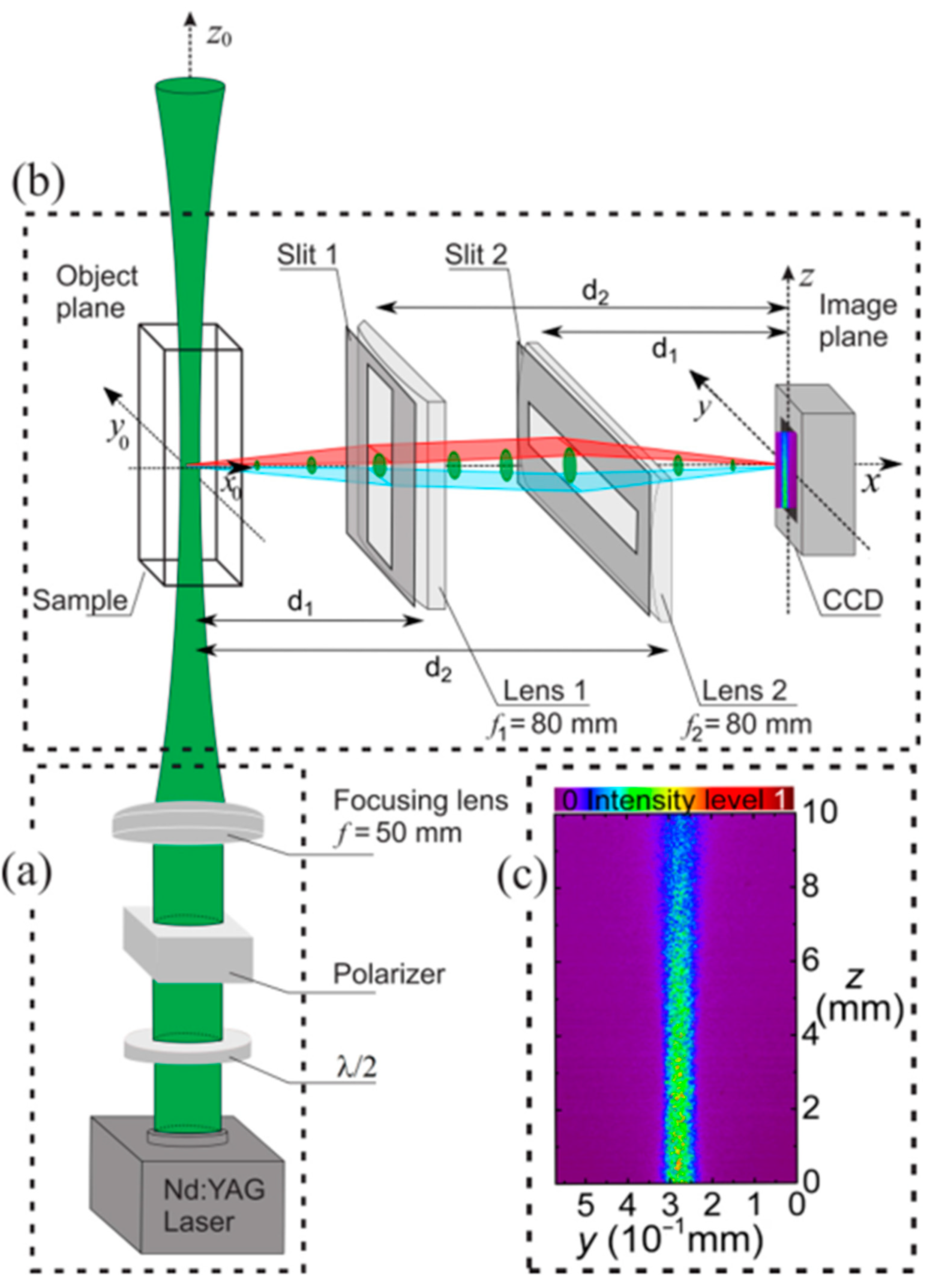
Figure 1. A schematic of the SLIM setup. (a) The λ / 2 and polarizer ensure a laser beam with controlled power and y 0 polarization. The laser propagates along the z 0 axis and is focused on a scattering NL sample. (b) The scattered light profile is imaged using an astigmatic imaging system to obtain distinct magnifications along the y and z axes on the detector. (c) From the beam profile across the entire sample, it is possible to determine the NL refractive and absorptive optical constants. (Reproduced from ref. [18] with permission).
Photonics 11 00650 g001
Figure 2. (a,b) indicate the SLIM measurements of the divergence angle for solutions containing distinct concentrations of ethanol and acetone. Each point corresponds to the average over 50 measurements, and the line represents the data fit. The points in (c) show the measured n 2 value for each individual pulse used in (b) and the mean value (dashed). (d) compares SLIM measurements (red squares) with Z-scan measurements using several solutions of ethanol and acetone. The solid line corresponds to the expected n 2 value considering the literature values for acetone and ethanol and the volume fraction of acetone (adapted from ref. [19] with permission).
Photonics 11 00650 g002
Figure 3. (a) shows the open aperture Z-scan signals at I 0 = 9.4   GW / cm 2 for samples containing different concentrations of TiO2 nanoparticles. z represents the thin sample position along the optical beam propagation. (b) shows the ln P z for a given position z in the sample with the larger particle concentration [green squares in (a)], as detected by a camera. Each curve represents a different initial intensity, varying from 2.5   GW / cm 2 to 12.5   GW / cm 2 . The power extinction model (dashed lines) does not describe the excess scattering near the sample input. (c) shows that the excess scattering not explained by the forward propagating model in (b) has an exponential growth along z . (Adapted from ref. [18] with permission).
Photonics 11 00650 g003
Figure 4. (a,b) Experimental and theoretical fits of closed aperture (CA) Z-scan curves for colloids with SiO2 NPs suspended in ethanol at 22.2 kW/cm2 and varying the NP volume fractions, f. (c) Intensity dependence of the NL phase shift from CA Z-scan measurements, including a comparison with pure ethanol (f = 0). (Adapted from ref. [20] with permission).
Photonics 11 00650 g004
Figure 5. (a,b) IC-scan curves for silica colloids obtained from 2D spatial intensity self-correlation (blue) and cross-correlation (black) functions at various intensities and volume fractions of f1 = 8.2 × 10⁻³ and f2 = 4.1 × 10⁻². (c,d) Behavior of Δ g m a x 2 obtained from (c) self-correlation and (d) cross-correlation functions with increasing incident intensity for volume fractions f1 (red circles), f2 (black squares), and f = 0 (pure ethanol—blue triangles). (e) Scattered light intensity as a function of incident intensity for two concentrations of silica colloids. (Adapted from ref. [20] with permission).
Photonics 11 00650 g005
Figure 6. (a) Speckle pattern from an unpolished silicon surface and (b) its corresponding intensity correlation function. (c) Experimental and numerical fit of g m a x 2 versus incident intensity obtained from RICO-scan for the unpolished silicon surface. (d) RZ-scan curves for polished (blue circles) and unpolished (green squares) silicon surfaces at an intensity of 4.1 GW/cm2.
Photonics 11 00650 g006
Figure 7. (a) Speckle pattern generated by a silicon powder sample with corresponding intensity correlation (b) and probability density (c) functions. (d) g m a x 2 versus incident intensity obtained performing RICO-scan for the silicon powder, with the red curve representing the best numerical fit using the same NL refractive index measured for polished and unpolished silicon surfaces.
Photonics 11 00650 g007
Disclaimer/Publisher’s Note: The statements, opinions and data contained in all publications are solely those of the individual author(s) and contributor(s) and not of MDPI and/or the editor(s). MDPI and/or the editor(s) disclaim responsibility for any injury to people or property resulting from any ideas, methods, instructions or products referred to in the content.