Article

One-Dimensional Gadolinium (III) Complexes Based on Alpha- and Beta-Amino Acids Exhibiting Field-Induced Slow Relaxation of Magnetization

by
Marta Orts-Arroyo
,
Adrián Sanchis-Perucho
,
Nicolas Moliner
,
Isabel Castro
,
Francesc Lloret
and
José Martínez-Lillo
*
Departament de Química Inorgànica, Instituto de Ciencia Molecular (ICMol), Universitat de València, c/Catedrático José Beltrán 2, Paterna, 46980 València, Spain
*
Author to whom correspondence should be addressed.
Inorganics 2022, 10(3), 32; https://doi.org/10.3390/inorganics10030032
Submission received: 8 February 2022 / Revised: 24 February 2022 / Accepted: 28 February 2022 / Published: 3 March 2022
(This article belongs to the Special Issue Lanthanide Single-Molecule Magnets)

Abstract

:
Gadolinium (III) complexes exhibiting slow relaxation of magnetization are uncommon and have been much less studied than other compounds based on anisotropic lanthanide (III) ions. We prepared two one-dimensional gadolinium (III) complexes based on α-glycine (gly) and β-alanine (β-ala) amino acids, with the formula {[Gd2(gly)6(H2O)4](ClO4)6·5H2O}n (1) and {[Gd2(β-ala)6(H2O)4](ClO4)6·H2O}n (2), which were magneto-structurally characterized. Compounds 1 and 2 crystallize in the triclinic system (space group Pī). In complex 1, two Gd (III) ions are eight-coordinate and bound to six oxygen atoms from six gly ligands and two oxygen atoms from two water molecules, the metal ions showing different geometries (bicapped trigonal prism and square antiprism). In complex 2, two Gd (III) ions are nine-coordinate and bound to seven oxygen atoms from six β-ala ligands and two oxygen atoms from two water molecules in the same geometry (capped square antiprism). Variable-temperature dc magnetic susceptibility measurements performed on microcrystalline samples of 1 and 2 show similar magnetic behavior for both compounds, with antiferromagnetic coupling between the Gd (III) ions connected through carboxylate groups. Ac magnetic susceptibility measurements reveal slow relaxation of magnetization in the presence of an external dc field in both compounds, hence indicating the occurrence of the field-induced single-molecule magnet (SMM) phenomenon in both 1 and 2.

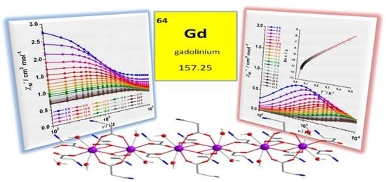
Graphical Abstract

1. Introduction

Since the discovery of the mononuclear phthalocyanine-based lanthanide (III) complexes, which exhibit the single-molecule magnet (SMM) phenomenon, in 2003 [1,2], many efforts have focused on the synthesis and development of lanthanide(III)-based complexes in order to study this singular magnetic behavior, which allows SMMs to become promising candidates for potential applications in high-density data storage, quantum computing, molecular refrigeration and spintronics investigations [3,4,5,6,7,8].
Heterometallic 3d–4f mixed systems, radical bridged compounds, mono- and polynuclear lanthanide (III) complexes containing highly anisotropic 4f ions, mainly Dy (III) and to a lesser extent Tb (III), Ho (III) and Er (III), were investigated during the last two decades in the field of molecular magnetism [9,10,11,12,13,14,15,16,17]. More recently, mononuclear SMMs, also known as single-ion magnets (SIMs), based on dysprosium metallocenes were reported displaying energy barriers of magnetization reversal exceeding the 1500 cm–1 value and blocking temperatures as high as that of the liquid nitrogen (>77 K), which exemplify the current progress in this research area [18,19].
In comparison with other members of the lanthanides family, the Gd (III) ion has been largely ignored in this type of study. This metal ion is considered magnetically isotropic due to the half-occupied 4f7 electron configuration and the lack of orbital contribution (S = 7/2, L = 0) with an 8S7/2 ground state and a spherical quadrupole moment. Hence, the number of reported Gd (III) complexes that exhibit slow relaxation of magnetization is quite scarce [20,21]. Nevertheless, in some cases, Gd (III) cations show a very low or negligible value of the zero-field splitting (D), which induces the occurrence of ac signals for complexes of this quasi-isotropic 4f metal ion. In this way, when an external magnetic field is applied, the degeneracy between energy levels can be removed and the Quantum Tunnelling of Magnetisation (QTM) can be suppressed, which could result in mixed mechanisms of spin–lattice, spin–phonon and spin–spin relaxations [20]. This fact makes this type of study on both new and old Gd(III) systems very appealing.
Herein, we report the synthesis, crystal structure and magnetic properties of two carboxylate-bridged GdIII 1D coordination polymers of the formula {[Gd2(gly)6(H2O)4](ClO4)6·5H2O}n (1) and {[Gd2(β-ala)6(H2O)4](ClO4)6·H2O}n (2) [gly = α-glycine and β-ala = β-alanine]. To the best of our knowledge, no magneto-structural study on homometallic gadolinium (III) complexes based on these amino acids has been reported so far (Scheme 1).

2. Results and Discussion

2.1. Synthetic Procedure

Compounds 1 and 2 are prepared from a mixture of Gd2O3 and glycine (1)/β-alanine (2). Both mixtures react in an aqueous solution acidulated with perchloric acid. However, the employed crystallization technique was different. While for preparing 1 the reaction mixture was heated at 80 °C for 48 h and then cooled at a rate of 4.5 °C/h to room temperature, the reaction mixture that generates compound 2 was heated at 60 °C for 1h and the resulting solution was left to evaporate at room temperature for 2 weeks. It is important to mention that, although no problems were encountered in this work, care should be taken when using the potentially explosive perchlorate anion (ClO4), which comes from the perchloric acid.

2.2. Description of the Crystal Structures

Crystal data and structure refinement parameters for 1 and 2 are summarized in Table 1, where we indicate that both compounds crystallize in the triclinic system with centrosymmetric space group Pī. A recent review of the Cambridge Structural Database (CSD) revealed that the crystal structures of 1 and 2 were previously deposited with identifiers UKIKIJ and TEHKUN, respectively. Nevertheless, they were collected at room temperature and were deposited with refinement and resolution levels lower than the ones reported in this work [22,23].
The crystal structures of 1 and 2 are better described as cationic dinuclear [GdIII2]6+ units which are connected through carboxylate groups from glycine (1) and β-alanine (2), forming one-dimensional {[GdIII2]6+}n systems, the positive charges being counterbalanced by means of ClO4 anions. H2O solvent molecules are also present in their crystal structure (Figure 1).
In complex 1, two GdIII ions of the dinuclear [Gd2(gly)6(H2O)4]6+ unit are linked between them through four bridging carboxylate groups of four glycinate ligands (gly). These two GdIII ions are separated by a distance of 4.223(1) Å. Another two glycinate ligands connect these two GdIII ions to adjacent dinuclear units with separations of 5.229 (1) [Gd (1)···Gd (1a); (a) = 2 − x, 1 − y, 1 − z] and 5.135 (1) Å [Gd (2)···Gd (2b); (b) = 3 − x, 2 − y, 2 − z], thus generating a 1D {[GdIII2]6+}n chain (Figure 2). Each GdIII ion of the dinuclear [Gd2(gly)6(H2O)4]6+ unit is eight-coordinate and bonded to six oxygen atoms from six glycinate ligands and two oxygen atoms of two water molecules (Figure 1).
The Gd–O bond lengths exhibit an average value of 2.419 (1) Å, which is somewhat shorter than that of the Gd–Ow bond lengths [2.506 (1) Å] [24]. The glycinate ligands are present in their zwitterionic form with C−C, C−N, and C−O bond lengths, which are in agreement with those found in the literature for similar lanthanide-based complexes [25,26].
In the packing of 1, the cationic {[GdIII2]6+}n chains are intercalated by ClO4 anions. The shortest interchain Gd···Gd distance is approximately 11.0(1) Å. The dinuclear [Gd2(gly)6(H2O)4]6+ units in the chains of 1 are connected through H-bonding interactions, which involve coordinated water molecules [O (1w)···O (2wa) and O (3w)···O (4wb) distances of 2.844 (1) and 2.780 (1) Å, respectively]. Further H-bonding interactions generated by protonated −NH2 groups of the glycinate ligands and ClO4 anions link the {[GdIII2]6+}n chains in the structure of 1, as previously reported in the study of other SMMs structures [27,28,29].
In complex 2, two GdIII ions are connected between them through four bridging carboxylate groups from four β-alanine (β-ala) ligands to form the dinuclear [Gd2(β-ala)6(H2O)4]6+ unit. The two GdIII ions are distanced from each other by an average separation of ca. 4.008 (1) Å (Figure 1), (the symmetry codes for the Gd (1)···Gd (1a) and Gd (2)···Gd (2b) distances being (a) = −x, 1 − y, −z and (b) = 1 − x, − y, 1 − z, respectively). Additional β-ala ligands link adjacent dinuclear units with separations of 5.196 (1) [Gd (1)···Gd (1c); (c) = 1 − x, 1 − y, −z] and 5.203 (1) Å [Gd (2)···Gd (2d); (d) = 2 − x, − y, 1 − z], generating a cationic 1D coordination polymer that grows along the crystallographic a-axis (Figure 3).
Each GdIII ion in 2 is nine-coordinate and bonded to seven oxygen atoms from six carboxylate groups of β-ala ligands and two oxygen atoms of two water molecules (Figure 1). The average value of the Gd–O bond lengths [2.388 (1) Å] is shorter than that of the Gd–Ow bond lengths [2.522 (1) Å]. The β-ala ligands are coordinated in 2 as zwitterionic molecules and the values of the C−C, C−N, and C−O bond lengths agree with those found in the literature for similar complexes based on other lanthanide (III) ions [26,30].
In the packing of 2, the cationic {[GdIII2]6+}n chains and ClO4 anions are arranged in an alternate way. They are linked through intermolecular H-bonding interactions involving non-coordinated water molecules and protonated −NH2 groups of β-ala ligands. The shortest interchain Gd···Gd distance in 2 is approximately 11.0 Å, which is for the Gd (2)···Gd (1c) separation. The supramolecular structure of 2 is generated through additional H-bonding interactions.

2.3. Analysis of the Polyhedral Structures

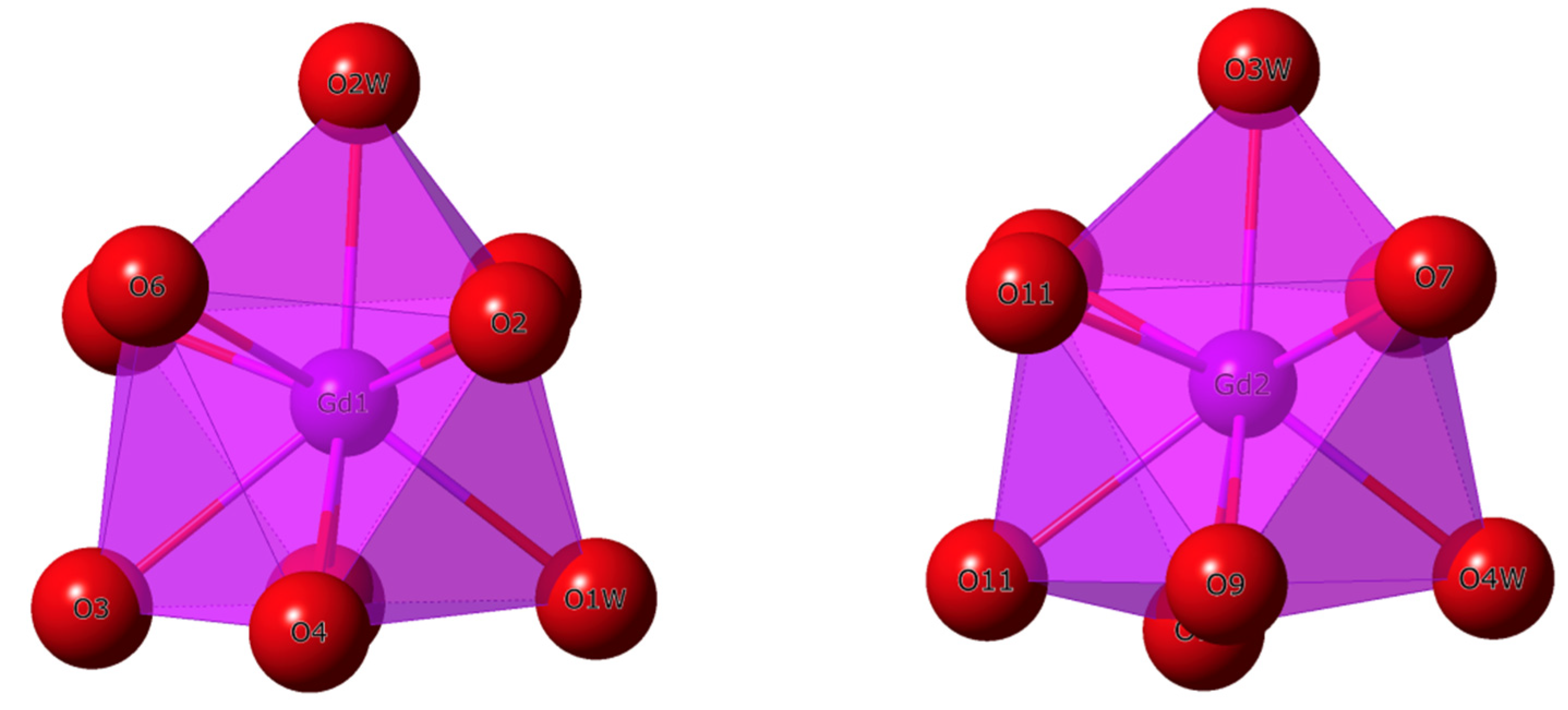
The coordination environment and geometry of the GdIII ions in 1 and 2 were further analyzed through the SHAPE program [31,32,33]. In 1, the two GdIII ions show a coordination number equal to eight (Figure 1). The lower computed value for Gd (1) was 0.732, which was associated with a bicapped trigonal prism (BCTPR) geometry (Table 2). For Gd (2), however, a value of 0.915 was assigned to a square antiprism (SAPR) geometry (Figure 4 and Table 2). These features would suggest different geometries for the metal centers Gd (1) and Gd (2) in compound 1 (Figure 4).
Unlike 1, the two GdIII ions of the dinuclear [GdIII2]6+ unit in compound 2 exhibit a coordination number equal to nine (Figure 1). The lower SHAPE values computed for these two GdIII ions were 1.117 and 1.208 for Gd (1) and Gd (2), respectively (Table 3). These calculated values were assigned to a capped square antiprism (CSAPR) geometry (Figure 5), hence indicating the same geometry for the GdIII ions in the dinuclear [GdIII2]6+ unit of 2.
As shown in Table 2 and Table 3, these computed values for 1 allow us to assign the C2v and D4d symmetries to the Gd (1) and Gd (2) ions, respectively, whereas both GdIII ions (Gd (1) and Gd (2)) exhibit C4v symmetry in 2. In any case, they would be approximate symmetries.

2.4. Magnetic Properties

Dc magnetic susceptibility measurements were carried out on microcrystalline samples of 1 and 2 in the 2–300 K temperature range and under an external magnetic field of 0.5 T. In order to keep the samples both immobilized and well isolated from the moisture of the air at all moments, the organic compound eicosene was used. The χMT versus T plots (χM being the molar magnetic susceptibility per two GdIII ions) for compounds 1 and 2 are given in Figure 6. At room temperature, the χMT values are ca. 15.7 (1) and ca. 15.9 cm3mol−1K (2), which are very close to that expected for two magnetically uncoupled GdIII ions (4f7 ion with gGd = 2.0, SGd = 7/2 and LGd = 0) [34]. Upon cooling, the χMT values approximately follow the Curie law with decreasing temperature to ca. 20 K, before they decrease reaching minimum values of approximately 13.4 (1) and 14.0 cm3mol−1K (2) at 2 K. The decrease in the χMT value observed for both compounds would likely be assignable to antiferromagnetic interactions and/or small zero-field splitting (ZFS) effects [20,21].
The field dependence of the molar magnetization (M) plots for 1 and 2 are given in the respective insets of Figure 6. The M values display a continuous increase with the applied magnetic field at 2 K. The higher M value is ca. 14.0 μB for both compounds, which is in agreement with those of similar GdIII compounds containing dinuclear units [35,36].
Taking into account the crystal structures described for 1 and 2, which are made up of linked dinuclear GdIII units, we considered them as magnetically isolated dinuclear GdIII systems. Thus, we performed the treatment of the experimental data of the χMT versus T plots through the isotropic Hamiltonian of Equation (1):
Ĥ = −JŜ1·Ŝ2 + μBgHŜ
The best least-squares fit gave the parameters J = −0.042 (1) cm−1 and g = 2.003(1) with R = 4.7 × 10⁻⁵ for 1, and J = −0.030 (3) cm−1 and g = 2.002 (1) with R = 5.2 × 10⁻⁵ for 2 {R being the agreement factor defined as Σi[(χMT)iobs − (χMT)icalcd]2/[(χMT)iobs]2}. As shown in Figure 6, the calculated curves (solid red lines) reproduce the experimental magnetic data in the whole temperature range quite well. The sign and magnitude of the J values indicate the presence of weak antiferromagnetic exchanges between the GdIII ions connected through carboxylate bridges of the α-glycine and β-alanine amino acids in 1 and 2, respectively. As far as we know, these J values are the first ones reported for GdIII complexes based on these two amino acids. Nevertheless, they are in agreement with those previously reported for GdIII systems linked through similar carboxylate bridges [36].
Ac magnetic susceptibility measurements were performed on 1 and 2 in the temperature range of 2–25 K and in a 5.0 G ac field oscillating at different frequencies. No out-of-phase ac signals (χ″M) were observed at Hdc = 0 G, which may be caused by a very fast Quantum Tunnelling of Magnetization (QTM) in 1 and 2. Nevertheless, out-of-phase ac signals were observed in both compounds when an external dc magnetic field (the optimal field being Hdc = 2500 G) was applied. This applied dc magnetic field suppresses QTM and breaks the Kramer’s doublet, leading to the observed slow relaxation [17,20,21]. In this way, both compounds show field-induced slow relaxation of magnetization, which is indicative of single-molecule magnet (SMM) behavior [4,7]. This magnetic relaxation observed for 1 and 2 was studied through both in-phase (χM’) and out-of-phase (χM″) ac susceptibilities versus frequency (ν/Hz) plots, which are given in Figure 7 and Figure 8, respectively. The experimental data of the maxima in 2 display higher intensity than those of 1, even though similar relaxation dynamics could be a priori expected for both compounds.
The insets in Figure 8 show the ln(τ) versus 1/T curves for 1 and 2. In both compounds, the experimental data draw a straight line along the ranges of ca. 0.03–0.15 (1) and ca. 0.04–0.07 K−1 (2) of the high-temperature region of 1 and 2, which connect with other straight-line behavior in the ranges of ca. 0.20–0.47 (1) and ca. 0.09–0.44 K−1 (2) of the low-temperature region. In order to fit the experimental data of the ln (τ) versus 1/T plots, several relaxation mechanisms were considered for both compounds [7,9]. Nevertheless, the whole ln(τ) versus 1/T curves were reasonably fitted through two mechanisms for the relaxation of magnetization, namely, Orbach [τo−1 exp (−Ueff/kBT)] and Raman [CTn], according to Equation (2):
τ−1 = τo−1 exp (−Ueff/kBT) + CTn
The least-squares fit of the experimental data of 1 and 2 through Equation (2) leads to the following set of parameters: Ueff = 26.3 (2) cm−1, τo = 3.1 (2) × 10−6 s, C = 168 (5) s−1K−n and n = 1.5 (2) for 1, and Ueff = 7.8 (2) cm−1, τo = 2.6 (1) × 10−5 s, C = 2.6 (2) s−1K−n, and n = 3.1 (2) for 2. Although these values are the first ones reported for one-dimensional homometallic GdIII complexes based on these amino acids, they are close to those previously reported for similar GdIII complexes [12,13]. The values of the effective energy barrier (Ueff) obtained for 1 and 2 are lower than those reported for the derivatives complexes containing DyIII ion [26]. Nevertheless, the Ueff values reported for 1 and 2 should be carefully considered as they could not correspond to any excited GdIII states and therefore would not be real effective energy barrier values [13,21].
The local symmetries displayed in the dinuclear GdIII units are C2v and D4d in 1 and C4v in 2. According to previous lanthanide (III) complexes studies, a priori, the local symmetry D4d found in 1 would lead to better SMM properties [5,6,7], as observed for its Ueff value when compared with that of 2, but this fact relies at last on the relative orientation of the magnetic anisotropy axes of all the spin carriers [26].
The τo values obtained for 1 and 2, being approximately 10−6–10−5 s, are in agreement with those previously reported for single-ion and single-molecule magnets [13,17], which supports our consideration that the predominant magnetic behavior in both compounds would be that of dinuclear single-molecule magnets, rather than a single-chain magnet.
Finally, according to our results, the relaxation pathway for 1 and 2 should be a combination of different processes, namely, Orbach (at a higher temperature) and Raman (at a lower temperature), both of them involving two phonons. The reported n value for 1 (n ≈ 2) would indicate the presence at least of a phonon bottleneck effect, whereas the n value for 3 (n ≈ 3) would indicate the contribution of a Raman mechanism, as previously reported [37]. These n values suggest that only a direct process would not be operative in the relaxation dynamics of 1 and 2. In any case, further detailed magnetic and theoretical studies performed on different GdIII complexes will be necessary to correctly understand the relaxation dynamics of GdIII SMMs.

3. Experimental Section

3.1. Preparation of the Complexes

3.1.1. Synthesis of {[Gd2(gly)6(H2O)4](ClO4)6·5H2O}n (1)

A solvothermal reaction of Gd2O3 (0.072 g, 0.20 mmol) and glycine (0.030 g, 0.40 mmol) was performed in an aqueous suspension (2 mL) acidulated with perchloric acid (1.0 mL, 2 M) at 80 °C for 48 h, followed by a cooling process at 4.5 °C/h to room temperature. Colourless parallelepipeds were obtained and were suitable for single-crystal X-ray diffraction studies. Yield: ca. 60%. Anal. Calcd. for C12H48N6O45Cl6Gd2 (1): C, 9.5; H, 3.2; N, 5.5. Found: C, 9.9; H, 3.0; N, 5.3. SEM-EDAX: a molar ratio of 1:3 for Gd/Cl was found for 1. IR (KBr pellet): peaks associated mainly to the glycine ligand and also to the perchlorate anion are observed at 3407 (s), 3080(m), 3006 (m), 2781 (w), 2708 (w), 1628 (vs), 1609 (vs), 1570 (m), 1499 (m), 1466 (m), 1413 (m), 1335 (m), 1144 (vs), 1109 (vs), 1088 (s), 905 (m), 626 (s), 536 (w), 507 (w) cm1.

3.1.2. Synthesis of {[Gd2(β-ala)6(H2O)4](ClO4)6·H2O)}n (2)

A mixture of Gd2O3 (0.090 g, 0.25 mmol) and β-alanine (0.022 g, 0.25 mmol) in an aqueous suspension (5 mL) acidulated with perchloric acid (1.0 mL, 2 M) was stirred and heated at 60 °C for 1h. The resulting solution was left to evaporate at room temperature for 2 weeks. Colourless needles were obtained, which were suitable for single-crystal X-ray diffraction. Yield: ca. 55%. Anal. Calcd for C18H52N6O41Cl6Gd2 (2): C, 14.1; H, 3.4; N, 5.5. Found: C, 14.0; H, 3.3; N, 5.6. SEM-EDAX: a molar ratio of 1:3 for Gd/Cl was found for 2. IR (KBr pellet): peaks associated to β-alanine ligand and perchlorate anion are observed at 3396 (s), 1622 (s), 1578 (s), 1460 (s), 1406 (m), 1336 (m), 1312 (w), 1264 (w), 1144 (vs), 1116 (vs), 1090 (s), 958 (m), 941 (w), 641 (m), 627 (s), 590 (w), 520 (w) cm1.

3.2. X-ray Data Collection and Structure Refinement

X-ray diffraction data collection on single crystals of dimensions 0.18 × 0.11 × 0.09 (1) and 0.18 × 0.09 × 0.06 mm3 (2) were collected on a Bruker D8 Venture diffractometer with PHOTON II detector and by using monochromatised Mo-Kα radiation (λ = 0.71073 Å). Crystal parameters and refinement results for 1 and 2 are summarized in Table 1. The structures were solved by standard direct methods and subsequently completed by Fourier recycling using the SHELXTL [38] software packages and refined by the full-matrix least-squares refinements based on F2 with all observed reflections. The final graphical manipulations were performed with the DIAMOND [39] and CRYSTALMAKER [40] programs. CCDC 2,149,741 and 2,149,742 for 1 and 2, respectively.

3.3. Physical Measurements

Elemental analyses (C, H, N) were performed in an Elemental Analyzer CE Instrument CHNS1100 and the molar ratio between heavier elements was found by means of a Philips XL-30 scanning electron microscope (SEM-EDAX), equipped with a system of X-ray microanalysis, in the Central Service for the Support to Experimental Research (SCSIE) at the University of Valencia. Infrared spectra (IR) of 1 and 2 were recorded with a PerkinElmer Spectrum 65 FT-IR spectrometer in the 4000–400 cm1 range. Variable-temperature, solid-state (dc and ac) magnetic susceptibility data were collected on Quantum Design MPMS-XL SQUID and Physical Property Measurement System (PPMS) magnetometers. Experimental magnetic data were corrected for the diamagnetic contributions of both the sample holder and the eicosene. The diamagnetic contribution of the involved atoms was corrected by using Pascal’s constants [41].

4. Conclusions

In summary, the synthesis, crystal structure and magnetic properties of two one-dimensional GdIII complexes based on the α-glycine (gly) and β-alanine (β-ala) amino acids, with the formula {[Gd2(gly)6(H2O)4](ClO4)6·5H2O}n (1) and {[Gd2(β-ala)6(H2O)4](ClO4)6·H2O}n (2), were reported. Their structures are described as cationic dinuclear [GdIII2]6+ units which are connected through carboxylate groups from glycine (1) and β-alanine (2), forming one-dimensional {[GdIII2]6+}n systems. Different symmetries of the GdIII ions, namely, C2v and D4d in 1 and C4v in 2, were found in the study of their coordination environment.
The investigation of the magnetic properties of 1 and 2 through dc magnetic susceptibility measurements reveals a similar magnetic behavior, with both compounds exhibiting weak antiferromagnetic exchange couplings between GdIII ions. In addition, ac magnetic susceptibility measurements show field-induced slow relaxation of magnetization for both 1 and 2, which indicates that the single-molecule magnet (SMM) phenomenon takes place in these novel one-dimensional GdIII complexes.

Supplementary Materials

The following supporting information can be downloaded at: https://www.mdpi.com/article/10.3390/inorganics10030032/s1, CIF files 2149741 (1) and 2149742 (2).

Author Contributions

Conceptualization, J.M.-L.; funding acquisition, I.C., F.L. and J.M.-L.; methodology, M.O.-A., A.S.-P., N.M., I.C. and J.M.-L.; investigation, M.O.-A., A.S.-P., N.M., F.L. and J.M.-L.; formal analysis, M.O.-A., A.S.-P., N.M., I.C. and J.M.-L.; writing—original draft preparation, J.M.-L.; writing—review and editing, J.M.-L. All authors have read and agreed to the published version of the manuscript.

Funding

This research was funded by the Spanish Ministry of Science and Innovation (Grant numbers PID2019-109735GB-I00 and CEX2019-000919-M (Excellence Unit “María de Maeztu”)) and also by the VLC-BIOMED Program of the University of Valencia (Grant number PI-2020-19-DIGABIO).

Institutional Review Board Statement

Not applicable.

Informed Consent Statement

Not applicable.

Data Availability Statement

The raw data that support the findings of this study are available from the corresponding author upon reasonable request.

Acknowledgments

The authors M.O.-A. and A.S.-P. thank the Spanish “FPI fellowships” and “FPU fellowships” programs, respectively.

Conflicts of Interest

The authors declare no conflict of interest.

References

  1. Ishikawa, N.; Sugita, M.; Ishikawa, T.; Koshihara, S.Y.; Kaizu, Y. Lanthanide double-decker complexes functioning as magnets at the single-molecular level. J. Am. Chem. Soc. 2003, 125, 8694–8695. [Google Scholar] [CrossRef]
  2. Ishikawa, N.; Sugita, M.; Ishikawa, T.; Koshihara, S.Y.; Kaizu, Y. Mononuclear Lanthanide Complexes with a Long Magnetization Relaxation Time at High Temperatures:  A New Category of Magnets at the Single-Molecular Level. J. Phys. Chem. B 2004, 108, 11265–11271. [Google Scholar] [CrossRef]
  3. Habib, F.; Murugesu, M. Lessons learned from dinuclear lanthanide nano-magnets. Chem. Soc. Rev. 2013, 42, 3278–3288. [Google Scholar] [CrossRef] [Green Version]
  4. Ferrando-Soria, J.; Vallejo, J.; Castellano, M.; Martínez-Lillo, J.; Pardo, E.; Cano, J.; Castro, I.; Lloret, F.; Ruiz-García, R.; Julve, M. Molecular magnetism, quo vadis? A historical perspective from a coordination chemist viewpoint. Coord. Chem. Rev. 2017, 339, 17–103. [Google Scholar] [CrossRef]
  5. Liu, J.-L.; Chen, Y.-C.; Tong, M.-L. Symmetry strategies for high performance lanthanide-based single-molecule magnets. Chem. Soc. Rev. 2018, 47, 2431–2453. [Google Scholar] [CrossRef] [PubMed]
  6. Dey, A.; Kalita, P.; Chandrasekhar, V. Lanthanide(III)-Based Single-Ion Magnets. ACS Omega 2018, 3, 9462–9475. [Google Scholar] [CrossRef] [PubMed]
  7. Zabala-Lekuona, A.; Seco, J.M.; Colacio, E. Single-Molecule Magnets: From Mn12-ac to dysprosium metallocenes, a travel in time. Coord. Chem. Rev. 2021, 441, 213984. [Google Scholar] [CrossRef]
  8. Gebrezgiabher, M.; Bayeh, Y.; Gebretsadik, T.; Gebreslassie, G.; Elemo, F.; Thomas, M.; Linert, W. Lanthanide-Based Single-Molecule Magnets Derived from Schiff Base Ligands of Salicylaldehyde Derivatives. Inorganics 2020, 8, 66. [Google Scholar] [CrossRef]
  9. McAdams, S.G.; Ariciu, A.-M.; Kostopoulos, A.K.; Walsh, J.P.S.; Tuna, F. Molecular single-ion magnets based on lanthanides and actinides: Design considerations and new advances in the context of quantum technologies. Coord. Chem. Rev. 2017, 346, 216–239. [Google Scholar] [CrossRef] [Green Version]
  10. Karotsis, G.; Evangelisti, M.; Dalgarno, S.J.; Brechin, E.K. A Calix[4 ]arene 3d/4f Magnetic Cooler. Angew. Chem. Int. Ed. 2009, 48, 9928–9931. [Google Scholar] [CrossRef]
  11. Ghosh, T.K.; Maity, S.; Mayans, J.; Ghosh, A. Family of Isomeric CuII–LnIII (Ln = Gd, Tb, and Dy) Complexes Presenting Field-Induced Slow Relaxation of Magnetization Only for the Members Containing GdIII. Inorg. Chem. 2020, 60, 438–448. [Google Scholar] [CrossRef] [PubMed]
  12. Borah, A.; Murugavel, R. Magnetic relaxation in single-ion magnets formed by less-studied lanthanide ions Ce(III), Nd(III), Gd(III), Ho(III), Tm(II/III) and Yb(III). Coord. Chem. Rev. 2022, 453, 214288. [Google Scholar] [CrossRef]
  13. Maciel, J.W.D.O.; Lemes, M.A.; Valdo, A.K.; Rabelo, R.; Martins, F.T.; Maia, L.J.Q.; Costa de Santana, R.; Lloret, F.; Julve, M.; Cangussu, D. Europium(III), Terbium(III), and Gadolinium(III) Oxamato-Based Coordination Polymers: Visible Luminescence and Slow Magnetic Relaxation. Inorg. Chem. 2021, 60, 6176–6190. [Google Scholar] [CrossRef] [PubMed]
  14. Rodríguez-Barea, B.; Mayans, J.; Rabelo, R.; Sanchis-Perucho, A.; Moliner, N.; Martínez-Lillo, J.; Julve, M.; Lloret, F.; Ruiz-García, R.; Cano, J. Holmium(III) Single-Ion Magnet for Cryomagnetic Refrigeration Based on an MRI Contrast Agent Derivative. Inorg. Chem. 2021, 60, 12719–12723. [Google Scholar] [CrossRef]
  15. Orts-Arroyo, M.; Rabelo, R.; Carrasco-Berlanga, A.; Moliner, N.; Cano, J.; Julve, M.; Lloret, F.; De Munno, G.; Ruiz-García, R.; Mayans, J.; et al. Field-induced slow magnetic relaxation and magnetocaloric effects in an oxalato-bridged gadolinium(III)-based 2D MOF. Dalton Trans. 2021, 50, 3801–3805. [Google Scholar] [CrossRef]
  16. Arauzo, A.; Lazarescu, A.; Shova, S.; Bartolomé, E.; Cases, R.; Luzón, J.; Bartolomé, J.; Turta, C. Structural and magnetic properties of some lanthanide (Ln = Eu(III), Gd(III) and Nd(III)) cyanoacetate polymers: Field-induced slow magnetic relaxation in the Gd and Nd substitutions. Dalton Trans. 2014, 43, 12342–12356. [Google Scholar] [CrossRef]
  17. Izuogu, D.C.; Yoshida, T.; Zhang, H.; Cosquer, G.; Katoh, K.; Ogata, S.; Hasegawa, M.; Nojiri, H.; Damjanović, M.; Wernsdorfer, W.; et al. Slow Magnetic Relaxation in a Palladium–Gadolinium Complex Induced by Electron Density Donation from the Palladium Ion. Chem. Eur. J. 2018, 24, 9285–9294. [Google Scholar] [CrossRef]
  18. Guo, F.-S.; Day, B.M.; Chen, Y.-C.; Tong, M.-L.; Mansikkamäki, A.; Layfield, R.A. Magnetic hysteresis up to 80 kelvin in a dysprosium metallocene single-molecule magnet. Science 2018, 362, 1400–1403. [Google Scholar] [CrossRef] [Green Version]
  19. Ullah, A.; Cerdá, J.; Baldoví, J.J.; Varganov, S.A.; Aragó, J.; Gaita-Ariño, A. In Silico Molecular Engineering of Dysprosocenium-Based Complexes to Decouple Spin Energy Levels from Molecular Vibrations. J. Phys. Chem. Lett. 2019, 10, 7678–7683. [Google Scholar] [CrossRef]
  20. Chen, Y.-C.; Peng, Y.-Y.; Liu, J.-L.; Tong, M.-L. Field-induced slow magnetic relaxation in a mononuclear Gd(III) complex. Inorg. Chem. Commun. 2019, 107, 107449. [Google Scholar] [CrossRef]
  21. Mayans, J.; Escuer, A. Correlating the axial Zero Field Splitting with the slow magnetic relaxation in GdIII SIMs. Chem. Commun. 2021, 57, 721–724. [Google Scholar] [CrossRef] [PubMed]
  22. Lu, J.-L.; Liu, B.-P.; Tan, Z.-C.; Zhang, D.-S.; Li, L.; Chen, Y.-D.; Shi, Q. Synthesis, Crystal Structure and Themochemical Study of [Gd2(Gly)6(H2O)4](ClO4)6(H2O)5. Acta Chim. Sin. Chin. Ed. 2007, 65, 2349–2355. [Google Scholar]
  23. Liang, F.-P.; Ma, L.-F.; Huang, S.-W.; Huang, Y.-Q. Synthesis, Crystal Structure and Antibacterial Activity of β-Alanine Complexes with Nd, Gd, Er. Chin. J. Appl. Chem. 2004, 21, 58–62. [Google Scholar]
  24. Orts-Arroyo, M.; Ten-Esteve, A.; Ginés-Cárdenas, S.; Castro, I.; Martí-Bonmatí, L.; Martínez-Lillo, J. A Gadolinium(III) Complex Based on the Thymine Nucleobase with Properties Suitable for Magnetic Resonance Imaging. Int. J. Mol. Sci. 2021, 22, 4586. [Google Scholar] [CrossRef]
  25. Legendziewicz, J.; Huskowska, E.; Waśkowska, A.; Argay, G. Spectroscopy and crystal structure of neodymium coordination compound with glycine: Nd2(Gly)6·(ClO4)6·9H2O. Inorg. Chim. Acta 1984, 92, 151–157. [Google Scholar] [CrossRef]
  26. Orts-Arroyo, M.; Castro, I.; Lloret, F.; Martínez-Lillo, J. Field-induced slow relaxation of magnetisation in two one-dimensional homometallic dysprosium(III) complexes based on alpha- and beta-amino acids. Dalton Trans. 2020, 49, 9155–9163. [Google Scholar] [CrossRef]
  27. Martínez-Lillo, J.; Dolan, N.; Brechin, E.K. A cationic and ferromagnetic hexametallic Mn(III) single-molecule magnet based on the salicylamidoxime ligand. Dalton Trans. 2013, 42, 12824–12827. [Google Scholar] [CrossRef] [Green Version]
  28. Martínez-Lillo, J.; Dolan, N.; Brechin, E.K. A family of cationic oxime-based hexametallic manganese(III) single-molecule magnets. Dalton Trans. 2014, 43, 4408–4414. [Google Scholar] [CrossRef]
  29. Rojas-Dotti, C.; Moliner, N.; Lloret, F.; Martínez-Lillo, J. Ferromagnetic Oxime-Based Manganese(III) Single-Molecule Magnets with Dimethylformamide and Pyridine as Terminal Ligands. Crystals 2019, 9, 23. [Google Scholar] [CrossRef] [Green Version]
  30. Jianxue, L.; Ninghai, H.; Chunji, N.; Qingbo, M. The crystal structure of a samarium complex with β-alanine. J. Alloys Compd. 1992, 184, L1–L3. [Google Scholar] [CrossRef]
  31. Llunell, M.; Casanova, D.; Cirera, J.; Alemany, P.; Alvarez, S. SHAPE 2.1; Universitat de Barcelona: Barcelona, Spain, 2013. [Google Scholar]
  32. Alvarez, S.; Alemany, P.; Casanova, D.; Cirera, J.; Llunell, M.; Avnir, D. Shape maps and polyhedral interconversion paths in transition metal chemistry. Coord. Chem. Rev. 2005, 249, 1693–1708. [Google Scholar] [CrossRef]
  33. Ruiz-Martínez, A.; Casanova, D.; Alvarez, S. Polyhedral Structures with an Odd Number of Vertices: Nine-Coordinate Metal Compounds. Chem. Eur. J. 2008, 14, 1291–1303. [Google Scholar] [CrossRef] [PubMed]
  34. Martínez-Lillo, J.; Cañadillas-Delgado, L.; Cano, J.; Lloret, F.; Julve, M.; Faus, J. A heteropentanuclear oxalato-bridged [ReIV4GdIII] complex: Synthesis, crystal structure and magnetic properties. Chem. Commun. 2012, 48, 9242–9244. [Google Scholar] [CrossRef]
  35. Roy, L.E.; Hughbanks, T. Magnetic Coupling in Dinuclear Gd Complexes. J. Am. Chem. Soc. 2006, 128, 568–575. [Google Scholar] [CrossRef] [PubMed]
  36. Cañadillas-Delgado, L.; Fabelo, O.; Cano, J.; Ruiz-Pérez, C. Magnetic Interactions in Oxo-Carboxylate Bridged Gadolinium(III) Complexes Synthesis, Crystal Structures and Magnetic Properties; Nova Science Publishers, Inc.: Hauppauge, NY, USA, 2010. [Google Scholar]
  37. Handzlik, G.; Magott, M.; Arczyński, M.; Sheveleva, A.M.; Tuna, F.; Baran, S.; Pinkowicz, D. Identical anomalous Raman relaxation exponent in a family of single ion magnets: Towards reliable Raman relaxation determination? Dalton Trans. 2020, 49, 11942–11949. [Google Scholar] [CrossRef]
  38. SHELXTL-2013/4, Bruker Analytical X-Ray Instruments; Sheldrick, G.M. (Ed.) Bruker Analytical X-ray Systems, Inc.: Madison, WI, USA, 2013. [Google Scholar]
  39. Diamond 4.5.0; Crystal Impact GbR; Crystal Impact: Bonn, Germany, 2018.
  40. CrystalMaker 8.7.5; CrystalMaker Software Ltd.: Oxford, UK, 2013.
  41. Bain, G.A.; Berry, J.F. Diamagnetic Corrections and Pascal’s Constants. J. Chem. Educ. 2008, 85, 532–536. [Google Scholar] [CrossRef]
Scheme 1. Molecular structure of the amino acids α-glycine (A) and β-alanine (B).
Inorganics 10 00032 sch001
Figure 1. (a) Detail of the dinuclear [Gd2(gly)6(H2O)4]6+ unit in 1; (b) Detail of the dinuclear [Gd2(β-ala)6(H2O)4]6+ unit in 2. In both cases, perchlorate anions and non-coordinating water molecules were omitted for clarity. Thermal ellipsoids are depicted at the 50% probability level.
Inorganics 10 00032 g001
Figure 2. View of the one-dimensional motif of the homometallic {[Gd2(gly)6(H2O)4](ClO4)6·5H2O}n chain in 1. Perchlorate anions and non-coordinating water molecules were omitted for clarity. Colour code: violet, Gd; red, O; blue, N; grey, C; white, H.
Inorganics 10 00032 g002
Figure 3. View of the one-dimensional motif of the homometallic {[Gd2(β-ala)6(H2O)4](ClO4)6·H2O)}n chain in 2. Perchlorate anions and non-coordinating water molecules were omitted for clarity. Colour code: violet, Gd; red, O; blue, N; grey, C; white, H.
Inorganics 10 00032 g003
Figure 4. Polyhedral view of the coordination sphere around the gadolinium (III) ions of the dinuclear [GdIII2]6+ unit in complex 1 [Gd (1), (left); Gd (2) (right)].
Inorganics 10 00032 g004
Figure 5. Polyhedral view of the coordination sphere around the gadolinium (III) ions of the dinuclear [GdIII2]6+ unit in complex 2 [Gd (1), (left); Gd (2) (right)].
Inorganics 10 00032 g005
Figure 6. Thermal variation of the χMT product for complexes 1 (left) and 2 (right). The solid red line represents the theoretical fit of the experimental data and the inset shows the M versus H plot at 2.0 K.
Inorganics 10 00032 g006
Figure 7. Frequency dependence of the in-phase ac susceptibility signals under a dc field of 2500 G for 1 (left) and 2 (right).
Inorganics 10 00032 g007
Figure 8. Frequency dependence of the out-of-phase ac susceptibility signals under a dc field of 2500 G for 1 (left) and 2 (right). The inset shows the ln (τ) versus 1/T plot with the fit considering the contribution of two mechanisms (Orbach + Raman).
Inorganics 10 00032 g008
Table 1. Summary of the crystal data and structure refinement parameters for 1 and 2.
Compound12
CIF21497412149742
FormulaC12H48Cl6N6O45Gd2C18H52Cl6N6O41Gd2
Fw/g mol−11523.761535.85
Temperature/K120 (2)120 (2)
Crystal systemTriclinicTriclinic
Space group
a11.401 (1)9.172 (1)
b13.986 (1)12.733 (1)
c15.506 (1)21.558 (1)
α/°96.47 (1)76.39 (1)
β/°102.59 (1)81.26 (1)
γ/°105.99 (1)82.47 (1)
V32280.1 (2)2406.9 (2)
Z22
Dc/g cm−32.2192.119
μ (Mo − Kα)/mm−13.3703.187
F (000)15041520
Goodness-of-fit on F21.0130.989
R1 [I > 2σ(I)]/all data0.0160/0.01750.0274/0.0302
wR2 [I > 2σ(I)]/all data0.0417/0.04260.0741/0.0760
Table 2. Selected values for possible geometries with coordination number (CN) equal to 8 obtained through the SHAPE program and from structural parameters of complex 1 a.
Metal IonHBPYCUSAPRTDDJGBFJETBPYBTPRJSDTT
Gd(1)17.11611.3171.2541.76313.13727.4060.7323.44011.813
Gd(2)13.9449.3360.9152.07712.46428.1791.1673.8899.984
a HBPY: Hexagonal bipyramid (D6h); CU: Cube (Oh); SAPR: Square antiprism (D4d); TDD: Triangular dodecahedron (D2d); JGBF: Johnson gyrobifastigum (D2d); JETBPY: Johnson elongated triangular bipyramid (D3h); BTPR: Biaugmented trigonal prism (C2v); JSD: Snub diphenoid (D2d); TT: Triakis tetrahedron (Td).
Table 3. Selected values for possible geometries with coordination number (CN) equal to 9 obtained through the SHAPE program and from structural parameters of complex 2 a.
Metal IonHPYJTCJCCUCSAPRJTCTPRTCTPRJTDICHHMFF
Gd(1)19.21315.40010.3401.1172.2551.54312.86610.1001.368
Gd(2)18.54514.84310.4771.2082.1341.56113.3759.5021.489
a HPY: Heptagonal bipyramid (D7h); JTC: Johnson triangular cupola (C3v); JCCU: Capped cube (C4v); CSAPR: Spherical capped square antiprism (C4v); JTCTPR: Tricapped trigonal prism (D3h); TCTPR: Spherical tricapped trigonal prism (D3h); JTDIC: Tridiminished icosahedron (C3v); HH: Hula-hoop (C2v); MFF: Muffin (Cs).
Publisher’s Note: MDPI stays neutral with regard to jurisdictional claims in published maps and institutional affiliations.