Article

Biodegradable Ca2+ Doped Mesoporous Silica Nanoparticles Promote Chemotherapy Synergism with Calcicoptosis and Activate Anti-Tumor Immunity

1
School of Medical Engineering and Technology, Xinjiang Medical University, Urumqi 830017, China
2
Core Facility, Xinjiang Medical University, Urumqi 830017, China
*
Author to whom correspondence should be addressed.
Inorganics 2024, 12(6), 152; https://doi.org/10.3390/inorganics12060152
Submission received: 14 April 2024 / Revised: 24 May 2024 / Accepted: 28 May 2024 / Published: 31 May 2024

Abstract

:
Mesoporous silica nanoparticles (MSNs), an excellent carrier material, have been widely used in tumor therapy as a vector for numerous therapeutic substances to boost therapeutical efficiency and specificity, such as loading them with chemotherapy drugs to improve the efficacy of chemotherapy. Nevertheless, they still face hurdles, such as lack of specificity and poor efficacy of monotherapy. The construction of multifunctional MSNs with excellent therapeutic effects by introducing metal ions has attracted the attention of many researchers. Herein, we demonstrated a calcium doped, chemotherapy drug doxorubicin (Dox) loaded, specific degradation nanoplatform, prepared using the sol–gel method by introducing calcium ions into an MSN framework, which enabled the doped nanoplatform to enhance chemotherapy and activate anti-tumor immune response. As a proof of concept, the doping of Ca2+ endowed MSNs with excellent specific degradation and pH responsive drug release, and enabled the synergy of chemotherapy and calcicoptosis. Furthermore, this nanoplatform also effectively elicited immunogenic cell death (ICD) and promoted the maturation of dendritic cells (DCs), realizing the activation of the anti-tumor immune system. The Ca2+ doped MSNs (CMSNs), that can activate immune response with specific degradation capability, demonstrate a practical strategy for the effective synergy between chemotherapy and calcicoptosis, providing a new paradigm for promoting chemotherapy-related treatment.

Graphical Abstract

1. Introduction

In the past few decades, chemotherapy has played a huge role in tumor treatment, and to this day, chemotherapy remains a first-line choice for the treatment of many advanced malignant tumors [1,2]. However, due to the low specificity, weak cell uptake efficiency and other problems, the clinical efficacy of chemotherapy is not satisfactory [3,4]. With the rapid development of nanotechnology, nanoparticle-based drug delivery systems (DDSs) have shown excellent properties in tumor therapy [5,6]. Loading chemotherapy drugs into nanoparticles can not only improve drug pharmacokinetic characteristics, but also increase drug accumulation in tumor cells and reduce toxic side effects [7]. Among numerous types of nanoparticles, due to their excellent properties such as a high specific surface area and easy functionalization, MSNs are widely utilized as carriers for various chemotherapy agents in tumor treatment research [8,9]. However, MSNs’ DDSs encounter some issues, such as unsatisfactory therapeutic effects due to the weak biodegradability of MSNs, as well as poor efficacy of monotherapy. Therefore, improving the specific degradation and granting MSNs a multimodal synergistic therapeutic function are highly desirable goals for enhancing the tumor treatment efficacy of MSNs’ DDSs.
To improve the specific degradation ability of MSNs’ DDSs, many components with tumor microenvironment (TME) responsiveness have been introduced into the MSNs’ framework, which not only could break the stable Si–O bonds in MSNs but also endows MSNs with TME responsive degradability [10,11,12,13]. Among various approaches to improve the specific degradation of MSNs, metal ion doping has attracted extensive attention. Utilizing metal ions, acid responsive metal–oxygen bonds can form in MSNs to replace the stable Si–O bonds, and the metal–O bonds in the MSNs’ framework break responsively under the stimulation of acidic TME, causing metal ions to escape from MSNs, and leading to the collapse of the MSNs’ framework [14,15]. Meanwhile, the degradation also accelerates the release of loaded therapeutic agents [16]. More importantly, while improving the specific degradation of MSNs, the metal ions also have therapeutic and diagnostic functions. For example, the reactive oxygen species (ROS), produced by doped metal ions such as Fe3+, Cu2+ and Mn4+ through Fenton or Fenton-like reaction, can be utilized for tumor treatment [17,18,19]. In addition to inducing tumor cell apoptosis through chemodynamic therapy (CDT), metal ions can induce tumor cell death through other means, for example, Fe2+ causes lipid peroxidation in tumor cells, leading to the occurrence of ferroptosis [20], and Cu2+ can disrupt the tricarboxylic acid cycle, resulting in the aggregation of lipoylated proteins and the loss of iron–sulfur clusters, which eventually leads to cuproptosis [21,22]. Furthermore, metal ions also exhibit an excellent immune activation effect, which could further augment the therapeutic effect [23,24,25]. Although metal ions such as copper and iron ions enable MSNs to acquire an excellent therapeutic effect, they are also more likely to cause cytotoxicity to normal cells. However, compared to other metal ions, Ca2+ can cause tumor cell death specifically, and their cytotoxicity to normal cells can be ignored, because tumor cells are more sensitive to changes in Ca2+ concentration than normal cells [26,27]. In this context, Ca2+ is a suitable and ideal candidate for improving the tumor treatment performance of MSNs’ nanoplatform.
Till now, there has been relatively little research utilizing CMSNs for tumor treatment, and the main focus of these investigations was to improve the degradability of the carrier [28,29,30,31,32]. Although these CMSNs, prepared by different approaches, have outstanding biodegradability and have been successfully used in tumor treatment research, they do not devote more attention to the therapeutic function of Ca2+ in tumor treatment, and can only achieve synergistic treatment by loading multiple therapeutic agents. Calcicoptosis, as a newly defined programmed cell death, is triggered by Ca2+ overload which leads to mitochondrial dysfunction [33,34]. Therefore, utilizing the Ca2+ can realize the specific biodegradation of MSNs and also endow the carrier with therapeutic function. Herein, we developed a multifunctional CMSNs nanoplatform for boosting the therapeutic effect of MSNs’ DDSs. Among various methods for MSNs’ preparation, the sol–gel method, as a classical and reliable preparation method, has attracted much attention [35,36]. This is because, compared with other methods such as the hydrothermal method, microwave method and self-assembly, the sol–gel method has been widely used for its outstanding stability, low cost and easy operation [37]. Moreover, despite the long-term anticancer immunity present in immunotherapy, the poor immunogenicity of MSNs’ DDSs hinders the immune response of the DDSs, and few studies have investigated the performance of Ca2+ doped MSNs’ DDSs in activating anti-tumor immunity. Thus, it is intriguing to explore whether the Ca2+ doped nanoplatform could be used to augment immune response.
In this investigation, a multifunctional CMSNs nanoplatform was constructed using the sol–gel method, and then the chemotherapeutic agent Dox was loaded into the as-prepared CMSNs (CMSNs@Dox). Under the stimulation of the acidic environment within tumor cells, Ca–O bonds in the framework of CMSNs@Dox broke responsively, which facilitated the biodegradation of nanoparticles, and also rendered the responsive release of Dox and Ca2+. The excessive Ca2+ within the tumor cells caused mitochondrial damage, leading to calcicoptosis, which then synergistically inhibited tumor cells with the chemotherapy drug Dox. In addition, the competence of CMSNs@Dox to trigger ICD and the maturation of DCs was also investigated, and it was found that CMSNs@Dox effectively elicited ICD and DCs’ maturation, demonstrating outstanding immune-activating abilities. Therefore, our Ca2+ doped nanoplatform could strengthen chemotherapy through synergy with calcicoptosis, and it also demonstrated a significant anti-tumor immunity activation.

2. Results and Discussion

2.1. Preparation and Characterization of CMSNs

In this work, CMSNs were prepared using the sol–gel method. The microstructure and particle size of the CMSNs were characterized by transmission electron microscopy (TEM) and, as illustrated in Figure 1a,b, CMSNs possessed a uniform spherical structure with a particle size of approximately 360 nm. To verify the successful doping of Ca2+ in CMSNs, TEM mapping was used to characterize the main elements in the CMSNs’ framework. As can be seen from Figure 1c, silicon, oxygen and calcium were uniformly distributed throughout the entire nanoparticles, which demonstrated the successful doping of Ca2+. To further certify the elemental composition of CMSNs, an X-ray photoelectron spectroscopy (XPS) analysis was performed. The results of XPS (Figure 1d) confirmed the successful doping of Ca2+ in CMSNs, and also indicated the formation of Ca–O bonds in CMSNs, in which a Ca 2p peak originated from Ca–O bonds (Figure 1e). However, there were not any Ca signals in the XPS results of MSNs (Figure S1). The XPS analysis of CMSNs also illustrated that the Ca content in CMSNs was about 3.22%, and the Ca content was also verified by Inductively Coupled Plasma Optical Emission Spectrometer (ICP-OES), which demonstrated that the Ca content per mg of CMSNs was 0.024 mg, similar to the results of XPS. Wide-angle X-ray diffraction (WAXRD) was used to analyze the structure of CMSNs, and as shown in the result (Figure 2c), only an amorphous peak was present near 20°, which indicated that CMSNs had an amorphous structure; this was consistent with other literature [38]. To have a comprehensive knowledge of the prepared CMSNs, nitrogen adsorption–desorption was also conducted. As shown in Figure 2a, CMSNs exhibited a classical type IV curve, which indicated that CMSNs had a typical mesoporous structure. The calculated BET (Brunauer–Emmett–Teller, BET) surface and the BJH (Barrett–Joiner–Halenda, BJH) pore volume were 805.95 m2/g and 0.77 cm3/g, respectively. Additionally, the pore distribution concentrated in 2.09 nm (Figure 2b). These characterizations demonstrated that doping with Ca2+ did not affect the loading features of CMSNs.

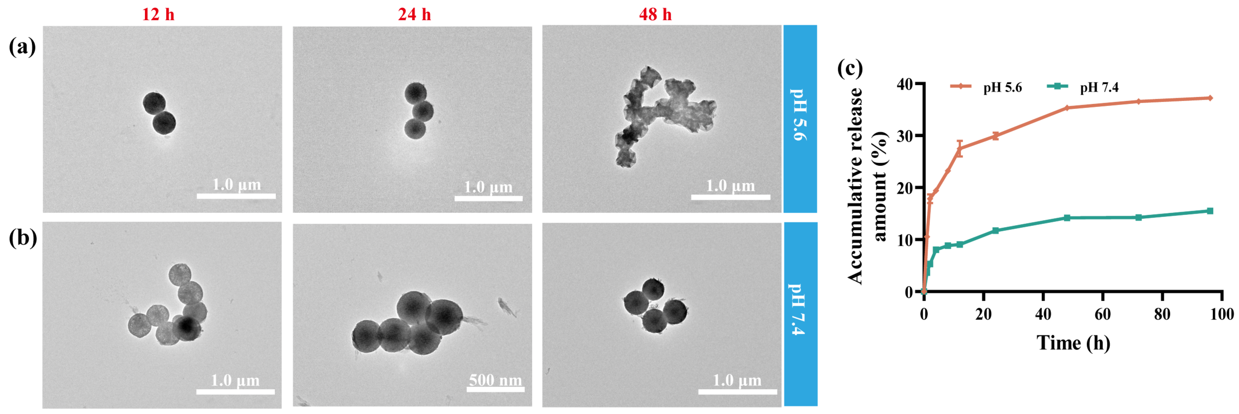
2.2. Biodegradability

Although the above characterizations demonstrated that CMSNs possessed excellent drug loading capability, it is still necessary to circumvent the problem of degradation, which affected the biomedical application of MSNs. Compared with the stable Si–O bonds in MSNs, Ca–O bonds in CMSNs were unstable under acidic conditions, which was due to the cation exchange with H+ and the cleavage of the Ca–O bond; what is more, the release of Ca2+ from Si–O–Ca could produce lots of non-bridging oxygens, which contained a large number of local sites for the nucleophilic attract primarily by OH-, leading to the breakdown of the framework [15,28,39]. The breaking of Ca–O bonds could destroy the structure of CMSNs and accelerate the degradation of CMSNs, characterized by the loss of regular spherical structures. To verify the pH responsive biodegradation behavior of CMSNs, an equal amount of CMSNs were immersed in PBS with different pH values (pH 7.4 and pH 5.6), respectively, and then the morphology of nanoparticles was tracked and observed. As illustrated in Figure 3a, the morphology of the CMSNs showed a changing trend over time when the PBS had a pH of 5.6. At 48 h, the morphology of the nanoparticles has changed from regular spherical to randomly blurred, indicating that the CMSNs possessed outstanding degradability under acidic conditions, which could alleviate the side effects caused by the accumulation of nanoparticles in the body. Considering that the CMSNs did not show significant degradation and structure collapse until 48 h, all cellular anti-tumor evaluations were conducted by co-culturing nanoparticles with cells for 48 h. However, when the CMSNs sample was placed in neutral buffer, it maintained a stable structure all the time (Figure 3b). For MSNs without Ca2+ doping, the sample maintained a stable spherical structure under both acidic and neutral conditions (Figure S2), indicating that Ca2+ doping could confer a brilliant pH responsive biodegradation ability to traditional MSNs. Of course, it also facilitated the precise release of loaded cargo.

2.3. Dox Loading and pH Responsive Drug Release

In view of the excellent structural characteristics of CMSNs, the chemotherapeutic drug Dox was loaded into the CMSNs, and the pH responsive drug release behavior was investigated. The drug loading efficiency and encapsulation efficiency of the CMSNs were 17.8% and 85%, respectively, which were significantly higher than the MSNs samples without doping, because the metal ions in the doped MSNs skeleton could form coordination complexes with drug molecules [16]. The incorporation of metal ions not only elevated the drug loading ability of CMSNs, but also granted the nanoplatform a pH-triggered drug release due to the excellent degradation ability of CMSNs under an acidic environment. To verify the pH responsive drug release behavior of the nanoplatform, CMSNs@Dox were placed in acidic (pH 5.6) and neutral (pH 7.4) PBS buffer to simulate an intracellular environment and a normal physiological environment. As depicted in Figure 3c, the CMSNs@Dox sample in the neutral buffer showed a slow and insignificant release, while under acidic conditions, the sample showed a more significant release behavior until 48 h, after which the amount of the drug released remained almost unchanged. This was due to the outstanding biodegradability of CMSNs under acidic conditions, and the collapse of the CMSNs’ structure facilitated the release of loaded cargo, which was consistent with the results of degradation experiment. Under neutral conditions, only about 15% of the loaded drug was released from CMSNs@Dox after 96 h, while about 37% of the drug was released under acidic conditions, which was twice as much as under neutral conditions. Therefore, CMSNs@Dox possessed a good specific drug release ability, which could effectively improve the precision of treatment and reduce toxic side effects on normal tissues.

2.4. Calcium Ions Dependent Mitochondrial Dysfunction

The incorporation of Ca2+ not only improved the biodegradability of CMSNs, but also endowed CMSNs@Dox with a responsive drug release ability. In addition, the degradation of CMSNs in tumor cells boosted the intracellular Ca2+ content, resulting in Ca2+ overload intracellularly. Ca2+ overload subsequently induced the calcicoptosis characterized by mitochondrial dysfunction (such as decreased mitochondrial membrane potential), thereby making the doped Ca2+ possess multiple functions. As shown in Figure 4a, compared with the control and MSNs treated groups, the intracellular Fluo-4 fluorescence intensity was significantly enhanced after CMSNs treatment. Furthermore, the Fluo-4 fluorescence intensity in 4T1 cells treated with different samples was semi-quantitatively by flow cytometry (FCM), and the mean fluorescence intensity (MFI) of Fluo-4 was about three times than the MSNs treatment group (Figure 4b), indicating that the intracellular Ca2+ content of 4T1 cells treated with CMSNs was significantly fortified, in other words, CMSNs as a Ca2+ reservoir significantly increased the intracellular Ca2+ content, facilitating the occurrence of calcicoptosis. In addition, there was no difference in Ca2+ signal between the MSNs treatment group and the control group, indicating that CMSNs could certainly enhance the intracellular Ca2+ content and cause a Ca2+ overload of tumor cells, which provided the prerequisite for calcicoptosis. The mitochondrial membrane potential (MMP) of 4T1 cells treated with various samples was evaluated by JC-1 probe. JC-1 can form aggregates which emit red fluorescence when the MMP is high, while when MMP is low, JC-1 can form monomers emitting a green fluorescence. In other words, when the MMP changed, the ratio of fluorescence intensity between the green fluorescence of monomers and the red fluorescence of aggregates would change [34,40]. As illustrated in Figure 4c, the MMP of cells treated with MSNs for 48 h was not different from that of the control group, indicating that MSNs did not have any impact on mitochondrial function; however, the MMP changed after CMSNs treatment. Moreover, the fluorescence ratio of the JC-1 monomer and polymer was also used to characterize the changes in MMP. As depicted in Figure 4d, different from the control and MSNs treatment group, after treatment with CMSNs, the fluorescence ratio of JC-1 polymer to monomer decreased significantly and was approximately one third of the control and MSNs treatment group (Figure 4d), indicating that CMSNs caused mitochondrial dysfunction, leading to a decrease in MMP. Moreover, 4T1 cells treated with CMSNs also demonstrated apparent early apoptosis (Figure 6), and mitochondrial dysfunction would exhibit significant early apoptosis according to literature [41], which further verified that CMSNs could cause mitochondrial dysfunction and lead to calcicoptosis.

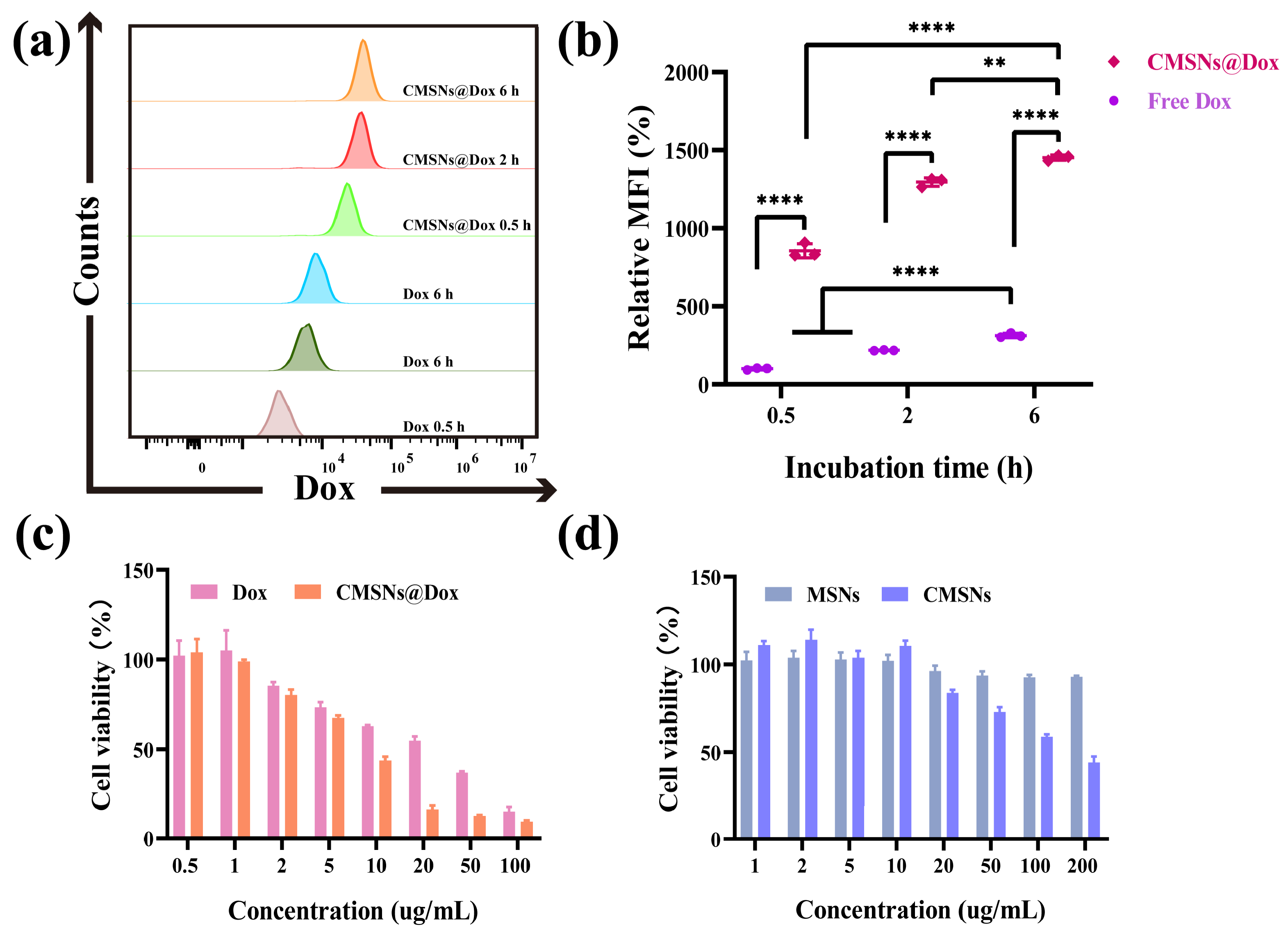
2.5. Cell Internalization

The uptake of therapeutic agents by tumor cells was a prerequisite for drugs to exert therapeutic effects. Unlike free drugs, nanoparticles that enter cells through endocytosis could improve the uptake efficiency of loaded drugs in tumor cells, thereby enhancing the therapeutic effect. Herein, the uptake behavior of CMSNs was investigated by FCM. As shown in Figure 5a,b, the uptake efficiency of nanoparticles and free Dox in tumor cells increased with time. More importantly, compared with free Dox, tumor cells exhibited higher uptake efficiency towards CMSNs@Dox at any time point, and the MFI of CMSNs@Dox was about five times that of free Dox at 6 h, indicating more chemotherapy drug molecules were internalized by cells with the assistance of CMSNs. Furthermore, CMSNs@Dox could release drugs promptly under the intracellular acidic environment, which would facilitate an enlargement in intracellular drug content and promote the efficacy of chemotherapy.

2.6. In Vitro Cytotoxicity and Anti-Tumor Efficiency

To evaluate the anti-tumor effect of CMSNs@Dox, we first investigated the cytotoxicity of MSNs, CMSNs, Dox and CMSNs@Dox against 4T1 cells. After co-incubating 4T1 cells with different samples at 37 °C for 48 h, the cell viability of 4T1 cells was measured by MTT assay. As predicted, CMSNs@Dox showed a superior cell inhibition effect at different concentrations than other treatment groups, and its IC50 value was significantly lower than free Dox (Figure 5c). It is worth noting that, unlike MSNs, which demonstrated excellent biocompatibility, CMSNs exhibited a certain degree of cytotoxicity when the concentration of CMSNs was increased (Figure 5d). This was because a large number of Ca2+ were released from CMSNs under the intracellular acidic environment, causing a Ca overload within tumor cells, which further elicited calcicoptosis. Hence, CMSNs@Dox could effectively realize the synergy of chemotherapy and calcicoptosis, which explained the more remarkable anti-tumor effect of CMSNs@Dox compared with free Dox from another perspective. Furthermore, the cytotoxicity of CMSNs and CMSNs@Dox to mouse embryo fibroblasts (3T3 cells) was also conducted, as shown in Figure S7, CMSNs have no cytotoxicity to 3T3 cells when the concentration reaches 200 μg/mL, and the cytotoxicity of CMSNs@Dox to 3T3 cells was significantly higher than that of tumor cells, demonstrating that CMSNs and CMSNs@Dox possess specificity to tumor cells. In a word, the reasons why CMSNs@Dox exhibited a significant anti-tumor effect mainly originated from two aspects: (1) CMSNs@Dox possessed excellent cellular internalization efficiency and outstanding pH responsive drug release ability, leading to an enlargement of intracellular drug accumulation, and (2) CMSNs@Dox enabled chemotherapy in tandem with calcicoptosis.
After the cytotoxicity assay, an Annexin V-FITC/7-AAD apoptosis assay was further utilized to assess the anti-tumor effect of CMSNs@Dox. As can be seen from Figure 6, the apoptosis percentage of 4T1 cells treated by CMSNs@Dox was more significant than the others, which further indicated that CMSNs@Dox posed a superior anti-tumor effect. Consistent with the MTT results, the CMSNs treatment group also showed a significant proportion of cell apoptosis compared with MSNs treatment group. This was because the Ca2+ in CMSNs elicited calcicoptosis, which was characterized by mitochondrial dysfunction, and the apparent early apoptosis further indicated the occurrence of calcicoptosis. The above experiments fully demonstrated that CMSNs@Dox could achieve the synergy between calcicoptosis and chemotherapy, and possessed outstanding anti-tumor effects. To demonstrate the synergistic therapeutic effect between chemotherapy and calcicoptosis, the Combination Index (CI) was calculated according to the IC50 value of each treatment, and the obtained CI was 0.116 and less than 1, which indicated that CMSNs@Dox possessed a brilliant synergy ability.

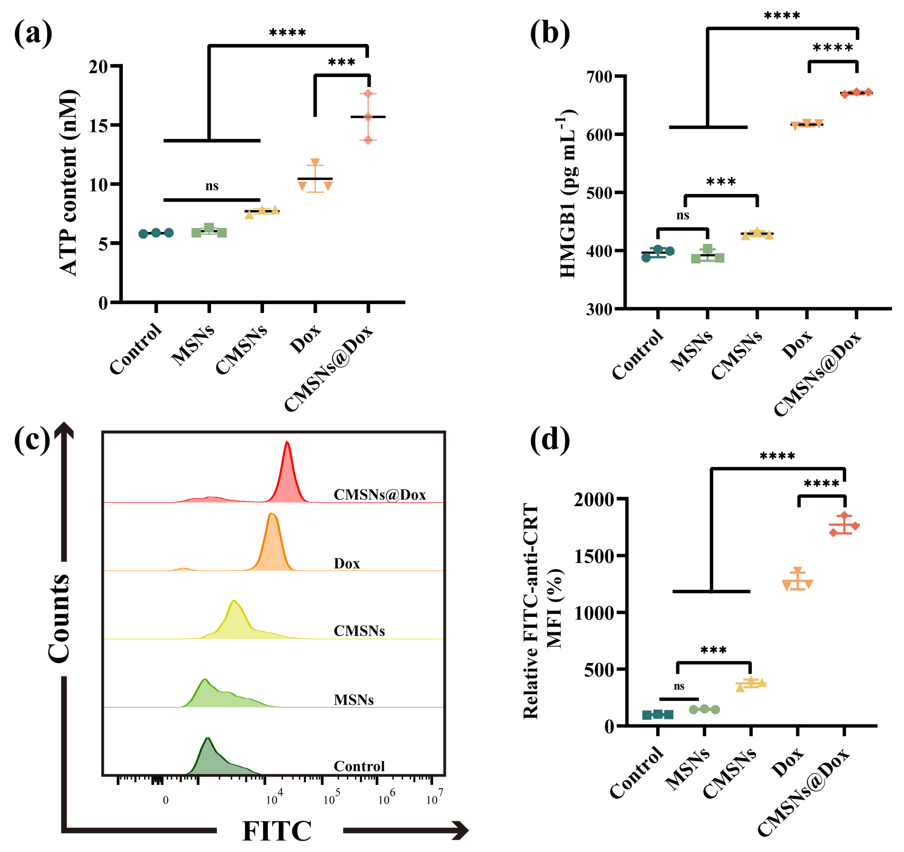
2.7. Immunogenic Cell Death and Immune Activation In Vitro

At present, much effort has been paid to the development of ICD-inducing agents due to adaptive immune responses can be driven by ICD [42], and recent investigations have shown that chemotherapy drugs could trigger adaptive immune response by inducing ICD [43]; therefore, it is meaningful to study whether the incorporation of Ca2+ could endow the drug delivery vector with the ability to induce ICD, so as to facilitate the CMSNs@Dox nanoplatform to elicit a more significant ICD effect and immune response. As an important indicator of ICD, damage associated molecule patterns (DAMPs) could activate the adaptive immune system by sending a “eat me” signal. Three main DAMPs (ATP, HMGB1, CRT) were evaluated after 4T1 cells were treated with different samples. As shown in Figure 7, compared with the control, MSNs and CMSNs treatment groups, extracellular ATP, HMGB1 and CRT were significantly elevated after free Dox treatment, which verified that chemotherapy could certainly induce ICD. On the other hand, the ATP, HMGB1 and CRT level of 4T1 cells treated with CMSNs was also raised compared with the control and MSNs treatment group, indicating that the Ca2+ doped carrier could also elicit ICD, although not as significant as chemotherapy. Among all treatment groups, cells treated with CMSNs@Dox exhibited the highest level of ATP secretion, HMGB1 release and CRT expression, which verified that the synergy of chemotherapy and calcicoptosis elicited ICD more effectively.
ICD is known to enhance anti-tumor immune responses by promoting dendritic cells’ maturation through antigen presentation [44]. Therefore, DC2.4 was co-incubated with the 4T1 cells, which were treated with different substances (MSNs, CMSNs, free Dox and CMSNs@Dox) to detect the population of CD80+CD86+ by FCM analysis. As shown in Figure 8a,b, after MSNs, CMSNs, free Dox and CMSNs@Dox treatment, the percentage of mature DCs was 0.93 ± 0.3%, 31.01 ± 2.52%, 41.1 ± 4.91%, 59.9 ± 4.23%, respectively. Except for the MSNs treatment group, the other groups were significantly higher than the control group. In addition, similar to the free Dox group, the CMSNs treatment group also induced a certain proportion of DCs’ maturation, demonstrating that the introduction of Ca2+ was essential for enhancing the immunogenicity of the carrier. Certainly, CMSNs@Dox remarkably amplified the percentage of DCs’ maturation than the others. The above results demonstrated that the synergy of chemotherapy and calcicoptosis can augment immunogenicity and DCs’ maturation, which could activate anti-tumor immunity and alleviate the tumor immunosuppressive microenvironment.

3. Materials and Methods

3.1. Materials

Tetraethylorthosilicate (TEOS) and Doxorubicin hydrochloride (Dox) were purchased from Aladdin (Shanghai, China). Cetyltrimethylammonium bromide (CTAB) was purchased from J&K Scientific (Beijing, China). Calcium nitrate tetrahydrate (Ca (NO3)2·4H2O), ammonia (NH3·H2O) and anhydrous ethanol were obtained from Sinopharm (Beijing, China). Roswell Park Memorial Institute (RPMI) 1640, penicillin-streptomycin and Fetal bovine serum (FBS) were purchased from Gibco Life Technologies (Grand Island, NE, USA). The 3-(4,5-dimethylthiazol-2-yl)-2,5-diphenyltetrazolium bromide (MTT) was purchased from Macklin (Shanghai, China). The enhanced mitochondrial JC-1 assay kit, Fluo-4 Calcium Assay kit, and enhanced ATP Assay kit were purchased from Beyotime Biotechnology (Shanghai, China). Annexin V-FITC/7AAD apoptosis detection kit was purchased from Yeasen Biotechnology (Shanghai, China). The FITC anti-Calreticulin antibody was purchased from Stressmarq Bioscience (Victoria, BC, Canada). The high mobility group box 1 (HMGB1) enzyme-linked immunosorbent assay (ELISA) kit was purchased from Wuhan Jiyinmei Biotech (Wuhan, China). All chemicals and reagents are used directly without any purification.

3.2. Synthesis of Virous MSNs

CMSNs were prepared using the classical sol–gel method with a minor modification [38]. A total of 0.08 g CTAB was added to 40 mL mixture of water and anhydrous ethanol (27 mL/13 mL), then 0.5 mL ammonia was added and stirred at 30 °C. After stirring for 30 min, 0.5 mL TEOS was added dropwise to the above mixture. A certain amount of aqueous solution containing 0.22 g Ca (NO3)2·4H2O was added to the mixture 30 min after adding TEOS. The reaction was terminated after continuous stirring for 5 h. Nanoparticles were obtained by centrifugation (8000 rpm, 10 min), and washed three times with deionized water and ethanol. The nanoparticles were dried at 60 °C for 12 h and then calcined at 550 °C for 6 h to remove the surfactant CTAB. The preparation procedure of MSNs was almost identical to CMSN, with the difference being that no Ca (NO3)2·4H2O added.

3.3. Structural Characterizations

The morphology and particle size of CMSNs were characterized by TEM (HT7700, HITACHI, Chiyoda, Japan). Element characterization was performed using field emission transmission electron microscopy (FETEM) (Tecnai F20, FEI, San Jose, CA, USA) and XPS (Thermofisher escalab 250xi, Thermofisher, Waltham, MA USA). The specific surface area, pore size and pore volume of the nanoparticles were measured through a nitrogen adsorption–desorption isotherm (Micromeritics ASAP2420, Micromeritics, Norcross, GA, USA). The Powder X-ray diffraction (XRD) mode was recorded on an X-ray diffractometer with Cu-Kα radiation (X′perT3 Powder, Almelo, The Netherlands) with a scanning step of 0.02°. Ultraviolet–visible (UV–vis) spectra were recorded using a spectrometer (TU-1901, PUXI, Shanghai, China).

3.4. In Vitro Biodegradation and Stability

An in vitro biodegradation evaluation of CMSNs was conducted by immersing the materials into PBS buffer that simulated the tumor microenvironment and normal physiological environment. Concretely speaking, 10 mg CMSNs and MSNs were dissolved in 30 mL phosphate-buffered saline (PBS) with a pH value of 5.6 (intracellular environment) and 7.4 (normal physiological environment), respectively. The mixture was placed in a shaker (37 °C, 100 rpm), and 1 mL mixture was collected at given time (12 h, 24 h, 48 h), the morphology and structure of the collected samples were observed subsequently by TEM.

3.5. Drug Loading and Release

Drug loading and pH triggered release of samples were carried out according to the published literature [45]. In brief, 15 mg Dox was added to PBS buffer containing 60 mg CMSNs and stirred at 25 °C for 24 h. The final mixtures were centrifuged (8000 rpm, 10 min) and washed with PBS several times to remove the unloaded drug molecule, and the final product CMSNs@Dox was obtained after lyophilization. At the same time, the absorbance of collected supernatant was measured by a UV–vis spectrophotometer to calculate drug loading efficiency (DL) and encapsulation efficiency (EE).
Drug release was performed by immersing CMSNs@Dox into a PBS buffer that mimics the pH value of tumor cells and the normal physiological environment, respectively. A total of 5 mg CMSNs@Dox was put into 2 mL PBS with pH 5.6 and 7.4, then transferred to a dialysis bag and immersed into 30 mL PBS buffer with corresponding pH value. Finally, the release system was placed in a shaker (37 °C, 100 rpm). At predetermined time points, the same volume of dialysate was taken and promptly supplemented with fresh PBS, an absorption value of dialysate at 480 nm was measured by UV–vis spectrophotometer, and the cumulative drug release was calculated.

3.6. Cell Culture

4T1 mouse breast cells and 3T3 mouse embryo fibroblasts were purchased from the Type Culture Collection of the Chinese Academy of Science (Shanghai, China), 4T1 cells were cultured in RPMI 1640 containing 10% FBS and 1% penicillin-streptomycin in a humidified incubator containing 5% CO2 at 37 °C, 3T3 cells were cultured in Dulbecco’s Modified Eagle Medium containing 10% FBS and 1% penicillin-streptomycin in a humidified incubator containing 5% CO2 at 37 °C.

3.7. Intracellular Ca2+ Detection

Intracellular Ca2+ levels were determined by a Fluo-4 Calcium Assay kit. 4T1 cells were inoculated (3 × 10⁵ cells per well) in a 6-well plate and incubated overnight at 37 °C. A medium containing 100 μg/mL CMSNs or MSNs was added and cultured at 37 °C for 48 h. Then, washing three times with PBS, cells were digested by trypsin and washed again. The collected cells were stained for 30 min by Fluo-4 according to the instruction of the Fluo-4 Calcium Assay kit. After washing the stained cells with PBS, cells were resuspended and analyzed by FCM (Cytoflex S, Beckman Coulter, Brea, CA, USA).

3.8. Cell Internalization

4T1 cells were inoculated with 3 × 10⁵ cells per well in a 6-well plate. After incubation for 24 h, the culture media was replaced with the same Dox concentration (5 μg/mL) of free Dox solution and CMSNs@Dox solution. At the predetermined time points (0.5 h, 2 h, 6 h), the culture medium was pipetted and washed with PBS three times, then digested and washed with PBS. FCM analysis was conducted after resuspending the collected cells in 500 μL PBS.

3.9. Mitochondrial Membrane Potential Analysis

4T1 cells (2 × 10⁵ cells per well) were added to a 6-well plate and cultures for 24 h. Then, they were subjected to various treatments including MSNs (100 μg/mL) and CMSNs (100 μg/mL) for 48 h. After washing with PBS three times, cells were digested by trypsin and washed with PBS. The collected cells were stained using a JC-1 assay kit according to the manufacturer’s instructions and subsequently analyzed by FCM.

3.10. In Vitro Cytotoxicity

The in vitro cell viability of the 4T1 and 3T3 cells was measured by MTT assay after incubating them with different samples. Cells were seeded in a 96-well plate at a density of 8 × 10⁴ per cell and incubated overnight at 37 °C. The culture medium was replaced with a fresh medium-containing sample (MSNs, CMSNs, Dox, CMSNS@Dox) with different concentrations. After co-incubation for 48 h, MTT working solution (10 μL, 5 mg/mL) was added to each well, and the incubation was continued for 4 h. The media was pipetted from the wells, followed by adding 150 μL DMSO to dissolve formazan. Finally, the cell viability was calculated according to the formazan absorbance at 570 nm.

3.11. Apoptosis

Annexin V-FITC/7-AAD apoptosis kit was used to evaluate the apoptosis of cells treated with different samples. 4T1 cells were inoculated in a 6-well plate at a density of 3 × 10⁵ cells per well and incubated overnight at 37 °C. The medium was replaced with fresh medium containing MSNs (100 μg/mL), CMSNs (100 μg/mL), free Dox (21 μg/mL) and CMSNs@Dox (121 μg/mL), respectively. After co-incubation for 48 h, cells were digested by trypsin and washed three times. According to the manufacture’s instruction, the collected cells were stained with Annexin V-FITC for 5 min, then 400 μL PBS containing 10 μL PI was added to each group and analyzed by FCM.

3.12. Immunogenic Cell Death

Adenosine triphosphate (ATP) and high mobility group box 1 (HMGB1) secretion and calreticulin (CRT) eversion were regarded as the main indicators of immunogenic cell death (ICD). Extracellular ATP content was evaluated by an ATP Assay kit. In detail, 4T1 cells were inoculated in a 12-well plate with 1 × 10⁵ cells per well and incubated overnight at 37 °C. Culture medium containing MSNs (100 μg/mL), CMSNs (100 μg/mL), free Dox (21 μg/mL) and CMSNs@Dox (121 μg/mL) was added. After incubation for 12 h, the culture medium supernatant in each group was collected and the ATP level was measured according to the manufacturer’s guidelines. Extracellular HMGB1 level was detected using an enzyme linked immunosorbent assay (ELISA) kit. Cells were inoculated in a 24-well plate at a density of 7 × 10⁴ per well. After 12 h incubation, the medium was replaced with fresh medium containing different samples (the same types and concentration as the ATP assay). The medium supernatant was collected and analyzed using an ELISA kit according to the manufacturer’s protocol. The fluorescence intensity of the FITC anti-Calreticulin antibody could reflect the expression level of CRT on the cell surface. 4T cells were seeded in 12-well plate with a density of 1 × 10⁵ cells per well and cultured overnight. After removing the cultured medium, cells were exposed to the fresh medium containing different samples (samples were identical to ATP assay) and incubated for 12 h continuously. Afterward, the cells were washed with PBS and digested by trypsin. After washing with PBS, the collected cells were co-incubated with FITC anti-Calreticulin antibody at room temperature for 30 min. Further, the stained cells were washed with PBS twice, then resuspended in 500 μL PBS and evaluated by FCM.

3.13. Dendritic Cells Maturation

To determine the maturation of dendritic cells, mouse bone marrow derived dendritic cell line 2.4 (DC2.4) was selected. The mouse bone marrow derived dendritic cell line 2.4 (DC2.4) was obtained from the Type Culture Collection of Chinese Academy of Science (Shanghai, China). DC2.4 cells were cultured in Dulbecco’s Modified Eagle Medium containing 10% FBS and 1% penicillin-streptomycin under a humidified atmosphere with 5% CO2 at 37 °C. 4T1 cells were inoculated (1.5 × 10⁵) in a 12-well plate overnight. The medium was replaced with fresh medium containing different samples (MSNs 100 μg/mL, CMSNs 100 μg/mL, Dox 21 μg/mL, CMSNs@Dox 121 μg/mL), respectively. After 12 h post treatment, the medium of each treatment group was added to immature DC2.4 cells and co-incubated for another 12 h. The collected DC2.4 cells were stained with BV610 anti-mouse CD80 antibody and APC anti-mouse CD86 antibody in the dark for 30 min. Subsequently, the stained DC2.4 cells were washed with PBS twice, and the proportion of mature DCs was measured by FCM.

3.14. Statistical Analysis

All experimental data were expressed as mean ± standard deviation (SD), and statistical analysis was performed using GraphPad Prism 8 Software (GraphPad Software, Inc., San Diego, CA, USA). Multiple sets of data were analyzed using one-way analysis of variance (ANOVA) following Tukey’s test, with p value less than 0.05 considered statistically significant.

4. Conclusions

In summary, CMSNs with excellent specific biodegradability were prepared using the sol–gel method. Due to the multiple functions of the Ca2+ exerted in CMSNs, the obtained CMSNs nanoplatform not only possessed excellent specific biodegradability, but also easily realized the synergy of calcicoptosis and chemotherapy, serving a double purpose and significantly magnifying the efficacy of traditional chemotherapy. More importantly, the doping of Ca2+ also enabled the CMSNs@Dox DDSs to elicit more significant ICD and DCs maturation, which could effectively activate the anti-tumor immune response. This newly prepared Ca2+ doped nanoplatform provided a practical and feasible strategy for improving chemotherapy efficacy, and will provide more alternatives for tumor treatment by loading different therapeutic agents in the cancer therapeutic field.

Supplementary Materials

The following supporting information can be downloaded at: https://www.mdpi.com/article/10.3390/inorganics12060152/s1, Figure S1: (a) Wide scan XPS of MSN and (b) high solution XPS of MSNs. Figure S2: In vitro degradation of MSNs in PBS at pH (a) 5.6 and (b) 7.4. Figure S3: TEM images of CMSNs. Figure S4: TEM mapping of CMSNs. Figure S5: TEM images of CMSNs under PBS buffer at pH 5.6. Figure S6: TEM images of CMSNs under PBS buffer at pH 7.4. Figure S7: Cytotoxicity of 3T3 cells treatment with CMSNs and CMSNs@Dox for 48 h.

Author Contributions

Conceptualization, C.L. and X.T.; methodology, C.L.; validation, C.L. and X.T.; investigation, C.L., X.T. and G.H.; resources, C.L.; writing—original draft preparation, C.L., X.T. and G.H.; supervision, C.L.; project administration, C.L.; funding acquisition, C.L. All authors have read and agreed to the published version of the manuscript.

Funding

This research was funded by the Natural Science Foundation of Xinjiang, grant number 2021D01C279.

Data Availability Statement

The data presented in the study are included in the article and Supplementary Materials, further inquiries can be directed to the corresponding author.

Acknowledgments

The authors thank Kun Yang for his kind assistance with the preparation of materials.

Conflicts of Interest

The authors declare no conflicts of interest.

References

  1. Anand, U.; Dey, A.; Chandel, A.K.S.; Sanyal, R.; Mishra, A.; Pandey, D.K.; De Falco, V.; Upadhyay, A.; Kandimalla, R.; Chaudhary, A.; et al. Cancer chemotherapy and beyond: Current status, drug candidates, associated risks and progress in targeted therapeutics. Genes. Dis. 2023, 10, 1367–1401. [Google Scholar] [CrossRef] [PubMed]
  2. Li, D.; Hu, D.; Xu, H.; Patra, H.K.; Liu, X.; Zhou, Z.; Tang, J.; Slater, N.; Shen, Y. Progress and perspective of microneedle system for anti-cancer drug delivery. Biomaterials 2021, 264, 120410–120421. [Google Scholar] [CrossRef] [PubMed]
  3. Johnson, Z.L.; Chen, J. ATP Binding Enables Substrate Release from Multidrug Resistance Protein 1. Cell 2018, 172, 81–89.e10. [Google Scholar] [CrossRef] [PubMed]
  4. Bianchini, G.; Balko, J.M.; Mayer, I.A.; Sanders, M.E.; Gianni, L. Triple-negative breast cancer: Challenges and opportunities of a heterogeneous disease. Nat. Rev. Clin. Oncol. 2016, 13, 674–690. [Google Scholar] [CrossRef] [PubMed]
  5. Ashrafizadeh, M.; Zarrabi, A.; Bigham, A.; Taheriazam, A.; Saghari, Y.; Mirzaei, S.; Hashemi, M.; Hushmandi, K.; Karimi-Maleh, H.; Nazarzadeh Zare, E.; et al. (Nano)platforms in breast cancer therapy: Drug/gene delivery, advanced nanocarriers and immunotherapy. Med. Res. Rev. 2023, 43, 2115–2176. [Google Scholar] [CrossRef] [PubMed]
  6. Li, Z.; Zou, J.; Chen, X. In Response to Precision Medicine: Current Subcellular Targeting Strategies for Cancer Therapy. Adv. Mater. 2023, 35, e2209529. [Google Scholar] [CrossRef]
  7. Palazzolo, S.; Bayda, S.; Hadla, M.; Caligiuri, I.; Corona, G.; Toffoli, G.; Rizzolio, F. The Clinical Translation of Organic Nanomaterials for Cancer Therapy: A Focus on Polymeric Nanoparticles, Micelles, Liposomes and Exosomes. Curr. Med. Chem. 2018, 25, 4224–4268. [Google Scholar] [CrossRef] [PubMed]
  8. Gao, Y.; Gao, D.; Shen, J.; Wang, Q. A Review of Mesoporous Silica Nanoparticle Delivery Systems in Chemo-Based Combination Cancer Therapies. Front. Chem. 2020, 8, 598722–598738. [Google Scholar] [CrossRef] [PubMed]
  9. Castillo, R.R.; Colilla, M.; Vallet-Regi, M. Advances in mesoporous silica-based nanocarriers for co-delivery and combination therapy against cancer. Expert. Opin. Drug Deliv. 2017, 14, 229–243. [Google Scholar] [CrossRef]
  10. Hadipour Moghaddam, S.P.; Yazdimamaghani, M.; Ghandehari, H. Glutathione-sensitive hollow mesoporous silica nanoparticles for controlled drug delivery. J. Control. Release 2018, 282, 62–75. [Google Scholar] [CrossRef]
  11. Theivendran, S.; Gu, Z.; Tang, J.; Yang, Y.; Song, H.; Yang, Y.; Zhang, M.; Cheng, D.; Yu, C. Nanostructured Organosilica Nitric Oxide Donors Intrinsically Regulate Macrophage Polarization with Antitumor Effect. ACS Nano 2022, 16, 10943–10957. [Google Scholar] [CrossRef] [PubMed]
  12. Yang, Y.; Chen, F.; Xu, N.; Yao, Q.; Wang, R.; Xie, X.; Zhang, F.; He, Y.; Shao, D.; Dong, W.F.; et al. Red-light-triggered self-destructive mesoporous silica nanoparticles for cascade-amplifying chemo-photodynamic therapy favoring antitumor immune responses. Biomaterials 2022, 281, 121368–121377. [Google Scholar] [CrossRef] [PubMed]
  13. Liu, B.; Feng, L.; Bian, Y.; Yuan, M.; Zhu, Y.; Yang, P.; Cheng, Z.; Lin, J. Mn2+/Fe3+/Co2+ and Tetrasulfide Bond Co-Incorporated Dendritic Mesoporous Organosilica as Multifunctional Nanocarriers: One-Step Synthesis and Applications for Cancer Therapy. Adv. Healthc. Mater. 2022, 11, e2200665. [Google Scholar] [CrossRef] [PubMed]
  14. Wang, X.; Li, X.; Ito, A.; Sogo, Y.; Watanabe, Y.; Tsuji, N.M.; Ohno, T. Biodegradable Metal Ion-Doped Mesoporous Silica Nanospheres Stimulate Anticancer Th1 Immune Response in Vivo. ACS Appl. Mater. Inter. 2017, 9, 43538–43544. [Google Scholar] [CrossRef] [PubMed]
  15. Vallet-Regi, M.; Schuth, F.; Lozano, D.; Colilla, M.; Manzano, M. Engineering mesoporous silica nanoparticles for drug delivery: Where are we after two decades? Chem. Soc. Rev. 2022, 51, 5365–5451. [Google Scholar] [CrossRef] [PubMed]
  16. Liu, C.-G.; Han, Y.-H.; Zhang, J.-T.; Kankala, R.K.; Wang, S.-B.; Chen, A.-Z. Rerouting engineered metal-dependent shapes of mesoporous silica nanocontainers to biodegradable Janus-type (sphero-ellipsoid) nanoreactors for chemodynamic therapy. Chem. Eng. J. 2019, 370, 1188–1199. [Google Scholar] [CrossRef]
  17. Yu, X.; Wang, X.; Yamazaki, A.; Li, X. Tumor microenvironment-regulated nanoplatforms for the inhibition of tumor growth and metastasis in chemo-immunotherapy. J. Mater. Chem. B 2022, 10, 3637–3647. [Google Scholar] [CrossRef] [PubMed]
  18. Shao, L.; Hu, T.; Fan, X.; Wu, X.; Zhou, F.; Chen, B.; Tan, S.; Xu, H.; Pan, A.; Liang, S.; et al. Intelligent Nanoplatform with Multi Therapeutic Modalities for Synergistic Cancer Therapy. ACS Appl. Mater. Inter. 2022, 14, 13122–13135. [Google Scholar] [CrossRef]
  19. Qin, G.; Xie, W.; Luo, X.; Zou, G.; Mo, Q.; Zhong, W. Manganese-doped stellate mesoporous silica nanoparticles: A bifunctional nanoplatform for enhanced chemodynamic therapy and tumor imaging. Microporous Mesoporous Mater. 2024, 370, 113012. [Google Scholar] [CrossRef]
  20. Lei, G.; Zhuang, L.; Gan, B. Targeting ferroptosis as a vulnerability in cancer. Nat. Rev. Cancer 2022, 22, 381–396. [Google Scholar] [CrossRef]
  21. Liu, T.; Zhou, Z.; Zhang, M.; Lang, P.; Li, J.; Liu, Z.; Zhang, Z.; Li, L.; Zhang, L. Cuproptosis-immunotherapy using PD-1 overexpressing T cell membrane-coated nanosheets efficiently treats tumor. J. Control. Release 2023, 362, 502–512. [Google Scholar] [CrossRef]
  22. Tsvetkov, P.; Coy, S.; Petrova, B.; Dreishpoon, M.; Verma, A.; Abdusamad, M.; Rossen, J.; Joesch-Cohen, L.; Humeidi, R.; Spangler, R.D.; et al. Copper induces cell death by targeting lipoylated TCA cycle proteins. Science 2022, 375, 1254–1261. [Google Scholar] [CrossRef]
  23. Lv, M.; Chen, M.; Zhang, R.; Zhang, W.; Wang, C.; Zhang, Y.; Wei, X.; Guan, Y.; Liu, J.; Feng, K.; et al. Manganese is critical for antitumor immune responses via cGAS-STING and improves the efficacy of clinical immunotherapy. Cell Res. 2020, 30, 966–979. [Google Scholar] [CrossRef] [PubMed]
  24. Yang, Y.; Fan, H.; Xu, X.; Yao, S.; Yu, W.; Guo, Z. Zinc Ion-Induced Immune Responses in Antitumor Immunotherapy. CCS Chem. 2024, 1–20. [Google Scholar] [CrossRef]
  25. Wang, X.; Li, X.; Onuma, K.; Sogo, Y.; Ohno, T.; Ito, A. Zn- and Mg-containing tricalcium phosphates-based adjuvants for cancer immunotherapy. Sci. Rep. 2013, 3, 2203. [Google Scholar] [CrossRef]
  26. An, J.; Zhang, K.; Wang, B.; Wu, S.; Wang, Y.; Zhang, H.; Zhang, Z.; Liu, J.; Shi, J. Nanoenabled Disruption of Multiple Barriers in Antigen Cross-Presentation of Dendritic Cells via Calcium Interference for Enhanced Chemo-Immunotherapy. ACS Nano 2020, 14, 7639–7650. [Google Scholar] [CrossRef]
  27. Awad, H.H.; El-Derany, M.O.; Mantawy, E.M.; Michel, H.E.; El-Naa, M.M.; Salah El-Din, R.A.; El-Brairy, A.I.; El-Demerdash, E. Comparative study on beneficial effects of vitamins B and D in attenuating doxorubicin induced cardiotoxicity in rats: Emphasis on calcium homeostasis. Biomed. Pharmacother. 2021, 140, 111679–111693. [Google Scholar] [CrossRef] [PubMed]
  28. He, Y.; Zeng, B.; Liang, S.; Long, M.; Xu, H. Synthesis of pH-Responsive Biodegradable Mesoporous Silica-Calcium Phosphate Hybrid Nanoparticles as a High Potential Drug Carrier. ACS Appl. Mater. Inter. 2017, 9, 44402–44409. [Google Scholar] [CrossRef]
  29. Choi, E.; Lim, D.-K.; Kim, S. Calcium-doped mesoporous silica nanoparticles as a lysosomolytic nanocarrier for amine-free loading and cytosolic delivery of siRNA. J. Ind. Eng. Chem. 2020, 81, 71–80. [Google Scholar] [CrossRef]
  30. Chen, M.; Hu, J.; Bian, C.; Zhu, C.; Chen, C.; Guo, Z.; Zhang, Z.; Agyekum, G.A.; Zhang, Z.; Cao, X. pH-Responsive and Biodegradable ZnO-Capped Mesoporous Silica Composite Nanoparticles for Drug Delivery. Materials 2020, 13, 3950. [Google Scholar] [CrossRef]
  31. Chang, L.; Yan, H.; Chang, J.; Gautrot, J.E. Cationic polymer brush-coated bioglass nanoparticles for the design of bioresorbable RNA delivery vectors. Eur. Polym. J. 2021, 156, 110593–110601. [Google Scholar] [CrossRef]
  32. Gao, S.; Liu, Y.; Liu, M.; Yang, D.; Zhang, M.; Shi, K. Biodegradable mesoporous nanocomposites with dual-targeting function for enhanced anti-tumor therapy. J. Control. Release 2022, 341, 383–398. [Google Scholar] [CrossRef] [PubMed]
  33. Guo, S.; Li, Z.; Feng, J.; Xiong, W.; Yang, J.; Lu, X.; Yang, S.; Xu, Y.; Wu, A.; Shen, Z. Cycloacceleration of ferroptosis and calcicoptosis for magnetic resonance imaging-guided colorectal cancer therapy. Nano Today 2022, 47, 101663–101677. [Google Scholar] [CrossRef]
  34. Gong, F.; Xu, J.; Liu, B.; Yang, N.; Cheng, L.; Huang, P.; Wang, C.; Chen, Q.; Ni, C.; Liu, Z. Nanoscale CaH2 materials for synergistic hydrogen-immune cancer therapy. Chem 2022, 8, 268–286. [Google Scholar] [CrossRef]
  35. de Barros, M.R.; Bittencourt, O.R.; Crocomo, P.Z.; Mafra, G.; Carasek, E.; Magosso, H.A.; Jost, C.L.; Winiarski, J.P. Adsorption of hazardous and noxious 4-nitrophenol by a silsesquioxane organic-inorganic hybrid material. J. Sol-Gel Sci. Technol. 2021, 99, 402–412. [Google Scholar] [CrossRef]
  36. Li, Z.; Hu, M.; Shen, K.; Zhou, F.; Chen, Z.; Cheng, X.; Liu, Q.; Wu, X. Enhancing thermal safety of hydrophobic silica aerogels by incorporating sodium dodecyl sulfate intercalated layered double hydroxides. J. Sol-Gel Sci. Technol. 2024. [Google Scholar] [CrossRef]
  37. Narayan, R.; Nayak, U.Y.; Raichur, A.M.; Garg, S. Mesoporous Silica Nanoparticles: A Comprehensive Review on Synthesis and Recent Advances. Pharmaceutics 2018, 10, 118. [Google Scholar] [CrossRef] [PubMed]
  38. Zhang, Y.; Huang, C.; Chang, J. Ca-Doped mesoporous SiO2/dental resin composites with enhanced mechanical properties, bioactivity and antibacterial properties. J. Mater. Chem. B 2018, 6, 477–486. [Google Scholar] [CrossRef] [PubMed]
  39. Li, X.; Zhang, L.; Dong, X.; Liang, J.; Shi, J. Preparation of mesoporous calcium doped silica spheres with narrow size dispersion and their drug loading and degradation behavior. Microporous Mesoporous Mater. 2007, 102, 151–158. [Google Scholar] [CrossRef]
  40. dos Santos, G.A.; Abreu e Lima, R.S.; Pestana, C.R.; Lima, A.S.; Scheucher, P.S.; Thome, C.H.; Gimenes-Teixeira, H.L.; Santana-Lemos, B.A.; Lucena-Araujo, A.R.; Rodrigues, F.P.; et al. (+)alpha-Tocopheryl succinate inhibits the mitochondrial respiratory chain complex I and is as effective as arsenic trioxide or ATRA against acute promyelocytic leukemia in vivo. Leukemia 2012, 26, 451–460. [Google Scholar] [CrossRef]
  41. Bock, F.J.; Tait, S.W.G. Mitochondria as multifaceted regulators of cell death. Nat. Rev. Mol. Cell Biol. 2020, 21, 85–100. [Google Scholar] [CrossRef] [PubMed]
  42. Kroemer, G.; Galassi, C.; Zitvogel, L.; Galluzzi, L. Immunogenic cell stress and death. Nat. Immunol. 2022, 23, 487–500. [Google Scholar] [CrossRef]
  43. Galluzzi, L.; Humeau, J.; Buque, A.; Zitvogel, L.; Kroemer, G. Immunostimulation with chemotherapy in the era of immune checkpoint inhibitors. Nat. Rev. Clin. Oncol. 2020, 17, 725–741. [Google Scholar] [CrossRef] [PubMed]
  44. Zhang, L.; Montesdeoca, N.; Karges, J.; Xiao, H. Immunogenic Cell Death Inducing Metal Complexes for Cancer Therapy. Angew. Chem. Int. Ed. Engl. 2023, 62, e202300662. [Google Scholar] [CrossRef] [PubMed]
  45. Li, X.; Wu, M.; Pan, L.; Shi, J. Tumor vascular-targeted co-delivery of anti-angiogenesis and chemotherapeutic agents by mesoporous silica nanoparticle-based drug delivery system for synergetic therapy of tumor. Int. J. Nanomed. 2016, 11, 93–105. [Google Scholar] [CrossRef]
Figure 1. Size and composition characterization. (a,b) TEM images of CMSNs. (c) TEM element mapping of CMSNs. (d) Wide scan XPS spectra of CMSNs, (e) Ca 2p high solution XPS spectra.
Inorganics 12 00152 g001
Figure 2. Structural characterization of CMSNs. (a) Nitrogen absorption desorption isotherms. (b) Pore size distribution of CMSNs. (c) WAXRD pattern of CMSNs.
Inorganics 12 00152 g002
Figure 3. In vitro degradation and drug release of CMSNs. (a) TEM images of CMSNs incubated in PBS at (a) pH 5.6 and (b) pH 7.4 for varied times. (c) Drug release profile of CMSNs@Dox at different times in PBS at pH 5.6 and 7.4.
Inorganics 12 00152 g003
Figure 4. Intracellular Ca2+ content and mitochondrial membrane potential analysis. (a,b) FCM analysis of intracellular Ca2+ content including flow cytometric histograms and mean fluorescence intensities (MFI) of Fluo-4 AM. (c,d) Flow cytometric histogram of JC-1 monomer and the corresponding fluorescence ratio of JC-1 polymer/monomer. Data are shown as mean ± SD, (n = 3, *** p < 0.001, **** p < 0.0001, ns, not significant).
Inorganics 12 00152 g004
Figure 5. Internalization and in vitro cytotoxicity. (a,b) FCM profiles and MFI of 4T1 cells co-incubated with CMSNs@Dox and free Dox for 0.5, 2 and 6 h. (c,d) Cell viability of 4T1 cells after treatment with MSNs, CMSNs, free Dox and CMSNs@Dox for 48 h. Data are shown as mean ± SD, (n = 3, ** p < 0.01, **** p < 0.0001).
Inorganics 12 00152 g005
Figure 6. FCM apoptosis analysis of 4T1 cells after treatment with MSNs, CMSNs, Dox and CMSNs@Dox.
Inorganics 12 00152 g006
Figure 7. In vitro ICD effect evaluation. (a) Extracellular APT and (b) HMGB1 level. (c,d) Expression of CRT on cell surface was analyzed by FCM, the histogram and corresponding MFI. Data are shown as mean ± SD, (n = 3, *** p < 0.001, **** p < 0.0001, ns, not significant).
Inorganics 12 00152 g007
Figure 8. FCM analysis of DCs maturation ratio after treatment with different substances. (a) Representative FCM patterns and (b) semi-quantification of maturation ratio. Data are shown as mean ± SD, (n = 3, ** p < 0.01, **** p < 0.0001, ns, not significant).
Inorganics 12 00152 g008
Disclaimer/Publisher’s Note: The statements, opinions and data contained in all publications are solely those of the individual author(s) and contributor(s) and not of MDPI and/or the editor(s). MDPI and/or the editor(s) disclaim responsibility for any injury to people or property resulting from any ideas, methods, instructions or products referred to in the content.

