Article

Utilizing Biomolecule-Rich Citrus Fruit Waste as a Medium for the Eco-Friendly Preparation of Silver Nanoparticles with Antimicrobial Properties

by
Ramesa Shafi Bhat
*,
Abeer M. Al-Dbass
,
Arwa Ishaq A. Khayyat
and
Sooad Al-Daihan
Biochemistry Department, College of Science, King Saud University, Riyadh 11495, Saudi Arabia
*
Author to whom correspondence should be addressed.
Inorganics 2024, 12(7), 180; https://doi.org/10.3390/inorganics12070180
Submission received: 5 June 2024 / Revised: 24 June 2024 / Accepted: 27 June 2024 / Published: 28 June 2024

Abstract

:
An ample amount of fruit waste is generated as agro-industrial waste, leading to significant nutritional, economic, and environmental challenges. Fruit peels are rich in many valuable bioactive compounds with the potential for developing nanoparticles. This study examined fresh juices of two citrus fruit peel wastes (Citrus sinensis: C. sinesis and Citrus limon: C. limon) for antioxidants and total protein. Then, we investigated their ability to produce silver nanoparticles, which were further analyzed for anti-microbial activity against thirteen pathogenic microbes. Both Citrus peel juices were rich in secondary metabolites. The formation of Ag nanoparticles was initially confirmed by UV-vis spectroscopy, with peaks at 400 nm for C. sinensis peel Ag nanoparticles and 430 nm for C. limon peel Ag nanoparticles. Further characterization was conducted using zeta sizer, zeta potential, Transmission Electron Microscopy (TEM), Scanning Electron Microscope (SEM), Energy-dispersive X-ray spectroscopy (EDX) and Fourier transform infrared (FTIR) spectroscopy. The antimicrobial activity was tested using the well diffusion method against 11 bacterial strains (five Gram-positive and six Gram-negative) and two fungal strains of Candida. TEM and SEM results revealed a spherical shape, with an average diameter of about 13 nm for C. sinensis and 21 nm for C. limon Ag. EDX confirmed the presence of silver in both nanoparticles. The FTIR spectrum of the extract indicated the presence of biomolecules, which facilitated the reduction and capping of the synthesized Ag nanoparticles. The prepared nanoparticles showed remarkable antimicrobial activity, but the nanoparticles from C. sinensis exhibited stronger antibacterial properties because of their smaller size. Citrus peel waste is a suitable medium for the eco-friendly production of silver nanoparticles.

Graphical Abstract

1. Introduction

With a broad spectrum of applications ranging from food science to nanobiotechnology, nanotechnology is a topic that is constantly expanding [1]. The world needs innovative solutions to deal with non-biodegradable garbage, which is more difficult and costly to dispose of than biodegradable waste [2]. Biodegradable trash can be converted into items that are useful to humans by modern technologies, particularly nanotechnology [3]. Researchers have used biodegradable trash as a source for nanomaterial preparation to investigate sustainable paths for nanotechnology [4,5]. Because of its ease of use, environmental friendliness, and natural biocompatibility, the biogenic production of metallic nanoparticles has emerged as a viable technology for producing nanoparticles over the past ten years, replacing traditional nanoparticle synthesis [6,7]. Fruit waste has a variety of bioactive molecules like flavonoids, phenols, tannins, steroids, triterpenoids, glycosides, anthocyanins, carotenoids, ellagitannins, vitamin C, and essential oils and has good antibacterial and antioxidant properties [7,8,9]. These bioactive molecules can act as reducing and capping agents and have the ability to interact with metal ions and aid in their reduction, resulting in metallic nanoparticles and reduced metal and metal oxide to nanoparticles [10,11]. Lactase and peroxidase in fruit waste also aid in metal nanoparticle synthesis [12]. These enzymes can both catalyze the reduction of metal ions to nanoparticles and stabilize metal ions to prevent them from aggregating [12].
The biosynthesis of nanomaterials from fruit waste has the potential to yield substantial economic and environmental advantages [13,14]. It is safe, environmentally friendly, and sustainable compared with conventional methods based on chemical precipitation and toxic solvents [15]. In addition, these biogenic nanoparticles have unique physicochemical properties and might be used in pharmacological and biomedical fields [16]. Fruit processing companies generate excess garbage from fruit peels. Typically, between 10% and 45% of the total fruit weight is from its peel, which is dumped as garbage, leading to serious pollution and disposal issues, mainly in solid waste management [17,18,19]. Fruit peels have a distinct chemical makeup due to the bioactive chemicals and functional groups with enormous potential for value because of their low cost and abundance [20,21]. Recently, the synthesis of silver (Ag) nanoparticles with different sizes and shapes using various fruit peels has been an interesting field for food packaging companies [22]. Fruit peel-mediated silver nanoparticles combat microbial contamination in food and extend the shelf life of food products by preventing the growth of bacteria and fungi within food, which is essential for the nutritional integrity of food [23]. Nanocomposite Ag nanoparticle film derived from fruit peel extended the shelf life of many delicate food items because of their anti-bacterial and UV-protection capabilities [24].
Citrus fruits are well known for their antioxidant properties and numerous health benefits. Overall, 30% of citrus fruits are processed for juices, resulting in thousands of tons of peels as agro-industrial waste [25]. Almost fifty to sixty percent of the weight of citrus fruits is peels rich in minerals, vitamins, micronutrients, and dietary fibers [26]. They also contain high concentrations of vitamin C and are excellent sources of flavonoids and phenols with high antibacterial, anti-inflammatory, anticancer, and cardioprotective properties [27]. Citrus fruits have chemicals in peels rather than pulps, giving them their antioxidant qualities [28]. In this study, the peel waste of two different citrus fruits (Citrus sinensis and Citrus limon) was examined for antioxidants and total protein and then used for silver nanoparticle synthesis. The synthesized particles were characterized and evaluated for antibacterial activity against several pathogenic microbes.

2. Materials and Methods

2.1. Preparation of Fresh Aqueous Peel Juice

Fresh Citrus sinensis and limon peels were washed and finely chopped into small pieces. Ten grams of the small pieces was blended with 100 mL of ultrapure water (Milli-Q® Reference) at 4 °C. After blending, the extracts were centrifuged at 4000 rpm for 10 min and filtered through 0.45 µm membranes. The fresh juices were assessed for antioxidant and protein contents on the same day and stored at 4 °C for the preparation of silver nanoparticles and the antibacterial assay.

2.2. Antioxidant and Total Protein Estimation of Fresh Peel Juice

2.2.1. Measurement of Total Phenols

The Folin–Ciocalteu method was used to measure total phenols [29]. Briefly, 2 mL of diluted Folin–Ciocalteu reagent was mixed with 200 μL of the test samples followed by adding 2.5 mL of 5% sodium bicarbonate solution. A UV–visible spectrophotometer (Thermo Scientific, Waltham, MA, USA) was used to detect absorbance at 725 nm. Gallic acid (GA) [Sigma—G7384] was used as a standard to ascertain the total phenolic content. The results were displayed as the mean value with standard deviation.

2.2.2. Measurement of Vitamin C

The method described by Jagota and Dani [30] was used to estimate ascorbic acid. First, 0.3 mL of the extracts and 0.7 mL of 10% trichloroacetic acid were mixed and incubated on ice for 5 min. The mixtures were centrifuged for five minutes at 3000 rpm. Then, 0.5 mL of supernatant was mixed with diluted Folin–Ciocalteu reagent (2:1 with distilled water). A UV–visible spectrophotometer was used to detect absorbance at 760 nm. Ascorbic acid (AA) was used as a standard to calculate the vitamin C concentration.

2.3. 2,2-Diphenyl-1-Picrylhydrazyl (DPPH) Radical-Scavenging Assay

The DPPH assay [31] was used to evaluate the free radical scavenging activity of the test samples. First, 20 mg of DPPH was dissolved in 100 mL of methanol. Then, 100 µL of the test sample was mixed with 3 mL of the DPPH solution. The absorbance was measured at 517 nm after incubation in dark for 30 min. The following formula was used to determine the antioxidant or radical scavenging assay percentage:
% of antioxidant activity = [(Ac − As) ÷ Ac] × 100
where Ac = control absorbance and As = sample absorbance.

2.4. Total Soluble Proteins

The protein content was measured using the Bradford method [32], which involved using the Bio-Rad protein assay reagent (Bio-Rad, USA) and bovine serum albumin (BSA) as a reference protein. After letting the mixture sit at room temperature for five minutes, absorbance measurements were performed at 595 nm.

2.5. Synthesis of Silver Nanoparticles

Silver nanoparticles were produced by slowly adding 5 mL of fresh juice to 45 mL of a 1.11 mM silver nitrate solution with constant stirring at room temperature to maintain the final concentration of silver nitrate at 1 mM. The rapid color transition from yellow to black for Citrus limon and from orange to brown for Citrus sinensis indicated the successful formation of Ag nanoparticles.

2.6. Characterization Techniques to Analyze the Synthesized Ag Particles

UV–Vis Spectra Analysis

UV-vis spectroscopic analysis is a rapid method to detect the formation of Ag nanoparticles. Spectral analyses were conducted by measuring maximum absorbance peaks caused by surface plasmon resonance from the 300 to 600 nm wavelength range using a UV-vis spectrophotometer.

2.7. The Average Size Analysis

2.7.1. Average Particle Size and Zeta Potential Analysis

Dynamic light scattering (DLS) was used to calculate the Z-average size of the Ag nanoparticles, while electrophoretic light scattering (ELS) was utilized to evaluate their zeta potential on NANO (Malvern panalytical). Using a red laser with a wavelength of 633 nm, a scattering angle of 173°, a measuring temperature of 25 °C, and a medium viscosity of 0.887 mPa s, DLS was used to analyze the size of the particles. Zeta potential values shed light on the interacting forces among particles. Smaller, less dense particles in the suspension with a high negative or positive zeta potential repel one another, creating comparatively stable systems resistant to aggregation.

2.7.2. Transmission Electron Microscopy (TEM) and Scanning Electron Microscopy (SEM)/Energy-Dispersive X-ray Spectroscopy (EDX)

TEM and SEM were used to determine the average size of the Ag nanoparticles. In TEM (JEOL JEM-1400 Plus), an electron beam passes through a tiny specimen to see inside the material on a copper-coated grid. This method is well known for its ability to deliver comprehensive information regarding nanoparticles. High-resolution three-dimensional imaging is produced by scanning samples with an electron beam using SEM (JSM-7610F-Field Emission Scanning Electron Microscope), which is useful for nanomedical research. EDX was utilized to examine the atomic-level composition of the synthesized nanoparticles.

2.8. Fourier-Transformed Infrared Spectroscopy (FTIR)

FTIR is frequently used to investigate different bio-reducing functional groups in a sample. In this work, C. sinensis and C. limon peel juices and the synthesized Ag nanoparticles from these juices were encapsulated for FTIR analysis using potassium bromide on a Perkin Elmer FTIR-Spectrometer. With a range of 4000 to 400 cm−1, the FTIR spectra made identifying the functional groups in both extracts easier.

2.9. Antimicrobial Activity

2.9.1. Microbial Strains

Thirteen clinical isolates provided by King Khalid Hospital, Riyadh, Saudi Arabia, including Gram-positive strains of Staphylococcus aureus, Enterococcus faecalis, Streptococcus pneumoniae, and Bacillus subtilis, were used in this investigation. Salmonella Typhi, Pseudomonas aeruginosa, Providencia stuartii, Escherichia coli, Enterobacter cloacae, and Klebsiella pneumoniae were among the Gram-negative bacterial strains. Candida albicans and Candida tropicalis were among the fungi strains. The strains were reactivated on Dextrose Agar (SDA) and Mueller–Hinton Agar (MHA) plates before use.

2.9.2. Well Diffusion Assay

We used the well diffusion method to measure antibacterial activity. All bacterial strains were cultured in broth for 18 h at roughly 106 CFU/mL density. Then, 20 µL of the microbial cultures was equally distributed on sterile plates using a sterile cotton swab. After drying for a few minutes, 100 µL of the test solution was added to each of the sterile, six-millimeter-diameter wells inserted into the plates. The diameter of the inhibitory zone (measured in mm) was used to quantify the suppression of microbial growth after a 24-h incubation period at 37 °C. Every test solution was examined three times, and the average values were presented.

3. Results and Discussion

3.1. Antioxidant and Total Protein Analysis

Table 1 shows the antioxidant and total protein in the freshly prepared C. sinensis and C. limon peel juices. C. sinensis peels showed higher antioxidant activity and total protein than C. limon. The total phenol content and ascorbic acid were 178 ± 1.1 µg/mL GA and 33 ± 1.0 µg/mL AA, respectively, for C. sinensis peel and 130 ± 1.5 µg/mL GA and 22 ± 1.6 µg/mL AA for C. limon peel juice. C. sinensis peel showed promising antioxidant activity, as indicated by its lower IC50 value of 30 ± 0.7 compared with C. limon peel, which showed an IC50 of 31.5 ± 0.5. Plant-based phenols and vitamin C have been used to aid metallic nanoparticles as stabilizing and reducing agents in an environmentally sustainable manner [33]. We noted a good amount of protein in both samples like 93 ug/mL BSA for C. sinensis and 75 ug/mL BSA for C. limon. Proteins can serve as templates for metal nanoparticle preparation because of their three-dimensional structure and ability to reduce amino acids. Proteins can encapsulate metal particles, eliminating the need for further chemical reduction agents and improving biological compatibility [34,35].

3.2. Biosynthesis of Ag Nanoparticles from Fresh Aqueous C. sinensis and C. limon Peel Juices

Using the peel juices of two different citrus fruits (Citrus sinensis and Citrus limon), we investigated the synthesis of Ag nanoparticles and evaluated their antibacterial capabilities. The first sign of the fabrication of Ag nanoparticles, because of the reduction process of silver ions, was the color change in the juices upon adding silver nitrate solution, as shown in Figure 1. We found a quick color change from yellow to black in C. limon peel juice and a color change from orange to dark brown in Citrus sinensis peel juice upon adding the silver nitrate solution, suggesting the successful formation of Ag nanoparticles in both samples. The reduction of silver can be achieved by the constituents of fruit peels described in Table 1. Citrus sinensis and Citrus limon are known for their antioxidant properties and contain various valuable substances, like vitamin C, carotenoids, limonoids, flavonoids, and polyphenols, that aid nanoparticle synthesis from metal salts [36]. This is explained by the reduction of silver ions by these phytoconstituents and reductive biomolecules. The proteins and peptides are also responsible for both the synthesis and capping of nanoparticles [37]. Proteins contain amine (–NH2) and carboxyl functional groups (–COOH), in addition to unique specific side chain functional groups that can modify the surface of nanoparticles [38,39]. Capping agents act as binding molecules that are used in small amounts during the synthesis of nanoparticles and mainly affect the nanoparticles’ size distribution, shape, and surface chemistry [11].

3.3. Characterization of Ag Nanoparticles

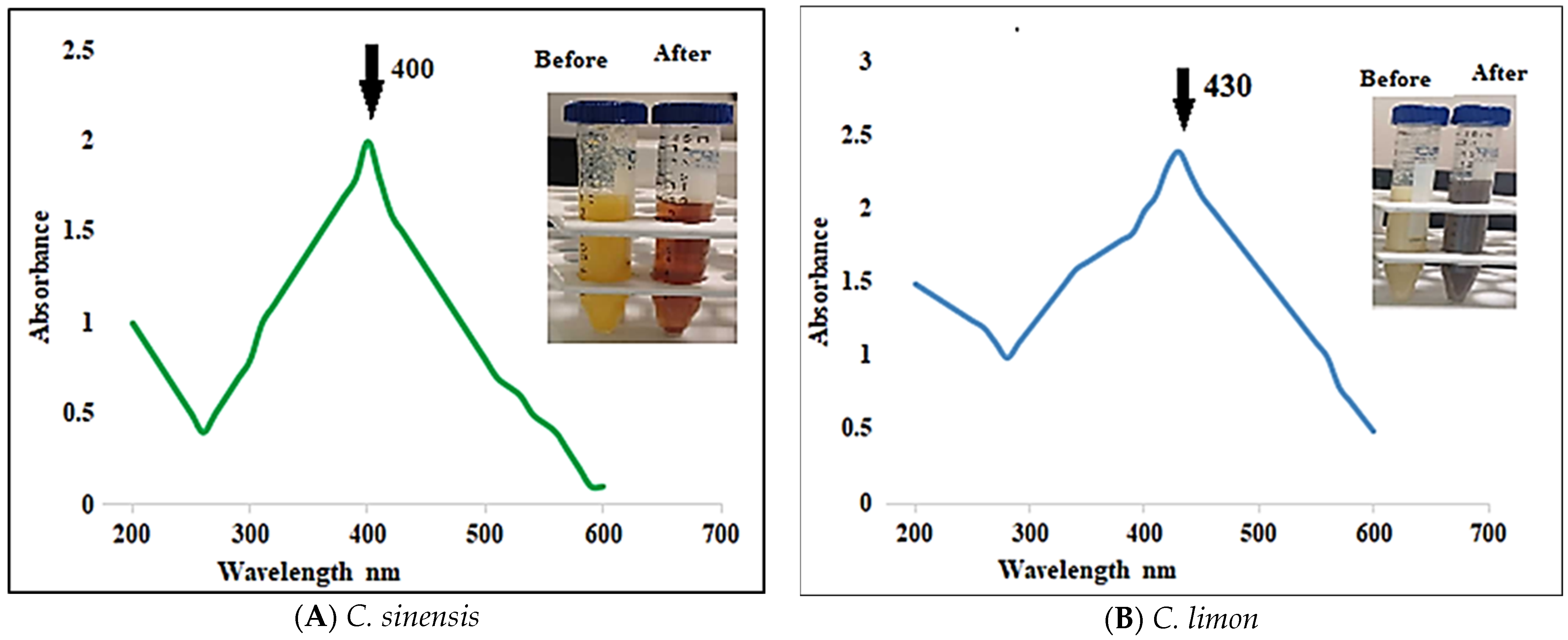
3.3.1. UV–Vis Spectra Analysis

Silver nanoparticles synthesized from fresh C. sinensis and C. limon peel juices showed a UV-vis absorbance spectra peak at 400 and 430 nm, respectively (Figure 1). Depending on their size, silver nanoparticles usually show absorption peaks in the 400–450 nm range. Because of the positively charged nucleus and unbound electrons, silver nanoparticles produce an SPR absorption band that resonantly absorbs the light wave. The SPR pattern depends on the size, shape, and dielectric characteristics of the metal nanoparticles. A single SPR peak is typical for spherical-shaped metal nanoparticles, suggesting the synthesis of spherical silver nanoparticles in the current investigation. Our results indicate the formation of different-sized silver nanoparticles from two samples, which can be due to the different content of antioxidants and protein in the peel juices, as shown in Table 1. The compositions and contents of phytochemical and nutritional in Citrus vary significantly among different varieties [40,41].

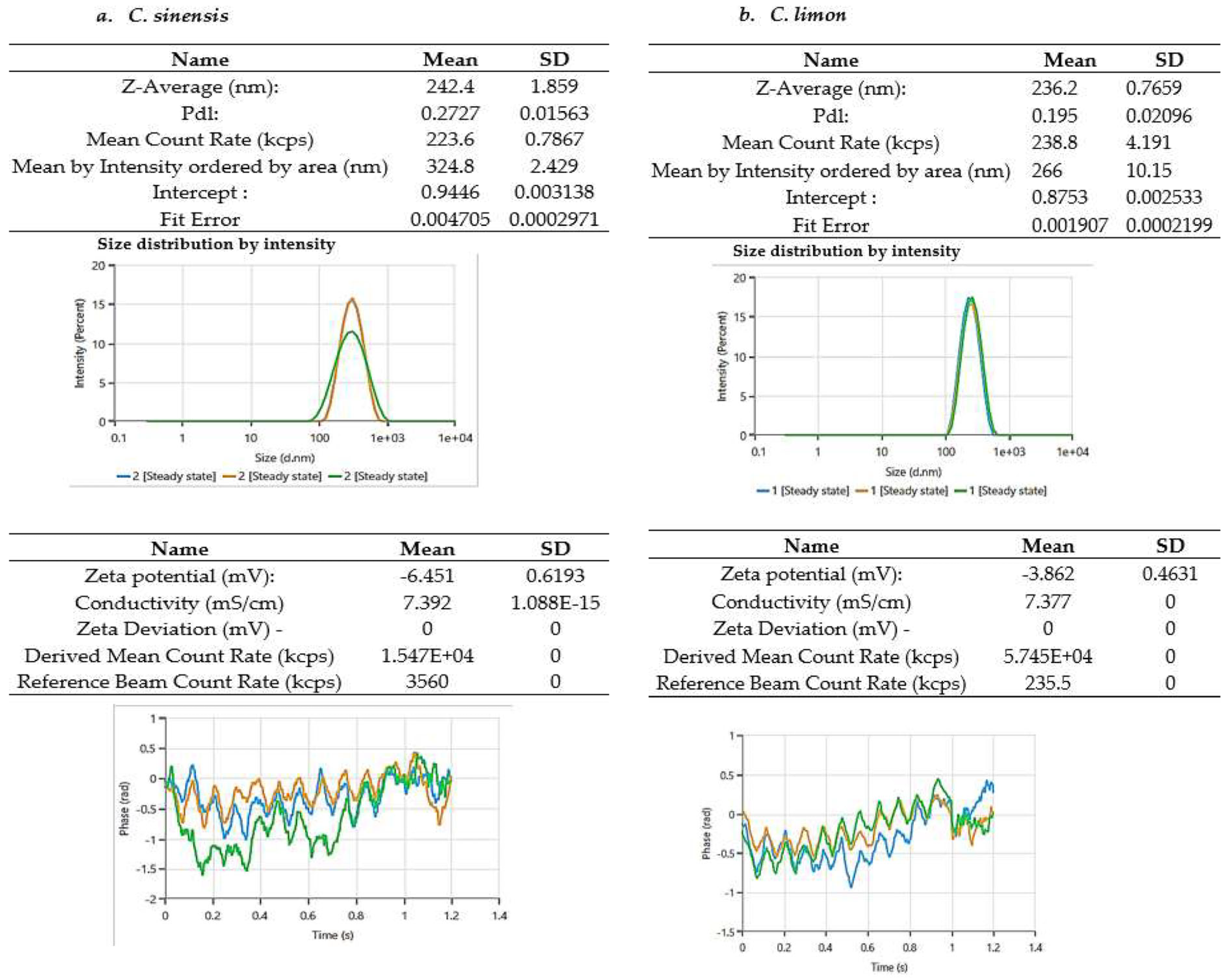
3.3.2. Zeta-Average Particle Size and Zeta Potential Analysis

Ag nanoparticles synthesized from the C. sinensis peels have a zeta-average mean size of 242.4 nm and a polydispersity index (PDI) of 0.272. Ag nanoparticles from the C. limon peels had a Z-average mean size of 236.2 nm and a PDI of 0.195, as shown in Figure 2. The PDI value represents the average uniformity of nanoparticles in a solution, representing their agglomeration and homogeneity across the sample. A wide size distribution is indicated by a PDI value greater than 0.7, whereas a monodispersed distribution is indicated by a value less than 0.1 [42]. The electrical charge of a nanoparticle is represented by its zeta potential. This value provides information about the stability of the particles. The graph of the zeta potential distribution is shown in Figure 2. The zeta potential of the C. sinensis peel Ag nanoparticle was found to be −6.45 mV, and the zeta potential of Ag nanoparticles from the C. limon peels was −3.862 mV. A negative zeta potential in our results indicated repulsion between the particles and stability of the colloidal state [43].

3.3.3. TEM and SEM/EDX Analysis

TEM is one of the best methods for analyzing the shape and size of nanoparticles [44]. The TEM image shown in Figure 3 confirms the synthesis of Ag nanoparticles using C. sinensis and C. limon peels. With an average diameter of roughly 13 nm for C. sinensis Ag nanoparticles and 21 nm for C. limon Ag nanoparticles, these particles showed a spherical form. These findings support our findings of the UV-vis spectra, as spherical-shaped Ag nanoparticles are reported to have UV-vis spectra in the 390–430 nm region [45]. Scanning electron microscopy images of the Ag nanoparticles revealed primarily spherical particles with a size below 100 nm for both samples. EDX analysis was used to assess the elemental composition and relative abundance of the Ag nanoparticles from C. sinensis and C. limon peels. EDX displayed a significant silver signal and a sharp peak, confirming the existence of Ag nanoparticles in both samples (Figure 4). The EDX spectrum illustrates the whole chemical composition and purity of Ag nanoparticles. The percentage relative composition of elements for Ag nanoparticles from C. sinensis is as follows: 15.34% carbon, 25.82% for oxygen, 1.38% sodium, 0.69% aluminum, 10.41% for silicon, 1.86% for potassium, 2.81% zinc, and 41.69% for silver; and for C. limon, the composition is as follows: 7.28% carbon, 11.36% nitrogen, 20.36% oxygen, 0.49% silicon, 0.29% chlorine, 0.46% potassium, 1.48% zinc, and 58.27% silver. The other elements can be phyto-constituents present in the peel that can serve as capping agents attached to the surface of silver nanoparticles [36,37].
Both C. sinensis and C. limon peel juices were rich in active chemicals (Table 1), which can help with the environmentally friendly formation of nanoparticles by capping them so that they do not aggregate as much [36,37,38,39]. These components in peel juices were further examined using FTIR spectroscopy (Figure 5). FTIR is used to find the distinctive functional groups from the spectral bands to determine the conjugation between the nanomaterial and adsorbed biomolecules [46]. Ag nanoparticles are produced with plant extracts by a three-step process. The first stage is called the activation phase, during which the extract’s reducing agents aid in the reduction of metal ions. Phase II is the growth phase, in which NPs are formed by reduced metal ions, capped at last for stability by metabolites found in the extract. Different functional peak stretches in peel juices were found between 3700 and 2300 cm−1. The components of flavonoids and phenolics, which extend their OH groups, resulted in a large absorption band in the higher energy range of the FTIR spectra at 3410.15, 3394.72, and 33,871.00 cm−1 in both samples [47]. The presence of absorption bands at 2924.09 cm−1 in both samples suggested the presence of hydroxyl compounds or –C–H stretching, while C=O or N–H stretching vibrations were attributed to the peaks that emerged at 2368.59 and 2376.30, cm−1. The peaks at 1627.92 and 1621.49 cm−1 corresponded to a C=O stretching vibration in the carboxyl group or a bending C=N vibration in the amide group. Flavonoids and phenolic chemicals have been linked to C–O bond stretching, or aromatic rings, as indicated by the emergence of strong intensity bands at 1072.42, 1064.71, 1041.56, and 1066.99 cm−1 [48]. FTIR is beneficial for characterizing a nanoparticle’s surface. It ascertains the chemical composition and reactive surface sites that give rise to the surface reactivity of the nanoparticle surface. The peak intensity of FTIR provides a clear indicator of the type of material present, making it a great technique for qualitative study [49].

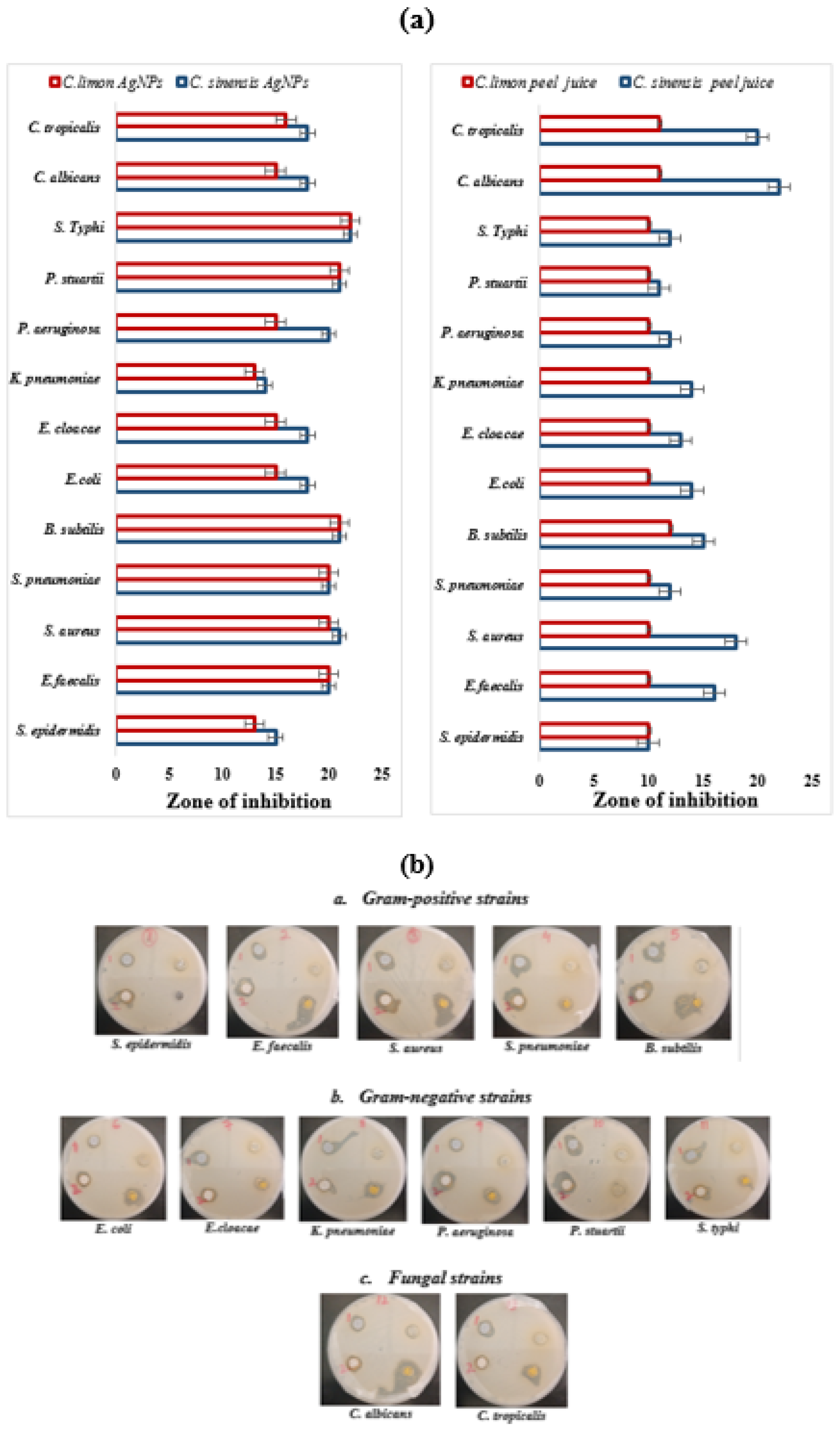
3.4. Antimicrobial Activity

Silver is regarded as a broad-spectrum antibacterial agent, but its nano form is beneficial because of its vast surface area, which enhances the duration of microbial exposure. We tested 13 pathogenic microorganisms for the antibacterial activity of C. sinensis and C. limon peel juices and prepared Ag nanoparticles. With varying degrees of efficacy, both the juices and nanoparticles effectively inhibited the growth of pathogens, as shown in Figure 6. However, C. sinensis peel juices exhibit higher antimicrobial activity against all test strains when compared with C. limon peel juices. Also, the Ag nanoparticles derived from C. sinensis peels were more antimicrobial than the ones prepared from C. limon peels. The size of the inhibition zone correlates with the degree of microbial susceptibility. Notably, the Gram-positive strains showed the highest sensitivity to all samples compared with the Gram-negative strains, as shown in Table 2. S. aureus was the most sensitive Gram-positive strain, showing an inhibition zone of 21 ± 1.5 mm for C. sinensis AgNPs. The best results among the Gram-negative strains were observed against S. typhi, with inhibition zones of 22 ± 1.0 mm for the synthesized Ag nanoparticles. C. sinensis peel juice showed remarkable antifungal activity against both candida strains, showing inhibition zones of 22 ± 20 against C. albicans and 20 ± 2.0 mm against C. tropicalis mm. Gram-negative bacteria exhibited greater resistance to the nanoparticles compared with Gram-positive bacteria. This discrepancy can be attributed to the unique morphology of Gram-negative bacterial cell walls, which have a hydrophilic lipopolysaccharide outer layer that is highly resistant to the infiltration of antibacterial agents [50]. Additionally, the presence of enzymes in the periplasmic space of Gram-negative bacteria contributes to the breakdown of antibacterial molecules [51,52]. Overall C. sinensis AgNPs were more effective antimicrobial agents than C. limon AgNPs. The reason can be their diminutive size. Because they have a comparatively bigger surface area than large NPs, small NPs are typically more hazardous [53]. The increased surface area also enhances reactive oxygen species (ROS) levels, which can damage essential biomolecules such as DNA, proteins, and lipids [54]. Numerous studies have highlighted the potent antimicrobial activity of small-sized nanoparticles [55,56,57]. Many pathogenic microbial strains have developed a resistance mechanism against antibiotics mainly because of their hydrophilic nature resulting in low intracellular retention and poor permeability. On the other hand, NPs can cross the hydrophobic barrier, particularly in phagocytic cells that absorb NPs and become more active inside the cell [58].

4. Conclusions

The ecological management of fruit peel waste has become crucial with the added necessity of achieving financial benefits. Synthesizing nanoparticles from this waste offers a sustainable and environmentally friendly solution. We successfully prepared silver nanoparticles from the peels of two different citrus fruits. The peel juices were rich in antioxidants and proteins, which can aid the reduction of Ag ions into Ag nanoparticles and the capping of synthesized nanoparticles. The ascorbic acid levels in both peel juices could be a key factor contributing to the antibacterial activity observed in this study. The biomedical applications of the prepared nanoparticles were demonstrated by their antibacterial properties.

Author Contributions

Conceptualization, R.S.B.; methodology, R.S.B. software, R.S.B. validation, S.A.-D. and R.S.B.; formal analysis, A.I.A.K. investigation, R.S.B. resources, A.M.A.-D. data curation, A.M.A.-D. and R.S.B.; writing—original draft preparation, R.S.B.; writing—review and editing, R.S.B.; visualization, supervision, R.S.B.; project administration, S.A.-D.; funding acquisition A.M.A.-D. All authors have read and agreed to the published version of the manuscript.

Funding

This research project was funded by Researchers Supporting Project number (RSP 2024R183), King Saud University, Riyadh, Saudi Arabia.

Institutional Review Board Statement

Not applicable.

Informed Consent Statement

Not applicable.

Data Availability Statement

The data are presented in this manuscript.

Acknowledgments

The authors extend their appreciation to the Researchers Support Project number RSP2024R183 King Saud University for supporting this research.

Conflicts of Interest

The authors declare no conflicts of interest.

References

  1. Malik, S.; Muhammad, K.; Waheed, Y. Nanotechnology: A Revolution in Modern Industry. Molecules 2023, 28, 661. [Google Scholar] [CrossRef]
  2. Ayilara, M.S.; Olanrewaju, O.S.; Babalola, O.O.; Odeyemi, O. Waste Management through Composting: Challenges and Potentials. Sustainability 2020, 12, 4456. [Google Scholar] [CrossRef]
  3. Abubakar, I.R.; Maniruzzaman, K.M.; Dano, U.L.; AlShihri, F.S.; AlShammari, M.S.; Ahmed, S.M.S.; Al-Gehlani, W.A.G.; Alrawaf, T.I. Environmental Sustainability Impacts of Solid Waste Management Practices in the Global South. Int. J. Environ. Res. Public Health 2022, 19, 12717. [Google Scholar] [CrossRef]
  4. Aswathi, V.P.; Meera, S.; Maria, C.G.A.; Nidhin, M. Green synthesis of nanoparticles from biodegradable waste extracts and their applications: A critical review. Nanotechnol. Environ. Eng. 2023, 8, 377–397. [Google Scholar] [CrossRef]
  5. Saratale, R.G.; Saratale, G.D.; Shin, H.S.; Jacob, J.M.; Pugazhendhi, A.; Bhaisare, M.; Kumar, G. New insights on the green synthesis of metallic nanoparticles using plant and waste biomaterials: Current knowledge, their agricultural and environmental applications. Environ. Sci. Pollut. Res. Int. 2018, 25, 10164–10183. [Google Scholar] [CrossRef]
  6. Osman, A.I.; Zhang, Y.; Farghali MRashwan, A.K.; Eltaweil, A.S.; Abd El-Monaem, E.M. Synthesis of green nanoparticles for energy, biomedical, environmental, agricultural, and food applications: A review. Environ. Chem. Lett. 2024, 22, 841–887. [Google Scholar] [CrossRef]
  7. Chopra, H.; Bibi, S.; Singh, I.; Hasan, M.M.; Khan, M.S.; Yousafi, Q.; Baig, A.A.; Rahman, M.M.; Islam, F.; Emran, T.B.; et al. Green Metallic Nanoparticles: Biosynthesis to Applications. Front. Bioeng. Biotechnol. 2022, 10, 874742. [Google Scholar] [CrossRef] [PubMed]
  8. Nirmal, N.P.; Khanashyam, A.C.; Mundanat, A.S.; Shah, K.; Babu, K.S.; Thorakkattu, P.; Al-Asmari, F.; Pandiselvam, R. Valorization of Fruit Waste for Bioactive Compounds and Their Applications in the Food Industry. Foods 2023, 12, 556. [Google Scholar] [CrossRef]
  9. Sha, S.P.; Modak, D.; Sarkar, S.; Roy, S.K.; Sah, S.P.; Ghatani, K.; Bhattacharjee, S. Fruit waste: A current perspective for the sustainable production of pharmacological, nutraceutical, and bioactive resources. Front. Microbiol. 2023, 14, 1260071. [Google Scholar] [CrossRef]
  10. Thatyana, M.; Dube, N.P.; Kemboi, D.; Manicum, A.E.; Mokgalaka-Fleischmann, N.S.; Tembu, J.V. Advances in Phytonanotechnology: A Plant-Mediated Green Synthesis of Metal Nanoparticles Using Phyllanthus Plant Extracts and Their Antimicrobial and Anticancer Applications. Nanomaterials 2023, 13, 2616. [Google Scholar] [CrossRef]
  11. Javed, R.; Zia, M.; Naz, S.; Ain, A.N.; Ao, Q. Role of capping agents in the application of nanoparticles in biomedicine and environmental remediation: Recent trends and future prospects. J. Nanobiotechnol. 2020, 18, 172. [Google Scholar] [CrossRef]
  12. Ashique, S.; Afzal, O.; Khalid, M.; Ahmad, M.F.; Upadhyay, A.; Kumar, S.; Garg, A.; Ramzan, M.; Hussain, A.; Altamimi, M.A.; et al. Biogenic nanoparticles from waste fruit peels: Synthesis, applications, challenges and future perspectives. Int. J. Pharm. 2023, 643, 123223. [Google Scholar] [CrossRef]
  13. Vera, J.; Herrera, W.; Hermosilla, E.; Díaz, M.; Parada, J.; Seabra, A.B.; Tortella, G.; Pesenti, H.; Ciudad, G.; Rubilar, O. Antioxidant Activity as an Indicator of the Efficiency of Plant Extract-Mediated Synthesis of Zinc Oxide Nanoparticles. Antioxidants 2023, 12, 784. [Google Scholar] [CrossRef]
  14. Abuzeid, H.M.; Julien, C.M.; Zhu, L.; Hashem, A.M. Green Synthesis of Nanoparticles and Their Energy Storage, Environmental, and Biomedical Applications. Crystals 2023, 13, 1576. [Google Scholar] [CrossRef]
  15. Ahmed, S.F.; Mofijur, M.; Rafa, N.; Chowdhury, A.T.; Chowdhury, S.; Nahrin, M.; Islam, A.S.; Ong, H.C. Green approaches in synthesising nanomaterials for environmental nano bioremediation: Technological advancements, applications, benefits and challenges. Environ. Res. 2022, 204, 111967. [Google Scholar] [CrossRef]
  16. Rai, M.; Ingle, A.P.; Trzcińska-Wencel, J.; Wypij, M.; Bonde, S.; Yadav, A.; Kratošová, G.; Golińska, P. Biogenic Silver Nanoparticles: What We Know and What Do We Need to Know? Nanomaterials 2021, 11, 2901. [Google Scholar] [CrossRef]
  17. Russo, C.; Maugeri, A.; Lombardo, G.E.; Musumeci, L.; Barreca, D.; Rapisarda, A.; Cirmi, S.; Navarra, M. The Second Life of Citrus Fruit Waste: A Valuable Source of Bioactive Compounds. Molecules 2021, 26, 5991. [Google Scholar] [CrossRef]
  18. Vasyliev, G.; Vorobyova, V. Valorization of Food Waste to Produce Eco-Friendly Means of Corrosion Protection and “Green” Synthesis of Nanoparticles. Adv. Mater. Sci. Eng. 2020, 2020, 1. [Google Scholar] [CrossRef]
  19. Moult, J.A.; Allan, S.R.; Hewitt, C.N.; Berners-Lee, M. Greenhouse Gas Emissions of Food Waste Disposal Options for UK Retailers. Food Policy 2018, 77, 50–58. [Google Scholar] [CrossRef]
  20. Vendruscolo, F.; Albuquerque, P.M.; Streit, F.; Esposito, E.; Ninow, J.L. Apple Pomace: A Versatile Substrate for Biotechnological Applications. Crit. Rev. Biotechnol. 2008, 28, 1–12. [Google Scholar] [CrossRef] [PubMed]
  21. Gowman, A.C.; Picard, M.C.; Lim, L.-T.; Misra, M.; Mohanty, A.K. Fruit Waste Valorization for Biodegradable Biocomposite Applications: A Review. Bioresources 2019, 14, 10047–10092. [Google Scholar] [CrossRef]
  22. Trotta, F.; Da Silva, S.; Massironi, A.; Mirpoor, S.F.; Lignou, S.; Ghawi, S.K.; Charalampopoulos, D. Silver Bionanocomposites as Active Food Packaging: Recent Advances & Future Trends Tackling the Food Waste Crisis. Polymers 2023, 15, 4243. [Google Scholar] [CrossRef] [PubMed]
  23. Ahmed, S.; Kaur, G.; Sharma, P.; Singh, S.; Ikram, S. Fruit waste (peel) as bio-reductant to synthesize silver nanoparticles with antimicrobial, antioxidant and cytotoxic activities. J. Appl. Biomed. 2018, 16, 221–231. [Google Scholar] [CrossRef]
  24. Xing, Y.; Li, W.; Wang, Q.; Li, X.; Xu, Q.; Guo, X.; Bi, X.; Liu, X.; Shui, Y.; Lin, H.; et al. Antimicrobial Nanoparticles Incorporated in Edible Coatings and Films for the Preservation of Fruits and Vegetables. Molecules 2019, 24, 1695. [Google Scholar] [CrossRef]
  25. Maqbool, Z.; Khalid, W.; Atiq, H.T.; Koraqi, H.; Javaid, Z.; Alhag, S.K.; Al-Shuraym, L.A.; Bader, D.M.D.; Almarzuq, M.; Afifi, M.; et al. Citrus Waste as Source of Bioactive Compounds: Extraction and Utilization in Health and Food Industry. Molecules 2023, 28, 1636. [Google Scholar] [CrossRef]
  26. Saeed, M.; Kamboh, A.A.; Huayou, C. Promising future of citrus waste into fermented high-quality bio-feed in the poultry nutrition and safe environment. Poult. Sci. 2024, 103, 103549. [Google Scholar] [CrossRef]
  27. Lv, X.; Zhao, S.; Ning, Z.; Zeng, H.; Shu, Y.; Tao, O.; Xiao, C.; Lu, C.; Liu, Y. Citrus fruits as a treasure trove of active natural metabolites that potentially provide benefits for human health. Chem. Cent. J. 2015, 9, 68. [Google Scholar] [CrossRef] [PubMed]
  28. Saleem, M.; Durani, A.I.; Asari, A.; Ahmed, M.; Ahmad, M.; Yousaf, N.; Muddassar, M. Investigation of antioxidant and antibacterial effects of citrus fruits peels extracts using different extracting agents: Phytochemical analysis with in silico studies. Heliyon 2023, 9, e15433. [Google Scholar] [CrossRef]
  29. Pérez, M.; Dominguez-López, I.; Lamuela-Raventós, R.M. The Chemistry Behind the Folin-Ciocalteu Method for the Estimation of (Poly)phenol Content in Food: Total Phenolic Intake in a Mediterranean Dietary Pattern. J. Agric. Food Chem. 2023, 71, 17543–17553. [Google Scholar] [CrossRef] [PubMed]
  30. Jagota, S.; Dani, H. A new colorimetric technique for the estimation of vitamin C using Folin phenol reagent. Anal. Biochem. 1982, 127, 178–182. [Google Scholar] [CrossRef]
  31. Baliyan, S.; Mukherjee, R.; Priyadarshini, A.; Vibhuti, A.; Gupta, A.; Pandey, R.P.; Chang, C.M. Determination of Antioxidants by DPPH Radical Scavenging Activity and Quantitative Phytochemical Analysis of Ficus religiosa. Molecules 2022, 27, 1326. [Google Scholar] [CrossRef]
  32. Bradford, M.M. A rapid and sensitive method for the quantitation of microgram quantities of protein utilizing the principle of protein-dye binding. Anal. Biochem. 1976, 72, 248–254. [Google Scholar] [CrossRef] [PubMed]
  33. Amini, S.M.; Akbari, A. Metal nanoparticles synthesis through natural phenolic acids. IET Nanobiotechnol. 2019, 13, 771–777. [Google Scholar] [CrossRef] [PubMed]
  34. Huang, Y.; Mosleh, I.; Abbaspourrad, A. Impact of protein/peptide templates on metallic nanoparticle synthesis and applications. Nano-Struct. Nano-Objects 2022, 30, 100864. [Google Scholar] [CrossRef]
  35. Saif, A.; Anjum, L.; Faisal, Z.; Akram, N.; Shah, Y.A.; Irfan, R.; Saeed, F.; Afzaal, M.; Asif Shah, M. Recent advances in protein-based nanoparticles and their applications in the delivery of bioactive compounds. Int. J. Food Prop. 2023, 26, 2866–2880. [Google Scholar] [CrossRef]
  36. Suhag, R.; Kumar, R.; Dhiman, A.; Sharma, A.; Prabhakar, P.K.; Gopalakrishnan, K.; Kumar, R.; Singh, A. Fruit peel bioactives, valorisation into nanoparticles and potential applications: A review. Crit. Rev. Food Sci. Nutr. 2023, 63, 6757–6776. [Google Scholar] [CrossRef] [PubMed]
  37. Chugh, G.; Singh, B.R.; Adholeya, A.; Barrow, C.J. Role of proteins in the biosynthesis and functioning of metallic nanoparticles. Crit. Rev. Biotechnol. 2022, 42, 1045–1060. [Google Scholar] [CrossRef] [PubMed]
  38. Nam, K.T.; Lee, Y.J.; Krauland, E.M.; Kottmann, S.T.; Belcher, A.M. Peptide-mediated reduction of silver ions on engineered biological scaffolds. ACS Nano 2008, 2, 1480–1486. [Google Scholar] [CrossRef] [PubMed]
  39. Selvakannan, P.R.; Swami, A.; Srisathiyanarayanan, D.; Shirude, P.S.; Pasricha, R.; Mandale, A.B.; Sastry, M. Synthesis of aqueous Au core-Ag shell nanoparticles using tyrosine as a pH-dependent reducing agent and assembling phase-transferred silver nanoparticles at the air-water interface. Langmuir 2004, 20, 7825–7836. [Google Scholar] [CrossRef]
  40. Liu, S.; Lou, Y.; Li, Y.; Zhang, J.; Li, P.; Yang, B.; Gu, Q. Review of phytochemical and nutritional characteristics and food applications of Citrus L. fruits. Front. Nutr. 2022, 9, 968604. [Google Scholar] [CrossRef]
  41. Pei, Y.; He, C.; Liu, H.; Shen, G.; Feng, J. Compositional Analysis of Four Kinds of Citrus Fruits with an NMR-Based Method for Understanding Nutritional Value and Rational Utilization: From Pericarp to Juice. Molecules 2022, 27, 2579. [Google Scholar] [CrossRef] [PubMed]
  42. Al-Dbass, A.M.; Daihan, S.A.; Al-Nasser, A.A.; Al-Suhaibani, L.S.; Almusallam, J.; Alnwisser, B.I.; Saloum, S.; Alotaibi, R.S.; Alessa, L.A.; Bhat, R.S. Biogenic Silver Nanoparticles from Two Varieties of Agaricus bisporus and Their Antibacterial Activity. Molecules 2022, 27, 7656. [Google Scholar] [CrossRef] [PubMed] [PubMed Central]
  43. Sennett, P.; Olivier, J.P. Colloidal Dispersions, Electrokinetic Effects, and the Concept of Zeta Potential. Ind. Eng. Chem. 1965, 57, 32–50. [Google Scholar] [CrossRef]
  44. Zhang, S.; Wang, C. Precise Analysis of Nanoparticle Size Distribution in TEM Image. Methods Protoc. 2023, 6, 63. [Google Scholar] [CrossRef] [PubMed]
  45. Mourdikoudis, S.; Pallares, R.M.; Thanh, N.T. Characterization techniques for nanoparticles: Comparison and complementarity upon studying nanoparticle properties. Nanoscale 2018, 10, 12871–12934. [Google Scholar] [CrossRef] [PubMed]
  46. Ganaie, T.A.; Masoodi, F.A.; Rather, S.A.; Wani, S.M. Physicochemical, antioxidant and FTIR-ATR spectroscopy evaluation of Kashmiri honeys as food quality traceability and Himalayan brand. J. Food Sci. Technol. 2021, 58, 4139–4148. [Google Scholar] [CrossRef] [PubMed]
  47. Bhat, R.S.; Alghamdi, J.M.; Aldbass, A.M.; Aljebrin, N.A.; Alangery, A.B.; Soliman, D.A.; Al-Daihan, S. Biochemical and FT-IR pro-filing of Tritium aestivum L. seedling in response to sodium fluoride treatment. Fluoride 2022, 55, 81–89. [Google Scholar]
  48. Bhat, R.S.; Al-Dbass, A.M.; Alghamdia, J.M.; Alonazia, M.A.; Al-Daihan, S. Trigonella foenum-graecum L. Seed germination under sodium halide salts exposure. Res. Rep. Fluoride 2023, 56, 169–179. [Google Scholar]
  49. Campanale, C.; Savino, I.; Massarelli, C.; Uricchio, V.F. Fourier Transform Infrared Spectroscopy to Assess the Degree of Alteration of Artificially Aged and Environmentally Weathered Microplastics. Polymers 2023, 15, 911. [Google Scholar] [CrossRef]
  50. Breijyeh, Z.; Jubeh, B.; Karaman, R. Resistance of gram-negative bacteria to current antibacterial agents and approaches to resolve it. Molecules 2020, 25, 1340. [Google Scholar] [CrossRef]
  51. Miller, S.I.; Salama, N.R. The gram-negative bacterial periplasm: Size matters. PLoS Biol. 2018, 16, e2004935. [Google Scholar] [CrossRef]
  52. Munita, J.M.; Arias, C.A. Mechanisms of antibiotic resistance. Virulence Mech. Bact. Pathog. 2016, 22, 481–511. [Google Scholar]
  53. Abbasi, R.; Shineh, G.; Mobaraki, M.; Doughty, S.; Tayebi, L. Structural parameters of nanoparticles affecting their toxicity for biomedical applications: A review. J. Nanopart. Res. 2023, 25, 43. [Google Scholar] [CrossRef]
  54. Yu, Z.; Li, Q.; Wang, J.; Yu, Y.; Wang, Y.; Zhou, Q.; Li, P. Reactive Oxygen Species-Related Nanoparticle Toxicity in the Biomedical Field. Nanoscale Res. Lett. 2020, 15, 115. [Google Scholar] [CrossRef]
  55. Bhat, R.S.; Almusallam, J.; Al Daihan, S.; Al-Dbass, A. Biosynthesis of silver nanoparticles using Azadirachta indica leaves: Characterisation and impact on Staphylococcus aureus growth and glutathione-S-transferase activity. IET Nanobiotechnol. 2019, 13, 498–502. [Google Scholar] [CrossRef]
  56. Al-Zahrani, S.A.; Bhat, R.S.; Al Rashed, S.A.; Mahmood, A.; Al Fahad, A.; Alamro, G.; Almusallam, J.; Al Subki, R.; Orfali, R.; Al Daihan, S. Green-synthesized silver nanoparticles with aqueous extract of green algae Chaetomorpha ligustica and its anticancer potential. Green Process. Synth. 2021, 10, 711–721. [Google Scholar] [CrossRef]
  57. Alduraihem, N.S.; Bhat, R.S.; Al-Zahrani, S.A.; Elnagar, D.M.; Alobaid, H.M.; Daghestani, M.H. Anticancer and Antimicrobial Activity of Silver Nanoparticles Synthesized from Pods of Acacia nilotica. Processes 2023, 11, 301. [Google Scholar] [CrossRef]
  58. Wang, L.; Hu, C.; Shao, L. The antimicrobial activity of nanoparticles: Present situation and prospects for the future. Int. J. Nanomed. 2017, 12, 1227–1249. [Google Scholar] [CrossRef] [PubMed]
Figure 1. Color change in juices ((A) C. sinensis and (B) C. limon) upon adding silver nitrate and the absorbance peak in UV–visual spectra shown by prepared Ag nanoparticles.
Inorganics 12 00180 g001
Figure 2. Zeta-average size (DLS) and zeta potential (ELS) values and graphs of the Ag nanoparticles from (a) C. sinensis and (b) C. limon peels. (Different colors in graph shows readings examined three times and Mean are presented above each graph in table form).
Inorganics 12 00180 g002
Figure 3. TEM and SEM images for Ag nanoparticles prepared from (a) C. sinensis and (b) C. limon peels.
Inorganics 12 00180 g003
Figure 4. (a) EDX spectrum, (b) weight%, and (c) elemental mapping of Ag nanoparticles prepared from C. sinensis (left side) and C. limon (right side) peels.
Inorganics 12 00180 g004
Figure 5. FTIR spectra of C. sinensis and C. limon peel juices and synthesized Ag nanoparticles. Arrows indicate the shift in the peaks during nanoparticle synthesis.
Inorganics 12 00180 g005
Figure 6. (a) Zone of inhibition (mm) of C. limon and C. sinensis peel juices and synthesized Ag nanoparticles. (b) Well diffusion assay demonstrating the antibacterial activity of 1. C. limon and 2. C. sinensis against a. Gram-positive strains; b. Gram-negative strains; and c. fungal strains. (right)—Ag NPS; (left)—peel juice.
Inorganics 12 00180 g006
Table 1. Antioxidants and total protein of fresh aqueous peel juice.
Parameter (ug/mL)C. sinensisC. limon
Total phenol (standard—GA)178 ± 1.1130 ± 1.5
Ascorbic acid (standard—AA)33 ± 1.022 ± 1.6
DPPH IC50 (standard AA-IC50-26)30 ± 0.731.5 ± 0.5
Total protein (standard—BSA)93 ± 2.075 ± 0.7
The values are presented as mean ± standard deviation.
Table 2. Zone of inhibition (mm) of C. limon and C. sinensis peel juices and synthesized Ag nanoparticles.
StrainsZone of Inhibition
C. sinensisC. limon
Peel JuicesAgNPsPeel JuicesAgNP
S. epidermidis10 ± 1.015 ± 1.510 ± 0.013 ± 2.0
E. faecalis16 ± 1.520 ± 1.510 ± 1.020 ± 1.0
S. aureus18 ± 1.021 ± 2.011 ± 1.020 ± 1.5
S. pneumoniae12 ± 1.020 ± 1.010 ± 1.520 ± 2.0
B. subtilis15 ± 2.021 ± 1.511 ± 1.020 ± 1.0
E. coli14 ± 1.018 ± 1.010 ± 1.515 ± 1.0
E. cloacae13 ± 1.018 ± 2.010 ± 2.015 ± 2.0
K. pneumoniae14 ± 1.514 ± 1.010 ± 1.513 ± 1.0
P. aeruginosa12 ± 1.520 ± 1.010 ± 1.015 ± 2.0
P. stuartii11 ± 1.021 ± 2.010 ± 1.521 ± 1.0
S. Typhi12 ± 1.522 ± 1.010 ± 2.022 ± 1.5
C. albicans22 ± 2.018 ± 1.511 ± 1.015 ± 1.0
C. tropicalis20 ± 2.018 ± 0011 ± 1.016 ± 1.0
Disclaimer/Publisher’s Note: The statements, opinions and data contained in all publications are solely those of the individual author(s) and contributor(s) and not of MDPI and/or the editor(s). MDPI and/or the editor(s) disclaim responsibility for any injury to people or property resulting from any ideas, methods, instructions or products referred to in the content.

