Article

Association of F. alocis and D. pneumosintes with Periodontitis Disease Severity and Red Complex Bacteria

by
Hawaabi F. M. Shaikh
1,
Pratima U. Oswal
1,
Manohar Suresh Kugaji
2,*,
Sandeep S. Katti
1,
Kishore Gajanan Bhat
3,
Eswar Kandaswamy
4 and
Vinayak M. Joshi
4,*
1
Department of Periodontology, Maratha Mandal’s Nathajirao G. Halgekar Institute of Dental Sciences & Research Centre, Belagavi 590019, India
2
Centre for Advanced Medical Research, BLDE Deemed to be University, Vijayapura 586103, India
3
Arihant Superspecialty Hospital, Belagavi 590010, India
4
Department of Periodontics, School of Dentistry, Louisiana State University Health Sciences Center, New Orleans, LA 70119, USA
*
Authors to whom correspondence should be addressed.
Dent. J. 2024, 12(4), 105; https://doi.org/10.3390/dj12040105
Submission received: 22 February 2024 / Revised: 21 March 2024 / Accepted: 10 April 2024 / Published: 12 April 2024
(This article belongs to the Special Issue Oral Health and Dysbiosis)

Abstract

:
Oral biofilms are considered the principal etiological agent in the development of periodontitis. Novel species that may contribute to periodontitis and dysbiosis have been identified recently. The study aims to evaluate the presence of F. alocis and D. pneumosintes in healthy and diseased patients and their association with clinical parameters and with red complex bacteria. The study included 60 subjects, with 30 patients each in the healthy and periodontitis groups. The clinical parameters were noted, and samples were subjected to DNA extraction followed by a polymerase chain reaction. Statistical analysis was performed using the Graph Pad Prism software. Results: F. alocis and D. pneumosintes were detected at a significantly higher percentage in the periodontitis group compared to the healthy group (p < 0.05). D. pneumosintes was significantly associated with T. forsythia in the periodontitis group (p < 0.05). Both of these organisms were present in sites with higher clinical attachment loss (p < 0.05). This study demonstrated that both F. alocis and D. pneumosintes were detected at a significantly higher percentage in periodontitis subjects and were detected more frequently in sites with a greater clinical attachment loss. It was also evident that both F. alocis and D. pneumosintes can be present independently of other putative periodontal pathogens.

1. Introduction

Microbial biofilms are considered to be the primary etiological agent for the initiation and progression of periodontitis, among other multiple contributing factors [1]. The biofilm is composed of both health-associated and pathogenic microorganisms [2,3,4]. Dental plaque is a biofilm that forms over the teeth, gingiva, and mucosa and can be composed of bacteria, fungi, and algae [5,6]. Socransky et al. elucidated that the bacteria in dental plaque form different complexes, of which the red complex bacteria consisting of Porphyromonas gingivalis (P. gingivalis), Treponema denticola (T. denticola), and Tannerella forsythia (T. forsythia) along with the orange complex bacteria are considered to be the most putative periodontal pathogens [7]. Currently, open-ended microbial identification techniques and next-generation sequencing techniques have greatly increased the understanding of microbial diversity and newer species existing in dental plaque [8,9]. This knowledge has helped us to understand the microorganisms that are involved in the dysbiosis that occurs as part of periodontal disease.
Paster et al., in 2001, pointed out the presence of novel species that could play a part in dysbiosis and periodontitis [10]. Recent studies and systematic reviews have found further proof of these new species and their putative role in periodontal pathogenesis [8,9,11]. Some of the newly discovered pathogens belong to the phyla Bacteroidetes, Firmicutes, Proteobacteria, Spirochaetes, Synergistetes, and Candidatus saccharibacteria [11]. According to a cross-sectional study, Filifactor alocis (F. alocis) was identified as one of the novel species (among the 39 other species) showing strong evidence as a periodontal pathogen [9]. F. alocis has unique virulence properties, which are evidenced by factors like extracellular vesicles, lipoteichoic acid, oxidative stress resistance, and protease secretion, which together trigger a pro-inflammatory cytokine response from the periodontal tissues [12,13,14]. F. alocis and P. gingivalis have been demonstrated to co-exist symbiotically, and this association intensifies the virulence properties of F. alocis [15,16,17]. F. alocis also has the ability to invade epithelial cells, similarly to P. gingivalis, using filapodial projections or vesicle-mediated internalization [16,17] and has been found in individuals with different grades of periodontitis [18]. F. alocis, being an obligate anaerobe, prefers to colonize deeper sites and is often found in the middle third and apical third of the pockets [19], which provide an ideal anaerobic environment [20]. Similar to F. alocis, Dialister pneumosintes (D. pneumosintes) is an obligate anaerobic bacterium that was first isolated from the nasopharynx during the flu epidemic [21]. D. pneumosintes has been isolated from the oral biofilm and is known to cause gingivitis, periodontitis, and other local infections [22,23,24,25,26]. Among the virulence characteristics, lipopolysaccharides in the cell wall of D. pneumosintes may be the most important as they trigger the release of proinflammatory cytokines and matrix metalloproteinases, leading to periodontal tissue and alveolar bone destruction [27]. The relationship between D. pneumosintes occurrence and the development of oral disease, its detection in young males and patients with severe periodontitis, its correlation with the pocket depth, clinical attachment loss, and active disease sites, suggests its role in the etiopathogenesis of periodontal disease [22,23,24,25,28]. D. pneumosintes’s association with other putative periodontal pathogens, such as Aggregatibacter actinomycetemcomitans and P. gingivalis, has also been correlated with the prevalence of severe periodontitis [29]. Recent genomic studies have increased our understanding of oral microbiota, emphasizing the need to investigate novel periodontal pathogens [9]. To the best of our understanding, this is the first study to establish the association of F. alocis and D. pneumosintes with red complex bacteria and with periodontitis disease severity in the Indian population.
The current study primarily aims to evaluate the presence of F. alocis and D. pneumosintes in periodontal health and disease, and its correlation with clinical parameters. The secondary aim is to evaluate if these novel species and the red complex bacteria are more frequently detected simultaneously or can exist independently in moderate to severe periodontitis.

2. Materials and Methods

The initial screening of this cross-sectional study consisted of one hundred eighty-five subjects aged between 18 and 70 years conducted between January 2016 and January 2017. The study group consisted of 87 subjects with a suspected diagnosis of moderate to severe periodontitis and 98 subjects periodontally healthy from the outpatient department. The institutional ethics review board approved the study (Certificate number: 2015-16/1118). All study participants were interviewed according to a standardized protocol, and a written informed consent was obtained from all subjects before the examination. All selected subjects who met the inclusion–exclusion criteria were divided into a healthy group (H group), consisting of subjects with a healthy periodontium, and a periodontitis group (P group) based on the periodontal parameters [27].
The sample size was estimated based on a previous paper [19] with a significance level of p = 0.05 and a power of 95% using a conservative two-tailed testing approach. The power analysis was accomplished using G*Power 3.1 [30]. Subjects were excluded from the study if they underwent periodontal therapy or had antimicrobial therapy in the previous 3 months, had a history of any systemic diseases/conditions, were pregnant and lactating women, and smoked or consumed smokeless tobacco. The subjects who matched the inclusion–exclusion criteria were included in the study (Figure 1). This manuscript was prepared according to the STROBE cross-sectional study checklist [31] (Supplement File).

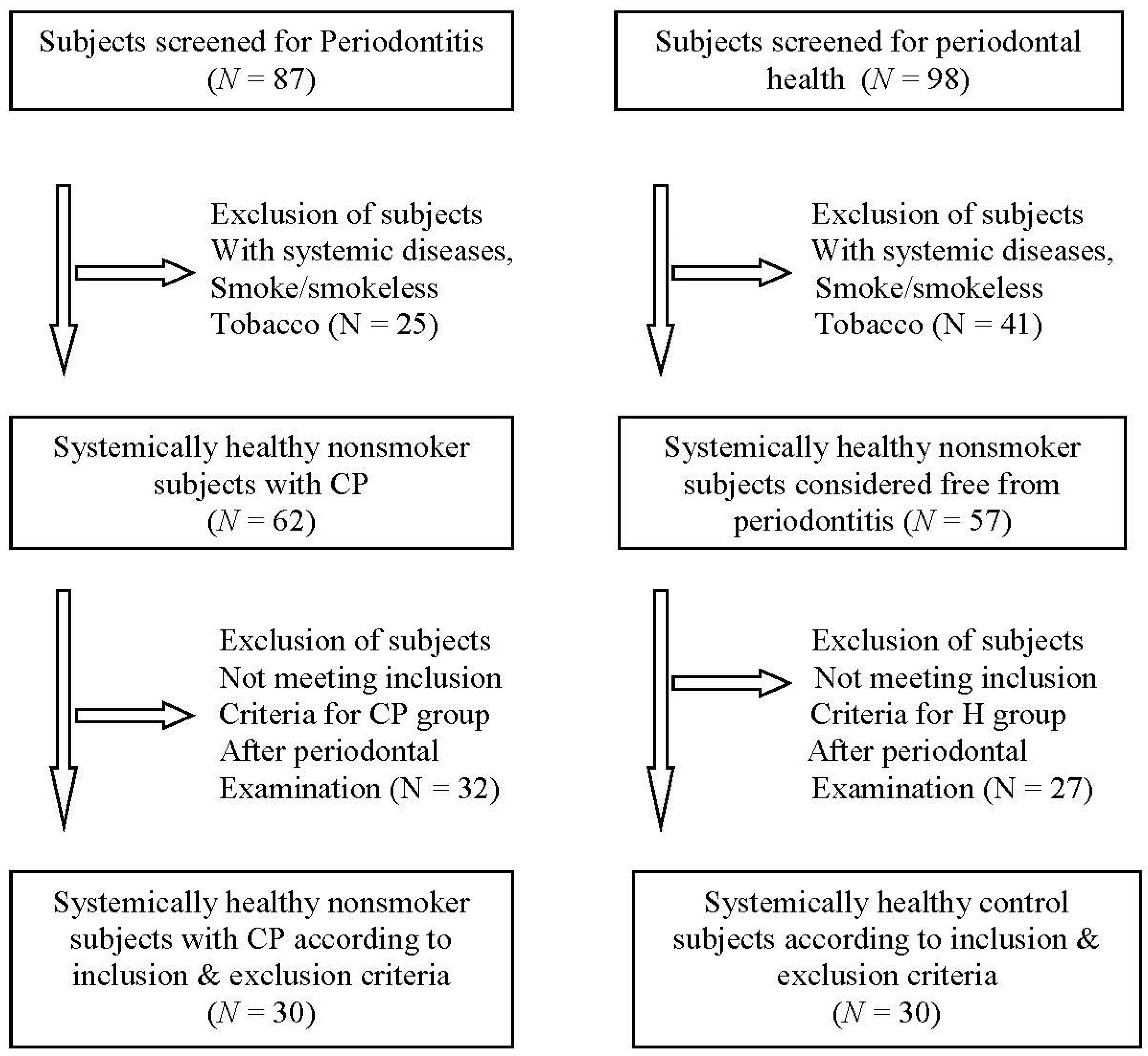
2.1. Clinical Examination and Subgingival Plaque Sample Collection

A comprehensive periodontal examination was carried out, and the clinical parameters recorded included plaque index (PI) [32], gingival index (GI) [33], bleeding index (BI) [34], PD, and CAL. The inclusion criteria for the H group were subjects with minimal signs of gingival inflammation or bleeding on probing (BOP) (BOP < 10%), absence of clinical attachment loss (CAL), and probing depth (PD) ≤ 3 mm. The P group included subjects with gingival inflammation with the presence of BOP, PD ≥ 5 mm, and CAL ≥ 3 mm (moderate to severe periodontitis). All participants needed to have a minimum of 20 teeth. Subgingival plaque samples were collected under strict asepsis using a sterile Gracey curette. In the P group, plaque samples were harvested from the three deepest sites with a probing depth of ≥5 mm, and in the H group, plaque samples were collected from the normal healthy gingival sulcus. The selected sites were isolated with sterile cotton rolls and air-dried, and supragingival plaque and calculus were removed. The plaque sample was then transferred into a 2 mL plastic vial containing the transport medium Tris-EDTA buffer (T.E.) and was then processed for DNA extraction. A single examiner did the clinical examination and sample collection.

2.2. DNA Extraction and Polymerase Chain Reaction

The DNA extraction for the plaque samples was carried out using a modified proteinase K method, as previously described [35]. Following the DNA extraction, a polymerase chain reaction (PCR) was carried out using the Ampliqon RED 2X master mix and specific primers to identify F. alocis, D. pneumosintes, P. gingivalis, T. denticola, and T. forsythia. Two multiplex PCR reactions were performed. One contained 16S rRNA primers specific for F. alocis and D. pneumosintes [36]. The other PCR reaction utilized 16S rRNA primers specific for P. gingivalis, T. denticola, and T. forsythia [37]. The primer sequences are presented in Table 1.
The PCR cycling conditions for the amplification of D. pneumosintes and F. alocis included an initial denaturation step at 95 °C for 5 min, followed by 36 cycles of a denaturation step at 94 °C for 30 s, primer annealing step at 55 °C for 1 min, and extension at 72 °C for 2 min. The thermal cycling conditions for the amplification of red complex bacteria encompassed an initial denaturation step at 95 °C for 5 min followed by 40 cycles of a denaturation step at 94 °C, primer annealing at 60 °C for 1 min, and an extension at 72 °C for 1 min. The final extension step was performed at 72 °C for 10 min.
The amplified products were separated on 2% agarose gel and subjected to electrophoresis in 1X Tris-Acetate EDTA buffer. The gel was stained using 0.5 µg/mL ethidium bromide and visualized using a Gel documentation system (Major Science, Saratoga, CA, USA). A 100 bp DNA ladder simultaneously loaded on the gel was used as a marker. The specific band size corresponding to each bacterium was identified and recorded as positive amplification.

2.3. Statistical Analysis

The demographic data and clinical parameters (GI, PI, BI, PD, and CAL) were compared using chi-squared test, Student’s t-test, and Mann–Whitney U test. The detection of F. alocis, D. pneumosintes and red complex bacteria in each group and the association of red complex bacteria with the presence of F. alocis and D. pneumosintes were analyzed using a Fisher’s exact test. The association of F. alocis and D. pneumosintes with CAL was analyzed using the Freeman–Halton extension of the Fisher’s exact test. Any p-value ≤ 0.05 was considered to be statistically significant. All calculations were performed using the Graph Pad Prism software (Version 5; GraphPad Software Inc., La Jolla, CA, USA).

3. Results

This current study consisted of 60 participants, with 30 individuals each in the H group and the P group. The H group consisted of 18 females and 12 males, with a mean age of 44.03 ± 10.11 years, and the P group consisted of 17 females and 13 males, with a mean age of 42.07 ± 10.60 years, with no statistical difference seen between the groups. All other clinical parameters recorded showed statistical differences between the H group and the P group (Table 2).
The comparison of the detection frequency of F. alocis, D. pneumosintes, and the red complex bacteria between the healthy and P groups was performed. F. alocis was detected in 20% of samples in the H group and 53.3% of samples in the P group, and the comparison was statistically significant (p < 0.05) (Table 3). The detection of D. pneumosintes was 20% and 66.67% in H and P groups, respectively, and the difference was significant (p < 0.05) (Table 3). Among the red complex bacteria, T. denticola was detected in 20% and 53.3% in the H and P groups, and the difference was significant (p < 0.05), while the detection of P. gingivalis and T. forsythia between the groups was non-significant (Table 3).
The presence of F. alocis was not significantly associated with the presence of any red complex bacteria (Table 4). However, D. pneumosintes showed a significant association with the co-presence of T. forsythia in the P group (p < 0.05) (Table 5). Both P. gingivalis and T. denticola did not show any association with the presence of D. pneumosintes (Table 5). The presence of F. alocis and D. pneumosintes at different clinical attachment loss levels showed that both of these organisms were detected at a higher frequency in sites with a greater attachment loss (p < 0.05) (Table 6).

4. Discussion

Periodontal infections are polymicrobial, and red complex and orange complex bacteria are frequently considered major periodontal pathogens [7]. Open-ended molecular approaches have been able to identify previously unidentified novel pathogens to be associated with periodontitis. The current study investigated the presence of F. alocis and D. pneumosintes in subgingival dental plaque in healthy gums and periodontitis and additionally their association with red complex bacteria in moderate to severe periodontitis.
This study demonstrated that both F. alocis and D. pneumosintes were detected at a significantly higher percentage in periodontitis subjects compared to healthy subjects. F. alocis and D. pneumosintes did not show a significant association with both P. gingivalis and T. denticola of the red complex bacteria. However, only D. pneumosintes demonstrated a significant association with T. forsythia in the P group. The detection of F. alocis and D. pneumosintes was significant at a higher CAL.
The demographic parameters were found to be comparable between the groups and the baseline clinical characteristics of the H and P groups were significantly different (p < 0.05). The P group demonstrated a higher occurrence of F. alocis compared to the H group (p < 0.05). This finding of our study is similar to those of previous studies that found periodontitis had an increased frequency and a higher number of F. alocis compared to healthy sites [38,39,40,41,42,43,44,45]. In 2021, Neelakandan et al. reported higher average counts of F. alocis in chronic periodontitis cases compared to healthy controls [46]. The evidence from an association and elimination study found a strong evidence of F. alocis being associated with periodontitis [9]. Similarly, another Brazilian study found F. alocis to be significantly increased in advanced periodontitis patients [29] (26). In our study, D. pneumosintes was detected more frequently in P subjects than in H subjects (p-value < 0.001). The results of our study are aligned with evidence from previous investigations of D. pneumosintes being associated with the oral microbiota of young adults and with advanced periodontal destruction [23,24,25]. A study by Ayala Herrera et al., in 2019, found that both F. alocis and D. pneumosintes were detected at higher frequencies (80% and 66.66%, respectively) in the Mexican population [47]. Ferraro et al. [22,48], in their study, found that periodontitis patients had a significantly greater mean prevalence of D. pneumosintes (62.1%) than periodontally healthy individuals (43.5%). In a recent study, D. pneumosintes was significantly more prevalent in periodontitis than in healthy individuals [29]. Nishiyama et al. did not detect D. pneumosintes in healthy samples but found this organism in 45.8% of the periodontitis samples [49]. In contrast, the association and elimination study using genomics demonstrated no specific association of D. pneumosintes with periodontitis [9]. This variation in the association of D. pneumosintes in different studies could also indicate that its prevalence varies among different populations. There is a need for more studies among these different populations to be able to determine the prevalence and role of D. pneumosintes in periodontitis.
The high incidence of F. alocis and D. pneumosintes in periodontitis patients, as seen in this study, could be because of their virulence traits. The unique virulence attributes of F. alocis include its tolerance to oxidative stress and its participation in periodontal biofilms that secrete different proteases to activate the host response. This host immune response then leads to the production of inflammatory mediators, like IL-1β, IL-6, and TNF-α, causing chronic inflammation and working synergistically with other periodontal pathogens [19,50]. On the other hand, D. pneumosintes is known to colonize both healthy sites and periodontitis sites, which along with its ability to interact with other organisms and known virulence factors can explain its high prevalence [22,27,28].
In the present study, an association of F. alocis with red complex bacteria in periodontally healthy and periodontitis subjects showed no significant difference. In a previous publication by our group, we found that F. alocis was positively correlated with T. forsythia in type 2 diabetes mellitus subjects with and without periodontitis [51]. A study by Chen et al. demonstrated that F. alocis was strongly correlated with P. gingivalis, T. forsythia, and five other bacteria in periodontitis subjects [52]. The results of the current study do not align with those of the previous studies, and this difference could be attributed to differences in techniques used to collect or process samples, periodontal disease status, and the population studied.
The study noted that both D. pneumosintes bacteria and T. Forsythia were detected more frequently in periodontitis. This finding is of great interest since, previously, T. forsythia is known to co-exist with Fusobacterium nucleatum [53], and this allows T. forsythia to obtain external N-acetylmuramic acid or other by-products from Fusobacterium nucleatum [54]. According to Ghayoumi et al. [55], T. forsythia may acquire growth factors from D. pneumosintes, or vice versa. Additionally, they outlined that some other organisms may provide growth factors to both T. forsythia and D. pneumosintes.
Among the clinical parameters, a higher clinical attachment loss has been associated with a greater severity of periodontal disease [1]. This study observed a significant association between clinical attachment loss and the presence of F. alocis and D. pneumosintes. The results of the study confirm the data from previous studies that have reported an increased prevalence of these bacteria with increased probing depths, higher clinical attachment loss, and increased disease severity [22,25,29,46,55,56,57]. The F. alocis genotype, which has an enhanced virulence, is found to be significantly associated with increased probing depths and progressive attachment loss [56,58]. The association of D. pneumosintes with periodontal inflammation and its probable interaction with the herpes virus have been associated with alveolar bone loss and attachment loss [22,28].
One main limitation of the current study was the small sample size, and therefore, caution must be exercised when interpreting the results. Secondly, a quantitative assessment of F. alocis and D. pneumosintes was not conducted because this investigation employed conventional PCR, which is a study limitation. The conventional PCR falls short because it cannot provide quantitative data. It would be intriguing to investigate how these bacteria’s different virulence factors contribute to the onset of inflammatory responses and their interaction with other microbial species in the oral ecological niche.
In conclusion, this study demonstrates that both F. alocis and D. pneumosintes are detected at a significantly higher percentage in periodontitis subjects compared to healthy subjects. Both F. alocis and D. pneumosintes had a higher frequency of being detected in sites with increased clinical attachment loss (moderate to severe periodontal disease). It was also evident that both F. alocis and D. pneumosintes can be present independently of other putative periodontal pathogens. Further studies are required to elucidate the role of these periodontal pathogens in the etiopathogenesis of periodontitis. There are limited data about the presence of these novel species in peri-implantitis, and further studies are required to fill this gap.

Supplementary Materials

The following supporting information can be downloaded at: https://www.mdpi.com/article/10.3390/dj12040105/s1. Table S1: STROBE Statement—Checklist of items that should be included in reports of cross-sectional studies.

Author Contributions

Conceptualization: V.M.J., K.G.B., M.S.K. and S.S.K. Data curation: H.F.M.S., P.U.O., V.M.J., K.G.B., M.S.K. and S.S.K. Methodology: V.M.J., K.G.B., M.S.K. and S.S.K. Investigation: H.F.M.S., P.U.O., K.G.B. and M.S.K. Writing—original draft: V.M.J., M.S.K., E.K., H.F.M.S. and P.U.O. Writing—review and editing: V.M.J., K.G.B., M.S.K., S.S.K., E.K., H.F.M.S. and P.U.O. All authors have read and agreed to the published version of the manuscript.

Funding

This research received no external funding.

Institutional Review Board Statement

The study was approved by the Institutional Review Board of Maratha Mandal’s NGH Institute of Dental Sciences and Research Centre, Belagavi (Certificate number 2015-16/1118/630).

Informed Consent Statement

Informed consent was obtained from all subjects involved in the study.

Data Availability Statement

The data presented in this study are available upon request from the corresponding author.

Acknowledgments

The authors would like to thank Ravi Shirhatti, Department of Community Dentistry and biostatistician, SDM College of Dental Sciences and Hospital, Karnataka, India, for his help in the statistical analysis.

Conflicts of Interest

The authors declare no conflicts of interest.

Abbreviations

DNADeoxyribonucleic acid
H groupHealthy group
P groupPeriodontitis group
CALClinical attachment loss
PDProbing depth
BOPBleeding on probing
PIPlaque index
GIGingival index
BIBleeding index
PCRPolymerase chain reaction
TETris-EDTA buffer
16SrRNA16S ribosomal ribonucleic acid
IL-1βInterleukin-1β
IL-6Interleukin-6
TNF-αTumour necrosis factor alpha

References

  1. Papapanou, P.N.; Sanz, M.; Buduneli, N.; Dietrich, T.; Feres, M.; Fine, D.H.; Flemmig, T.F.; Garcia, R.; Giannobile, W.V.; Graziani, F.; et al. Periodontitis: Consensus Report of Workgroup 2 of the 2017 World Workshop on the Classification of Periodontal and Peri-Implant Diseases and Conditions. J. Clin. Periodontol. 2018, 45 (Suppl. 20), S162–S170. [Google Scholar] [CrossRef] [PubMed]
  2. Abusleme, L.; Hoare, A.; Hong, B.-Y.; Diaz, P.I. Microbial Signatures of Health, Gingivitis, and Periodontitis. Periodontol. 2000 2021, 86, 57–78. [Google Scholar] [CrossRef]
  3. Naginyte, M.; Do, T.; Meade, J.; Devine, D.A.; Marsh, P.D. Enrichment of Periodontal Pathogens from the Biofilms of Healthy Adults. Sci. Rep. 2019, 9, 5491. [Google Scholar] [CrossRef]
  4. Bašić, K.; Peroš, K.; Bošnjak, Z.; Šutej, I. Subgingival Microbiota Profile in Association with Cigarette Smoking in Young Adults: A Cross-Sectional Study. Dent. J. 2021, 9, 150. [Google Scholar] [CrossRef]
  5. Bernardi, S.; Qorri, E.; Botticelli, G.; Scarano, A.; Marzo, G.; Gatto, R.; Greco Lucchina, A.; Mortellaro, C.; Lupi, E.; Rastelli, C.; et al. Use of Electrical Field for Biofilm Implant Removal. Eur. Rev. Med. Pharmacol. Sci. 2023, 27, 114–121. [Google Scholar] [CrossRef]
  6. Franco, R.; Rosa, A.; Lupi, E.; Capogreco, M. The Influence of Dental Implant Roughness on Biofilm Formation: A Comprehensive Strategy. Dent. Hypotheses 2023, 14, 90. [Google Scholar] [CrossRef]
  7. Socransky, S.S.; Haffajee, A.D.; Cugini, M.A.; Smith, C.; Kent, R.L. Microbial Complexes in Subgingival Plaque. J. Clin. Periodontol. 1998, 25, 134–144. [Google Scholar] [CrossRef]
  8. Antezack, A.; Etchecopar-Etchart, D.; La Scola, B.; Monnet-Corti, V. New Putative Periodontopathogens and Periodontal Health-Associated Species: A Systematic Review and Meta-Analysis. J. Periodontal Res. 2023, 58, 893–906. [Google Scholar] [CrossRef]
  9. Veras, E.L.; Castro Dos Santos, N.; Souza, J.G.S.; Figueiredo, L.C.; Retamal-Valdes, B.; Barão, V.A.R.; Shibli, J.; Bertolini, M.; Faveri, M.; Teles, F.; et al. Newly Identified Pathogens in Periodontitis: Evidence from an Association and an Elimination Study. J. Oral Microbiol. 2023, 15, 2213111. [Google Scholar] [CrossRef] [PubMed]
  10. Aas, J.A.; Paster, B.J.; Stokes, L.N.; Olsen, I.; Dewhirst, F.E. Defining the Normal Bacterial Flora of the Oral Cavity. J. Clin. Microbiol. 2005, 43, 5721–5732. [Google Scholar] [CrossRef]
  11. Pérez-Chaparro, P.J.; Gonçalves, C.; Figueiredo, L.C.; Faveri, M.; Lobão, E.; Tamashiro, N.; Duarte, P.; Feres, M. Newly Identified Pathogens Associated with Periodontitis: A Systematic Review. J. Dent. Res. 2014, 93, 846–858. [Google Scholar] [CrossRef]
  12. Hiranmayi, K.V.; Sirisha, K.; Ramoji Rao, M.V.; Sudhakar, P. Novel Pathogens in Periodontal Microbiology. J. Pharm. Bioallied Sci. 2017, 9, 155–163. [Google Scholar] [CrossRef]
  13. Kim, H.Y.; Song, M.-K.; Gho, Y.S.; Kim, H.-H.; Choi, B.-K. Extracellular Vesicles Derived from the Periodontal Pathogen Filifactor Alocis Induce Systemic Bone Loss through Toll-like Receptor 2. J. Extracell. Vesicles 2021, 10, e12157. [Google Scholar] [CrossRef]
  14. Yoo, H.-J.; Lee, S.-H. Virulence of Filifactor Alocis Lipoteichoic Acid on Human Gingival Fibroblast. Arch. Oral Biol. 2022, 135, 105370. [Google Scholar] [CrossRef]
  15. Aruni, A.W.; Zhang, K.; Dou, Y.; Fletcher, H. Proteome Analysis of Coinfection of Epithelial Cells with Filifactor Alocis and Porphyromonas Gingivalis Shows Modulation of Pathogen and Host Regulatory Pathways. Infect. Immun. 2014, 82, 3261–3274. [Google Scholar] [CrossRef]
  16. Aruni, A.W.; Roy, F.; Fletcher, H.M. Filifactor Alocis Has Virulence Attributes That Can Enhance Its Persistence under Oxidative Stress Conditions and Mediate Invasion of Epithelial Cells by Porphyromonas Gingivalis. Infect. Immun. 2011, 79, 3872–3886. [Google Scholar] [CrossRef]
  17. Moffatt, C.E.; Whitmore, S.E.; Griffen, A.L.; Leys, E.J.; Lamont, R.J. Filifactor Alocis Interactions with Gingival Epithelial Cells. Mol. Oral Microbiol. 2011, 26, 365–373. [Google Scholar] [CrossRef]
  18. Dahlén, G.; Claesson, R.; Aberg, C.H.; Haubek, D.; Johansson, A.; Kwamin, F. Subgingival Bacteria in Ghanaian Adolescents with or without Progression of Attachment Loss. J. Oral Microbiol. 2014, 6, 23977. [Google Scholar] [CrossRef]
  19. Schlafer, S.; Riep, B.; Griffen, A.L.; Petrich, A.; Hübner, J.; Berning, M.; Friedmann, A.; Göbel, U.B.; Moter, A. Filifactor Alocis--Involvement in Periodontal Biofilms. BMC Microbiol. 2010, 10, 66. [Google Scholar] [CrossRef]
  20. Ozuna, H.; Snider, I.; Belibasakis, G.N.; Oscarsson, J.; Johansson, A.; Uriarte, S.M. Aggregatibacter Actinomycetemcomitans and Filifactor Alocis: Two Exotoxin-Producing Oral Pathogens. Front. Oral Health 2022, 3, 981343. [Google Scholar] [CrossRef]
  21. Olitsky, P.K.; Gates, F.L. Experimental Studies of the Nasopharyngeal Secretions From Influenza Patients: I. Transmission Experiments With Nasopharyngeal Washings. J. Exp. Med. 1921, 33, 125–145. [Google Scholar] [CrossRef]
  22. Ferraro, C.T.L.; Gornic, C.; Barbosa, A.S.; Peixoto, R.J.M.; Colombo, A.P.V. Detection of Dialister Pneumosintes in the Subgingival Biofilm of Subjects with Periodontal Disease. Anaerobe 2007, 13, 244–248. [Google Scholar] [CrossRef]
  23. Doan, N.; Contreras, A.; Flynn, J.; Slots, J.; Chen, C. Molecular Identification of Dialister Pneumosintes in Subgingival Plaque of Humans. J. Clin. Microbiol. 2000, 38, 3043–3047. [Google Scholar] [CrossRef]
  24. Contreras, A.; Doan, N.; Chen, C.; Rusitanonta, T.; Flynn, M.J.; Slots, J. Importance of Dialister Pneumosintes in Human Periodontitis. Oral Microbiol. Immunol. 2000, 15, 269–272. [Google Scholar] [CrossRef]
  25. Zhao, Y.; Ye, Q.; Feng, Y.; Chen, Y.; Tan, L.; Ouyang, Z.; Zhao, J.; Hu, J.; Chen, N.; Su, X.; et al. Prevotella Genus and Its Related NOD-like Receptor Signaling Pathway in Young Males with Stage III Periodontitis. Front. Microbiol. 2022, 13, 1049525. [Google Scholar] [CrossRef]
  26. Drago, L.; Vassena, C.; Saibene, A.M.; Del Fabbro, M.; Felisati, G. A Case of Coinfection in a Chronic Maxillary Sinusitis of Odontogenic Origin: Identification of Dialister Pneumosintes. J. Endod. 2013, 39, 1084–1087. [Google Scholar] [CrossRef]
  27. Jumas-Bilak, E.; Jean-Pierre, H.; Carlier, J.-P.; Teyssier, C.; Bernard, K.; Gay, B.; Campos, J.; Morio, F.; Marchandin, H. Dialister micraerophilus sp. nov. and Dialister propionicifaciens sp. nov., Isolated from Human Clinical Samples. Int. J. Syst. Evol. Microbiol. 2005, 55, 2471–2478. [Google Scholar] [CrossRef]
  28. Slots, J.; Sugar, C.; Kamma, J.J. Cytomegalovirus Periodontal Presence Is Associated with Subgingival Dialister Pneumosintes and Alveolar Bone Loss. Oral Microbiol. Immunol. 2002, 17, 369–374. [Google Scholar] [CrossRef]
  29. Araújo, L.L.; Lourenço, T.G.B.; Colombo, A.P.V. Periodontal Disease Severity Is Associated to Pathogenic Consortia Comprising Putative and Candidate Periodontal Pathogens. J. Appl. Oral Sci. 2023, 31, e20220359. [Google Scholar] [CrossRef]
  30. Faul, F.; Erdfelder, E.; Buchner, A.; Lang, A.-G. Statistical Power Analyses Using G*Power 3.1: Tests for Correlation and Regression Analyses. Behav. Res. Methods 2009, 41, 1149–1160. [Google Scholar] [CrossRef]
  31. von Elm, E.; Altman, D.G.; Egger, M.; Pocock, S.J.; Gøtzsche, P.C.; Vandenbroucke, J.P. Strengthening the Reporting of Observational Studies in Epidemiology (STROBE) Statement: Guidelines for Reporting Observational Studies. BMJ 2007, 335, 806–808. [Google Scholar] [CrossRef]
  32. Silness, J.; Loe, H. Periodontal Disease In Pregnancy. Ii. Correlation Between Oral Hygiene and Periodontal Condtion. Acta Odontol. Scand. 1964, 22, 121–135. [Google Scholar] [CrossRef]
  33. Loe, H.; Silness, J. Periodontal Disease In Pregnancy. I. Prevalence and Severity. Acta Odontol. Scand. 1963, 21, 533–551. [Google Scholar] [CrossRef]
  34. Carter, H.G.; Barnes, G.P. The Gingival Bleeding Index. J. Periodontol. 1974, 45, 801–805. [Google Scholar] [CrossRef]
  35. Kugaji, M.S.; Bhat, K.G.; Joshi, V.M.; Pujar, M.; Mavani, P.T. Simplified Method of Detection of Dialister Invisus and Olsenella Uli in Oral Cavity Samples by Polymerase Chain Reaction. J. Adv. Oral Res. 2017, 8, 47–52. [Google Scholar] [CrossRef]
  36. Siqueira, J.F.; Rôças, I.N. Simultaneous Detection of Dialister Pneumosintes and Filifactor Alocis in Endodontic Infections by 16S RDNA-Directed Multiplex PCR. J. Endod. 2004, 30, 851–854. [Google Scholar] [CrossRef] [PubMed]
  37. Ashimoto, A.; Chen, C.; Bakker, I.; Slots, J. Polymerase Chain Reaction Detection of 8 Putative Periodontal Pathogens in Subgingival Plaque of Gingivitis and Advanced Periodontitis Lesions. Oral Microbiol. Immunol. 1996, 11, 266–273. [Google Scholar] [CrossRef]
  38. Kumar, P.S.; Griffen, A.L.; Moeschberger, M.L.; Leys, E.J. Identification of Candidate Periodontal Pathogens and Beneficial Species by Quantitative 16S Clonal Analysis. J. Clin. Microbiol. 2005, 43, 3944–3955. [Google Scholar] [CrossRef] [PubMed]
  39. Kumar, P.S.; Leys, E.J.; Bryk, J.M.; Martinez, F.J.; Moeschberger, M.L.; Griffen, A.L. Changes in Periodontal Health Status Are Associated with Bacterial Community Shifts as Assessed by Quantitative 16S Cloning and Sequencing. J. Clin. Microbiol. 2006, 44, 3665–3673. [Google Scholar] [CrossRef]
  40. Dahlén, G.; Leonhardt, A. A New Checkerboard Panel for Testing Bacterial Markers in Periodontal Disease. Oral Microbiol. Immunol. 2006, 21, 6–11. [Google Scholar] [CrossRef]
  41. Colombo, A.P.V.; Boches, S.K.; Cotton, S.L.; Goodson, J.M.; Kent, R.; Haffajee, A.D.; Socransky, S.S.; Hasturk, H.; Van Dyke, T.E.; Dewhirst, F.; et al. Comparisons of Subgingival Microbial Profiles of Refractory Periodontitis, Severe Periodontitis, and Periodontal Health Using the Human Oral Microbe Identification Microarray. J. Periodontol. 2009, 80, 1421–1432. [Google Scholar] [CrossRef]
  42. Lindholm, M.; Claesson, R.; Löf, H.; Chiang, H.-M.; Oscarsson, J.; Johansson, A.; Åberg, C.H. Radiographic and Clinical Signs of Periodontitis and Associated Bacterial Species in a Swedish Adolescent Population. J. Periodontol. 2023, 94, 630–640. [Google Scholar] [CrossRef]
  43. Ikeda, E.; Shiba, T.; Ikeda, Y.; Suda, W.; Nakasato, A.; Takeuchi, Y.; Azuma, M.; Hattori, M.; Izumi, Y. Japanese Subgingival Microbiota in Health vs. Disease and Their Roles in Predicted Functions Associated with Periodontitis. Odontology 2020, 108, 280–291. [Google Scholar] [CrossRef]
  44. Ko, Y.; Lee, E.-M.; Park, J.C.; Gu, M.B.; Bak, S.; Ji, S. Salivary Microbiota in Periodontal Health and Disease and Their Changes Following Nonsurgical Periodontal Treatment. J. Periodontal Implant Sci. 2020, 50, 171–182. [Google Scholar] [CrossRef] [PubMed]
  45. Ji, S.; Kook, J.-K.; Park, S.-N.; Lim, Y.K.; Choi, G.H.; Jung, J.-S. Characteristics of the Salivary Microbiota in Periodontal Diseases and Potential Roles of Individual Bacterial Species to Predict the Severity of Periodontal Disease. Microbiol. Spectr. 2023, 11, e0432722. [Google Scholar] [CrossRef]
  46. Neelakandan, A.; Potluri, R.; Yadalam, P.K.; Chakraborty, P.; Saravanan, A.V.; Arunraj, R. The Varied Proportion of Filifactor Alocis in Periodontal Health and Disease in the South Indian Subpopulation. Contemp. Clin. Dent. 2021, 12, 433–438. [Google Scholar] [CrossRef]
  47. Ayala Herrera, J.L.; Apreza Patrón, L.; Martínez Martínez, R.E.; Domínguez Pérez, R.A.; Abud Mendoza, C.; Hernández Castro, B. Filifactor Alocis and Dialister Pneumosintes in a Mexican Population Affected by Periodontitis and Rheumatoid Arthritis: An Exploratory Study. Microbiol. Immunol. 2019, 63, 392–395. [Google Scholar] [CrossRef]
  48. Nishiyama, S.A.B.; Nakano, V.; Velásquez-Melendez, G.; Avila-Campos, M.J. Occurrence of Herpes Simplex Virus 1 and Three Periodontal Bacteria in Patients with Chronic Periodontitis and Necrotic Pulp. Can. J. Microbiol. 2008, 54, 326–330. [Google Scholar] [CrossRef] [PubMed]
  49. Aruni, A.W.; Mishra, A.; Dou, Y.; Chioma, O.; Hamilton, B.N.; Fletcher, H.M. Filifactor Alocis—A New Emerging Periodontal Pathogen. Microbes Infect. 2015, 17, 517–530. [Google Scholar] [CrossRef] [PubMed]
  50. Shaikh, H.F.M.; Oswal, P.U.; Kugaji, M.S.; Katti, S.S.; Bhat, K.G.; Joshi, V.M. Co-Occurrence of Filifactor Alocis with Red Complex Bacteria in Type 2 Diabetes Mellitus Subjects with and without Chronic Periodontitis: A Pilot Study. Int. J. Transl. Med. 2023, 3, 97–107. [Google Scholar] [CrossRef]
  51. Chen, H.; Liu, Y.; Zhang, M.; Wang, G.; Qi, Z.; Bridgewater, L.; Zhao, L.; Tang, Z.; Pang, X. A Filifactor Alocis-Centered Co-Occurrence Group Associates with Periodontitis across Different Oral Habitats. Sci. Rep. 2015, 5, 9053. [Google Scholar] [CrossRef]
  52. Wyss, C. Dependence of Proliferation of Bacteroides Forsythus on Exogenous N-Acetylmuramic Acid. Infect. Immun. 1989, 57, 1757. [Google Scholar] [CrossRef]
  53. Haffajee, A.D.; Socransky, S.S. Microbial Etiological Agents of Destructive Periodontal Diseases. Periodontol. 2000 1994, 5, 78–111. [Google Scholar] [CrossRef]
  54. Ghayoumi, N.; Chen, C.; Slots, J. Dialister Pneumosintes, a New Putative Periodontal Pathogen. J. Periodontal Res. 2002, 37, 75–78. [Google Scholar] [CrossRef]
  55. Oliveira, R.R.D.S.; Fermiano, D.; Feres, M.; Figueiredo, L.C.; Teles, F.R.F.; Soares, G.M.S.; Faveri, M. Levels of Candidate Periodontal Pathogens in Subgingival Biofilm. J. Dent. Res. 2016, 95, 711–718. [Google Scholar] [CrossRef]
  56. Razooqi, Z.; Höglund Åberg, C.; Kwamin, F.; Claesson, R.; Haubek, D.; Oscarsson, J.; Johansson, A. Aggregatibacter Actinomycetemcomitans and Filifactor Alocis as Associated with Periodontal Attachment Loss in a Cohort of Ghanaian Adolescents. Microorganisms 2022, 10, 2511. [Google Scholar] [CrossRef]
  57. Gonçalves, C.; Soares, G.M.S.; Faveri, M.; Pérez-Chaparro, P.J.; Lobão, E.; Figueiredo, L.C.; Baccelli, G.T.; Feres, M. Association of Three Putative Periodontal Pathogens with Chronic Periodontitis in Brazilian Subjects. J. Appl. Oral Sci. 2016, 24, 181–185. [Google Scholar] [CrossRef] [PubMed]
  58. Feres, M.; Retamal-Valdes, B.; Gonçalves, C.; Cristina Figueiredo, L.; Teles, F. Did Omics Change Periodontal Therapy? Periodontol. 2000 2021, 85, 182–209. [Google Scholar] [CrossRef] [PubMed]
Figure 1. Flow diagram showing the selection of patients with periodontitis (P group) and healthy periodontium (H group), according to the inclusion and exclusion criteria.
Dentistry 12 00105 g001
Table 1. Primer sequences used in the study with their specific amplification length in base pairs.
TargetPrimer Sequence (5′-3′) aAmplification Length (bp)
Filifactor alocisCAGGTGGTTTAACAAGTTAGTGG
CTAAGTTGTCCTTAGCTGTCTCG
594 [36]
Dialister pneumosintesTTCTAAGCATCGCATGGTGC
GATTTCGCTTCTCTTTGTTG
1105 [36]
Porphyromonas gingivalisAGGCAGCTTGCCATACTGCG
ACTGTTAGCAACTACCGATGT
404 [37]
Treponema denticolaTAATACCGAATGTGCTCATTTACAT
TCAAAGAAGCATTCCCTCTTCTTCTTA
316 [37]
Tannerella forsythiaGCGTATGTAACCTGCCCGCA
TGCTTCAGTGTCAGTTATACCT
641 [37]
Table 2. Comparison of the demographic and clinical parameters in the healthy and periodontitis groups.
ParameterH GroupP Groupp-Value
Gender (Male/Female)12/1813/170.7934 *
Age (years)44.03 ± 10.1142.07 ± 10.600.465 #
GI0.100 ± 0.0432.09 ± 0.188<0.0001 γ
PI0.094 ± 0.0402.23 ± 0.193<0.0001 γ
BI3.098 ± 2.19193.30 ± 6.517<0.0001 γ
PD1.465 ± 0.3335.361 ± 0.252<0.0001 γ
CAL0.000 ± 0.0005.014 ± 0.566<0.0001 γ
* Chi-squared test; # T-test; γ Mann–Whitney U test. GI: gingival index, PI: plaque index, BI: bleeding index, PD: probing depth, CAL: clinical attachment loss, H group: healthy group, P group: periodontitis group.
Table 3. Comparison of the healthy and periodontitis groups for the occurrence of F. alocis, D. pneumosintes, and red complex bacteria by Fisher’s exact test.
OrganismsNegative/
Positive
H GroupP GroupTotalFisher’s Exact Test
p-Value
F. alocisNegative24 (80%)14 (46.7)38 (63.3%)0.015 *
Positive6 (20%)16 (53.3%)22 (36.7%)
D. pneumosintesNegative24 (80%)10 (33.33%)34 (56.67)0.0006 *
Positive6 (20%)20 (66.67%)26 (43.33)
P. gingivalisNegative24 (80%)17 (56.67)41 (68.33)0.09
Positive6 (20%)13 (43.33)19 (31.67)
T. denticolaNegative24 (80%)14 (46.7)38 (63.3%)0.015 *
Positive6 (20%)16 (53.3%)22 (36.7%)
T. forsythiaNegative22 (73.33)21 (70.00)43 (71.67)1.0
Positive8 (26.67)9 (30.00)17 (28.33)
H group: healthy group, P group: periodontitis group. Statistical analysis is based on the comparison of the positive sites of the H and P groups only. * has been added to only the signifcant data sets.
Table 4. Correlation of F. alocis with red complex bacteria in the healthy and periodontitis groups by Fisher’s exact test.
GroupRed Complex Bacteria F. alocisTotalFisher’s Exact Test
p-Value
NegativePositive
H group (N = 30)P. gingivalisNegative19 (79.2%)5 (20.8%)24 (100.0%)1.0
Positive5 (83.3%)1 (16.7%)6 (100.0%)
T. denticolaNegative20 (83.3%)4 (16.7%)24 (100.0%0.5705
Positive4 (66.7%)2 (33.3%)6 (100.0%)
T. forsythiaNegative16 (72.7%)6 (27.3%)22 (100.0%)0.1550
Positive8 (100.0%)0 (0%)8 (100.0%)
P group (N = 30)P. gingivalisNegative6 (35.3%)11 (64.7%)17 (100.0%)0.2685
Positive8 (61.5%)5 (38.5%)13 (100.0%)
T. denticolaNegative9 (64.3%)5 (35.7%)14 (100.0%)0.1414
Positive5(31.3%)11 (68.8%)16 (100.0%)
T. forsythiaNegative12 (57.1%)9 (42.9%)21(100.0%)0.1184
Positive2 (22.2%)7 (77.8%)9 (100.0%)
H group: healthy group, P group: periodontitis group. Statistical analysis is based on the comparison of the positive sites of the H and P groups only.
Table 5. Correlation of D. pneumosintes with red complex bacteria in the healthy and periodontitis groups by Fisher’s exact test.
GroupRed Complex Bacteria D. pneumosintesTotalFisher’s Exact Test
p-Value
NegativePositive
H group (N = 30)P. gingivalisNegative20 (83.33%)4 (16.67%)24 (100.0%)0.5705
Positive4 (66.67%)2 (33.33%)6 (100.0%)
T. denticolaNegative19 (79.17%)5 (20.83%)24 (100.0%1.0000
Positive5 (83.33%)1 (16.67%)6 (100.0%)
T. forsythiaNegative17 (77.27%)5 (22.73%)22 (100.0%)1.0000
Positive7 (87.50%)1 (12.50%)8 (100.0%)
P group (N = 30)P. gingivalisNegative4 (23.53%)13 (76.47%)17 (100.0%)0.2553
Positive6 (46.15%)7 (53.85%)13 (100.0%)
T. denticolaNegative6 (42.86%)8 (57.14%)14 (100.0%)0.4421
Positive4 (25.00%)12 (75.00%)16 (100.0%)
T. forsythiaNegative10 (47.62%)11 (52.38%)21(100.0%)0.0134 *
Positive0 (0.00%)9 (100%)9 (100.0%)
H group: healthy group, P group: periodontitis group. Statistical analysis is based on the comparison of the positive sites of the H and P groups only. * has been added to only the signifcant data sets.
Table 6. Comparison of clinical attachment loss with F. alocis- and D. pneumosintes-positive and -negative cases by Fisher’s exact test.
Clinical Attachment LossF. alocisFisher’s Exact Test p-ValueD. pneumosintesFisher’s Exact Test p-Value
NegativePositiveNegativePositive
<3 mm24
(80.0%)
6
(20.0%)
0.024 *24
(80%)
6
(20%)
0.0007 *
3–5 mm8 (44.4%)10 (55.6%)5
(27.77%)
13
(72.23%)
>5 mm6 (50.0%)6 (50.0%)5
(41.66%)
7
(58.34%)
* has been added to only the signifcant data sets.
Disclaimer/Publisher’s Note: The statements, opinions and data contained in all publications are solely those of the individual author(s) and contributor(s) and not of MDPI and/or the editor(s). MDPI and/or the editor(s) disclaim responsibility for any injury to people or property resulting from any ideas, methods, instructions or products referred to in the content.

