Article

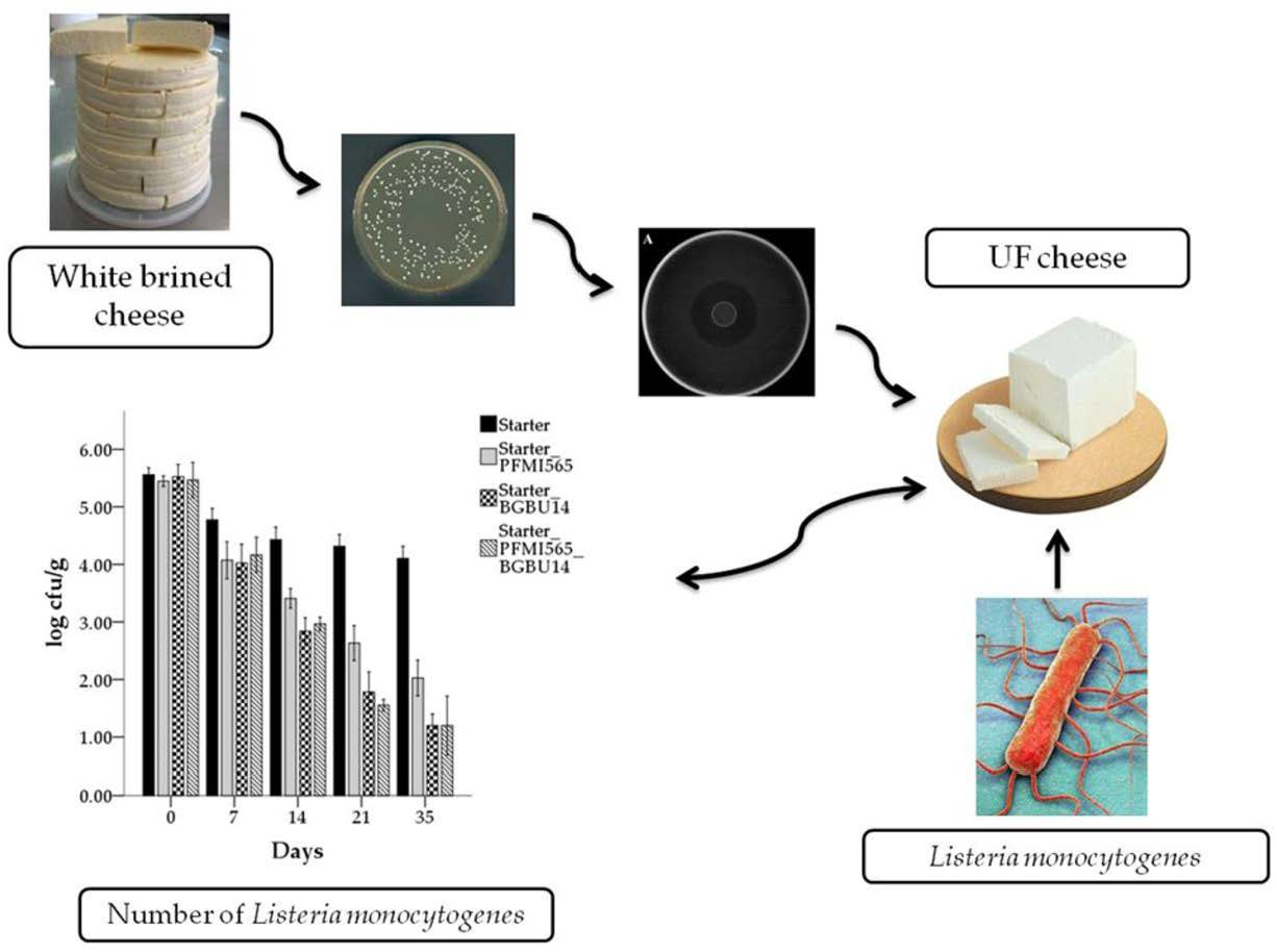
Autochthonous Enterococcus durans PFMI565 and Lactococcus lactis subsp. lactis BGBU1–4 in Bio-Control of Listeria monocytogenes in Ultrafiltered Cheese

1
Faculty of Agriculture, University of Belgrade, 11080 Belgrade, Serbia
2
Institute of Chemistry, Technology and Metallurgy, University of Belgrade, 11000 Belgrade, Serbia
*
Author to whom correspondence should be addressed.
Foods 2021, 10(7), 1448; https://doi.org/10.3390/foods10071448
Submission received: 10 May 2021 / Revised: 31 May 2021 / Accepted: 18 June 2021 / Published: 22 June 2021
(This article belongs to the Section Food Microbiology)

Abstract

:
Nowadays, consumers are interested in cheese produced without chemical additives or high-temperature treatments, among which, protective lactic acid bacteria (LAB) cultures could play a major role. In this study, the aims were to isolate, identify and characterize antilisterial LAB from traditionally produced cheese, and utilize suitable LAB in cheese production. Among 200 isolated LAB colonies, isolate PFMI565, with the strongest antilisterial activity, was identified as Enterococcus durans. E. durans PFMI565 was sensitive to clinically important antibiotics (erytromicin, tetracycline, kanamycin, penicillin, vancomycin) and had low acidifying activity in milk. E. durans PFMI565 and the previously isolated bacteriocin producer, Lactococcus lactis subsp. lactis BGBU1–4, were tested for their capability to control Listeria monocytogenes in experimentally contaminated ultrafiltered (UF) cheeses during 35 days of storage at 4 °C. The greatest reductions of L. monocytogenes numbers were achieved in UF cheese made with L. lactis subsp. lactis BGBU1–4 or with the combination of L. lactis subsp. lactis BGBU1–4 and E. durans PFMI565. This study underlines the potential application of E. durans PFMI565 and L. lactis subsp. lactis BGBU1–4 in bio-control of L. monocytogenes in UF cheese.

Graphical Abstract

1. Introduction

Cheeses are a significant part of human diets because of their chemical composition and high contents of vitamins, fatty acids, minerals, bioactive compounds, and probiotic bacteria [1,2]. Cheeses are classified based on several factors: type of milk used for production (whey cheese, ultrafiltration, soured milk), fat content, consistency, type of fermentation, and texture [1]. Cheeses made from ultrafiltered milk (UF cheeses), a type of soft cheese, are very popular in Serbia, and can be stored in brine or with salt added to the milk or curd during production. However, UF cheeses with pH > 4.3 and high water activity are suitable matrices for growth of the pathogen Listeria monocytogenes [3,4]. Although UF cheeses are manufactured using pasteurized milk, contamination of this type of cheese sometimes occurs, usually during the production process or in post-processing manipulation of cheeses [5].
Despite the fact that the incidence of L. monocytogenes infections accounts for a low proportion of foodborne illnesses, the high mortality rate of listeriosis (20–30%) means this pathogen is responsible for many of the fatalities linked to food [6]. The infective dose of L. monocytogenes in food is high, typically ˃104 cfu g−1(mL−1), but in the case of immunocompromised individuals, the infective dose ranges from 102–104 cfu g−1(mL−1) [7,8].
According to European Union (EU) and Serbian regulations, the number of L. monocytogenes in ready-to-eat products has to be less than 102 cfu g−1 (mL−1) of food product during the entire shelf life [9,10]. L. monocytogenes is ubiquitous in the environment and it is very difficult to eliminate once it is established in food production plants. Thus, elimination and control of this pathogen in foods and food plants are imperative aims in food production [11]. Contamination of food by L. monocytogenes can be decreased by controlling the pathogen in the environment or during food production [12].
During food production, high heat or chemical treatments are very useful in control of L. monocytogenes, although these methods can cause some changes in the sensory and nutritional qualities of the food [13]. Apart from these traditional methods of L. monocytogenes control in food production, a potentially different approach could be using lactic acid bacteria (LAB) with antilisterial activity. LAB can produce different antimicrobial substances, such as antimicrobial peptides (bacteriocins), diacetyl, reuterin, hydrogen peroxide, and organic acids (lactic acid, propionic acid, acetic acid, benzoic acid) [14,15]. Any of these antimicrobial substances can be used in food production as partially purified compounds or can be delivered into food via antimicrobial-producing LAB cultures that are added as starter or adjunct cultures [16,17].
Traditional cheeses are a potential pool of new LAB strains with desirable biological and metabolic properties such as: acidogenic activity, production of antimicrobial compounds, production of proteinases, and probiotic properties [18,19,20]. Among LAB, the genera Lactococcus and Enterococcus are most commonly used as adjunct cultures in different products to control foodborne pathogens or to improve product quality [17,21,22,23]. However, to date, among many potential antimicrobial substances, only two bacteriocins (nisin and pediocin PA-1) have FDA approval and are commercially used in a variety of food products as natural preservatives [24,25].
In this study, LAB with potential antilisterial activity were isolated from traditionally made white brined cheeses. Their acidification activity and antibiotic susceptibility were determined in order to investigate their potential for application as components of starter or protective cultures in cheese production. According to their activities, one isolate was selected, identified and tested for its inhibitory activity against L. monocytogenes in UF cheese. Additionally, the efficacy of Lactococcus lactis subsp. lactis bv. diacetylactis BGBU1–4 in controlling L. monocytogenes growth in UF cheese was examined. This organism was previously isolated from the same cheese type and produces lactolisterin BU, a thermostable bacteriocin with antilisterial activity [26,27]. In a previous study, the strong antilisterial activity of L. lactis subsp. lactis BGBU1–4 in quark-type cheese was proven [28]. The aim of this study was to examine the effect of two LAB isolates exhibiting antilisterial activity on L. monocytogenes during storage of UF cheese.

2. Materials and Methods

2.1. Cheese Samples, Isolation of Lactic Acid Bacteria with Antilisterial Activity

White brined cheeses were produced in Sjenica, western Serbia, from unpasteurized cow’s milk and according to a traditional procedure without addition of starter cultures. Three cheese samples were taken from each cheese interior, individually homogenized using a sterile mortar and pestle, and 20 g was transferred to a stomacher bag with 180 mL of sodium citrate (2% w/v). The contents were homogenized in a Stomacher (Interlab, BagMixer 400P), and serial tenfold dilutions of the homogenates were prepared with sterile sodium chloride (0.85% w/v) and were surface plated onto two different growth media to isolate LAB: M17 agar (Merck GmbH, Darmstadt, Germany) supplemented with glucose (0.5% w/v; GM17) incubated at 30 °C and 37 °C for lactococci and enterococci, respectively, and de Man Rogosa and Sharp agar (MRS agar, Merck GmbH, Darmstadt, Germany) incubated on 30 °C and 37 °C for 24 h under aerobic conditions for lactobacilli. The plates with individual colonies were then overlaid with GM17 soft agar containing L. monocytogenes ATCC19111 and incubated for a further 24 h at 37 °C. Colonies with antilisterial activity were detected by the appearance of a zone of inhibition, and were purified using the medium on which they were originally cultivated. Antilisterial activity was confirmed by the agar well diffusion assay [29]. L. monocytogenes ATCC19111 (5 log cfu mL−1) was inoculated into GM17 soft agar, and wells were cut in the plates. Wells were filled with 50 µL of whole culture from an antilisterial producer (after 16 h incubation at appropriate temperature and medium) or cell-free supernatant (CFS). CFS of each whole culture was obtained by centrifugation (3500× g, room temperature, 10 min) and filtration of supernatant using 0.2 µm filter (Thermo Fisher Scientific, Waltham, MA, USA).

2.2. Proteinaceous Nature of Antilisterial Molecule

To determine the proteinaceous nature of antilisterial compounds, CFS and neutralized CFS (pH 7.00, adjusted with 1 M NaOH) were used. The test was performed by placing a crystal of proteolytic enzyme, pronase E (Sigma, St. Louis, MO, USA) close to the edge of the CFS-containing well; reduction of activity was taken as positive proteinaceous nature.

2.3. Identification of Lactic Acid Bacteria with Antilisterial Activity

Total DNA from antilisterial LAB isolates was obtained by the method of Hopfwood et al. (1986) [30] with minor modification. Bacterial cells grown in appropriate medium to early logarithmic (OD600 = 0.6–0.8) were collected by centrifugation (3500× g, room temperature, 10 min). Pellet was washed twice using TEN buffer (50 mM Tris-HCl pH 8; 10mM EDTA pH 8; 50 mM NaCl), further resuspended in solution of PP buffer (0.5 M saharoze; 40 mM NH4-acetata; 10 mM Mg-acetata; pH 7) with lysozyme (4 mg/mL) and incubated for 15 min at 37 °C. Identification of selected isolates was performed by 16s rRNA gene sequencing previously described by Golic et al. (2013) [27]. Platinum Taq DNA polymerase (Thermo Fisher Scientific, Waltham, MA, USA) was used to amplify the 16s rRNA gene using a GeneAmp PCR System 2700 thermal cycler (Applied Biosystems, Foster City, CA, USA) with specific primers, 16S—Fw (GAATCTTCCACAATGGACG) and 16S—Rev (TGACGGGCGGTGTGTACAAG) [31]. PCR products were visualized on 1% agarose gel at constant voltage of 80V. Excepted size of PCR products is 1500 bp. PCR products were purified using a Thermo Scientific PCR Purification Kit (Thermo Scientific, Lithuania) according to the manufacturer’s instructions. Purified PCR products were sequenced by the Macrogen Sequencing Service (Macrogen Europe, Amsterdam, The Netherlands). The BLAST algorithm was used for analyzing nucleotide sequences [32].

2.4. Detection of Hydrogen Peroxide

The ability of antilisterial isolates to produce H2O2 was confirmed using the method previously described by María Silvina Juárez Tomás et al. (2004) [33]. Solution A was prepared by mixing 12.5 mg 3,3’,5,5’-tetramethyl-benzidine (TMB; Sigma) and 3 mL of methanol (Merck, Darmstadt, Germany). Solution B was prepared with peroxidase (0.5 mg/mL, Sigma) and 1 mL sterilized mili-Q water. GM17 agar (10 mL held at 45 °C) was mixed with 0.6 mL of solution A and 0.2 mL of solution B, then poured into petri dishes to make TMB plates. Isolates were streaked on the surface of TMB plates and incubated at an appropriate temperature for 24 h. Colonies able to produce hydrogen peroxide turn blue or brown under ambient light, as H2O2 reacts with horseradish peroxidase in the agar to oxidize the TMB. All analyses of H2O2 production by antilisterial LAB were conducted in triplicate.

2.5. Detection of Diacetyl

Analyses of butanedione (diacetyl) in milk and antilisterial LAB culture were conducted using an Agilent 7890A gas chromatograph (GC) connected to an Agilent 7697A headspace device (HSS) and an electron capture detector (ECD), using the method previously described by Richelieu et al. (1997) [34]. Whole culture from an antilisterial producer (after 16 h incubation at appropriate temperature and medium) was washed twice in PBS buffer (0.1 M, pH 7) and finally resuspended in sterile reconstituted skim milk (10% w/v) (Nilac, The Netherlands). The samples were transferred to 20 mL glass headspace vials (5 mL of sample per vial) and hermetically sealed with crimped aluminum caps. High-purity nitrogen (>99.999%) was used to pressurize vials in the HSS device. The vials were equilibrated over 30 min at 70 °C. After equilibration, the samples were extracted using the single extraction mode, and transferred to the GC unit via the fused silica capillary transfer line (inner diameter 0.25 mm) at 100 °C loop temperature and 110 °C transfer line temperature. The GC was equipped with a Thermo Scientific™ TraceGOLD™ TG-5MT capillary column (60 m × 0.25 mm ID × 0.25 μm). For analyses of butanedione in the samples, the following oven temperature program was used: 45 °C for 2 min, then 10 °C/min to 150 °C, then hold at 150 °C for 27 min. The ECD operated at 250 °C with make-up gas flow of 30 mL/min. Butanedione in the samples was quantified using an external calibration method based on the concentration of the analyte in a standard series and the corresponding peak areas. For that purpose, butanedione analytical standard (97% purity; purchased from Sigma-Aldrich) was dissolved in water (milli Q ultrapure water) containing 4 ethanol (HPLC grade; purity ≥99.9%; purchased from Sigma-Aldrich). A standard series of butanedione solutions (0, 2, 5, 10, 15, 25, and 50 μg/L) was used to construct the calibration curve. All analyses were conducted in triplicate.

2.6. Antibiotic Resistance

The antibiotic resistance of selected isolates was determined by the Kirby-Bauer disk diffusion method, according to the Clinical and Laboratory Standards Institute [35]. Nine antibiotics were examined: streptomycin 300 µg (STR), ampicillin 10 µg (AMP), gentamicin 120 µg (GEN), vancomycin 30 µg (VAN), tetracycline 30 µg (TET), neomycin 30 µg (NEO), penicillin 10 U (PEN), erythromycin 15 µg (ERY), and chloramphenicol 30 µg (CHL). LAB isolate were classified as sensitive (S) or resistant (R) phenotype by the appearance of a zone of inhibition around antibiotic discs (BBL Sensi-Disc Antimicrobial Susceptibility Test Disc, Becton, Dickinson and Company, Franklin Lakes, NJ, USA).

2.7. Acidifying Activity

Acidifying activity of LAB isolates was examined using the International Dairy Federation Standard (IDF standard, 1995) [36]. The isolates were subcultured in appropriate medium (GM17 for lactococci or MRS for lactobacilli) and temperature (30 °C for lactococci or 37 °C for lactobacilli) for 16 h. Then, isolates were inoculated into sterile reconstituted skim milk (10% w/v) (Nilac, The Netherlands) at a level of 1 (v/v). The pH of milks was determined after 6, 12, and 24 h of incubation at the appropriate temperature. Analysis of acidifying activity of tested LAB isolates were done in triplicate.

2.8. UF Cheese Making Procedure

Control UF cheese (C) was made according to the procedure of Mazinani et al. (2014) [37] with minor modifications. Coagulation and fermentation was conducted at 30 °C during 17–18 h, and then 20 g/kg salt was added onto the cheese surface. Cheese was ripened at 12 °C during the next 7 days and stored at 4 °C for 35 days.
Experimental cheeses were produced using an identical procedure as for control cheese, except one LAB with antilisterial activity isolated in this study and/or L. lactis subsp. lactis BGBU1–4 was added to cheese at the same time as the starter culture. Those antilisterial LAB were previously labeled by streptomycin and rifampicin resistance, respectively, using the procedure described by Frece et al. (2005) [38], with minor modification. Both antilisterial LAB were cultured at appropriate conditions for 24h. The cultured cells of BGBU1–4 and antilisterial LAB from this study were added to plates containing 500 µg/mL of rifampicin (Sigma-Aldrich Chemie GmbH, Deisenhofen, Germany) 1 mg/mL of streptomycin, respectively, and incubated for 72 h at appropriate temperatures. Labeled antilisterial LAB were further used for cheese production. Overnight cultures were washed twice in PBS buffer (0.1 M, pH 7) and finally resuspended in the same buffer. The final suspensions were serially diluted and were added in suitable amounts to obtain 6 log cfu mL−1. At the same time, cheeses were artificially contaminated with L. monocytogenes ATCCC19111 (at three different contamination levels: ~3, 4, 5 log cfu mL−1), obtaining the 12 cheese variants presented in (Table 1). Control cheeses were produced with no added L. monocytogenes ATCCC19111 in order to obtain four additional variants (Table 1). All cheeses were made in triplicate.

2.9. Sampling and Microbiological Analysis of Experimental UF Cheeses

Experimental cheeses were analyzed five times during 35 days of storage at 4 °C: (i) immediately after ripening (day 0); (ii) after 7 days of storage; (iii) after 14 days of storage; (iv) after 21 days of storage; v) after 35 days of storage. Viable cell counts of antilisterial LAB and L. monocytogenes ATCC19111 were determined. Each experimental UF cheese was aseptically sampled (10 g), diluted in 90 mL sterile Ringer’s solution (0.85% w/v), and homogenized for 2 min in a Stomacher (Interlab, BagMixer 400P). After homogenization, tenfold dilutions were prepared for microbiological analysis. Antilisterial LAB isolated in this study was cultivated on plates containing 1 mg/mL streptomycin (Sigma-Aldrich Chemie GmbH, Deisenhofen, Germany) at appropriate conditions, while BGBU1–4rif was cultivated on GM17 plates containing 500 µg/mL of rifampicin (Sigma-Aldrich Chemie GmbH, Deisenhofen, Germany) at 30 °C for 24 h. L. monocytogenes ATCC19111 was enumerated on Palcam Listeria selective agar base with Palcam Listeria selective supplement (Merck, Darmstadt, Germany), incubated at 37 °C for 48 h (ISO 11290-2:1998).

2.10. Statistical Analysis

All experiments were performed in triplicate. Two-way ANOVA was used to determine differences between experimental groups of milk and cheeses. The differences between means were compared using Student’s t-test, and were considered significant if p < 0.05.

3. Results and Discussion

3.1. Isolation and Identification of LAB with Antilisterial Activity

Among around 200 colonies isolated from cheeses, 20 were purified and selected according their zones of inhibition and antilisterial activity against L. monocytogenes ATCC19111. The percentage of colonies with antilisterial activity (among total LAB on the plates) was around 10%, which correlated with a previous study [39]. However, it differs from results previously reported by Campagnollo et al. [40], where up to 48.1% of tested bacterial colonies isolated from Minas cheese presented antilisterial activity. The reason for positive rate of antilisterial isolates could be a choice of type of cheese for isolation of bacteria, type of media used for isolation, type of antilisterial compound produced by isolates, and culture method. One isolate (PFMI565) had the strongest activity against L. monocytogenes ATCC19111 (zone of inhibition was 10 mm), and was chosen for further experiments. According to 16s rRNA gene sequencing, PFMI565 was identified as Enterococcus durans, showing 97.7% identity with many Entorococcus durans strains, including 4599, 4541, 4292, XT-1, and 3901. Enterococcus isolates can produce many antimicrobial substances including lactic acid, H2O2, bacteriocins, and bacteriocin-like substances (BLIS) [41,42,43,44]. The genera Enterococcus and Listeria are quite close according to molecular taxonomy and phylogenetic position, which could be a potential explanation for the antilisterial activity of enterococcoci [45,46,47].

3.2. Proteinaceous Nature of Antilisterial Compounds

Many strains of enterococci can produce bacteriocins, antimicrobial substances of protein nature [48]. To determine the nature of the antilisterial compound expressed by PFMI565, a crystal of pronase E was used in the agar well diffusion assay. After incubation, antilisterial activity was detected in the vicinity of the pronase E, indicating the antilisterial activity was not proteinaceous, so not bacteriocin or bacteriocin-like substances (Figure 1A). Similar results published previously indicated the antilisterial activity could be due to organic acid [43]. However, results from this study indicated that organic acid was not the only antilisterial compound, since although the zone of inhibition of neutralized CFS was smaller than that of CFS, it was clearly visible (Figure 1B).

3.3. Hydrogen Peroxide and Diacetyl Production

E. durans PFMI565 produced H2O2, as colonies turned blue. Previously, it has been shown that H2O2 produced and released by bacteria has the ability to inhibit other competent bacteria and host bacteria as well [49]. In some studies, LAB that are H2O2 producers inhibited the growth of foodborne and clinical pathogens such as Staphylococcus aureus, Neiseria gonorrhea, and Gardnerella vaginalis [50,51,52,53]. The antilisterial activity of neutralized CFS from E. durans PFMI565 is shown (Figure 1B), so the antilisterial activity of E. durans PFMI565 is likely to be due, at least in part, to H2O2.
Diacetyl is a flavor-producing compound that can be produced by LAB and has antimicrobial activity against L. monocytogenes and S. aureus in liquid [54]. The diacetyl concentration of E. durans PFMI565 whole culture was 11.79 ± 0.44 µg/L, which corresponds to 0.01179 ppm. The minimum concentration of diacetyl required to achieve an antilisterial effect is 300 ppm [55]. In the specific conditions of packaging food in an atmosphere with 20% CO2, 50 ppm diacetyl showed antilisterial activity [56], which is still higher than the concentration detected in our pure culture of E. durans PFMI565. Comparing the results obtained in this study with others, it can be concluded that the concentration of diacetyl produced by E. durans PFMI565 was insufficient to achieve an antilisterial effect.

3.4. Antibiotic Resistance

Antibiotic resistance testing revealed E. durans PFMI565 was resistant to three out of nine antibiotics (gentamicin, streptomycin and neomycin), but was sensitive to six out of nine antibiotics (chloramphenicol ampicillin, vancomycin, tetracycline, penicillin and erythromycin). These results are partially in agreement with Amarel et al. 2017 [57], who showed that E. durans SJRP14, SJRP17 and SJRP26 were sensitive to clinically important antibiotics: erytromicin, tetracycline, kanamycin, penicillin, and vancomycin. Resistances to these antibiotics are usually a product of transformable genetic elements that are responsible for the transmission of antibiotic resistance determinants [57,58,59]. On the other hand, enterococci usually possess chromosomally encoded enzymes responsible for resistance to aminoglycosides (strepotmycin, gentamicin, neomycin), so transmission of these resistance genes is impossible [60]. Therefore, according to antibiotic susceptibility testing, the presence of E. durans PFMI565 in cheese as a starter or adjunct culture does not represent a risk for the spread of antibiotic resistance.

3.5. Acidifying Activity

In order to test the suitability of antilisterial strains Lactococcus lactis subsp. lactis BGBU1–4 and Enterococcus durans PFMI565 as starter culture components, acidifying activity was studied. The initial pH of skim milk was 6.58, and after 6 h of incubation with L. lactis subsp. lactis BGBU1–4 and E. durans PFMI565, pH was 5.82 (6 h) and 5.92 (6 h), respectively (Figure 2). After 24 h of incubation, pH of skim milk was reduced to 4.45 and 4.73 for L. lactis subsp. lactis BGBU1–4 and E. durans PFMI565, respectively. The pH of milk are statistically difference in the case of these two strains (Figure 2). Previously, it was reported that selected strains of E. durans and L. lactis subsp. lactis biovar diacetylactis can reduce the pH of skim milk to pH < 5, after 4 h of incubation [59,61,62], unlike the LAB used in this study. However, it is known that acidifying activity of LAB varies considerably [63]. A possible reason for differences in acidifying activity between strains of the same species could be related to levels of expression of ß-galactosidase or phosphor-ß-galastosidase [64]. Autochthonous LAB isolated from cheese could be used as a starter culture, important for standardization of production of autochthonous food products, as an adjunct culture for production of healthy and functional food product, or as a protective culture for prolonging shelf life [65]. Starter cultures used in cheese production and containing LAB usually can reduce milk pH to below 5.3 in 6 h [66]. According to the results of this study, L. lactis subsp. lactis BGBU1–4 and E. durans PFMI565 had only low acidifying activity in skim milk in the first 6 h of incubation, and therefore, they are not suitable to be used as starter cultures for cheese production. Nevertheless, L. lactis subsp. lactis BGBU1–4 and E. durans PFMI565 could be used as adjunct cultures in cheese and other dairy products, since both LAB showed antilisterial activity in vitro.

3.6. Microbiological Analysis of UF Cheeses

The ability of a lactolisterin BU producer (L. lactis subsp. lactis BGBU1–4) and a non-bacteriocin producer (E. durans PFMI565) that has antilisterial activity, to inhibit growth of L. monocytogenes in UF cheese, was studied. L. lactis subsp. lactis BGBU1–4 and E. durans PFMI565 were added during cheese production along with starter cultures, since both these LAB had average acidifying and strong antilisterial activities which are good attributes for adjunct cultures.
L. monocytogenes was not detected in control cheeses C, CB, C565, and B565C. In cheeses CL3, CL4 and CL5 produced with starter culture, without adjunct cultures BGBU1–4rif and PFMI565str, and artificially contaminated with L. monocytogenes ATCC19111, numbers of this pathogen on the first day after ripening (day 0) were ~3.3 log cfu g−1, 4.4 log cfu g−1, and 5.5 log cfu g−1, respectively. At the end of storage, L. monocytogenes numbers dropped to ~2 log cfu g−1 in cheese CL3, ~2.2 log cfu g−1 in cheese CL4 and ~4.1 log cfu g−1 in cheese CL5. Results from previous studies indicate that L. monocytogenes decreased by more than 2 log cfu g−1 in soft feta-type cheeses during 90 days of storage at 4 °C [67,68]. This reduction in L. monocytogenes numbers during storage could be due to deleterious effects of fats and proteins on antilisterial molecules, influence of sodium chloride concentration and pH on activity of antilisterial molecules, or the artificially high initial level of inoculated L. monocytogenes [13,69]. Still, numbers of L. monocytogenes in all cheeses remained at levels that are not allowed by law in Serbia or in the EU (cheese must contain less than 102 cfu g−1 (mL−1) [9,10].
Results of antimicrobial activity of strains BGBU1–4rif and PFMI565str against L. monocytogenes ATCC19111 in experimental cheeses are shown in (Figure 3). Statistical analysis has shown that both factors (type of strain and time of storage) and their interaction had significant effect on the number of L. monocytogenes ATCC19111 counted. In the control cheese variants, made without BGBU1–4Rif and PFMI565str, statistically significant decrease in cells number of L. monocytogenes ATCC19111 was found during storage. However, the intensity of reduction rate was different, depending on whether cheese made with addition of BGBU1–4rif, PFMI565str or their combination. The trend of a decreasing number of L. monocytogenes ATCC19111 did not depend on the initial number inoculated into cheeses. In cheeses with BGBU1–4str and PFMI565rif, decreases in the number of L. monocytogenes followed different trends depending on whether cheese was produced with BGBU1–4rif, PFMI565str, or a combination of these two strains. The greatest reductions in L. monocytogenes numbers were achieved in cheese with BGBU1–4str and in cheese with a combination of BGBU1–4rif and PFMI565str. After 14 days of storage at 4 °C, L. monocytogenes counts in these two cheeses were decreased statistically significant, 1 log more than in cheese without antilisterial LAB. By the end of 21 days of storage, L. monocytogenes counts were statistically significant lower in all cheeses produced with antilisterial LAB than in control cheeses. In a previous study, it was shown that L. lactis subsp. lactis BGBU1–4 (a lactolisterin BU producer) has strong inhibitory activity against L. monocytogenes ATCC19111 in quark-type cheese during storage [28]. The results in our current study showed reductions of L. monocytogenes ATCC19111 in UF cheeses with L. lactis subsp. lactis BGBU1–4 and E. durans PFMI565 (separately) and in cheese with the combination of these two LAB, but still, viable L. monocytogenes remained in cheeses at detectable levels at the end of storage. Similar incomplete reductions of L. monocytogenes in cottage and quark-type cheese using nisin A, nisin Z, lacticin 481, and lactolisterin BU producers were also published [23,28]. The reason for this effect could be possible inactivation of bacteriocin or antimicrobial compound by its interaction with proteases or pH of cheese [70,71]. In some studies, it was also concluded that the control of L. monocytogenes in cheeses depends on LAB strain, nature of antimicrobial compounds and type of cheese [21,72].
Numbers of BGBU1–4rif and PFMI565str were also followed during 35 days of storage in all cheeses where they were used. After ripening, on the first day of storage (day 0), numbers of both LAB in all cheeses were ~8.30 log cfu g−1. Numbers of BGBU1–4rif increased in cheeses during storage and reached ~8.75 cfu g−1 after 35 days of storage. On the other hand, numbers of PFMI565str decreased somewhat and were ~7.5 log cfu g−1 after 35 days of storage. In general, both examined LAB showed good ability to survive in UF cheese during storage. These results confirm previous findings indicating very good survival of enterococci and BGBU1–4 in cheese during storage [21,22,29,40]. In both cases, numbers of BGBU1–4rif and PFMI565str did not depend on the initial number of L. monocytogenes. Numbers of BGBU1–4rif were higher than numbers of PFMI565str at the end of storage, which could be due to the stronger antilisterial effect in of lactolisterin BU producer compared with E. durans PFMI565 in the UF cheese. Our study confirmed in vitro results [26] (Figure 1A,B) and showed that BGBU1–4rif and PFMI565str produce antilisterial effects in UF cheese during 35 days of storage at 4 °C.

4. Conclusions

In total, 20 LAB colonies with antilisterial activity were isolated and purified from Serbian white cheese. Among the 20 LAB, the isolate with the strongest antilisterial activity was identified as Enterococcus durans PFMI565.
E. durans PFMI565 produces little acidifying activity in milk and is sensitive to clinically important antibiotics, making it a good candidate for application in cheese production as an adjunct culture. Furthermore, addition of E. durans PFMI565 and Lactococcus lactis susp lactis BGBU1–4 (bacteriocin producer) into UF cheeses artificially contaminated with Listeria monocytogenes resulted in antilisterial effects during 35 days of storage. The reduction of L. monocytogenes is significantly greater in UF cheeses made with L. lactis susp lactis BGBU1–4 and in cheese with a combination of both L. lactis susp lactis BGBU1–4 and E. durans PFMI565.
The findings in this study indicate that the autochthonous LAB, E. durans PFMI565 and L. lactis subsp. lactis BGBU1–4, used as protective cultures in production of UF cheeses, would provide protection against growth of L. monocytogenes in UF cheese. However, the nature of the antilisterial compound(s) produced by E. durans PFMI565 is not yet resolved, although bacteriocin has been ruled out. Additionally, the presence of virulence factors in E. durans PFMI565 must be determined. Therefore, additional investigation of E. durans PFMI565 is required before this LAB could be used in any applications for commercial UF cheese production.

Author Contributions

Conceptualization, Data curation, Writing—original draft, M.I. and N.M.; Methodology, Writing—review and editing, M.M.; Software, Writing—review and editing, J.M.; Methodology, A.R. and T.S.K.; Writing-original draft, funding acquisition, supervision, Z.R. All authors have read and agreed to the published version of the manuscript.

Funding

This research was funded by the Ministry for Education, Science and Technological Development of the Republic of Serbia through agreement 451-03-9/2021-14/ 200116.

Institutional Review Board Statement

Not applicable.

Informed Consent Statement

Not applicable.

Data Availability Statement

Data available in a publicly accessible repository.

Conflicts of Interest

The authors declare no conflict of interest.

References

  1. Walther, B.; Schmid, A.; Sieber, R.; Wehrmüller, K. Cheese in nutrition and health. Dairy Sci. Technol. 2008, 88, 389–405. [Google Scholar] [CrossRef] [Green Version]
  2. Santiago-López, L.; Aguilar-Toalá, J.E.; Hernández-Mendoza, A.; Vallejo-Cordoba, B.; Liceaga, A.M.; González-Córdova, A.F. Invited review: Bioactive compounds produced during cheese ripening and health effects associated with aged cheese consumption. J. Dairy Sci. 2018, 101, 3742–3757. [Google Scholar] [CrossRef] [PubMed] [Green Version]
  3. Miočinović, J.; Puđa, P.; Radulović, Z.; Pavlović, V.; Miloradović, Z.; Radovanović, M.; Paunović, D. Development of low fat UF cheese technology. Hrvatska Mljekarska Udruga 2011, 61, 33–44. [Google Scholar]
  4. Miloradovic, Z.; Miocinovic, J.; Kljajevic, N.; Tomasevic, I.; Pudja, P. The influence of milk heat treatment on composition, texture, colour and sensory characteristics of cows’ and goats’ Quark-type cheeses. Small Rumin. Res. 2018, 169, 154–159. [Google Scholar] [CrossRef]
  5. Melo, J.; Andrew, P.W.; Faleiro, M.L. Listeria monocytogenes in cheese and the dairy environment remains a food safety challenge: The role of stress responses. Food Res. Int. 2015, 67, 75–90. [Google Scholar] [CrossRef]
  6. Thakur, M.; Asrani, R.K.; Patial, V. Listeria monocytogenes: A food-borne pathogen. In Foodborne Diseases; Elsevier: Amsterdam, The Netherlands, 2018; Volume 15, pp. 157–192. [Google Scholar]
  7. McLauchlin, J.; Mitchell, R.T.; Smerdon, W.J.; Jewell, K. Listeria monocytogenes and listeriosis: A review of hazard characterisation for use in microbiological risk assessment of foods. Int. J. Food Microbiol. 2004, 92, 15–33. [Google Scholar] [CrossRef]
  8. Vázquez-Boland, J.A.; Kuhn, M.; Berche, P.; Chakraborty, T.; Domínguez-Bernal, G.; Goebel, W.; González-Zorn, B.; Wehland, J.; Kreft, J. Listeria pathogenesis and molecular virulence determinants. Clin. Microbiol. Rev. 2001, 14, 584–640. [Google Scholar] [CrossRef] [Green Version]
  9. The European Union summary report on trends and sources of zoonoses, zoonotic agents and food-borne outbreaks in 2013. EFSA J. 2015, 13. [CrossRef] [Green Version]
  10. Regulation on the General and Special Conditions of Food Hygiene at any Stage of Production, Processing and Marketing” 33/2010. Official Bulletin of the Republic of Serbia. 2010. Available online: https://www.vet.minpolj.gov.rs/legislativa/pravilnici/Pravilnik_mikrobioloski_kriterijumi.pdf (accessed on 20 June 2021).
  11. Schlech, W.F. Foodborne listeriosis. Clin. Infect. Dis. 2000, 31, 770–775. [Google Scholar] [CrossRef]
  12. Oh, H.; Kim, S.; Lee, S.; Lee, H.; Ha, J.; Lee, J.; Choi, Y.; Choi, K.H.; Yoon, Y. Prevalence and genetic characteristics of meatborne listeria monocytogenes isolates from livestock farms in Korea. Korean J. Food Sci. Anim. Resour. 2016, 36, 779–786. [Google Scholar] [CrossRef]
  13. Jung, D.S.; Bodyfelt, F.W.; Daeschel, M.A. Influence of Fat and Emulsifiers on the Efficacy of Nisin in Inhibiting Listeria monocytogenes in Fluid Milk. J. Dairy Sci. 1992, 75, 387–393. [Google Scholar] [CrossRef]
  14. Reis, J.A.; Paula, A.T.; Casarotti, S.N.; Penna, A.L.B. Lactic Acid Bacteria Antimicrobial Compounds: Characteristics and Applications. Food Eng. Rev. 2012, 4, 124–140. [Google Scholar] [CrossRef]
  15. Guillier, L.; Stahl, V.; Hezard, B.; Notz, E.; Briandet, R. Modelling the competitive growth between Listeria monocytogenes and biofilm microflora of smear cheese wooden shelves. Int. J. Food Microbiol. 2008, 128, 51–57. [Google Scholar] [CrossRef]
  16. Rodríguez, E.; Calzada, J.; Arqués, J.L.; Rodríguez, J.M.; Nuñez, M.; Medina, M. Antimicrobial activity of pediocin-producing Lactococcus lactis on Listeria monocytogenes, Staphylococcus aureus and Escherichia coli O157:H7 in cheese. Int. Dairy J. 2005, 15, 51–57. [Google Scholar] [CrossRef]
  17. Gálvez, A.; López, R.L.; Abriouel, H.; Valdivia, E.; Omar, N. Ben Application of bacteriocins in the control of foodborne pathogenic and spoilage bacteria. Crit. Rev. Biotechnol. 2008, 28, 125–152. [Google Scholar] [CrossRef] [PubMed]
  18. Radulović, Z.; Miočinović, J.; Mirković, N.; Mirković, M.; Paunović, D.; Ivanović, M.; Seratlić, S. Survival of spray-dried and free-cells of potential probiotic Lactobacillus plantarum 564 in soft goat cheese. Anim. Sci. J. 2017, 88, 1849–1854. [Google Scholar] [CrossRef] [PubMed]
  19. Topisirovic, L.; Kojic, M.; Fira, D.; Golic, N.; Strahinic, I.; Lozo, J. Potential of lactic acid bacteria isolated from specific natural niches in food production and preservation. Int. J. Food Microbiol. 2006, 112, 230–235. [Google Scholar] [CrossRef] [PubMed]
  20. Castro, J.M.; Tornadijo, M.E.; Fresno, J.M.; Sandoval, H. Biocheese: A food probiotic carrier. Biomed. Res. Int. 2015. [Google Scholar] [CrossRef]
  21. Vera Pingitore, E.; Todorov, S.D.; Sesma, F.; Gombossy de Melo Franco, B.D. Application of bacteriocinogenic Enterococcus mundtii CRL35 and Enterococcus faecium ST88Ch in the control of Listeria monocytogenes in fresh Minas cheese. Food Microbiol. 2012, 32, 38–47. [Google Scholar] [CrossRef] [PubMed]
  22. Aspri, M.; Field, D.; Cotter, P.D.; Ross, P.; Hill, C.; Papademas, P. Application of bacteriocin-producing Enterococcus faecium isolated from donkey milk, in the bio-control of Listeria monocytogenes in fresh whey cheese. Int. Dairy J. 2017, 73, 1–9. [Google Scholar] [CrossRef]
  23. Dal Bello, B.; Cocolin, L.; Zeppa, G.; Field, D.; Cotter, P.D.; Hill, C. Technological characterization of bacteriocin producing Lactococcus lactis strains employed to control Listeria monocytogenes in Cottage cheese. Int. J. Food Microbiol. 2012, 153, 58–65. [Google Scholar] [CrossRef]
  24. Delves-Broughton, J. Nisin and its application as a food preservative. Int. J. Dairy Technol. 1990, 43, 73–76. [Google Scholar] [CrossRef]
  25. Rodríguez, J.M.; Martínez, M.I.; Kok, J. Pediocin PA-1, a wide-spectrum bacteriocin from lactic acid bacteria. Crit. Rev. Food Sci. Nutr. 2002, 42, 91–121. [Google Scholar] [CrossRef] [Green Version]
  26. Lozo, J.; Mirkovic, N.; O’Connor, P.M.; Malesevic, M.; Miljkovic, M.; Polovic, N.; Jovcic, B.; Cotter, P.D.; Kojic, M. Lactolisterin BU, a novel class II broad-spectrum bacteriocin from Lactococcus lactis subsp. lactis bv. diacetylactis BGBU1–4. Appl. Environ. Microbiol. 2017, 83. [Google Scholar] [CrossRef] [Green Version]
  27. Golić, N.; Čadež, N.; Terzić-Vidojević, A.; Šuranská, H.; Beganović, J.; Lozo, J.; Kos, B.; Šušković, J.; Raspor, P.; Topisirović, L. Evaluation of lactic acid bacteria and yeast diversity in traditional white pickled and fresh soft cheeses from the mountain regions of Serbia and lowland regions of Croatia. Int. J. Food Microbiol. 2013, 166, 294–300. [Google Scholar] [CrossRef] [PubMed]
  28. Mirkovic, N.; Kulas, J.; Miloradovic, Z.; Miljkovic, M.; Tucovic, D.; Miocionovic, J.; Jovcic, B.; Mirkov, I.; Kojic, M. Lactolisterin BU-producer Lactococcus lactis subsp. lactis BGBU1–4: Bio-control of Listeria monocytogenes and Staphylocococcus aureus in fresh soft cheese and effect on immunological response of rats. Food Control 2020, 111, 107076. [Google Scholar] [CrossRef]
  29. Kojic, M.; Svircevic, J.; Banina, A.; Topisirovic, L. Bacteriocin-Producing Strain of Lactococcus lactis subsp. diacitilactis S50. Appl. Environ. Microbiol. 1991, 57, 1835–1837. [Google Scholar] [CrossRef] [Green Version]
  30. Hopwood, D.A.; Bibb, M.J.; Chater, K.F.; Kjeser, T.; Bruton, C.J.; Kieser, H.M.; Lydiate, D.J.; Smith, C.P.; Ward, J.M. Genetic Manipulation of Streptomyces: A Laboratory Manual; The John Innes Foundation: Norwich, UK, 1985. [Google Scholar]
  31. Jovčić, B.; Begović, J.; Lozo, J.; Topisirović, L.; Kojić, M. Dynamics of sodium dodecyl sulfate utilization and antibiotic susceptibility of strain Pseudomonas sp. ATCC19151. Arch. Biol. Sci. 2009, 61, 159–164. [Google Scholar] [CrossRef]
  32. Altschul, S.F.; Madden, T.L.; Schäffer, A.A.; Zhang, J.; Zhang, Z.; Miller, W.; Lipman, D.J. Gapped BLAST and PSI-BLAST: A new generation of protein database search programs. Nucleic Acids Res. 1997, 25, 3389–3402. [Google Scholar] [CrossRef] [PubMed] [Green Version]
  33. Tomás, M.S.J.; Claudia Otero, M.; Ocaña, V.; Elena Nader-Macías, M. Production of antimicrobial substances by lactic acid bacteria I: Determination of hydrogen peroxide. Methods Mol. Biol. 2004, 268, 337–346. [Google Scholar] [CrossRef]
  34. Richelieu, M.; Houlberg, U.; Nielsen, J.C. Determination of α-Acetolactic Acid and Volatile Compounds by Headspace Gas Chromatography. J. Dairy Sci. 1997, 80, 1918–1925. [Google Scholar] [CrossRef]
  35. Clinical & Laboratory Standards Institute. CLSI Guidelines. Available online: https://clsi.org/ (accessed on 24 March 2021).
  36. International Dairy Federation. IDF guideline-determination of acidifying activity of dairy cultures. Bull. Int. Dairy Fed. 1995, 306, 34–36. [Google Scholar]
  37. Mazinani, S.; Fadaei, V.; Khosravi-Darani, K. Impact of Spirulina platensis on Physicochemical Properties and Viability of Lactobacillus acidophilus of Probiotic UF Feta Cheese. J. Food Process. Preserv. 2016, 40, 1318–1324. [Google Scholar] [CrossRef]
  38. Frece, J.; Kos, B.; Beganović, J.; Vuković, S.; Šušković, J. In vivo testing of functional properties of three selected probiotic strains. World J. Microbiol. Biotechnol. 2005, 21, 1401–1408. [Google Scholar] [CrossRef]
  39. Cintas, L.M.; Casaus, P.; Håvarstein, S.; Hernandez, P.E.; Hernandez, H.; Nes, I.F. Biochemical and genetic characterization of enterocin P, a novel sec-dependent bacteriocin from enterococcus faecium P13 with a broad antimicrobial spectrum. Appl. Environ. Microbio. 1997, 63, 4321–4330. [Google Scholar] [CrossRef] [PubMed] [Green Version]
  40. Campagnollo, F.B.; Margalho, L.P.; Kamimura, B.A.; Feliciano, M.D.; Freire, L.; Lopes, L.S.; Alvarenga, V.O.; Cadavez, V.A.P.; Gonzales-Barron, U.; Schaffner, D.W.; et al. Selection of indigenous lactic acid bacteria presenting anti-listerial activity, and their role in reducing the maturation period and assuring the safety of traditional Brazilian cheeses. Food Microbiol. 2018, 73, 288–297. [Google Scholar] [CrossRef] [PubMed]
  41. Ahmed Sheikh, H.M. Antimicrobial activity of certain bacteria and fungi isolated from soil mixed with human saliva against pathogenic microbes causing dermatological diseases. Saudi J. Biol. Sci. 2010, 17, 331–339. [Google Scholar] [CrossRef] [Green Version]
  42. Foulquié Moreno, M.R.; Callewaert, R.; Devreese, B.; Van Beeumen, J.; De Vuyst, L. Isolation and biochemical characterisation of enterocins produced by enterococci from different sources. J. Appl. Microbiol. 2003, 94, 214–229. [Google Scholar] [CrossRef]
  43. Zheng, W.; Zhang, Y.; Lu, H.M.; Li, D.T.; Zhang, Z.L.; Tang, Z.X.; Shi, L.E. Antimicrobial activity and safety evaluation of Enterococcus faecium KQ 2.6 isolated from peacock feces. BMC Biotechnol. 2015, 15, 30. [Google Scholar] [CrossRef] [PubMed] [Green Version]
  44. Daeschel, M.A. Antimicrobial substances from lactic acid bacteria for use as food preservatives. Food Technol. 1989, 49, 164–1677. [Google Scholar]
  45. Ghrairi, T.; Manai, M.; Berjeaud, J.M.; Frère, J. Antilisterial activity of lactic acid bacteria isolated from rigouta, a traditional Tunisian cheese. J. Appl. Microbiol. 2004, 97, 621–628. [Google Scholar] [CrossRef]
  46. Ribeiro, S.C.; Coelho, M.C.; Todorov, S.D.; Franco, B.D.G.M.; Dapkevicius, M.L.E.; Silva, C.C.G. Technological properties of bacteriocin-producing lactic acid bacteria isolated from Pico cheese an artisanal cow’s milk cheese. J. Appl. Microbiol. 2014, 116, 573–585. [Google Scholar] [CrossRef]
  47. Rivas, F.P.; Castro, M.P.; Vallejo, M.; Marguet, E.; Campos, C.A. Antibacterial potential of Enterococcus faecium strains isolated from ewes’ milk and cheese. LWT-Food Sci. Technol. 2012, 46, 428–436. [Google Scholar] [CrossRef]
  48. Ness, I.F.; Diep, D.B.; Ike, Y. Enterococcal Bacteriocins and Antimicrobial Proteins that Contribute to Niche Control; Massachusetts Eye and Ear Infirmary: Boston, MA, USA, 2014. [Google Scholar]
  49. Moy, T.I.; Mylonakis, E.; Calderwood, S.B.; Ausubel, F.M. Cytotoxicity of hydrogen peroxide produced by Enterococcus faecium. Infect. Immun. 2004, 72, 4512–4520. [Google Scholar] [CrossRef] [Green Version]
  50. Ito, A.; Sato, Y.; Kudo, S.; Sato, S.; Nakajima, H.; Toba, T. The screening of hydrogen peroxide-producing lactic acid bacteria and their application to inactivating psychrotrophic food-borne pathogens. Curr. Microbiol. 2003, 47, 231–236. [Google Scholar] [CrossRef] [PubMed]
  51. St. Amant, D.C.; Valentin-Bon, I.E.; Jerse, A.E. Inhibition of Neisseria gonorrhoeae by Lactobacillus species that are commonly isolated from the female genital tract. Infect. Immun. 2002, 70, 7169–7171. [Google Scholar] [CrossRef] [PubMed] [Green Version]
  52. Ocaña, V.S.; Pesce de Ruiz Holgado, A.A.; Nader-Macà as, M.E. Growth inhibition of Staphylococcus aureus by H 2 O 2-producing Lactobacillus paracasei subsp. paracasei isolated from the human vagina. FEMS Immunol. Med. Microbiol. 1999, 23, 87–92. [Google Scholar] [CrossRef] [PubMed] [Green Version]
  53. Hawes, S.E.; Hillier, S.L.; Benedetti, J.; Stevens, C.E.; Koutsky, L.A.; Wølner-Hanssen, P.; Holmes, K.K. Hydrogen peroxide-producing lactobacilli and acquisition of vaginal infections. J. Infect. Dis. 1996, 174, 1058–1063. [Google Scholar] [CrossRef] [PubMed] [Green Version]
  54. Lee, W. Production of Diacetyl (2,3, butanediol) by Continuous Fermentation with Simultaneous Product Separation; Purdue University: West Lafayette, IN, USA, 1991. [Google Scholar]
  55. Lanciotti, R.; Patrignani, F.; Bagnolini, F.; Guerzoni, M.E.; Gardini, F. Evaluation of diacetyl antimicrobial activity against Escherichia coli, Listeria monocytogenes and Staphylococcus aureus. Food Microbiol. 2003, 20, 537–543. [Google Scholar] [CrossRef]
  56. Williams-Campbell, A.M.; Jay, J.M. Effects of diacetyl and carbon dioxide on spoilage microflora in ground beef. J. Food Prot. 2002, 65, 523–527. [Google Scholar] [CrossRef] [PubMed]
  57. Amaral, D.M.F.; Silva, L.F.; Casarotti, S.N.; Nascimento, L.C.S.; Penna, A.L.B. Enterococcus faecium and Enterococcus durans isolated from cheese: Survival in the presence of medications under simulated gastrointestinal conditions and adhesion properties. J. Dairy Sci. 2017, 100, 933–949. [Google Scholar] [CrossRef] [Green Version]
  58. Werner, G.; Coque, T.M.; Franz, C.M.A.P.; Grohmann, E.; Hegstad, K.; Jensen, L.; van Schaik, W.; Weaver, K. Antibiotic resistant enterococci-Tales of a drug resistance gene trafficker. Int. J. Med. Microbiol. 2013, 303, 360–379. [Google Scholar] [CrossRef]
  59. Morandi, S.; Brasca, M.; Andrighetto, C.; Lombardi, A.; Lodi, R. Technological and molecular characterisation of enterococci isolated from north-west Italian dairy products. Int. Dairy J. 2006, 16, 867–875. [Google Scholar] [CrossRef]
  60. Miller, W.R.; Munita, J.M.; Arias, C.A. Mechanisms of antibiotic resistance in enterococci. Expert Rev. Anti. Infect. Ther. 2014, 12, 1221–1236. [Google Scholar] [CrossRef] [PubMed]
  61. Herreros, M.A.; Fresno, J.M.; González Prieto, M.J.; Tornadijo, M.E. Technological characterization of lactic acid bacteria isolated from Armada cheese (a Spanish goats’ milk cheese). Int. Dairy J. 2003, 13, 469–479. [Google Scholar] [CrossRef]
  62. Jamaly, N.; Benjouad, A.; Comunian, R.; Daga, E.; Bouksaim, M. Characterization of Enterococci isolated from Moroccan dairy products. Afr. J. Microbiol. Res. 2010, 4, 1768–1774. [Google Scholar]
  63. Cogan, T.M.; Barbosa, M.; Beuvier, E.; Bianchi-Salvadori, B.; Cocconcelli, P.S.; Fernandes, I.; Gomez, J.; Gomez, R.; Kalantzopoulos, G.; Ledda, A.; et al. Characterization of the lactic acid bacteria in artisanal dairy products. J. Dairy Res. 1997, 64, 409–421. [Google Scholar] [CrossRef]
  64. Iskandar, C.F.; Cailliez-Grimal, C.; Borges, F.; Revol-Junelles, A.M. Review of lactose and galactose metabolism in Lactic Acid Bacteria dedicated to expert genomic annotation. Trends Food Sci. Technol. 2019, 88, 121–132. [Google Scholar] [CrossRef]
  65. Radulović, Z.; Miočinović, J.; Petrović, T.; Dimitrijević-Branković, S.; Nedović, V. Traditional and emerging technologies for autochthonous lactic acid bacteria application. In Emerging and Traditional Technologies for Safe, Healthy and Quality Food; Springer: Cham, Switzerland, 2016; pp. 237–256. [Google Scholar]
  66. Beresford, T.P.; Fitzsimons, N.A.; Brennan, N.L.; Cogan, T.M. Recent advances in cheese microbiology. Int. Dairy J. 2001, 11, 259–274. [Google Scholar] [CrossRef]
  67. Manolopoulou, E.; Sarantinopoulos, P.; Zoidou, E.; Aktypis, A.; Moschopoulou, E.; Kandarakis, I.G.; Anifantakis, E.M. Evolution of microbial populations during traditional Feta cheese manufacture and ripening. Int. J. Food Microbiol. 2003, 82, 153–161. [Google Scholar] [CrossRef]
  68. Papageorgiou, D.K.; Marth, E.H. Fate of Listeria monocytogenes during the manufacture, ripening and storage of feta cheese. J. Food Prot. 1989, 52, 82–87. [Google Scholar] [CrossRef] [PubMed]
  69. Yang, E.; Fan, L.; Yan, J.; Jiang, Y.; Doucette, C.; Fillmore, S.; Walker, B. Influence of culture media, pH and temperature on growth and bacteriocin production of bacteriocinogenic lactic acid bacteria. AMB Express 2018, 8, 10. [Google Scholar] [CrossRef] [PubMed] [Green Version]
  70. Maisnier-Patin, S.; Deschamps, N.; Tatini, S.R.; Richard, J. Inhibition of Listeria monocytogenes in Camembert cheese made with a nisin-producing starter. Lait 1992, 72, 249–263. [Google Scholar] [CrossRef] [Green Version]
  71. Rai, M.; Pandit, R.; Gaikwad, S.; Kövics, G. Antimicrobial peptides as natural bio-preservative to enhance the shelf-life of food. J. Food Sci. Technol. 2016, 53, 3381–3394. [Google Scholar] [CrossRef] [Green Version]
  72. Izquierdo, E.; Marchioni, E.; Aoude-Werner, D.; Hasselmann, C.; Ennahar, S. Smearing of soft cheese with Enterococcus faecium WHE 81, a multi-bacteriocin producer, against Listeria monocytogenes. Food Microbiol. 2009, 26, 16–20. [Google Scholar] [CrossRef] [PubMed]
Figure 1. Antilisterial activity of Enterococcus durans PFMI565 against Listeria monocytogenes ATCC19111; (A) Cell-free supernatant (CFS) of E. durans PFMI565; (B) Neutralized CFS (pH 7.00) of E. durans PFMI565.
Foods 10 01448 g001
Figure 2. Acidifying activity of Lactococcus lactis subsp. lactis BGBU1–4 and Enterococcus durans PFMI565 in skim milk during 24 h of incubation at 30 °C. Values represent mean value +/– standard deviation (n = 3). Small letter indicated statistical significant difference between pH of milk of same strain during storage. Big letter indicate statistical significant difference between pH of milk with L. lactis subsp lactis BGBU1–4 and E. durans PFMI565 in same hour of incubation.
Foods 10 01448 g002
Figure 3. The effect of Lactococcus lactis subsp. lactis BGBU1–4 and Enterococcus durans PFMI565 on survival of Listeria monocytogenes ATCC19111 in UF cheeses with (A) Initial number of L. monocytogenes ATCC19111 ~3 log cfu g−1; (B) Initial number of L. monocytogenes ATCC19111 ~4 log cfu g−1; (C) Initial number of L. monocytogenes ATCC19111 ~5 log cfu g−1. Bars represent means +/– standard deviations (n = 3). Same small letter indicated there is no statistical significant difference in cell number of L. monocytogenes ATCC1911 in same sample during storage. Big letter indicate statistically significant differences in cell number of L. monocytogenes ATCC19111 between different samples at the same day of storage.
Foods 10 01448 g003aFoods 10 01448 g003b
Table 1. Starter and adjunct culture used for UF cheese production and level of Listeria monocytogenes ATCC19111 contamination.
Cheese DesignationBacterial Cultures and Level of Contamination
CCHN 11
CBCHN11, BGBU1–4
CBL3CHN11, BGBU1–4, L.monocyt. 3 log cfu g−1
CBL4CHN11, BGBU1–4, L.monocyt. 4 log cfu g−1
CBL5CHN11, BGBU1–4, L.monocyt. 5 log cfu g−1
C565CHN11, isolate-PFMIX*
C565L3CHN11, isolate-PFMIX*, L.monocyt. 3log cfu g−1
C565L4CHN11, isolate-PFMIX*, L.monocyt. 4 log cfu g−1
C565L5CHN11, isolate-PFMIX*, L.monocyt. 5 log cfu g−1
B565CCHN11, BGBU1–4, isolate-PFMIX*
B565CL3CHN11, BGBU1–4, isolate-PFMIX*, L.monocyt. 3 log cfu g−1
B565CL4CHN11, BGBU1–4, isolate-PFMIX*, L.monocyt. 4 log cfu g−1
B565CL5CHN11, BGBU1–4, isolate-PFMIX*, L.monocyt. 5 log cfu g−1
Isolate-PFMIX*—isolate with the strongest activity against Listeria monocytogenes ATCC19111 obtained in this study.
Publisher’s Note: MDPI stays neutral with regard to jurisdictional claims in published maps and institutional affiliations.

