Article

Resistance-Nodulation-Cell Division (RND) Transporter AcrD Confers Resistance to Egg White in Salmonella enterica Serovar Enteritidis

1
School of Health Science and Engineering, University of Shanghai for Science and Technology, Shanghai 200093, China
2
Molecular Characterization of Foodborne Pathogens Research Unit, Eastern Regional Research Center, Agricultural Research Service, United States Department of Agriculture, Wyndmoor, PA 19038, USA
3
School of Agriculture and Biology, Shanghai Jiao Tong University, Shanghai 200240, China
*
Author to whom correspondence should be addressed.
Foods 2022, 11(1), 90; https://doi.org/10.3390/foods11010090
Submission received: 22 November 2021 / Revised: 11 December 2021 / Accepted: 29 December 2021 / Published: 30 December 2021
(This article belongs to the Special Issue Foodborne Pathogens and Food Safety)

Abstract

:
The excellent survival ability of Salmonella enterica serovar Enteritidis (S. Enteritidis) in egg white leads to outbreaks of salmonellosis frequently associated with eggs and egg products. Our previous proteomic study showed that the expression of multidrug efflux RND transporter AcrD in S. Enteritidis was significantly up-regulated (4.06-fold) in response to an egg white environment. In this study, the potential role of AcrD in the resistance of S. Enteritidis to egg white was explored by gene deletion, survival ability test, morphological observation, Caco-2 cell adhesion and invasion. It was found that deletion of acrD had no apparent effect on the growth of S. Enteritidis in Luria-Bertani (LB) broth but resulted in a significant (p < 0.05) decrease in resistance of S. Enteritidis to egg white and a small number of cell lysis. Compared to the wild type, a 2-log population reduction was noticed in the ΔacrD mutant with different initial concentrations after incubation with egg white for 3 days. Furthermore, no significant difference (p > 0.05) in the adhesion and invasion was found between the wild type and ΔacrD mutant in LB broth and egg white, but the invasion ability of the ΔacrD mutant in egg white was significantly (p < 0.05) lower than that in LB broth. This indicates that acrD is involved in virulence in Salmonella. Taken together, these results reveal the importance of AcrD on the resistance of S. Enteritidis to egg white.

Graphical Abstract

1. Introduction

Eggs are an important and integral part of the human diet. These are consumed all over the world and possess natural physical and chemical defenses to prevent the contamination of microorganisms [1]. Egg white, as a chemical barrier, is generally a hostile environment for bacterial survival and growth because of its unfavorable conditions, such as alkaline pH, nutritional limitations and antibacterial molecules [2,3]. However, the risk of Salmonella contamination is a serious threat to human health as well as egg production and processing. In particular, Salmonella enterica serovar Enteritidis (S. Enteritidis) represents the predominant serotype that is involved in food-borne diseases due to the consumption of eggs and egg products. More importantly, S. Enteritidis presents an exceptional ability to survive in egg white in contrast to other Salmonella serotypes [4,5,6].
It is important to understand the resistance mechanisms of S. Enteritidis to egg white. Previous workers have revealed key information through the use of molecular biological techniques such as site-directed mutagenesis, transposon-mediated insertional mutagenesis, in vivo expression and DNA arrays at the transcriptional level, which may be helpful in explaining the underlying survival mechanism in this foodborne pathogen [7,8,9]. While genes identified in those studies were mainly involved in iron transport, biotin synthesis, energy metabolism, cell envelope maintenance, DNA synthesis and repair, motility and pathogenicity. Furthermore, genes such as outer membrane channel-related gene tolC [10], DNA repair-related gene yafD [7] and stress response-related genes uspAB [11] were identified by mutagenic analysis and were considered as main players for the survival of S. Enteritidis in egg white. On the other hand, according to our previous study, the transcriptomic and proteomic profiles of S. Enteritidis exposed to egg white were analyzed using RNA-Seq and iTRAQ analysis to reveal potential important metabolic pathways, such as stress response, iron acquisition, amino acid and biotin synthesis, transport and regulation [6,12]. A highly up-regulated expression (4.06-fold) of the stress response related protein AcrD was found in S. Enteritidis in response to whole egg white at the protein level [12].
Gram-negative bacteria such as Salmonella usually have multidrug efflux transporters, which have been found to recognize and excrete various structurally unrelated compounds from the cell. Among the multidrug efflux pumps, members of the RND (Resistance-Nodulation-cell Division) family appear to be the most effective efflux systems in those bacteria. Salmonella has five RND-type efflux systems: AcrAB, AcrAD, AcrEF, MdtABC and MdsABC [13]. The RND transporter AcrD has a unique biological role, which can remove antimicrobial compounds, such as aminoglycosides, from the bacterial cell. Inactivation of acrD resulted in changes in the expression of 403 genes involved in basic metabolism, stress responses and virulence [14]. Furthermore, the deletion of acrD led to a significant reduction in biofilm formation and down-regulated expression of key biofilm formation-related proteins encoded by csgBD [15]. Previously, the deletion of acrD resulted in an increased sensitivity to antibiotics, dyes and detergents in S. Typhimurium [16,17]. Furthermore, AcrD also contributes to copper and zinc resistance in Salmonella [18]. Previous works have demonstrated that Salmonella usually infects the human host through the ingestion of contaminated food products. This bacterium is able to resist the adverse environment of the gastrointestinal tract and then adhere, colonize and invade host intestinal epithelial cells, leading to human infections and diseases [19,20]. To our knowledge, the role of acrD in S. Enteritidis resistance to antibacterial egg white is not yet clear.
Hence, this study aimed to uncover the role of acrD in the resistance and virulence of S. Enteritidis to egg white by gene expression analysis, gene deletion, survival ability test, cellular morphology analysis, Caco-2 cell adhesion and invasion assays. These results will provide new information to help elucidate the resistance mechanisms of S. Enteritidis to egg white.

2. Materials and Methods

2.1. Bacterial Strains

S. Enteritidis strain SJTUF10978, isolated from chicken wings, was used as the wild-type (WT) strain in this study. Escherichia coli DH5α and Salmonella MRL0026 were utilized as reference strains for cell adhesion and invasion assays. These strains were stored at −80 °C in LB (Luria-Bertani) broth, including 50% (v/v) glycerol. All strains were propagated overnight at 37 °C on LB agar before experiments.

2.2. Caco-2 Cell Culture Preparation

The human colon adenocarcinoma cell line Caco-2, obtained from Shanghai Fuheng Biotechnology Co., Ltd. (Shanghai, China) (FH0029), were routinely maintained in DMEM (Dulbecco’s Modified Eagle’s Medium, Gibco, Pittsburgh, PA, USA) medium containing 1% non-essential amino acids (Coolaber, Beijing, China), 10% fetal bovine serum (Fuheng biology, Shanghai, China), 100 U/mL penicillin (Hyclone, Shanghai, China) and 100 μg/mL streptomycin (Hyclone, Shanghai, China) at 37 °C with 5% CO2. Meanwhile, cells were sub-cultured every 2–3 days and used between passages 5 and 10.

2.3. Egg White and Its Filtrate Preparation

SPF (Specific Pathogen Free) eggs used in this study were purchased from Boehringer Ingelheim Vital Biotechnology Co., Ltd. (Beijing, China). Fifty eggs were stored in a 37 °C incubator with 65% RH (Relative Humidity) for 5 days as required for every independent biological repeat. Egg white was collected by homogenization and centrifugation as previously described [12]. Egg white filtrate (FEW, less than 10 kDa) was acquired by centrifugation using ultrafiltration tubes with the cut-off limit of 30 kDa and 10 kDa according to our previous method [21].

2.4. Gene Expression Analysis

Total RNA from log-phase cells of S. Enteritidis in whole egg white and LB broth was extracted using Trizol reagent (Invitrogen, Carlsbad, CA, USA) following the manufacturer’s instructions. The RNA concentrations were determined using a NanoDrop 2000c spectrophotometer (Thermo scientific, South Logan, UT, USA), and the quality of RNA was evaluated using 1% agarose gel electrophoresis. Furthermore, genomic DNA treatment and cDNA synthesis was conducted using the PrimeScript RT reagent kit following the manufacturer’s instructions (TaKaRa, Dalian, China). The gene expression of acrD was tested by RT-qPCR analysis (Eppendorf, Hamburg, Germany) as previously described [12], using primer pair of acrD-F (5′-ACGCAACAGCAGACCC-3′) and acrD-R (5′-GCCCAGACCGCTAATT-3′). The relative expression of acrD in S. Enteritidis was calculated by the comparative cycle threshold method [22]. For data normalization, 16S rRNA was utilized as a reference gene.

2.5. Construction of acrD Deletion Mutant Strain

In-frame deletion of acrD was generated based on the previously described homologous recombination knockout method [23]. The primers used are shown in Table 1. In addition, strains and plasmids utilized for the deletion are listed in Table 2. Firstly, the fragment of homologous arms (i.e., upper arm and lower arm) was amplified from the genomic DNA of wild-type S. Enteritidis SJTUF10978 by overlap extension PCR. Secondly, this fragment was cloned into the pMD19-T plasmid carrying an ampicillin resistance gene to produce pMD19-ΔacrD. The correct pMD19-ΔacrD plasmid was digested with Sac I and Xba I and then ligated into the pRE112 plasmid carrying a chloramphenicol resistance gene and a sucrose-sensitive gene. Then, the obtaining pRE112-ΔacrD plasmid was imported into E. coli SM10 λpir using CaCl2 transformation method. The recombinant plasmid was then extracted and transformed into the S. Enteritidis wild-type strain by electroporation to obtain a single-crossover strain. The resulting strain was induced by 8%(w/v) sucrose to finish a second crossover. Finally, suspected colonies were chosen and confirmed by DNA sequencing and PCR analysis to acquire the acrD deletion mutant (ΔacrD).

2.6. Measurement of Bacterial Growth

Overnight cultures of S. Enteritidis wild-type and ΔacrD strains in LB broth were collected by centrifugation and diluted to the cell density of OD600 ≈ 0.1. Then, cultures were incubated at 37 °C with continuous shaking at 200 rpm. The bacterial growth curve was measured at regular time (1 h) intervals by a Bioscreen C Analyzer (OY Growth Curves, Finland).

2.7. Survival Ability of S. Enteritidis Strains in Egg White and Its Filtrate

The survival of S. Enteritidis wild-type and ΔacrD strains in egg white and its filtrate was measured according to our previously described method [12]. Bacteria (1 mL) at logarithmic phase were collected, washed twice using sterile PBS (Phosphate-Buffered Saline, 1.8 mM KH2PO4, 10 mM Na2HPO4, 2.7 mM KCl, 137 mM NaCl, pH 7.2) and suspended in PBS. The bacterial suspension was adjusted to approximately 1 × 10⁷ CFU/mL and 1 × 10⁴ CFU/mL by dilution in PBS. Then in a 96-well microplate, 20 μL aliquots of the bacterial suspensions were inoculated into 180 μL of egg white and 180 μL of its filtrate, respectively. It was mixed to give a final concentration of 1 × 10⁶ CFU/mL and 1 × 10³ CFU/mL, respectively. The above mixtures were incubated at 37 °C for 24 h. Viable bacteria after incubation were enumerated by plating 100 μL of the treated cell suspensions on LB agar and incubated at 37 °C for 24 h.

2.8. Scanning Electron Microscopy (SEM) Analysis

The cell morphology of S. Enteritidis wild-type and ΔacrD strains in LB broth, egg white and egg white filtrate at 37 °C for 1 day was observed using a Sirion 200 SEM (FEI Company, Hillsboro, OR, USA) as previously described [21].

2.9. Adhesion and Invasion Assays

The adhesive and invasive ability of Salmonella strains and E. coli DH5α were investigated according to a previous method [26] with some modifications. The 48-h, 80% confluent Caco-2 monolayers were sub-cultured and placed into a 12-well plate at a density of approximately 1 × 10⁵ cells/well. Bacterial strains were inoculated in LB broth overnight at 37 °C. Bacterial cells were recovered by centrifugation at 13,800× g for 5 min, washed twice using DEME medium and suspended in DEME medium to a final concentration of 107 CFU/mL. Then, bacterial suspensions and Caco-2 cells were mixed at a ratio of 100:1 and then incubated for 1 h at 37 °C in a 5% CO2 incubator. The unattached bacteria after incubation were removed after incubation by washing with PBS. 1% Triton X-100 was added to release the attached bacteria at 37 °C for 5 min. Then, the suspensions were serially diluted, and 20 μL of each dilution was plated on LB agar and then incubated at 37 °C for 24 h. Counted colonies were recorded as the total adhesive bacterial population. The adhesion rate of bacteria was represented as the ratio of the number of adhesive bacteria compared to that of initial inoculated bacteria.
Similarly, in the invasion test, infected Caco-2 cells were incubated in a DMEM medium containing 1% penicillin and streptomycin for 1 h at 37 °C to kill extracellular bacteria. Serial dilutions of the lysates were plated on LB agar to enumerate invading bacterial populations. The invasion rate of bacteria to Caco-2 cells was represented as the ratio of the number of invading bacteria compared to that of cell-adhesion bacteria.

2.10. Statistical Analysis

Three independent experiments were conducted in all assays, and each treatment was carried out in triplicate. Data were evaluated via one-way ANOVA using SAS software. Duncan’s multiple range test (p < 0.05, p < 0.001) was used to identify the difference in survival, cell adhesion and invasion ability between wild-type S. Enteritidis and the mutant.

3. Results and Discussion

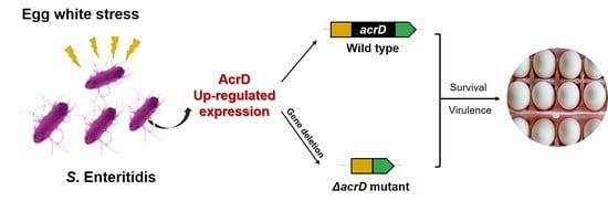
3.1. Expression of acrD in S. Enteritidis in Response to Whole Egg White and Construction of acrD Mutant

In previous studies, up-regulated expression of acrD at the mRNA level has been demonstrated in S. Enteritidis in response to low concentrations of egg white, e.g., 10% egg white and 80% egg white [6,9]. To test whether acrD was up-regulated in the whole egg white (i.e., 100% egg white), the expression of acrD in S. Enteritidis exposed to the whole egg white was further analyzed using RT-qPCR in this study. As shown in Figure 1A, the expression of acrD was significantly (p < 0.001) up-regulated (16.09-fold) in whole egg white compared with that in LB broth at the mRNA level. This gene expression data at the mRNA level was consistent with the proteomic data, which also showed up-regulated expression of AcrD in S. Enteritidis exposed to whole egg white at the protein level [12]. Hence, these results suggest that acrD may play a potential role in the resistance of S. Enteritidis to egg white.
Salmonella can resist adverse conditions through gene expression regulation. For example, two universal stress-related genes, uspA and uspB, of S. Enteritidis 147str are highly expressed in egg white, and a decreased colonization ability was observed to the magnum and isthmus of the oviduct when these genes were deleted [11]. The promoter of out membrane channel gene tolC was activated by egg white at 42 °C, and mutagenic analysis showed that tolC had an important role in S. Enteritidis survival in egg white [10]. Furthermore, the specific gene SEN1393 results in higher survivability of S. Enteritidis in egg white [27]. Although the acrD gene was expressed under different environmental stress conditions (e.g., antibiotics, detergents, metal) and seemed to contribute to stress resistance [15,16,18,20], there is no concrete evidence on the functional role of acrD in S. Enteritidis under egg white stress.
To better understand the role of acrD (encoding 1037 amino acids, a multidrug efflux RND transporter) in the resistance of S. Enteritidis to egg white, this gene (3114 bp) was deleted successfully in S. Enteritidis strain SJTUF10978 to obtain a ΔacrD deletion mutant (Figure 1B,C). The in-frame deletion mutant was confirmed by PCR and DNA sequencing (Figure 1B,C). Moreover, similar growth patterns of wild-type and ΔacrD strains in LB broth were found at 37 °C (Figure 1D), indicating that acrD is not required for S. Enteritidis growth in LB broth.

3.2. Survival Study of S. Enteritidis ΔacrD Mutant in Egg White and Its Filtrate

To explore the role of acrD in the survival of S. Enteritidis in egg white, the wild type and ΔacrD mutant were exposed to egg white and surviving bacteria were enumerated via plate counts on LB agar. As shown in Figure 2, the survival ability of ΔacrD mutant was significantly lower than that of the wild type in egg white (p < 0.05) (Figure 2A,B). More importantly, a 2-log population reduction was observed for the ΔacrD mutant after incubation in egg white for 3 days with different initial concentrations (i.e., 103 and 106 CFU/mL) (Figure 2A,B). These results demonstrate that acrD confers resistance to egg white in S. Enteritidis.
Generally, egg white filtrate (FEW, less than 10 kDa) has been used as a food matrix to reveal the antibacterial activity of egg white proteins [10,28]. Therefore, to explore the role of acrD in the resistance of S. Enteritidis to antibacterial egg white proteins in the present study, the wild-type strain and its ΔacrD mutant were exposed to egg white (containing various types of proteins) and egg white filtrate without the main antibacterial proteins. As shown in Figure 2C,D, the loss of acrD had no significant (p > 0.05) effect on the resistance of S. Enteritidis in egg white filtrate regardless of the initial cell concentrations, indicating that acrD plays a critical role in S. Enteritidis resistance to egg white proteins.
The RND transporter AcrD has a unique biological role in multidrug resistance, which can remove antimicrobial drugs such as aminoglycosides from the bacterial cell, and the RND family requires interaction with outer membrane channel TolC to function [13,29]. In addition, a previous study has demonstrated that RND transporters, such as AcrD, are necessary for the secretion of enterobactin, which chelates iron to enable bacterial growth under iron-limiting conditions [30]. Meanwhile, TolC has an important role in the resistance of S. Enteritidis to egg white ovotransferrin at 42 °C [10]. These results suggest that AcrD may contribute to Salmonella iron homeostasis to resist ovotransferrin in egg white. Antibacterial component experiments are needed to further confirm this hypothesis.

3.3. Cellular Morphology of S. Enteritidis in Egg White under SEM

The cellular morphology of S. Enteritidis strains (wild type and ΔacrD mutant) in LB broth, egg white and egg white filtrate at 37 °C for 1 day was observed by SEM. As shown in Figure 3, no significant morphological change was found between WT and ΔacrD in LB broth. However, a small number of ΔacrD cells exposed to egg white were lysed, compared with that of the WT. In contrast, there was no apparent morphological difference between WT and ΔacrD in egg white filtrate.
It has been commonly suggested that antibacterial proteins and peptides (e.g., lysozyme, ovotransferrin, defensins) are the main antibacterial factors of egg white that prevent bacterial growth, and the bacterial cell membrane is the main target of these antibacterial components [3,31]. For example, the bactericidal mechanisms of egg white lysozyme are mainly involved in hydrolyzing the β-1,4 glycosidic bonds of bacterial peptidoglycan, whereas the peptidoglycan layer is a key shape determining factor of the bacterial cell membrane [32,33]. Cationic peptides produced by the degradation of lysozyme and ovotransferrin, as well as other antibacterial peptides from egg white such as β-defensins, could interact electrostatically with negative charges on the outer membrane (e.g., anionic phospholipids, lipopolysaccharides and lipoteichoic acid) of bacterial cells, leading to bacterial death due to the leakage of substances [34]. Hence, in combination with the results of survival ability of the S. Enteritidis WT and its ΔacrD mutant in egg white filtrate, we speculated that damaged cells of ΔacrD are caused by antibacterial components in egg white.

3.4. The Adhesion and Invasion Ability of S. Enteritidis to Caco-2 Cells

Cell adhesive and invasive ability are usually used to evaluate the potential virulence of bacteria [31,35,36]. In this study, the adhesion and invasion abilities of Caco-2 cells by the S. Enteritidis wild type and its ΔacrD mutant were further investigated in LB broth and egg white. Non-adherent/invasive E. coli DH5α and the highly invasive Salmonella MRL0026 strain were used as negative and positive controls, respectively. The results showed that no significant difference (p > 0.05) in the adhesion rate was observed between the wild type and ΔacrD mutant in LB broth or egg white (Figure 4), indicating that the loss of acrD had no significant effect on the adhesion ability of S. Enteritidis. Similarly, the invasion rate of the ΔacrD mutant was basically consistent with that of the wild type in LB and egg white (p > 0.05). However, the invasion rate of the ΔacrD mutant in egg white (4.54%) was significantly (p < 0.05) lower than that in LB broth (9.30%) (Figure 4). These results indicate that the invasion ability of S. Enteritidis was influenced by egg white for cells lacking acrD.
Previous studies have confirmed that AcrD is related to the virulence of bacteria. For example, the infected ability of Salmonella was significantly reduced in its ability to infect INT 407 cells when either AcrD, AcrB or AcrF were missing [37]. Inactivation of acrD resulted in changes in the expression of some virulence-related genes [14]. Although no significant differences (p > 0.05) in the adhesion and invasion rates between the wild-type and ΔacrD mutant in LB broth or egg white were found in this study, there was a significant difference (p < 0.05) between the invasion rate of the ΔacrD mutant when in egg white versus LB broth (Figure 4). Combined with the other authors’ findings, the results of this study indicated that acrD is involved in virulence in Salmonella in response to egg white; however, the extent of this role requires further investigation.

4. Conclusions

This study revealed that AcrD conferred resistance to egg white in S. Enteritidis strain SJTUF10978 by analyzing an acrD deletion mutant. Meanwhile, this protein appears to be involved in virulence in S. Enteritidis in response to egg white. These findings broaden the understanding of the RND protein related to efflux pumps that mediates the resistance of Salmonella in egg white. Collectively, this study provides some novel insights into the resistance mechanism of S. Enteritidis to egg white.

Author Contributions

Conceptualization, X.Q.; methodology, X.Q.; validation, X.Q.; formal analysis, X.Q.; investigation, X.Q.; writing—original draft preparation, X.Q.; writing—review and editing, X.Q., Y.L. and X.S.; visualization, X.Q.; supervision, X.Q. and X.S.; project administration, X.Q.; funding acquisition, X.Q. All authors have read and agreed to the published version of the manuscript.

Funding

This work was supported by the National Natural Science Foundation of China (Grant No. 32102111), the Project funded by the China Postdoctoral Science Foundation (2021M702194) and the Shanghai Post-Doctoral Excellence Program (2020338).

Data Availability Statement

All data related to the research are presented in the article.

Acknowledgments

We thank Qiyun Zhuo and Xinxin Lu for the Caco-2 cell culture preparation and Muhammad Zohaib Aslam for his constructive suggestions on the manuscript.

Conflicts of Interest

The authors declare no conflict of interest.

References

  1. Li, Y.; Yang, X.R.; Zhang, H.N.; Jia, H.Y.; Liu, X.G.; Yu, B.; Zeng, Y.C.; Zhang, Y.; Pei, X.Y.; Yang, D.J. Prevalence and antimicrobial susceptibility of Salmonella in the commercial eggs in China. Int. J. Food Microbiol. 2020, 325, 108623. [Google Scholar] [CrossRef]
  2. Kang, H.; Loui, C.; Clavijo, R.I.; Riley, L.W.; Lu, S. Survival characteristics of Salmonella enterica serovar Enteritidis in chicken egg albumen. Epidemiol. Infect. 2006, 134, 967–976. [Google Scholar] [CrossRef]
  3. Baron, F.; Nau, F.; Guérin-Dubiard, C.; Bonnassie, S.; Gautier, M.; Andrews, S.C.; Jan, S. Egg white versus Salmonella Enteritidis! A harsh medium meets a resilient pathogen. Food Microbiol. 2016, 53, 82–93. [Google Scholar] [CrossRef] [PubMed]
  4. Clavijo, R.I.; Loui, C.; Andersen, G.L.; Riley, L.W.; Lu, S. Identification of genes associated with survival of Salmonella enterica serovar Enteritidis in chicken egg albumen. Appl. Environ. Microbiol. 2006, 72, 1055–1064. [Google Scholar] [CrossRef] [PubMed] [Green Version]
  5. De Vylder, J.; Raspoet, R.; Dewulf, J.; Haesebrouck, F.; Ducatelle, R.; Van Immerseel, F. Salmonella Enteritidis is superior in egg white survival compared with other Salmonella serotypes. Poult. Sci. 2013, 92, 842–845. [Google Scholar] [CrossRef]
  6. Huang, X.Z.; Zhou, X.J.; Jia, B.; Li, N.; Jia, J.Y.; He, M.; He, Y.C.; Qin, X.J.; Shi, C.L.; Shi, X.M. Transcriptional sequencing uncovers survival mechanisms of Salmonella enterica Serovar Enteritidis in antibacterial egg white. mSphere 2019, 4, e00700-18. [Google Scholar] [CrossRef] [PubMed] [Green Version]
  7. Lu, S.W.; Killoran, P.B.; Riley, L.W. Association of Salmonella enterica Serovar Enteritidis YafD with resistance to chicken egg albumen. Infect. Immun. 2003, 71, 6734–6741. [Google Scholar] [CrossRef] [Green Version]
  8. Gantois, I.; Ducatelle, R.; Pasmans, F.; Haesebrouck, F.; Van Immerseel, F. Salmonella enterica serovar Enteritidis genes induced during oviduct colonization and egg contamination in laying hens. Appl. Environ. Microbiol. 2008, 74, 6616–6622. [Google Scholar] [CrossRef] [Green Version]
  9. Baron, F.; Bonnassie, S.; Alabdeh, M.; Cochet, M.; Nau, F.; Guérin-Dubiard, C.; Gautier, M.; Andrews, S.C.; Jan, S. Global gene-expression analysis of the response of Salmonella Enteritidis to egg white exposure reveals multiple egg white-imposed stress responses. Front. Microbiol. 2017, 8, 829. [Google Scholar] [CrossRef]
  10. Raspoet, R.; Eeckhaut, V.; Vermeulen, K.; De Smet, L.; Wen, Y.; Nishino, K.; Haesebrouck, F.; Ducatelle, R.; Devreese, B.; Van Immerseel, F. The Salmonella Enteritidis TolC outer membrane channel is essential for egg white survival. Poult. Sci. 2019, 98, 2281–2289. [Google Scholar] [CrossRef]
  11. Raspoet, R.; Gantois, I.; Devloo, R.; Martel, A.; Haesebrouck, F.; Pasmans, F.; Ducatelle, R.; Van Immerseel, F. Salmonella Enteritidis universal stress protein (usp) gene expression is stimulated by egg white and supports oviduct colonization and egg contamination in laying hens. Vet. Microbiol. 2011, 153, 186–190. [Google Scholar] [CrossRef] [PubMed]
  12. Qin, X.J.; He, S.K.; Zhou, X.J.; Cheng, X.; Huang, X.Z.; Wang, Y.Y.; Wang, S.Y.; Cui, Y.; Shi, C.L.; Shi, X.M. Quantitative proteomics reveals the crucial role of YbgC for Salmonella enterica serovar Enteritidis survival in egg white. Int. J. Food Microbiol. 2019, 289, 115–126. [Google Scholar] [CrossRef] [PubMed]
  13. Blair, J.M.A.; Richmond, G.E.; Piddock, L.J.V. Multidrug efflux pumps in Gram-negative bacteria and their role in antibiotic resistance. Future Microbiol. 2014, 9, 1165–1177. [Google Scholar] [CrossRef] [PubMed]
  14. Buckner, M.M.C.; Blair, J.M.A.; Ragione, R.M.L.; Newcombe, J.; Dwyer, D.J.; Lvens, A.; Piddock, L.J.V. Beyond antimicrobial resistance: Evidence for a distinct role of the AcrD efflux pump in Salmonella biology. mBio 2016, 7, e01916-16. [Google Scholar] [CrossRef] [PubMed] [Green Version]
  15. Stephanie, B.; Ekanayaka, A.S.; Piddock, L.J.; Webber, M.A. Loss of or inhibition of all multidrug resistance efflux pumps of Salmonella enterica serovar Typhimurium results in impaired ability to form a biofilm. J. Antimicrob. Chemother. 2012, 67, 2409–2417. [Google Scholar] [CrossRef] [Green Version]
  16. Eaves, D.J.; Ricci, V.; Piddock, L.J.V. Expression of acrB, acrF, acrD, marA, and soxS in Salmonella enterica Serovar Typhimurium: Role in multiple antibiotic resistance. Antimicrob. Agents Chemother. 2004, 48, 1145–1150. [Google Scholar] [CrossRef] [PubMed] [Green Version]
  17. Nishino, K.; Latifi, T.; Groisman, E.A. Virulence and drug resistance roles of multidrug efflux systems of Salmonella enterica serovar Typhimurium. Mol. Microbiol. 2006, 59, 126–141. [Google Scholar] [CrossRef]
  18. Nishino, K.; Nikaido, E.; Yamaguchi, A. Regulation of multidrug efflux systems involved in multidrug and metal resistance of Salmonella enterica Serovar Typhimurium. J. Bacteriol. 2007, 189, 9066–9075. [Google Scholar] [CrossRef] [PubMed] [Green Version]
  19. Álvarez-Ordóñez, A.; Begley, M.; Prieto, M.; Messens, W.; López, M.; Bernardo, A.; Hill, C. Salmonella spp. survival strategies within the host gastrointestinal tract. Microbiology 2011, 157, 3268–3281. [Google Scholar] [CrossRef] [Green Version]
  20. Budak, G.; Ozsoy, O.E.; Son, Y.A.; Can, T.; Tuncbag, N. Reconstruction of the temporal signaling network in Salmonella-infected human cells. Front. Microbiol. 2015, 6, 730. [Google Scholar] [CrossRef] [Green Version]
  21. Qin, X.J.; Dong, R.; He, S.K.; Zhou, X.J.; Zhang, Z.F.; Cui, Y.; Shi, C.L.; Liu, Y.H.; Shi, X.M. Characterization of the role of ybgC in lysozyme resistance of Salmonella Enteritidis. Food Control 2020, 109, 106732. [Google Scholar] [CrossRef]
  22. Livak, K.J.; Schmittgen, T.D. Analysis of relative gene expression data using real-time quantitative PCR and the 2−ΔΔCT method. Methods 2001, 25, 402–408. [Google Scholar] [CrossRef]
  23. Zhou, X.J.; Liu, B.; Shi, C.L.; Shi, X.M. Mutation of a Salmonella serogroup-C1-specific gene abrogates O7-antigen biosynthesis and triggers NaCl-dependent motility deficiency. PLoS ONE 2014, 9, e106708. [Google Scholar] [CrossRef] [PubMed] [Green Version]
  24. Rubirés, X.; Saigi, F.; Piqué, N.; Climent, N.; Merino, S.; Alberti, S.; Tomas, J.M.; Regué, M. A gene (wbbL) from Serratia marcescens N28b (O4) complements the rfb-50 mutation of Escherichia coli K-12 derivatives. J. Bacteriol. 1997, 179, 7581–7586. [Google Scholar] [CrossRef] [PubMed] [Green Version]
  25. Edward, R.A.; Keller, L.H.; Schifferli, D.M. Improved allelic exchange vectors and their use to analyze 987P fimbria gene expression. Gene 1998, 207, 149–157. [Google Scholar] [CrossRef]
  26. Chakroun, I.; Cordero, H.; Mahdhi, A.; Morcillo, P.; Fedhila, K.; Cuesta, A.; Bakhrouf, A.; Mahdouani, K.; Esteban, M.Á. Adhesion, invasion, cytotoxic effect and cytokine production in response to atypical Salmonella Typhimurium infection. Microb. Pathog. 2017, 106, 40–49. [Google Scholar] [CrossRef]
  27. Liu, B.; Hou, W.W.; Li, K.; Chen, Q.; Liu, Y.X.; Yue, T.L. Specific gene SEN 1393 contributes to higher survivability of Salmonella Enteritidis in egg white by regulating sulfate assimilation pathway. Int. J. Food Microbiol. 2019, 337, 108927. [Google Scholar] [CrossRef] [PubMed]
  28. Alabdeh, M.; Lechevalier, V.; Nau, F.; Gautier, M.; Cochet, M.; Gonnet, F.; Jan, S.; Baron, F. Role of incubation conditions and protein fraction on the antimicrobial activity of egg white against Salmonella Enteritidis and Escherichia coli. J. Food Protect. 2011, 74, 24–31. [Google Scholar] [CrossRef]
  29. Zhang, C.Z.; Chen, P.X.; Yang, L.; Li, W.; Chang, M.X.; Jiang, H.X. Coordinated expression of acrAB-tolC and eight other functional efflux pumps through activating ramA and marA in Salmonella enterica serovar Typhimurium. Microb. Drug Resist. 2018, 24, 120–125. [Google Scholar] [CrossRef]
  30. Horiyama, T.; Nishino, K. AcrB, AcrD, and MdtABC multidrug efflux systems are involved in enterobactin export in Escherichia coli. PLoS ONE 2014, 9, e108642. [Google Scholar] [CrossRef] [Green Version]
  31. McWhorter, A.R.; Chousalkar, K.K. Comparative phenotypic and genotypic virulence of Salmonella strains isolated from Australian layer farms. Front. Microbiol. 2015, 6, 12. [Google Scholar] [CrossRef] [PubMed] [Green Version]
  32. Mine, Y.; Ma, F.; Lauriau, S. Antimicrobial peptides released by enzymatic hydrolysis of hen egg white lysozyme. J. Agric. Food Chem. 2004, 52, 1088–1094. [Google Scholar] [CrossRef]
  33. Rojas, E.R.; Billings, G.; Odermatt, P.D.; Auer, G.K.; Zhu, L.; Miguel, A.; Chang, F.; Weibel, D.B.; Theriot, J.A.; Huang, K.C. The outer membrane is an essential load-bearing element in Gram-negative bacteria. Nature 2018, 559, 617–621. [Google Scholar] [CrossRef] [PubMed]
  34. Hervé-Grépinet, V.; Réhault-Godbert, S.; Labas, L.; Magallon, T.; Derache, C.; Lavergne, M.; Gautron, J.; Lalmanach, A.C.; Nys, Y. Purification and characterization of avian β-defensin 11, an antimicrobial peptide of the hen egg. Antimicrob. Agents Chemother. 2010, 54, 4401–4408. [Google Scholar] [CrossRef] [PubMed] [Green Version]
  35. Liu, Y.H.; Yoo, B.B.; Hwang, C.; Suo, Y.J.; Sheen, S.; Khosravi, P.; Huang, L.H. LMOf2365_0442 encoding for a fructose specific PTS permease IIA may be required for virulence in L. monocytogenes strain F2365. Front. Microbiol. 2017, 8, 1611. [Google Scholar] [CrossRef]
  36. Pradhan, D.; Negi, V.D. Stress-induced adaptations in Salmonella: A ground for shaping its pathogenesis. Microbiol. Res. 2019, 229, 126311. [Google Scholar] [CrossRef]
  37. Blair, J.M.A.; Smith, H.E.; Vito, R.; Lawler, A.J.; Thompson, L.J.; Piddock, L.J.V. Expression of homologous RND efflux pump genes is dependent upon AcrB expression: Implications for efflux and virulence inhibitor design. J. Antimicrob. Chemother. 2015, 70, 424–431. [Google Scholar] [CrossRef]
Figure 1. (A) Relative expression level of acrD of S. Enteritidis in whole egg white (EW) compared to that of Luria-Bertani (LB) broth at the mRNA level. Vertical bars show standard deviation. Asterisk indicates statistical differences according to Duncan’s multiple range test at p < 0.001 (***) level. (B) The in-frame deletion of acrD. P1: upstream fragment, P2: downstream fragment. (C) Confirmation of the successful construction of ΔacrD mutant by PCR using F1 and R2 primers. M: DNA marker, 1/2: ΔacrD mutant, 3: wild type. (D) The growth curve of S. Enteritidis wild type and ΔacrD mutant in LB broth.
Foods 11 00090 g001
Figure 2. Survival ability of S. Enteritidis wild type and ΔacrD mutant in whole egg white (A,B) and its filtrate (C,D). Survival of S. Enteritidis in whole egg white with initial concentrations of 106 CFU/mL (A) and 103 CFU/mL (B). Survival of S. Enteritidis in egg white filtrate with initial concentrations of 106 CFU/mL (C) and 103 CFU/mL (D). Three independent experiments were performed, and the results of representative experiments were shown. Error bars indicate the standard deviation of three replicates. Asterisks (*) indicate significant differences in the survival ability between wild type and ΔacrD mutants (p < 0.05).
Foods 11 00090 g002
Figure 3. SEM micrographs of S. Enteritidis wild type and ΔacrD mutants in LB broth, egg white (EW) and egg white filtrate (FEW) at 37 °C for 1 day. Red arrows highlight clear examples of morphology changes. Magnification = 50,000×, bar marker = 1 μm.
Foods 11 00090 g003
Figure 4. Adhesion (A) and invasion (B) ability of Caco-2 cells by S. Enteritidis wild type (WT), ΔacrD mutant and control strains in egg white (EW) and LB broth at 37 °C. Non-adherent/invasive E. coli DH5α and highly invasive Salmonella MRL0026 were used as negative and positive controls, respectively. Mean values and standard deviations were calculated from three replicates. Asterisk (*) indicates that there is a significant difference between the invasion rate of ΔacrD in egg white and that in LB broth (p < 0.05). ns, no significant difference.
Foods 11 00090 g004
Table 1. Primers used for ΔacrD mutant construction.
PrimerSequence (5′ to 3′)
acrD-F1GCTCTAGACTCTACGCCGCTGCTGA (Xba I)
acrD-R1GGCCGGGAGCTAAAGGGGAACCTCGTGTTT
acrD-F2TTCCCCTTTAGCTCCCGGCCAGCCTGATAC
acrD-R2CGAGCTCGGCGACGAATAAGTTGCTGTG (Sac I)
The 20-bp overlap sequences for amplification of the fragments of homologous arms is shown in bold. Restriction enzyme sites are underlined.
Table 2. Strains and plasmids used in this study.
Strains or PlasmidsRelevant CharacteristicsReference or Source
S. Enteritidis SJTUF10978Wild-type strainLab stock
ΔacrDacrD deletion mutant of S. Enteritidis SJTUF10978This study
E. coli DH5α Host for cloningLab stock
E. coli SM10 (λpir)thi thr-1 leu6 proA2 his-4 arg E2 lacY1 galK2, ara14xyl5 supE44, λpir[24]
pMD19-TCloning vector, AmprTaKaRa, China
pMD19-ΔacrDpMD19-T containing a 3113 bp acrD deletion PCR productThis study
pRE112pGP704 suicide plasmid, pir dependent, oriT oriV sacB, Cmr[25]
pRE112-ΔacrDpRE112 containing a 3113 bp acrD deletion PCR productThis study
Publisher’s Note: MDPI stays neutral with regard to jurisdictional claims in published maps and institutional affiliations.

