Article

Prevalence, Virulence Genes, Phylogenetic Analysis, and Antimicrobial Resistance Profile of Helicobacter Species in Chicken Meat and Their Associated Environment at Retail Shops in Egypt

1
Department of Food Hygiene and Control, Faculty of Veterinary Medicine, Benha University, Benha 13511, Egypt
2
Department of Food Hygiene and Control, Faculty of Veterinary Medicine, Mansura University, Mansura 35516, Egypt
3
Department of Nutrition, Benha University Hospital, Benha University, Benha 13511, Egypt
4
Department of Animal Wealth Development, Genetics and Genetic Engineering, Faculty of Veterinary Medicine, Benha University, Benha 13511, Egypt
5
Department of Hygiene and Preventive Medicine (Zoonoses), Faculty of Veterinary Medicine, Kafrelsheikh University, Kafrelsheikh 33516, Egypt
6
Department of Animal Production and Veterinary Public Health, Faculty of Veterinary Medicine, Banat’s University of Agricultural Sciences and Veterinary Medicine “King Michael I of Romania”, 300645 Timisoara, Romania
7
Department of Infectious Diseases and Preventive Medicine, Faculty of Veterinary Medicine, Banat’s University of Agricultural Sciences and Veterinary Medicine “King Michael I of Romania”, 300645 Timisoara, Romania
8
Department of Animal Hygiene and Zoonoses, Faculty of Veterinary Medicine, Alexandria University, Alexandria 21526, Egypt
*
Authors to whom correspondence should be addressed.
Foods 2022, 11(13), 1890; https://doi.org/10.3390/foods11131890
Submission received: 19 May 2022 / Revised: 23 June 2022 / Accepted: 24 June 2022 / Published: 26 June 2022

Abstract

:
Helicobacter pylori (H. pylori) and Helicobacter pullorum (H. pullorum) are frequently reported pathogens in humans and poultry, respectively. Nevertheless, the source of H. pylori is still unclear. This study aimed to detect Helicobacter spp. in chicken carcasses and to assess the antibiogram and the virulence genes of Helicobacter isolates. Three hundred chicken meat samples (100 each of chicken breast, liver, and gizzard), besides 60 swab samples from chicken processing surfaces, were collected from retail shops in Qalyubia Governorate, Egypt, and examined for the prevalence of H. pylori and H. pullorum. The 16S rRNA of three H. pylori and two H. pullorum isolates were sequenced to determine the genetic relationship between these two Helicobacter spp. Of the 300 chicken samples tested, 16 (5.33%) and 14 (4.67%) were positive for H. pylori and H. pullorum, respectively. Multiplex PCR revealed that the virulence genes vacuolating cytotoxin A (vacA)s1, cytotoxin-associated gene A (cagA), and restriction endonuclease-replacing gene A (hrgA) were detected in 66.7%, 77.8%, and 100% of H. pylori strains tested, respectively. H. pylori showed the highest resistance for clarithromycin, while H. pullorum exhibited the highest resistance towards erythromycin and ciprofloxacin. The study concluded that the chicken meat and giblets are potential sources of the virulent and antimicrobial-resistant strains of H. pylori of human origin.

1. Introduction

The poultry industry has been growing massively over the preceding two decades [1]. Chicken meat is considered one of the most commonly consumed food worldwide; thus, the hygienic procedures for providing chickens are tremendously related to public health and may be associated with many foodborne zoonotic pathogens of a substantial concern [2].
Meat-borne zoonotic diseases could be transmitted to humans by eating undercooked or inappropriately processed poultry meat. Furthermore, meat may become contaminated during poultry raising, handling, and slaughtering processes [3]. Among the major foodborne bacterial pathogens such as Salmonella enterica, Yersinia enterocolitica, Campylobacter spp., and Shiga toxigenic Escherichia coli (E. coli), Helicobacter spp. has been identified as an unusual infective agent. These enteric microorganisms are the most important causes of bacterial gastroenteritis, and also the most significant reasons for morbidity and mortality, especially in childhood [4,5]. As a result, Helicobacter spp. should be regarded as a hazardous foodborne pathogen.
Helicobacter pylori (H. pylori) is a microaerophilic, Gram-negative spiral bacterium, found in the stomachs of approximately half of the world’s population. It is strongly linked to peptic ulcer disease, duodenal ulcer, gastric adenocarcinoma, type B gastritis, and mucosa-associated lymphoid tissue (MALT) lymphoma [6,7,8]. A unique trait of H. pylori is its ability to colonize the gastric mucosa and thrive in the very acidic environment of the human stomach by producing huge amounts of urease enzyme, which raises the pH inside the stomach [9].
Helicobacter pullorum (H. pullorum) is a Gram-negative, microaerophilic, fastidious, slightly curved, non-spore-forming motile bacillus with monotrichous flagella [10]. H. pullorum is categorized as enterohepatic Helicobacter species [11]. This bacterium inhabits the intestinal tract of poultry and was initially detected in the liver and the duodenum of asymptomatic birds. It was also isolated from the liver and cecal contents of broiler chickens and laying hens suspected of vibrionic hepatitis [12,13]. In poultry slaughterhouses, H. pullorum has been found to be present on chicken carcasses, possibly due to its high concentration in the cecum and consequent contamination during slaughtering and evisceration [14]. Subsequently, the raw chicken meat may become contaminated. For this reason, the pathogenic potential role of this microorganism, as an emergent foodborne human pathogen, needs to be put into consideration. Moreover, H. pullorum was involved in several gastrointestinal disorders in humans, such as gastroenteritis, chronic liver disease, and inflammatory bowel disease [15,16,17].
H. pylori pathogenicity is linked to several virulence markers, including cytotoxin-associated gene A (cagA), vacuolating cytotoxin A (vacA), as well as restriction endonuclease-replacing gene A (hrgA) [18,19]. CagA is found in roughly half of all H. pylori strains and is responsible for the development of severe gastroduodenal disorders and gastric cancerous lesions [20]. Furthermore, vacA is present in all strains of H. pylori and can induce pores in the gastric host cells, resulting in the formation of vacuoles inside it [21]. In addition, hrgA is a novel potential virulence marker that was discovered during the characterization of the restriction-modification system (R-M) of H. pylori and was predominant among gastric cancer patients and cagA-positive H. pylori isolates [22].
Until now, treatment of H. pylori infections has been recommended by triple therapy, which is composed of clarithromycin, amoxicillin, or metronidazole, and a proton pump inhibitor [23]. The emergence of antibiotic-resistant H. pylori strains has become a global issue, and many published studies revealed that H. pylori strains isolated from food sources, as well as clinical specimens, show a high rate of resistance against various types of antimicrobial medications, including macrolide, fluoroquinolones, metronidazole, tetracyclines, penicillin, aminoglycosides, and sulfonamides [24,25,26].
In Egypt, although few studies concerning the occurrence of H. pylori [27,28] and H. pullorum [29] in chickens were reported, up to date, no study concerning the assessment of the sequence analysis and phylogenetic approach of H. pylori genotypes has been conducted. Therefore, the current research aimed to determine the prevalence of H. pylori and H. pullorum in chickens in Egypt, investigate the frequency of vacA, cagA, and hrgA virulence genes in H. pylori strains and evaluate the antimicrobial resistance phenotypes to clarithromycin, amoxicillin, metronidazole, tetracycline, and levofloxacin antibiotics in H. pylori and H. pullorum isolates, as well as to study the phylogenetic analysis and genetic relatedness of H. pylori and H. pullorum isolates.

2. Materials and Methods

2.1. Samples Collection

This study was conducted to investigate the prevalence and antibiotic resistance characteristics of H. pylori and H. pullorum in chicken meat, giblets and their associated environment, at retail shops in Qalyubia Governorate, Egypt. A total of 330 samples were collected from 10 retail shops of different sanitation levels distributed at a distance of 1–7 km in Benha and its suburbs during the period from February to April 2019. The 300 chicken samples included 100 breast fillets, 100 livers, and 100 gizzard samples. All samples were collected as freshly slaughtered chickens within 12 h from slaughter and were chilled at 4 °C. Additionally, 30 environmental pooled swab samples were collected from chicken processing surfaces at the retail shops as follows: 10 cutting board surfaces (3 swabs per board; 10 cm2 per swab), 10 knives (2 swabs per knife), and 10 workers’ hands (4 swabs per worker; 2 swabs per each hand) samples. The pooled swabs per sample were suspended in 10 mL buffered peptone water (BPW; Oxoid, Hampshire, UK). Samples were collected during 10 occasions of visits to the poultry retail shops and were transported in a cold icebox to the laboratory to be tested within 1 h from arrival.

2.2. Isolation and Identification of Helicobacter spp.

For selective pre-enrichment, 25 g of each sample (chicken meat, liver, or gizzard), were added to 225 mL of a Brucella broth supplemented with 5% sheep defibrinated blood, and DENT selective supplement (Oxoid, Hampshire, UK), and the mixture was homogenized in Stomacher® 400 (Seward, Worthing, UK). The mixture was then divided into two 250 mL flasks and incubated at 37 °C for 48 h under a microaerophilic condition using BBL GasPak™ jars (Becton Dickinson, Franklin Lakes, NJ, USA), supplemented with CampyGen bags (Oxoid, Hampshire, UK). After incubation, 100 µL of the mixture were inoculated onto Columbia blood agar base (Oxoid, Hampshire, UK), supplemented with 5% sheep blood and DENT selective supplements. The plates were incubated for up to 7 days at 37 °C under a microaerophilic condition, as previously described. Suspected colonies were further identified using Gram’s staining (Gram-negative for Helicobacter spp.), oxidase (positive for Helicobacter spp.), urease (positive for H. pylori), and nitrate reduction (positive for H. pullorum) tests.

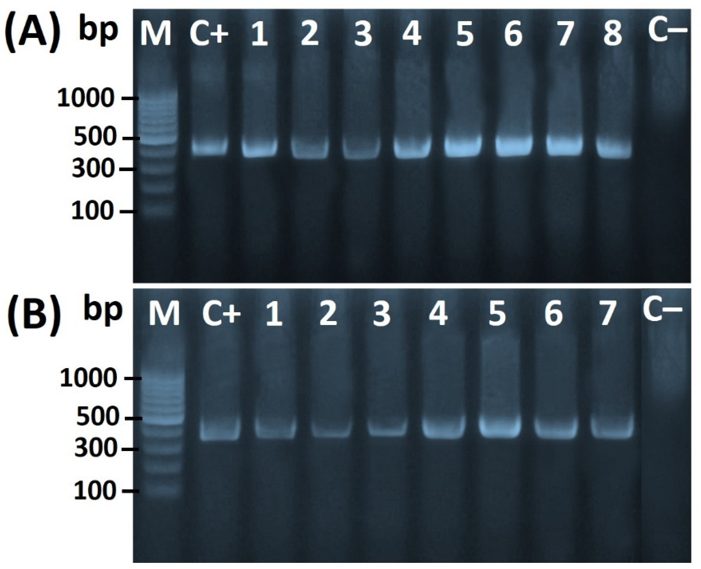
2.3. Molecular Confirmation of Helicobacter spp.

Specific primer sets (Table 1) were synthesized by Metabion, Steinkirchen, Germany to be used for the amplification of the 16S rRNA of Helicobacter genus and H. pullorum, as well as the specific primers of H. pylori-specific phosphoglucosamine mutase gene (glmM), and virulence factors including hrgA, cagA, and vacA genes of for glmM gene.
In summary, a QIAamp DNA Mini kit (Qiagen, Hilden, Germany) was used to extract DNA from a pure culture. For genus confirmation through 16S rRNA amplification, 5 µL of DNA template were mixed with 12.5 µL of EmeraldAmp Max PCR Master Mix (Takara Bio, Kusatsu, Japan), 1 µL of each primer (20 pmol), and 5.5 µL of water. The PCR mix was transferred to theApplied Biosystems 2720 Thermal Cycler (Applied Biosystems, Foster City, CA, USA), under the following conditions: 94 °C, for 5 min, followed by 35 cycles of 94 °C for 1 min, 54 °C for 1 min, and 72 °C for 1 min, and finally, an extension for 10 min at 72 °C. The H. pylori ATCC 43,504 strain and distilled water were used as positive and negative controls, respectively.
For species identification, primers targeting the glmM gene and H. pullorum-specific 16S rRNA were used for the identification of H. pylori and H. pullorum, respectively. The PCR mix and cycling conditions were the same as previously described in genus-specific 16S rRNA except for annealing temperatures.
A multiplex PCR was used to detect the virulence genes hrgA, cagA, and vacA in H. pylori isolates as previously described [32]. The PCR mixture included a 5 µL DNA template, 12.5 µL of EmeraldAmp Max PCR Master Mix (Takara Bio), 1 µL of each primer (10 pmol), and water up to 25 µL. The PCR cycling conditions were identical to those previously described in genus-specific 16S rRNA except for the annealing temperature, which was set at 52 °C for 1 min. The DNA of H. pylori was used as a positive control, while the DNA isolated from E. coli K12DH5α served as a negative control. PCR products were electrophoresed in a 1.5% agarose gel with 0.3% ethidium bromide in a 10% Tris–borate–EDTA buffer. DNA signals in the gel were visualized under a UV transilluminator.

2.4. Antimicrobial Susceptibility Testing

The agar dilution model was used to evaluate antibiotic susceptibility profiles, according to the Clinical and Laboratory Standards Institute guidelines [33]. The Helicobacter isolates were collected from 72 h culture on Blood agar and suspended in saline, to reach the 2.0 McFarland opacity standard. Then, 2 µL of each suspension were spot inoculated in Mueller-Hinton agar (Oxoid, Hampshire, UK), supplemented with 5% sheep blood, and serial two-fold dilutions of eight antibiotics (Sigma-Aldrich, St. Louis, MO, USA). The inoculated plates were incubated under microaerophilic conditions, at 37 °C for 72 h, as previously described. For H. pylori, the antibiotic resistance breakpoints for amoxicillin, metronidazole, tetracycline, and levofloxacin were determined according to the European Committee on Antimicrobial Susceptibility Testing [34], while the breakpoint for clarithromycin was adopted from CLSI [33]. For H. pullorum, the breakpoints for ampicillin, erythromycin, tetracycline, and ciprofloxacin were assumed according to Campylobacter and related species [29,35,36]. For quality control, H. pylori ATCC 43504 reference strain was used for H. pylori isolates. For H. Pullorum isolates, C. jejuni ATCC 33560 was used as a control for ciprofloxacin, erythromycin, and tetracycline, while Staphylococcus aureus ATCC 43300 was used as a control for ampicillin.

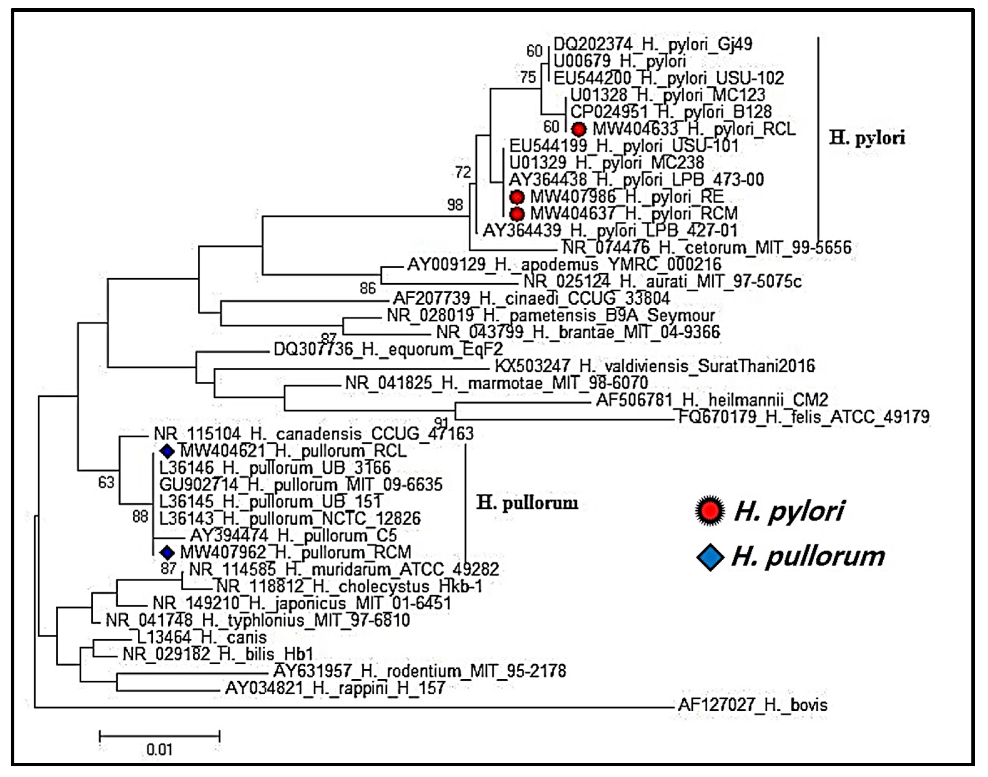
2.5. Helicobacter Species 16S rRNA Gene Sequencing and Phylogenetic Analysis

Using the QIAquick gel extraction kit (Qiagen, Valencia, CA, USA), the 16S rRNA PCR products of three H. pylori isolates (one isolate from each of meat, liver, and environmental swab) and two H. pullorum isolates (one isolate from each of meat and liver samples) were purified. The purified products were sequenced in both directions, using a Big-Dye Terminator v3.1 cycle sequencing kit (Applied Biosystems, Foster City, CA, USA) in an Applied Biosystems 3130 genetic analyzer (Applied Biosystems), according to the manufacturer’s instructions. The BLAST 2.2 program (National Center for Biotechnology Information; NCBI) was used to confirm the nucleotide sequence identity. The phylogenetic tree was generated using the MegAlign module of DNASTAR Lasergene software V.12.1 [37], and phylogenetic analyses were performed in MEGA6 software using maximum likelihood, neighbor-joining, and maximum parsimony [38].

2.6. Statistical Analysis

Fisher’s exact test was applied to determine the significant difference between the prevalence of Helicobacter species in chicken meat and their associated environment. Significance was determined at p < 0.05.

3. Results and Discussions

3.1. Prevalence of Helicobacter spp. in Chicken Meats and Swab Samples

Livestock (particularly poultry) is considered a crucial reservoir of many pathogenic microorganisms. Helicobacter has recently developed a public health concern as an emerging foodborne pathogen [4]. For that reason, the microbial quality assessment of chicken meat is important to reduce the load of Helicobacter in meat. Information regarding chickens as an essential reservoir for H. Pylori dissemination to humans is very limited [28]. In the current study, H. pylori was detected in 4% (4/100) of chicken breast meat samples. In chicken liver and gizzard samples, however, H. pylori was isolated from 10% (10/100) and 2% (2/100) samples, respectively (Table 2). The overall prevalence of H. pylori among the 300 broiler chicken samples (meat and giblets) was 5.33% (16/300) (Table 2). This result was consistent with that of Dairouty et al. [27], who revealed that 5% (1/20) of raw poultry meat were positive for H. pylori. Conversely, a much higher prevalence rate of 36% (4/11) was reported for H. pylori by Meng et al. [39], in fresh raw chickens. H. pylori contamination in the chicken meat samples tested in the present study could be attributed to the contaminated hands of the butchers, veterinarians, and slaughterhouse workers during handling, chicken portions, and giblets preparation and packaging. Besides, usage of unclean water during the washing of the chicken carcasses could be another potential reason for the presence of H. pylori in the chicken meats. Furthermore, the incidence of H. pylori in chicken specimens may be due to cross-contamination from knives or other slaughterhouse equipment.
Regarding the prevalence of H. pullorum in chicken meat and giblets, H. pullorum was detected in 2% (2/100) meat samples. In chicken liver and gizzard samples, however, H. pullorum was detected in 6% (6/100) and 6% (6/100) of the samples tested, respectively. The overall prevalence of H. pullorum among the 300 broiler chicken samples (meat and giblets) was 4.67% (14/300) (Table 2). A lower prevalence rate was reported by Gholami-Ahangaran et al. [40], who indicated that H. pullorum was present in 2% (2/100) of the examined liver samples. On the contrary, higher prevalence rates of 23.5% (4/17) and 24% (12/50) were recorded for H. pullorum in raw fresh chicken meat [4] and thigh chicken samples [41], respectively. The presence of H. pullorum in chicken meat samples could be attributed to its dissemination from the poultry cecum and consequent contamination of chicken carcasses during poultry processing, and this suggests that this organism may be a potential risk factor for zoonotic foodborne transmission to human consumers. Furthermore, the isolation of H. pullorum from the chicken liver could be attributed to the bacterium’s ability to enter the liver via retrograde transfer from the duodenum. H. pullorum may also translocate from the gut lumen to the portal circulation [42].
Concerning the presence of H. pylori and H. pullorum in environmental swab samples, just two H. pylori isolates were isolated from two different cutting boards, while H. pullorum was only detected in one environmental sample, specifically from a worker’s hand (Table 2). This finding may be due to infected workers’ hands and infrequent cleaning and disinfection of the cutting boards before use.
The frequencies of the different types of Helicobacter (pylori, pullorum, and others) were compared among retail chicken organs (breast meat, liver, and gizzard) and a significant difference were detected at p < 0.05, with the highest frequency noticed for H. pylori in liver, while there was no significant difference between the frequencies of different types of Helicobacter (pylori, pullorum, and others) in the environmental swap samples (cutting board, knives, and working hands).

3.2. Phylogenetic Analysis of Partial 16S rRNA Gene Sequencing of Helicobacter Species

The PCR amplicon for the 16S rRNA gene of Helicobacter spp. Was detected at the particular expected size of 398 bp (Figure 1A,B). Additionally, H. pullorum-specific 16S rRNA was detected at the expected size of 447 bp (Figure 2A), while the PCR product specific of glmM gene-specific for the characterization of H. pylori was detected at the expected size of 294 bp (Figure 2B).
The resultant 16S rRNA gene sequences of the selected three H. pylori and two H. pullorum in this study were submitted to the GenBank nucleotide database, under the following accession numbers MW404637, MW404633, and MW407986 for H. pylori isolates and MW407962 and MW404621 for H. pullorum isolates (Table 3).
The 16S rRNA gene sequencing of the selected three H. pylori indicated that two isolates were clustered together with a 100% genetic similarity and were supported with a bootstrap value of 98%, but the third one was distantly related with a 99.2% sequence similarity. On the other hand, the nucleotide sequences of the 16S rRNA gene from the selected two H. pullorum isolates were located in the same cluster with a 100% genetic identity. It was also found that these two H. pullorum isolates of the current study were highly related to the other H. pullorum sequences, which were retrieved from the NCBI GenBank databases. It means that H. pullorum isolates had a very low genetic diversity. The identities of the H. pylori and H. pullorum isolates are shown in Figure 3.
The homology search (BLAST) of the sequenced 16S rRNA gene was conducted for determining their identities and knowing their phylogeny trees. The phylogenetic analysis of the three H. pylori sequences in the current study showed high genetic identity to all the retrieved H. pylori sequences, which were of human origin, and this finding emphasized that humans could be the main source of H. pylori contamination in chicken meat and livers. Furthermore, the phylogenetic analysis of the two H. pullorum sequences of the present study revealed a 100% homology to one isolate (L36145), which was obtained from a human patient with gastroenteritis, and also showed a 99.8% identity to another isolate (AY394474), which was recovered from the cirrhotic liver of human with hepatitis C. This finding indicated that H. pullorum could be a potential foodborne zoonotic pathogen. The phylogenetic analysis of the H. pylori and H. pullorum isolates is shown in Figure 4.
The phylogenic diversity among the analyzed Helicobacter isolates might be attributed to the difference in sampling sites from long-distance localities in Qalyubia Governorate from which chicken meat and giblets were collected, and hence, a variation of the strains isolated from these different samples. On the other hand, the close identity between the 16S rRNA sequences of the strains analyzed could be attributed to the cross-contamination of the chicken samples.

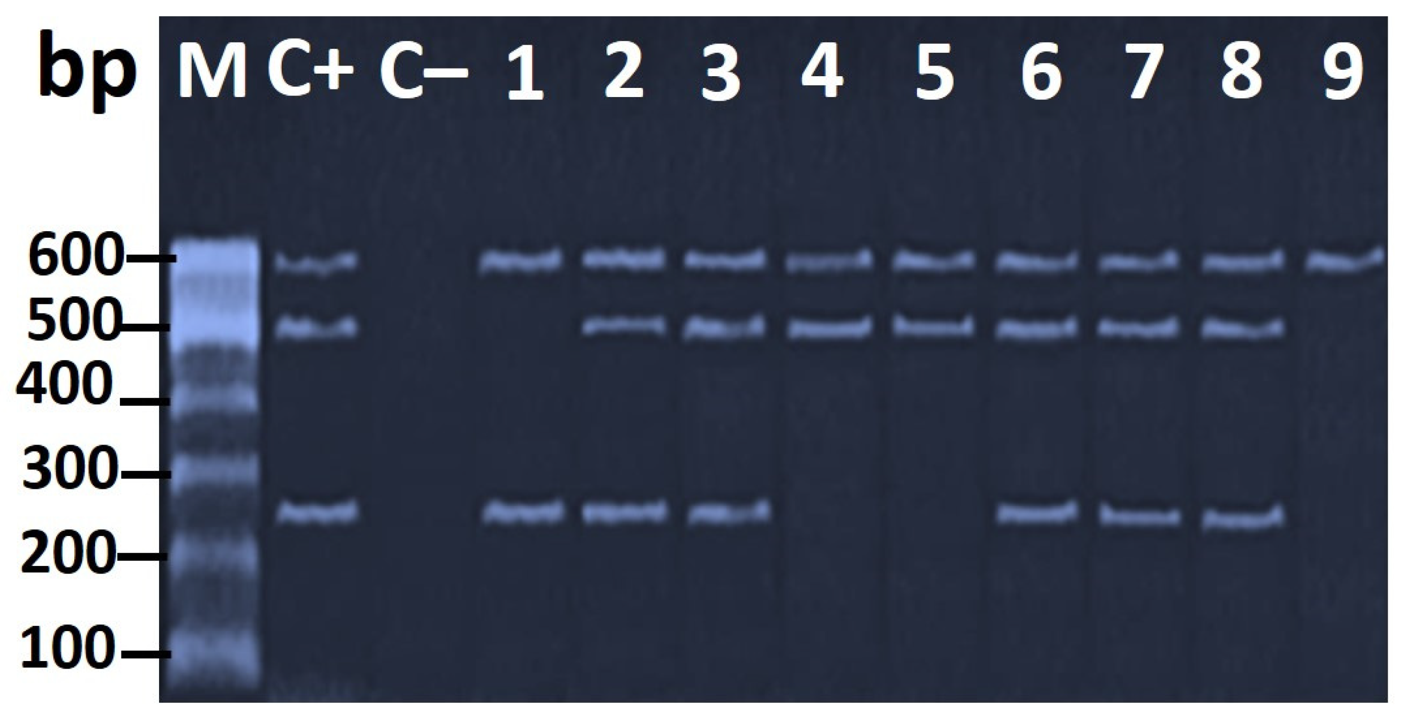
3.3. Genotypic Characterization of H. pylori Virulence Genes

The multiplex PCR verified the presence of the virulence genes vacA, cagA, and hrgA at the expected molecular size of 259, 499, and 594 bp, respectively, in the H. pylori isolates (Figure 5).
The frequency distributions of vacA, cagA, and hrgA virulence genes of H. pylori isolates in this study are shown in Table 4. Based on the molecular analysis, vacAs1, cagA, and hrgA were detected in 66.7% (12/18), 77.8% (14/18), and 100% (18/18) of the 18 H. pylori isolated strains, respectively. Accordingly, the most common virulence marker was the hrgA gene. Additionally, the highest frequency of the tested virulence genotypes was detected in the chicken isolates from the liver and breast meat of broiler chickens which are commonly consumed food for humans.
The existence of vacAs1 and cagA genes among the isolates in the present investigation was in agreement with that of Hamada et al. [28], who found that vacA and cagA genes were detected in 57.1% and 42.9% of H. pylori isolates recovered from 90 chicken meat, liver, and gizzard samples. Similarly, Hemmatinezhad et al. [43] reported that vacA and cagA were the most commonly identified genes in the H. pylori isolates from ready-to-eat foods since they existed in 78.37% and 41.89% of the tested isolates. On the other hand, a much lower existence rate of 20% was determined for both vacAs1 and cagA in H. pylori isolates from minced meat samples [44]. Likewise, a considerable prevalence of vacAs1 and cagA virulence factors were shown previously in many foods of animal origin [45,46].
The existence of the virulence genes in H. pylori evoked adverse effects on human consumers. The presence of vacAs1 and cagA virulence factors enables the colonization and survival of H. pylori within the gastric mucosa through complex mechanisms, such as adhesion to gastric epithelial cells, interleukin-8 production, stimulation of inflammatory response, formation of intracellular vacuoles, induction of apoptosis of gastric epithelial cells, and lastly gastritis, gastroduodenal ulcers, and even gastric cancer in individuals who eat these examined contaminated chicken meat samples [47]. In addition, a significant relationship between vacAs1 expression and peptic ulcer disease (PUD) has previously been found [18,48].
Concerning the hrgA gene, the current study is considered the first record of its detection in H. pylori from a food source. Moreover, it had the highest frequency (100%) in the studied H. pylori isolates of chicken meat, giblets, and swab samples. It is a novel gene that was identified during the examination of the hpyIIIR−hpyIIIM locus in Western and Asian H. pylori strains, and this gene was found in place of the hpyIIIR gene and located upstream of hpyIIIM in 34% (70/208) of the examined strains. From this time, it is named restriction endonuclease-replacing gene A [22]. This recently identified gene was more predominant in western countries than in Asia and more prevalent in gastric cancer patients in comparison to in patients without gastric cancer in East Asian countries. In addition, it was more abundant in cagA+ than cagA isolates in western strains [49]. The virulent role of hrgA was also investigated, and it was observed that it had a direct function in interleukin-8 induction and the apoptosis of gastric epithelial cells [22]. Thus, our report suggests that hrgA could potentially be a public health hazard for humans who consumes chicken meat contaminated with hrgA+ H. pylori strains.

3.4. Antimicrobial Resistance Profiles of the H. pylori and H. pullorum Isolates

The antibacterial agents that were selected for evaluation are commonly used in human medicine to treat patients suffering from Helicobacter infection. In this study, four H. pylori isolates (44.4%) were resistant to at least one antibiotic (Table 5), while two isolates showed multiple drug resistance (MDR) to more than three classes of antibiotics. The highest resistance rates of 44.4% and 33.3% were for clarithromycin and metronidazole, respectively, while the lowest rate was determined for amoxicillin (11.1%) (Table 5).
In the case of H. pullorum, six isolates were resistant to at least one antibiotic (85.7%), and three isolates (42.9%) were MDR (Table 5). All isolates were susceptible to ampicillin but showed high resistance rates for erythromycin (85.7%) and ciprofloxacin (71.4%).
In respect of H. pylori isolates, we found that H. pylori strains exhibited a high rate of resistance toward clarithromycin, metronidazole, tetracycline, levofloxacin, and amoxicillin. Our findings were consistent with those of Hamada et al. [28], who found high levels of H. pylori resistance to amoxicillin, penicillin, oxytetracycline, nalidixic acid, ampicillin, and norfloxacin in chicken meat, liver, and gizzard; Mashak et al. [50] who reported that H. pylori strains were resistant towards tetracycline, erythromycin, levofloxacin, and amoxicillin in raw meat samples; Gilani et al. [44], who found that H. pylori bacteria displayed a high resistance against ampicillin, erythromycin, amoxicillin, tetracycline, and clarithromycin in meat product samples; Mousavi et al. [44], who stated that H. pylori isolates from milk displayed strong resistance toward ampicillin, tetracycline, erythromycin, and metronidazole; and Ranjbar et al. [46], who mentioned that H. pylori strains from traditional dairy products harbored a high incidence of resistance against ampicillin, amoxicillin, tetracycline, erythromycin, and metronidazole. Besides, previous studies reported by Secka et al. [51] and Yahaghi et al. [52] revealed that H. pylori isolated from food specimens regularly showed an incidence of resistance against amoxicillin, metronidazole, ampicillin, and oxytetracycline. In addition, epidemiological studies conducted in China, Taiwan, the Kingdom of Saudi Arabia, Egypt, Nigeria, Iran, India, Brazil, Argentina, and Colombia revealed that H. pylori strains obtained from medical samples are highly resistant to amoxicillin, metronidazole, quinolones, and tetracycline [53,54].
The emergence of multidrug-resistant foodborne bacterial pathogens in humans is mainly attributed to the frequent misuse and overuse of antibiotics for prophylaxis and growth promotion in the poultry industry. Additionally, the abuse of antimicrobial agents and self-medication by human beings may be considered an additional source. It is likely that the H. pylori bacteria can be transmitted from infected butchers and workers to meat samples through cross-contamination during meat handling in poultry slaughterhouses. Our finding of the antibiotic resistance pattern of H. pylori isolates revealed that the meat, livers, and gizzards of the examined poultry may be a possible vehicle for antibiotic-resistant H. pylori and subsequently could be hazardous to human health.
Concerning H. pullorum strains, three H. pullorum isolates collected in this study showed high resistance rates for erythromycin, ciprofloxacin, and tetracycline (42.9–85.7%), which agreed with other studies [4,36,41]. All H. pullorum isolates were sensitive to ampicillin, which was in line with the results of Zanoni et al. [36] and Hassan et al. [29]; nonetheless, Ceelen et al. [15] showed a higher rate of ampicillin resistance among poultry isolates in Europe. This observed incidence of antimicrobial resistance in chicken samples was attributable to the frequent use of quinolones and tetracyclines for prophylaxis and growth promotion in the Egyptian poultry industry.

4. Conclusions

The present study concluded that approximately 5% of chicken meat and giblets marketed in Egypt were contaminated with H. pylori and/or H. pullorum which are resistant to the antimicrobials recommended for human treatment, and hence, the consumption of undercooked chicken meat and giblets is considered a potential public health hazard to humans. Contamination of chicken carcasses can occur during slaughtering and/or processing or when they come into contact with contaminated hands or contaminated water in poultry abattoirs. It is critical for slaughterhouses to maintain good hygienic measures and sanitary practices to limit contamination of meat with H. pylori and H. pullorum. The present study also found a very high (100%) frequency of the hrgA gene in the isolated H. pylori strains. In addition, more than 20% of H. pylori isolates were resistant to three or more antibiotics. This highlights the significance of evaluating the antibiotic susceptibility profile of Helicobacter infections to recommend using the most effective antimicrobial agents for its eradication.

Author Contributions

Conceptualization, A.M.E., K.I.S., and K.I.; data curation, A.M.E., K.I.S., K.I., and V.H.; formal analysis, A.M.E., W.S.A., W.A.E., F.E., W.E., and A.M.; funding acquisition, K.I.S., K.I., and H.E.; investigation, A.M.E., W.S.A., W.A.E., F.E., W.E., and A.M.; methodology, W.S.A., W.A.E., F.E., W.E., and A.M.; resources, V.H.; software, A.M.E., W.S.A., W.A.E., F.E., W.E., and A.M.; supervision, K.I.S., K.I., and H.E.; visualization, A.M.E., W.S.A., W.A.E., F.E., W.E., and A.M.; writing—original draft, A.M.E., K.I.S., and K.I.; writing—review & editing, K.I.S., K.I., and H.E. All authors have read and agreed to the published version of the manuscript.

Funding

This research received no external funding.

Data Availability Statement

Data is contained within the article.

Acknowledgments

This research paper is supported by the project “Increasing the impact of excellence research on the capacity for innovation and technology transfer within USAMVB Timișoara” code 6PFE, submitted in the competition Program 1—Development of the national system of research—development, Subprogram 1.2—Institutional performance, Institutional development projects—Development projects of excellence in RDI.

Conflicts of Interest

The authors declare no conflict of interest.

References

  1. Marouf, S.; Khalf, M.A.; Alorabi, M.; El-Shehawi, A.M.; El-Tahan, A.M.; Abd El-Hack, M.E.; El-Saadony, M.T.; Salem, H.M. Mycoplasma gallisepticum: A devastating organism for the poultry industry in Egypt. Poult. Sci. 2021, 101, 101658. [Google Scholar] [CrossRef] [PubMed]
  2. Jennings, J.L.; Sait, L.C.; Perrett, C.A.; Foster, C.; Williams, L.K.; Humphrey, T.J.; Cogan, T.A. Campylobacter jejuni is associated with, but not sufficient to cause vibrionic hepatitis in chickens. Vet. Microbiol. 2011, 149, 193–199. [Google Scholar] [CrossRef] [PubMed]
  3. Qumar, S.; Majid, M.; Kumar, N.; Tiwari, S.K.; Semmler, T.; Devi, S.; Baddam, R.; Hussain, A.; Shaik, S.; Ahmed, N. Genome dynamics and molecular infection epidemiology of multidrug-resistant Helicobacter pullorum isolates obtained from broiler and free-range chickens in India. Appl. Environ. Microbiol. 2016, 83, e02305–e02316. [Google Scholar] [CrossRef] [PubMed] [Green Version]
  4. Borges, V.; Santos, A.; Correia, C.B.; Saraiva, M.; Ménard, A.; Vieira, L.; Sampaio, D.A.; Pinheiro, M.; Gomes, J.P.; Oleastro, M. Helicobacter pullorum isolated from fresh chicken meat: Antibiotic resistance and genomic traits of an emerging foodborne pathogen. Appl. Environ. Microbiol. 2015, 81, 8155–8163. [Google Scholar] [CrossRef] [PubMed] [Green Version]
  5. Javed, K.; Gul, F.; Abbasi, R.; Zaidi, R.A.; Noreen, Z.; Bokhari, H.; Javed, S. Prevalence and role of Type six secretion system in pathogenesis of emerging zoonotic pathogen Helicobacter pullorum from retail poultry. Avian. Pathol. 2019, 48, 557–563. [Google Scholar] [CrossRef]
  6. Atherton, J.C. The pathogenesis of Helicobacter pylori-induced gastro-duodenal diseases. Annu. Rev. Pathol. 2006, 1, 63–96. [Google Scholar] [CrossRef]
  7. Kennemann, L.; Didelot, X.; Aebischer, T.; Kuhn, S.; Drescher, B.; Droege, M.; Reinhardt, R.; Correa, P.; Meyer, T.F.; Josenhans, C. Helicobacter pylori genome evolution during human infection. Proc. Natl. Acad. Sci. 2011, 108, 5033–5038. [Google Scholar] [CrossRef] [Green Version]
  8. Zamani, M.; Ebrahimtabar, F.; Zamani, V.; Miller, W.; Alizadeh-Navaei, R.; Shokri-Shirvani, J.; Derakhshan, M. Systematic review with meta-analysis: The worldwide prevalence of Helicobacter pylori infection. Aliment Pharmacol. Ther. 2018, 47, 868–876. [Google Scholar] [CrossRef] [Green Version]
  9. Ruggiero, P. Helicobacter pylori and inflammation. Curr. Pharm. Des. 2010, 16, 4225–4236. [Google Scholar] [CrossRef]
  10. Stanley, J.; Linton, D.; Burnens, A.P.; Dewhirst, F.E.; On, S.L.; Porter, A.; Owen, R.J.; Costas, M. Helicobacter pullorum sp. nov.-genotype and phenotype of a new species isolated from poultry and from human patients with gastroenteritis. Microbiology 1994, 140, 3441–3449. [Google Scholar] [CrossRef] [Green Version]
  11. Hameed, K.G.A.; Sender, G. Prevalence of Helicobacter pullorum in Egyptian hen’s eggs and in vitro susceptibility to different antimicrobial agents. Anim. Sci. Pap. Rep. 2011, 29, 257–264. [Google Scholar]
  12. Atabay, H.; Corry, J.; On, S.L. Diversity and prevalence of Arcobacter spp. in broiler chickens. J. Appl. Microbiol. 1998, 84, 1007–1016. [Google Scholar] [CrossRef] [PubMed]
  13. Wai, S.S.; Abdul-Aziz, S.; Bitrus, A.A.; Zunita, Z.; Abu, J. Helicobacter pullorum in broiler chickens and the farm environment: A One Health approach. Int. J. One Health 2019, 5, 20–25. [Google Scholar] [CrossRef] [Green Version]
  14. González, A.; Piqueres, P.; Moreno, Y.; Cañigral, I.; Owen, R.J.; Hernández, J.; Ferrús, M.A. A novel real-time PCR assay for the detection of Helicobacter pullorum-like organisms in chicken products. Int. Microbiol. 2008, 11, 203–208. [Google Scholar] [PubMed]
  15. Ceelen, L.; Decostere, A.; Verschraegen, G.; Ducatelle, R.; Haesebrouck, F. Prevalence of Helicobacter pullorum among patients with gastrointestinal disease and clinically healthy persons. J. Clin. Microbiol. 2005, 43, 2984–2986. [Google Scholar] [CrossRef] [PubMed] [Green Version]
  16. Fox, J. The non-H pylori helicobacters: Their expanding role in gastrointestinal and systemic diseases. Gut 2002, 50, 273–283. [Google Scholar] [CrossRef]
  17. Taylor, L.H.; Latham, S.M.; Woolhouse, M.E. Risk factors for human disease emergence. Philos. Trans. R Soc. Lond B Biol. Sci. 2001, 356, 983–989, Series B: Biological Sciences. [Google Scholar] [CrossRef]
  18. Akeel, M.; Shehata, A.; Elhafey, A.; Elmakki, E.; Aboshouk, T.; Ageely, H.; Mahfouz, M. Helicobacter pylori vacA, cagA and iceA genotypes in dyspeptic patients from southwestern region, Saudi Arabia: Distribution and association with clinical outcomes and histopathological changes. BMC Gastroenterol. 2019, 19, 16. [Google Scholar] [CrossRef]
  19. Sedaghat, H.; Moniri, R.; Jamali, R.; Arj, A.; Zadeh, M.R.; Moosavi, S.G.A.; Rezaei, M. Prevalence of Helicobacter pylori vacA, cagA, cagE, iceA, babA2, and oipA genotypes in patients with upper gastrointestinal diseases. Iran. J. Microbiol. 2014, 6, 14–21. [Google Scholar]
  20. Yong, X.; Tang, B.; Li, B.-S.; Xie, R.; Hu, C.-J.; Luo, G.; Qin, Y.; Dong, H.; Yang, S.-M. Helicobacter pylori virulence factor cagA promotes tumorigenesis of gastric cancer via multiple signaling pathways. Cell Commun. Signal. 2015, 13, 30. [Google Scholar] [CrossRef] [Green Version]
  21. Foegeding, N.J.; Caston, R.R.; McClain, M.S.; Ohi, M.D.; Cover, T.L. An overview of Helicobacter pylori vacA toxin biology. Toxins 2016, 8, 173. [Google Scholar] [CrossRef] [PubMed] [Green Version]
  22. Ando, T.; Wassenaar, T.M.; Peek, R.M.; Aras, R.A.; Tschumi, A.I.; van Doorn, L.-J.; Kusugami, K.; Blaser, M.J. A Helicobacter pylori restriction endonuclease-replacing gene, hrgA, is associated with gastric cancer in Asian strains. Cancer Res. 2002, 62, 2385–2389. [Google Scholar]
  23. Goderska, K.; Pena, S.A.; Alarcon, T. Helicobacter pylori treatment: Antibiotics or probiotics. Appl. Microbiol. Biotechnol. 2018, 102, 1–7. [Google Scholar] [CrossRef] [PubMed]
  24. Alba, C.; Blanco, A.; Alarcón, T. Antibiotic resistance in Helicobacter pylori. Curr. Opin. Infect. Dis. 2017, 30, 489–497. [Google Scholar] [CrossRef] [PubMed]
  25. Suzuki, S.; Esaki, M.; Kusano, C.; Ikehara, H.; Gotoda, T. Development of Helicobacter pylori treatment: How do we manage antimicrobial resistance? World J. Gastroenterol. 2019, 25, 1907–1912. [Google Scholar] [CrossRef]
  26. Yousefi-Avarvand, A.; Vaez, H.; Tafaghodi, M.; Sahebkar, A.H.; Arzanlou, M.; Khademi, F. Antibiotic resistance of Helicobacter pylori in Iranian children: A systematic review and meta-analysis. Microb. Drug Resist. 2018, 24, 980–986. [Google Scholar] [CrossRef]
  27. El Dairouty, R.; Murad, H.; El Shenawy, M.; Hosny, I.; Okda, A.; El Shamy, S. Helicobacter pylori and its interrelations with other foodborne pathogenic bacteria in Egyptian meat and some meat products. Curr. Sci. Int. 2016, 5, 139–146. [Google Scholar]
  28. Hamada, M.; Elbehiry, A.; Marzouk, E.; Moussa, I.M.; Hessain, A.M.; Alhaji, J.H.; Heme, H.A.; Zahran, R.; Abdeen, E. Helicobacter pylori in a poultry slaughterhouse: Prevalence, genotyping and antibiotic resistance pattern. Saudi J. Biol. Sci. 2018, 25, 1072–1078. [Google Scholar] [CrossRef]
  29. Hassan, A.K.; Shahata, M.A.; Refaie, E.M.; Ibrahim, R.S. Detection and identification of Helicobacter pullorum in poultry species in upper Egypt. J. Adv. Vet. Res. 2014, 4, 42–48. [Google Scholar]
  30. Tabrizi, S.A.; Derakhshandeh, A.; Esfandiari, A.; Atashi, A.Z. Identification of Helicobacter spp. in gastrointestinal tract, pancreas and hepatobiliary system of stray cats. Iran J. Vet. Res. 2015, 16, 374–376. [Google Scholar]
  31. Safaei, H.; Rahimi, E.; Zandi, A.; Rashidipour, A. Helicobacter pylori as a zoonotic infection: The detection of H. pylori antigens in the milk and faeces of cows. J. Res. Med. Sci. 2011, 16, 184–187. [Google Scholar] [PubMed]
  32. Tiwari, S.; Khan, A.; Manoj, G.; Ahmed, S.; Abid, Z.; Habeeb, A.; Abid, Z.; Habibullah, C. A simple multiplex PCR assay for diagnosing virulent Helicobacter pylori infection in human gastric biopsy specimens from subjects with gastric carcinoma and other gastro-duodenal diseases. J. Appl. Microbiol. 2007, 103, 2353–2360. [Google Scholar] [CrossRef] [PubMed]
  33. CLSI 2015; Performance Standards for Antimicrobial Susceptibility Testing: Twenty-Fifth Informational Supplement. CLSI document M100-S25; Clinical and Laboratory Standards Institute: Wayne, PA, USA, 2015.
  34. EUCAST 2020; The European Committee on Antimicrobial Susceptibility Testing. Breakpoint Tables for Interpretation of MICs and Zone Diameters. Version 10.0; The European Society of Clinical Microbiology and Infectious Diseases (ESCMID): Basel, Switzerland, 2020. Available online: http://www.eucast.org (accessed on 23 April 2020).
  35. CLSI 2005; Performance Standards for Antimicrobial Susceptibility Testing. Fifteenth Informational Supplement. M100-S15; Clinical and Laboratory Standards Institute: Wayne, PA, USA, 2005.
  36. Zanoni, R.G.; Rossi, M.; Giacomucci, D.; Sanguinetti, V.; Manfreda, G. Occurrence and antibiotic susceptibility of Helicobacter pullorum from broiler chickens and commercial laying hens in Italy. Int. J. Food Microbiol. 2007, 116, 168–173. [Google Scholar] [CrossRef] [PubMed]
  37. Thompson, J.D.; Higgins, D.G.; Gibson, T.J. CLUSTAL W: Improving the sensitivity of progressive multiple sequence alignment through sequence weighting, position-specific gap penalties and weight matrix choice. Nucleic Acids Res. 1994, 22, 4673–4680. [Google Scholar] [CrossRef] [PubMed] [Green Version]
  38. Tamura, K.; Stecher, G.; Peterson, D.; Filipski, A.; Kumar, S. MEGA6: Molecular evolutionary genetics analysis version 6.0. Mol. Biol. Evol. 2013, 30, 2725–2729. [Google Scholar] [CrossRef] [PubMed] [Green Version]
  39. Meng, X.; Zhang, H.; Law, J.; Tsang, R.; Tsang, T. Detection of Helicobacter pylori from food sources by a novel multiplex PCR assay. Food Saf. 2008, 28, 609–619. [Google Scholar] [CrossRef]
  40. Gholami-Ahangaran, M.; Haddadi, I.; Karimi, Y.; Omrani, E. Molecular evidence of Helicobacter pullorum, as a foodborne pathogen in broiler carcasses in Iran. Europ. Poult. Sci. 2015, 79. [Google Scholar] [CrossRef]
  41. Jebellijavan, A.; Emadi Chashmi, S.H.; Staji, H.; Akhlaghi, H. Comparison of the Culture and PCR Methods to Determine th Prevalence and Antibiotic Resistance of Helicobacter pullorum Isolated from Chicken Thigh Samples in Semnan, Iran. J. Hum. Environ. Health Promot. 2020, 6, 167–172. [Google Scholar] [CrossRef]
  42. Ceelen, L.M.; Decostere, A.; Van den Bulck, K.; On, S.L.W.; Baele, M.; Ducatelle, R.; Haesebrouck, F. Helicobacter pullorum in Chickens, Belgium. Emerg. Infect. Dis. 2006, 12, 263–267. [Google Scholar] [CrossRef] [Green Version]
  43. Hemmatinezhad, B.; Momtaz, H.; Rahimi, E. vacA, cagA, iceA and oipA genotypes status and antimicrobial resistance properties of Helicobacter pylori isolated from various types of ready to eat foods. Ann. Clin. Microbiol. Antimicrob. 2016, 15, 1–9. [Google Scholar] [CrossRef] [Green Version]
  44. Gilani, A.; Razavilar, V.; Rokni, N.; Rahimi, E. vacA and cagA genotypes status and antimicrobial resistance properties of Helicobacter pylori strains isolated from meat products in Isfahan province, Iran. Iran J. Vet. Res. 2017, 18, 97–102. [Google Scholar] [PubMed]
  45. Mousavi, S.; Dehkordi, F.S. Virulence factors and antibiotic resistance of Helicobacter pylori isolated from raw milk and unpasteurized dairy products in Iran. J. Venom. Anim. Toxins Incl. Trop. Dis. 2018, 20, 1–7. [Google Scholar] [CrossRef] [PubMed] [Green Version]
  46. Ranjbar, R.; Yadollahi Farsani, F.; Safarpoor Dehkordi, F. Antimicrobial resistance and genotyping of vacA, cagA, and iceA alleles of the Helicobacter pylori strains isolated from traditional dairy products. J. Food Saf. 2019, 39, e12594. [Google Scholar] [CrossRef]
  47. Baj, J.; Forma, A.; Sitarz, M.; Portincasa, P.; Garruti, G.; Krasowska, D.; Maciejewski, R. Helicobacter pylori virulence factors—mechanisms of bacterial pathogenicity in the gastric microenvironment. Cells 2021, 10, 27. [Google Scholar] [CrossRef] [PubMed]
  48. Idowu, A.; Mzukwa, A.; Harrison, U.; Palamides, P.; Haas, R.; Mbao, M.; Mamdoo, R.; Bolon, J.; Jolaiya, T.; Smith, S. Detection of Helicobacter pylori and its virulence genes (cag A, dup A, and vac A) among patients with gastroduodenal diseases in Chris Hani Baragwanath Academic Hospital, South Africa. BMC Gastroenterol. 2019, 19, 1–10. [Google Scholar] [CrossRef]
  49. Ando, T.; Aras, R.; Kusugami, K.; Blaser, M.; Wassenaar, T. Evolutionary history of hrgA, Which Replaces the Restriction Gene hpy IIIR in the hpy III Locus of Helicobacter pylori. J. Bacteriol. 2003, 185, 295–301. [Google Scholar] [CrossRef] [Green Version]
  50. Mashak, Z.; Jafariaskari, S.; Alavi, I.; Shahreza, M.S.; Dehkordi, F.S. Phenotypic and genotypic assessment of antibiotic resistance and genotyping of vacA, cagA, iceA, oipA, cagE, and babA2 alleles of Helicobacter pylori bacteria isolated from raw meat. Infect Drug Resist. 2020, 13, 257–272. [Google Scholar] [CrossRef] [Green Version]
  51. Secka, O.; Berg, D.E.; Antonio, M.; Corrah, T.; Tapgun, M.; Walton, R.; Thomas, V.; Galano, J.J.; Sancho, J.; Adegbola, R.A. Antimicrobial susceptibility and resistance patterns among Helicobacter pylori strains from The Gambia, West Africa. Antimicrob. Agents Chemother. 2013, 57, 1231–1237. [Google Scholar] [CrossRef] [Green Version]
  52. Yahaghi, E.; Khamesipour, F.; Mashayekhi, F.; Safarpoor Dehkordi, F.; Sakhaei, M.H.; Masoudimanesh, M.; Khameneie, M.K. Helicobacter pylori in vegetables and salads: Genotyping and antimicrobial resistance properties. BioMed. Res. Int. 2014, 2014, 757941. [Google Scholar] [CrossRef] [Green Version]
  53. Hunt, R.; Xiao, S.; Megraud, F. World Gastroenterology Organization (WGO) global guideline. Helicobacter pylori in developing countries. . J. Gastrointestin. Liver Dis. 2011, 20, 299–304. [Google Scholar] [CrossRef]
  54. Sallam, K.I.; Abd-Elghany, S.M.; Imre, K.; Morar, A.; Herman, V.; Hussein, M.A.; Mahros, M.A. Ensuring safety and improving keeping quality of meatballs by addition of sesame oil and sesamol as natural antimicrobial and antioxidant agents. Food Microbiol. 2021, 99, 103834. [Google Scholar] [CrossRef] [PubMed]
Figure 1. (A) H. pylori (lanes 1–8). (B) H. pullorum (lanes 1–7). Agarose gel electrophoresis showing 16S rRNA PCR amplicon (398 bp) for Helicobacter genus. Five microliters from the PCR product were separated by electrophoresis on a 1.5% agarose gel and visualized under UV light. M: DNA marker (Gene Ladder 100) used as a reference for a fragment size; Lane C+: positive control from H. pylori ATCC 43,504 strain. C−: negative control of Escherichia coli (E. coli) K12 DH5α as a negative control.
Foods 11 01890 g001
Figure 2. (A) Agarose gel electrophoresis of PCR amplicon (447 bp) of the 16S rRNA specific for H. pullorum strains (lanes 1–7). (B) Agarose gel electrophoresis of PCR product of glmM gene (294 bp) specific for the characterization of H. pylori strains (lanes 1–9). Lane M: 100 bp ladder as a molecular-size DNA marker. Lane 1: H. pylori positive control for the glmM gene. Lane 2: negative control from E. coli K12DH5α. Lanes from 1 to 9: positive H. pylori strains.
Foods 11 01890 g002
Figure 3. Identity percentages detected through the homology search (BLAST-N) of the 16S rRNA sequences of the isolated H. pylori and H. pullorum.
Foods 11 01890 g003
Figure 4. Phylogeny analysis of the 16S rRNA sequences of the isolated three H. pylori (a red circular shape) and the two H. pullorum (a blue rhombus shape) isolates.
Foods 11 01890 g004
Figure 5. Agarose gel electrophoresis of the multiplex PCR of vacA (259 bp), cagA (499 bp), and hrgA (594 bp) as virulence genes of H. pylori strains. Lane M: 100 bp ladder as a molecular-size DNA marker. Lane C+: H. pylori ATCC 43504 strain positive control. Lane C−: E. coli K12 DH5α negative control. Lanes 2, 3, 6, 7, and 8: H. pylori positive control for vacA, cagA, and hrgA genes. Lanes 4 and 5: positive control of H. pylori for cagA and hrgA genes. Lane 1: positive control of H. pylori for vacA and hrgA genes. Lane 9: positive control of H. pylori for the hrgA gene. Lanes 1 and 2 represent chicken meat; lanes 3 and 4 represent chicken gizzard; lanes 5 to 9 represent chicken liver.
Foods 11 01890 g005
Table 1. Primers used in this study.
Target GenePrimers Sequences (5′–3′)Product Size (bp)Reference
Helicobacter spp. 16S rRNA5′-AAGGATGAAGCTTCTAGCTTGCTA-3′
5′-GTGCTTATTCGTGAGATACCGTCAT-3′
398Tabrizi et al. [30]
Helicobacter pullorum (H. pullorum)-specific 16S rRNA5′-ATG AAT GCTAGTTGTTGTCAG-3′
5′-GATTGGCTCCACTTCACA-3′
447Stanley et al. [10]
Helicobacter pylori (H. pylori)-specific phosphoglucosamine mutase gene (glmM)5′-GAATAAGCTTTTAGGGGTGTTAGGGG-3′
5′-GCTTACTTTCTAACACTAACGCGC-3′
294Safaei et al. [31]
Restriction endonuclease-replacing gene A (hrgA)5′-TCTCGTGAAAGAGAATTTCC-3′
5′-TAAGTGTGGGTATATCAATC-3′
594Tiwari et al. [32]
Cytotoxin-associated gene A (cagA)5′-GCGATTGTTATTGTGCTTGTAG-3′
5′-GAAGTGGTTAAAAAACAATGCCCC-3′
499
Vacuolating cytotoxin A (vacA)5′-ATGGAAATACAACAAACACAC-3′
5′-CTGCTTGAATGCGCCAAAC-3′
259
Table 2. Prevalence of Helicobacter species in chicken meat and their associated environment at the retail shops.
SourceSample TypeHelicobacter spp.Total
H. pyloriH. pullorumOthers
No.%No.%No.%No.%
Retail chicken
(n = 300)
Breast meat (100)44220066
Liver (100)101066442020
Gizzard (100)2266221010
Environmental swabs (n = 30) *Cutting boards (10)2200000220
Knives (10)00000000
Workers’ hands (10)0000110110
Total330185.45144.2472.123911.82
* Each sample is a pool of 2–4 swabs.
Table 3. The accession numbers of 16S rRNA gene sequence of the selected five Helicobacter species.
GeneIsolate spp.Isolate IDSource of IsolatesAccession Number
16S rRNAH. pyloriH. pylori_RCMChicken meatMW404637
H. pylori_RCLChicken liverMW404633
H. pylori_RERetail shop environment
(Cutting board swabs)
MW407986
H. pullorumH. pullorum_RCMChicken MeatMW407962
H. pullorum_RCLChicken LiverMW404621
Table 4. Frequency distributions of the virulence genes in H. pylori isolates from chicken meat and environmental samples.
SourceNumber of IsolatesvacAs1cagAhrgA
Number%Number%Number%
Breast441002504100
Liver1066088010100
Gizzard20021002100
Environment2210021002100
Total181266.71477.818100
Table 5. Antimicrobial resistance profiles of H. pylori and H. pullorum isolates from chicken meat and environmental samples.
IsolatesAntibioticBpIsolates Number According to the Results of MIC (µg/mL)ABR
No. (%)
MDR
No. (%)
˂0.120.120.250.51248163264128256˃256
H. pylori
(n = 18)
Amoxicillin˃0.125142 2 2 (11.1)2
(22.2)
Clarithromycin≥1226 2 22 2 8 (44.4)
Metronidazole˃8 31134 4 2 6 (33.3)
Tetracycline>1 437 2 2 4 (22.2)
Levofloxacin>14334 2 2 4 (22.2)
H. pullorum
(n = 14)
Ampicillin≥32 237 2 0 (0)3
(42.9)
Erythromycin≥8 2 13233 12 (85.7)
Tetracycline≥16 353 1 26 (42.9)
Ciprofloxacin≥4 133124 10 (71.4)
Bp: breakpoints for antibiotic resistance; ABR: antibiotic resistance isolates; MDR: multiple drugs-resistant isolates (≥3 classes of antibiotics); No.: isolates number.
Publisher’s Note: MDPI stays neutral with regard to jurisdictional claims in published maps and institutional affiliations.

