Review

Gelatin-Based Film as a Color Indicator in Food-Spoilage Observation: A Review

by
Mannur Ismail Shaik
,
Muhammad Farid Azhari
and
Norizah Mhd Sarbon
*
Faculty of Fisheries and Food Science, Universiti Malaysia Terengganu, Kuala Nerus 21030, Terengganu, Malaysia
*
Author to whom correspondence should be addressed.
Foods 2022, 11(23), 3797; https://doi.org/10.3390/foods11233797
Submission received: 4 October 2022 / Revised: 12 November 2022 / Accepted: 15 November 2022 / Published: 24 November 2022
(This article belongs to the Special Issue Edible Film Based on Polysaccharides, Proteins and Lipids)

Abstract

:
The color indicator can monitor the quality and safety of food products due to its sensitive nature toward various pH levels. A color indicator helps consumers monitor the freshness of food products since it is difficult for them to depend solely on their appearance. Thus, this review could provide alternative suggestions to solve the food-spoilage determination, especially for perishable food. Usually, food spoilage happens due to protein and lipid oxidation, enzymatic reaction, and microbial activity that will cause an alteration of the pH level. Due to their broad-spectrum properties, natural sources such as anthocyanin, curcumin, and betacyanin are commonly used in developing color indicators. They can also improve the gelatin-based film’s morphology and significant drawbacks. Incorporating natural colorants into the gelatin-based film can improve the film’s strength, gas-barrier properties, and water-vapor permeability and provide antioxidant and antimicrobial properties. Hence, the color indicator can be utilized as an effective tool to monitor and control the shelf life of packaged foods. Nevertheless, future studies should consider the determination of food-spoilage observation using natural colorants from betacyanin, chlorophyll, and carotenoids, as well as the determination of gas levels in food spoilage, especially carbon dioxide gas.

Graphical Abstract

1. Introduction

Recently, there has been a great concern among consumers to detect the quality and safety of food products in the market due to the increasing awareness of high-quality and safe food, especially perishable food, such as seafood, poultry, and meat. Perishable food contains essential nutrients (water, glucose, and amino acids) required for bacteria such as Pseudomonas spp., Enterobacteriaceae, Enterococcus spp., lactic acid bacteria, and mesophilic total aerobic bacteria to grow [1]. The degradation of glucose by the bacteria is the main factor that causes off-flavor and off-odor in food products during storage. The spoiled food caused by the metabolic process can appear undesirable and unacceptable for consumers due to the alteration in sensory characteristics, i.e., texture, smell, taste, and appearance [2]. Conventionally, food-spoilage observation is undergone through sensory evaluation and chemical experiments [3]. In the last decade, scientists have been working on an alternative way to detect food spoilage by using a color indicator to monitor the microbial metabolites in food. It is helpful for food-spoilage observation to replace the conventional method, which can lead to human errors due to its inefficiency and low precision [4].
The use of color indicators plays a vital role in evaluating the freshness of the food product due to its sensitive nature toward various pH levels. It can offer significant advantages as it is low-cost, rapid, reliable, noninvasive, and non-destructive [5,6]. Usually, two sources of color indicators are present: synthetic sources (bromophenol blue, chlorophenol red, and alizarin) and natural sources (anthocyanin, curcumin, and chlorophyll). The criteria for the color indicator to be used in a food-spoilage observation is that the indicator must be easily distinguished by the naked eye, sensitive to various pH levels, and have a low response time between the appearance of distinctive color and change in pH [7]. During the decomposition of food by the microorganisms, the pH of the food can be altered due to the presence of total volatile basic nitrogen (TVB-N), fatty acids, ketones, etc. The mechanism of the color indicator has been proven to change its color due to the changes in pH when tested on the food product, as conducted in prior studies on beef [3,4], chicken and shrimp [8], and fish [9]. This color indicator tremendously impacts the packaging industry to improve food packaging and fulfill consumer needs. Recently, researchers have focused on developing color indicators from natural sources to detect food spoilage that may not adversely affect food. Food packaging is vital because it functions as a physical barrier between food and the environment to maintain the quality and prolong the shelf life of food products [10]. Biodegradable packaging is commonly used as food packaging to replace plastics from nonrenewable resources such as petrochemicals, which have some significant drawbacks due to being nonbiodegradable and raising critical environmental concerns among consumers [11]. Active packaging and smart packaging have significant contributions to the packaging industry due to the advantages of extending the shelf life of food products, food-spoilage observation, portable size, and low cost [8]. Active packaging is usually used to extend the shelf life of food products and improve the safety and quality of the food product. In contrast, smart packaging can monitor packaged food and indicate the product’s quality and freshness information for manufacturers, retailers, and consumers [12]. Commonly, the biopolymers used to immobilize the color pigment in functioning as smart packagings are protein-based film (chitosan, gelatin, and agar) and polysaccharide-based film (starch, carboxymethyl cellulose, and konjac glucomannan) [11,13].
Gelatin is a good biopolymer for film formation due to its film-forming ability, biocompatibility, and biodegradability, making it suitable for food packaging [14]. The gelatin-based film has been proven to protect against aroma, oxygen, and light. However, it has some significant drawbacks, such as poor barrier properties against water vapor and high moisture sensitivity [15]. Thus, it gives some negative feedback when the film is applied to food because it may dissolve, swell, or be integrated with water molecules [16]. The negative feedback of the gelatin-based film can be improved by incorporating natural extract and improving the functionalities, such as antioxidant and antimicrobial properties and color indicators [17]. The essential characteristic of a color indicator is that the film needs to be good in gas-barrier properties against carbon dioxide, oxygen gas, ammonia, and others for food-spoilage observation [18]. For instance, microorganism degradation from meat spoilage will release carbon dioxide, organic acids, and total volatile basic nitrogen (TVB-N), which will be detected by the color indicator and will change its color according to the pH inside the packaging [8]. Nowadays, consumers demand nontoxic, affordable, and environmentally possible products. The need for a pollution-free environment, the rise in crude oil prices, the demand of consumers for longer food shelf lives, and the proliferation of convenience packaging are contributing to the development of biodegradable packaging material for the market [19]. Consumers’ awareness of biodegradable packaging material is increasing as people are more educated. This sparks the interest of many researchers to produce safe, environmentally friendly, and affordable packaging material. Hence, research has increased on gelatin-based film as a biodegradable packaging material. Gelatin is the most widely used biodegradable packaging material because of the development of high-performance films [20]. Gelatin-based films can preserve, protect, or lengthen the shelf life of food products [21]. The gelatin-based film market in the U.S. is anticipated to rise rapidly during the anticipated period. In addition, the U.K., France, and Germany are expected to see high growth due to the wide range of uses for gelatin films in food. In contrast, China and India are anticipated to see high growth during the projected period due to the rapid adoption of new technologies and products in these countries [22]. Gelatin-based films are already produced widely in many developed countries. However, there is still no commercialized gelatin-based film in Malaysia, and the application of biodegradable films in packaging material is not yet widely applied.
This review would be able to provide alternative suggestions to solve the food-spoilage determination, especially for perishable food, by giving adequate exposure to the usage of the color indicators of the gelatin-based film. However, to date, no published review has been carried out on gelatin-based film as a color indicator in food-spoilage observation. Therefore, this review aims to critically investigate, discuss, and provide clear information about color indicators from gelatin-based films and natural colorants to observe food products’ quality and safety.

2. Food Spoilage

Food spoilage is leading, due to various mechanisms, which include microbial, chemical, and physical reactions, to rendering the food product undesirable or unacceptable for the consumer. Food spoilage mainly results from microbial growth and/or metabolism of spoilage bacteria, molds, and yeasts during storage [23]. Therefore, food spoiling from microbial contamination is advised to be thrown away.

2.1. Type of Food Spoilage

Food spoilages occur due to the transmission of infectious agents or pathogens, including bacteria, viruses, fungi, and protozoa, which can contaminate food products and cause foodborne illness in humans. The pathogens usually transmit to meat products due to preslaughter husbandry practices, animal age at the time of slaughtering, handling during slaughtering, evisceration and processing, temperature controls during slaughtering, processing and distribution, preservation method, type of packaging, and storage by the consumer [24]. Thus, the pathogens will cause damage to the food products, which is known as microbial spoilage. There is previous research conducted by various researchers who found that the specific microorganisms that induce meat spoilage in poultry meat are Pseudomonas, Enterobacteriaceae, lactic acid bacteria, and Brochothrix thermosphacta [25,26]; in lamb meat are Pseudomonas, Shewanella putrefaciens, Brochothrix thermosphacta, Enterobacteriaceae, lactic acid bacteria, and Clostridium spp. [27]; and in beef meat is Carnobacterium, Brochothrix, and Leuconostoc spp. [28]. Therefore, microbial spoilage is one type of food spoilage involving lactic acid bacteria (LAB), spore-forming bacteria, and Gram-negative bacteria.
Common genera of LAB include Lactobacillus, Weisella, Leuconostoc, Lactococcus, Pediococcus, Streptococcus, and Enterococcus. LAB, also known as facultative anaerobic bacteria (tolerating the presence or absence of oxygen), are usually involved in the metabolic activity of food and can cause undesirable spoilage to foods, such as greening and slime production to meat, off-flavor, bloating, or exploding of vacuum-sealed pouches, and gas formation [29]. For example, it is reported that the presence of Lactobacillus curvatus, Lactobacillus sakei, and Leunostoc mesenteroides can lead to carbon dioxide production in the sliced vacuum-packed cooked ham, causing the packaging to lose the vacuum condition [30]. In addition, LAB can also cause off-odors (pungent, fishy, rotten egg, and ammonia-like) and slime production due to the secretion of exopolysaccharides by the bacteria on refrigerated chicken meat, as reported by Katiyo et al. [31].
Spore-forming bacteria such as Alicyclobacillus spp., Bacillus spp., and Clostridium spp. are categorized as heat-resistant bacteria because they survive at high temperatures or pasteurization processes. Clostridium tagluense, Clostridium putrefaciens, Clostridium algidicarnis, Clostridium frigoris, and Clostridium gasigenes are detected on spoiled meat, while Clostridium estertheticum is detected on vacuum-packed chilled stored meat [32]. A study has reported that Dezhou-braised chicken treated at 121 °C for 30 min shows positive effects of maintaining fresh odor and texture and improving the shelf-life compared to Dezhou-braised chicken treated at 84 °C for 35 min due to the presence of Clostridium spp. and Bacillus spp. [33]. Thus, it is proved that the spore-forming bacteria cannot withstand sterilization temperatures. In addition, Gram-negative bacteria, including Acinetobacter, Aeromonas, Pseudomonas, Salmonella, Enterobacteria, Alcaligenes, Alteromonas, Flavobacterium, Moraxella, and Archromobacter, can contribute to the spoilage of poultry, pork, lamb, mutton, and beef [34]. Gram-negative bacteria play an important role in producing biogenic amines such as putrescine. Putrescine is considered carcinogenic because it can contribute to undesirable toxicological effects when consuming food in an excessive amount, such as headaches, hypertension, low blood pressure, edema, etc. Putrescine production happens in three metabolic pathways: direct decarboxylation of ornithine, biosynthetic, and biodegradation of arginine. The last two pathways usually occur in the presence of Pseudomonas and Enterobacteria with the catalyzation of indigenous enzymes such as arginine decarboxylase and agmatinase [35].
Subsequently, enzymatic spoilage also contributes to the type of food spoilage that involves hydrolases (involving water) and oxidoreductases (involving oxygen) in food, resulting in hydrolytic reactions and oxidation-reduction reactions by breaking the chemical bond in the food composition [29]. Hydrolase enzymes, such as α-amylases, β-amylases, and glucoamylases, can hydrolyze the α-1,4-glycosidic bond, β-1,4-glycosidic bond, and both (α-1,4- and β-1,4-glycosidic bond) of meat glycogen, respectively, to produce simpler molecules such as glucose, maltose, dextrin, and maltotriose. The presence of glucose contributes to food spoilage because it is an essential energy source for the growth of microorganisms [36]. In addition, oxidoreductase enzymes such as lipoxygenases can catalyze lipid oxidation, leading to oxidative rancidity in lipid-containing food [37]. The activity of enzymes is a natural process that leads to meat deterioration (known as enzymatic spoilage) because the enzymes remain active and participate in biochemical changes in the muscles of meat products during and after slaughter [1]. In addition, the enzymes present naturally in food or produced by microorganisms can act as a catalyst for the breakdown of complex food composition (carbohydrates, fats, and proteins) into simpler forms, resulting in softening and greenish discoloration in meat [24]. It is also reported that the protease enzyme is involved in the proteolytic activity to produce TVB-N, which results in off-odors of meat products [1].

2.2. Factors Affecting Food Spoilage

2.2.1. Extrinsic Factor

The extrinsic factor depends on the external environment of the food products that affect the growth of the microorganisms. The parameters involved are storage temperature, moisture, and atmosphere conditions (aerobic, anaerobic, and modified atmosphere packaging) [29]. Understanding time and temperature management to control food spoilage are essential to improve the shelf-life of food products because they are the main factors contributing to the growth of microorganisms. The growth of microorganisms depends on the survivability of the microbes (optimum temperature) that are classified into psychrotroph (2–7 °C), mesophile (10–40 °C), and thermophile (43–66 °C) [29,38]. At below or above optimum temperature, it will retard the microorganisms from multiplying. A study on fish meat (sturgeon fillet) by Li et al. [39] found that only Acinetobacter johnsonii can multiply at chilled temperatures (4 °C) compared to Shewanella baltica, Pseudomonas fragile, Pseudomonas koreensis, and Pseudomonas Antarctica. Thus, it is proven that bacteria can only survive by depending on the survivability of the microbes. The gaseous atmosphere within the packaging is also crucial in maintaining the quality of the food and retard the growth of microbial spoilage. Aerobic bacteria such as Pseudomonas spp. (P. fragi, P. ludensis, and P. putida), Acinetobacter spp., and Moraxella spp., are the common bacterial spoilage in aerobically packaged meat within the temperature range from −1 °C to 25 °C, while anaerobic microbes such as Shewanella spp., Brochothrix spp. (B. thermosphacta and B. Compestris), Serratia spp. and lactic acid bacteria species will become dominant and retard the aerobic bacteria from overgrowth in vacuum-packed meat [29,40].

2.2.2. Intrinsic Factor

The intrinsic factor depends on the internal environment of the food products that affect the growth of microorganisms. The parameters involved are the physical and chemical composition of the substrate, water activity, pH, nutrient availability, initial microflora, and the presence of natural antimicrobial substances [29]. Meat muscles are rich in proteins, lipids, minerals, and vitamins, which are essential energy sources for the microorganisms present in meat [17]. Usually, the pH of the meat after slaughter is slightly lower (pH 5.4–5.8) due to the presence of lactic acid from the hydrolysis of glycogen during anaerobic glycolysis, which inhibits the growth of microbes. During storage, the pH will increase to a pH of 6.0, which leads the microbes to multiply and results in spoilage [29]. Water activity is vital in providing a favorable environment for microbial growth in meat. Bacteria can grow at a higher water activity (from 0.75 to 1.0) than mold and yeast (0.62). It is also reported that reducing water activity from 0.99 to 0.97 can inhibit the growth of Pseudomonas and Enterobacteriaceae in meat [41].

2.3. Characteristics of Spoiled Food

The characteristics of spoiled food are discoloration, slime production, off-odor, and off-flavor. Discoloration occurs due to the growth of lipolytic yeast, such as Saccharomycopsis lipolytica, which can cause brown-black spots on meat fat. The bacterial production of hydrogen sulfide, which derives from cysteine, happens due to the limitation of glucose and oxygen, which can cause the conversion of muscle pigment to sulfmyoglobin (green pigment). Hydrogen peroxide oxidation of nitroso hemochrome to Chloe myoglobin also forms green spots on meat [29]. Lactic acid bacteria, Leuconostoc spp., Enterococcus spp., Pediococcus spp., and Carnobacterium spp., synthesize the production of hydrogen peroxide. (C. divergens), and Weisella spp. (W. viridescens). Next, slime production on the meat surface is due to Leuconostoc, lactic acid bacteria, Carno-bacterium, and Weisella during cold storage [40]. Moreover, the presence of esters, ketones, aldehydes, hydrocarbons, alcohols, benzenoids, terpenoids, nitrogen–sulfur compounds, amines, and volatile fatty acids are the main factor that causes off-odor and off-flavor in the meat. Lactic acid bacteria and B. thermosphacta can metabolize glucose in aerobic conditions and ribose, glycerol, and amino acids in anaerobic conditions, leading to the volatile compound’s production. The presence of volatile compounds will result in sour smells and sour flavors. In contrast, a rotten-egg smell is produced due to the presence of S. putrefaciens, S. liguefaciens, E. agglomerans, and H. alvei due to hydrogen sulfide production [42].

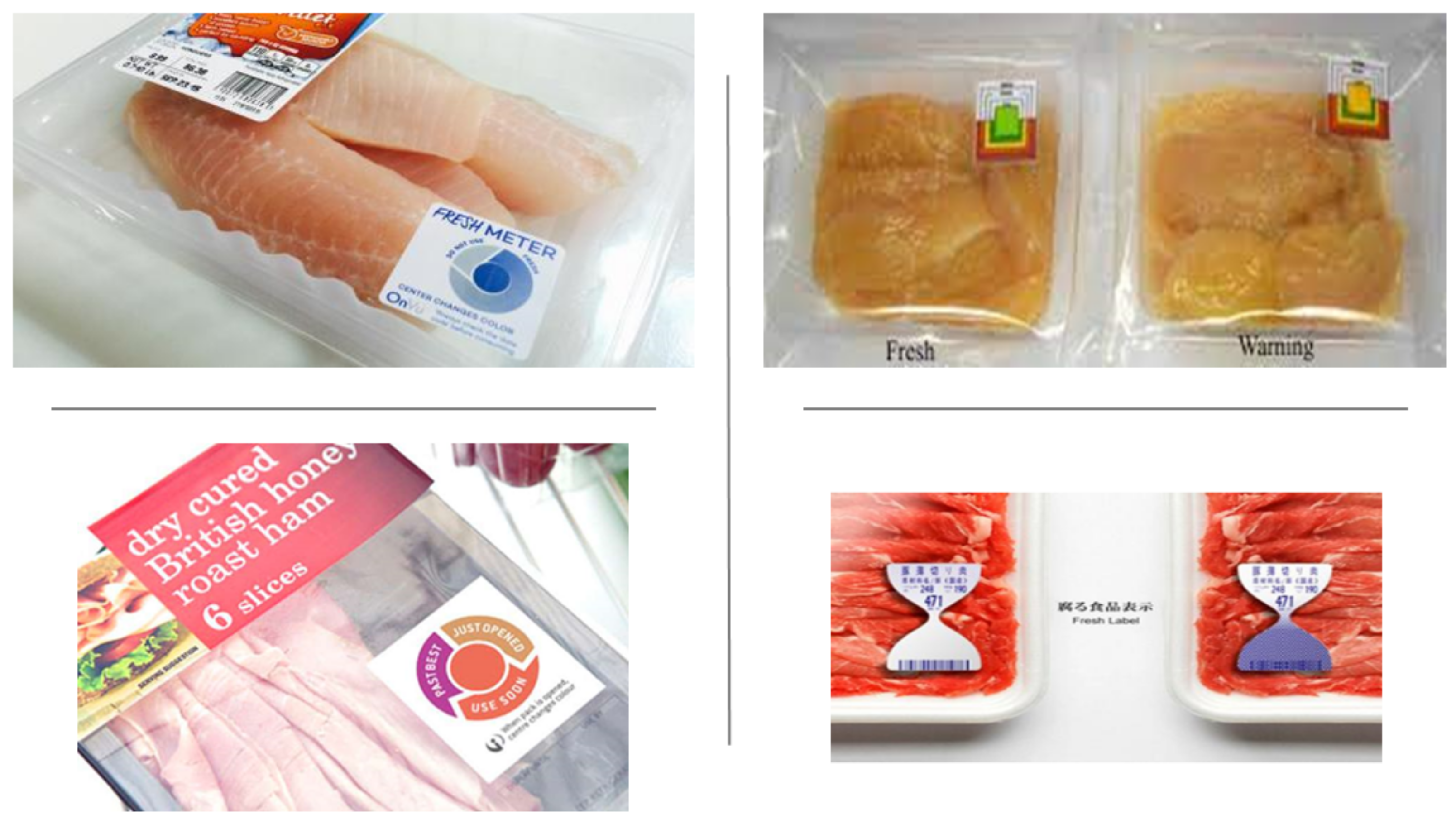
3. Color Indicator for Food Spoilage

3.1. Introduction to Color Indicators in Food

A color indicator is a new technology for the real-time monitoring of freshness to notify the level of food spoilage that helps consumers to choose the best quality food products [4]. Conventionally, the identification of food spoilage detected by a chemical experiment, which involves the determination of total volatile basic nitrogen (TVB-N), triphenyl tetrazolium chloride (TTC), and biological methods (total viable counts, TVC), as well as lactic acid bacteria, Brochothrix thermosphacta, Pseudomonas spp., and Enterobacteriaceae determination [4]. In addition, food spoilage is also determined by changes in sensory characteristics such as color, texture, smell, and appearance of the food products [3]. Sometimes, the uses of the conventional methods to identify food spoilage are rejected due to the inefficiency and low precision, because they lead to human errors, and because consumers have difficulty differentiating the product quality by simply relying on sensory characteristics such as color [4]. For example, fresh and spoiled meat colors have an insignificant difference; thus, it is difficult for consumers to choose the desired quality to fulfill their needs [1]. Furthermore, it is also reported that the biological method using Fourier transform infrared spectroscopy to monitor spoilage on minced beef is high cost, only relies on trained panelists, is lengthy, and is not able to give the immediate answer required compared to the color indicator [43]. Furthermore, the color indicator needs to meet several criteria for freshness monitoring, e.g., easy to distinguish the color changes with the naked eye, sensitivity towards various pH levels, and low response time between the appearance of distinctive color and changes in pH [44]. Different types of color indicators in food packaging that are available in the market are shown in Figure 1. Commonly, the color indicator has two essential parts, i.e., solid support and colorants [7].
The function of the solid support is to immobilize the colorants, which usually use natural biopolymers (biopolymer-based film), such as polysaccharides and proteins [46]. However, the biopolymer-based film can be easily degraded by microorganisms and other volatile compounds in the packaging environment. Therefore, combinations with other polymers have been introduced, i.e., chitosan–starch [9], starch–gelatin [8,47], starch–polyvinyl alcohol [48], starch–nanomaterial [49,50], and carboxymethyl cellulose–polyvinyl alcohol [51], and combinations with colorants [3,6,52] to overcome the negative feedbacks of the packaging film. Moreover, the colorants are the main character as a color indicator that undergoes color changes to evaluate the quality of food products by detecting the changes in pH, gas level, and microbial activity in the packaging [53]. Synthetic and natural sources are two types of colorants commonly used in developing color indicators.

3.2. Type of Color Indicator

3.2.1. Synthetic Sources

Synthetic colorants, also known as acid-base indicators, are affected by a wide range of intermolecular interactions (Bronsted and Lewis acid-base, hydrogen bonding, di-polar, and π-π interactions) between the analyte and colorants [54]. The critical element for synthetic dyes to be used as a color indicator is the ability to change their color in an acidic or basic environment. Various types of artificial colorants are commonly used as food spoilage indicators, such as tetraphenylethylene (TPE), bromocresol green, methyl red, xylenol blue, crystal violet lactone, bromophenol blue, cresol red, Bromo-cresol purple, bromothymol blue sodium salt, alizarin, phenol red, chlorophenol red, and rosolic acid, that can indicate meat spoilage during bacterial decomposition due to the ability to change color at a different level of pH [55,56]. A study by Liu et al. [57] found that TPE dye that is immobilized to polyaniline polymer (PANI) will change its color from emerald-green to peacock-blue when exposed to the spoiled fish due to the releasing of volatile amines (triethylamine, trimethylamine, and ammonia). In addition, bromocresol green dye is also used as a colorimetric sensor to indicate fish spoilage. It is reported that the bromocresol green turns yellow-green from yellow in four hours, then turns green in ten hours, and finally turns dark green in 22 h due to the increase of TVB-N in fish from 10 mg/100 g to 23.11 ± 2.22 mg/100 g. The color will turn dark blue when TVB-N exceeds 25 mg/100 g [58]. However, studies using other sources of synthetic colorants are still lacking.

3.2.2. Natural Sources
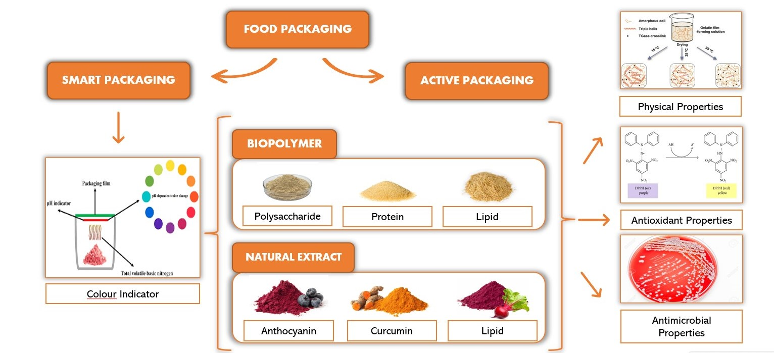
Colorants from natural sources have been suggested as a suitable alternative to be used as a color indicator over synthetic colorants in food packaging due to their obvious side effects, toxicity in the short and long term, possible carcinogenic effects, and allergic reactions, especially in children (ADHD-like hyperactivity), and their ability to disrupt biological systems and environment [59]. Natural colorants can be used as an indicator to detect food spoilage due to their ability to see changes in pH, gas level, and microbial activity in the packaging [53]. So far, a color indicator that detects changes in gas levels, especially carbon dioxide, is still lacking. Natural colorants are usually sensitive to various pH levels, which can change their color according to the pH. So far, a color indicator that detects changes in gas levels, especially carbon dioxide, is still lacking. Natural colorants are usually sensitive to various pH levels and can change their color according to the pH, as shown in Figure 2. In addition, the colorants immobilized in the polymer matrix will change color due to the rise of volatile amines by pathogenic microbes in spoiled food [60]. This positive impact of natural colorants has prompted various researchers to develop color indicators due to the excellent potential for the development of real-time monitoring of freshness from natural sources such as anthocyanin, curcumin, chlorophyll, carotenoid, and betacyanin [52,61].
Anthocyanin is a natural phenolic indicator dye that can be extracted from several sources, such as red cabbage, blueberry, black soybean, raspberry, and purple cauliflower. It is soluble in water and appears red, pink, purple, green-yellow, and colorless depending on the pH [62]. There are various anthocyanin types, such as cyanidin, pelargonidin, peonidin, delphinidin, malvidin, and petunidin, consisting of 3-glucoside of the aglycone anthocyanidin, which is the sugar-free analog of anthocyanins [59]. The changes in the color of anthocyanin at different pH levels are due to the mechanism reaction of cations, such as flavylium cation (red pigment) that appears at strong acid conditions at pH < 4, carbinol and pseudo-base chalcone (colorless dye) appear at pH 4–6, quinoidal anhydrous-base (blue/purple pigment) appears at pH 6–8. Colorless chalcone (light-yellow pigment) appears at pH > 8 [63]. In addition, the substitution configuration of anthocyanin also leads to the color changes of anthocyanin. For example, increasing methoxy groups will generate a reddish color, while hydroxy groups will generate bluish color [59]. Incorporating anthocyanin as a natural additive to the pH-sensitive film becomes an innovative tool for the indication of real-time monitoring of freshness [61]. Curcumin can be found in Curcuma longa, which is known as a bright yellow pigment due to the presence of curcuminoid pigment. The curcuminoid pigment consist of three types of curcumin, which are curcumin I (curcumin), curcumin II (desmeth-oxy-curcumin), and curcumin III (bisdemethoxycurcumin) [64]. Curcumin has poor solubility in water and hydrocarbon solvents but is very soluble in polar solvents such as ethanol. Curcumin can undergo hydrolytic degradation due to its instability at high temperatures (>190 °C). The color stability of curcumin in an aqueous solution depends on the pH of the solution, where the optimum pH is 1–6. The color change from yellow to red occurs at a pH lower than pH 3 and is more significant than pH 7 [65]. The color changes of curcumin at different pHs give the ability to indicate the quality of food since pH changes occur resulting from food spoilage [18]. Nonetheless, there is also a lack of studies using natural colorants from chlorophyll, carotenoid, and betacyanin.

4. Food Packaging

Food packaging is critical in containment, protection, communication, and convenience to prevent physical, chemical, and biological damage and provide product information [66]. There are various types of food packaging present in the market that is innovated to offer more convenience to consumers and also to meet consumers’ lifestyles. Usually, food packaging acts as passive protection for food products, providing a protective barrier between food and environmental influences (oxygen, moisture, light, dust, pest, and volatiles) [66]. Food packaging technology significantly impacts food products to minimize the possible deterioration or degradation of quality and improve the shelf life of packaged food [67]. Scientists have worked hard to enhance packaging by developing various technologies for food packaging, such as vacuum packaging, modified atmosphere packaging (MAP), active packaging, and smart packaging.

4.1. Active Packaging

Active packaging is commonly used to extend the shelf life of food products, improve safety, and maintain the quality of the product. The active packaging usually performs monolayered by incorporating an active compound with the polymer and a multilayered system by entrapping the active compound between the polymer layer [68,69]. A monolayered film can be used if the film has good antioxidant packaging requirements, such as transparency and good sealing condition. Still, this film has disadvantages in controlling the active compound from loss to the environment. Thus, the multilayered film is used to minimize the migration of active compounds to the environment because the active layer is protected with a protective layer [70]. The development of active packaging film can be classified into three methods, which are the casting method (by mixing the active compound and polymer matrix into an appropriate solvent), the extrusion method (by incorporating the active compound into the melted polymer), and the coating method (by treating the active compound with a physical or chemical process to favor the adhesion of the active compound) [71].
Biopolymer or nonsynthetic polymers, such as proteins, polysaccharides, and lipids, are widely used in film packaging to replace or minimize the use of synthetic polymers, i.e., polycarbonate (PC), low-density polyethylene (LDPE), high-density polyethylene (HDPE), polystyrene (PS), polyethylene terephthalate (PET), and polypropylene (PP) [72]. This is because synthetic polymer has disadvantages, such as being nonbiodegradable, challenging to recycle, and can cause environmental damage (air and water pollution) [73,74]. Various sources of proteins, polysaccharides, and lipids have been used for the development of active packaging film to improve the quality and shelf life of meat products such as gelatin [75], starch [76,77], chitosan [78], carboxymethyl cellulose [79,80], and propolis extract [81]. The use of biopolymer in developing active packaging film should be good in mechanical and physical properties and have good properties of sealing, hermeticity, barrier, etc. [70]. Plasticizers (sorbitol, glycerol, polyethylene glycol, etc.) are incorporated into the polymer due to the drawbacks of the polymers, such as being too brittle and rigid, which leads to limitations for application in the food industry. The plasticizers will react with the polymer’s chain, increasing flexibility and improving mechanical and physical properties [82]. Moreover, active packaging can absorb/scavenge properties and release/emit properties [67]. The scavenging system of active packaging is responsible for absorbing volatile compounds that can cause off-flavor, oxidation, and discoloration to the food product by incorporating absorbing materials. For example, a study by Byun et al. [83] found that the incorporation of oxygen scavenger (Iron (II) chloride) into gelatin film can reduce the oxygen level of active packaging from 20.90% to 4.56% after 50 days of storage. In addition, the releasing system of active packaging can provide various advantages. For example, a smaller number of antioxidants is needed than direct addition to the food product and can indirectly produce a no-preservative food product. Moreover, the antioxidant activity can also be centered on the more sensitive product surface through the migration from the packaging to the food matrix [84]. It has been proved that the incorporation of antioxidants can give benefits to the food product, especially meat products (beef, foal, pork, and turkey) such as polyvinylpolypyrrolidone washing solution (PVPP-WS) [85], citric acid [86], rosemary extract [85], green tea extract [87], green, black and oolong tea extract [73], oregano essential oil [87], encapsulated green tea extract [88], citrus extract [89], and cinnamon + rosemary [90] to reduce lipid and protein oxidation, prevent discoloration, and improve sensory characteristics of the meat product.

4.2. Smart Packaging

Smart packaging is a type of innovative packaging that is extended from traditional packaging. Traditional packaging aims to prevent the products from leaking, breaking, or being contaminated by pathogenic microbes; to communicate to the consumers the information about the food products (nutritional content or cooking instruction); to provide convenience; and to provide containment during transportation [91]. The presence of smart packaging may accommodate and fulfill consumer needs because traditional packaging is no longer sufficient (increasing awareness of biodegradable packaging, increasing product complexity, and minimizing the carbon footprints of manufactured products) by enhancing the functionality of traditional packaging [92]. In addition, smart packaging can provide product quality information due to microbial growth or chemical changes in the food product inside the packaging. The smart packaging system can also be classified as direct and passive indicators attached to the packaging labels, incorporated into a polymer matrix, or printed onto food packaging [12,93].
Moreover, smart packaging can monitor the quality of food products due to the presence of sensors inside the packaging to sense the freshness of food products, which, in turn, also shows whether the product is safe to consume. Usually, pH-sensitive dyes or colorants from natural or synthetic sources are used as a sensor that is entrapped within the polymer matrix (e.g., gelatin, starch, agar, tara gum, cellulose, and k-carrageenan) that will change its color when alteration of the pH occurs due to the presence of volatile compounds from food spoilage [94]. The reaction of the pH sensor can be classified into three types: firstly, the physical adsorption of immobilized colorants on a solid support, such as an ion exchanger. Secondly, the colorants, such as cellulose polymer, are covalently attached to a hydrophilic support on the packaging. Thirdly, the colorants are physically entrapped in a polymer matrix [95]. Natural pigments replace synthetic colors (e.g., bromocresol, methyl red, and chlorophenol) because they are safer and eco-friendlier. Various types of bioactive compounds from natural sources have been incorporated into the smart packaging by the researchers, such as potatoes/sweet potatoes [96], purple potatoes [97], red cabbage [98], roselle calyx [48], turmeric [99], and cactus pears [100].

5. Gelatin-Based Film

5.1. Sources of Gelatin

Gelatin is widely used in the food industry due to its ability to serve as a thickener, emulsifier, plasticizer, binding agent, etc., that is extracted from animals such as pigskin (46%), bovine hides (29.4%), bovine bones (23.1%), and other sources (1.5%), which consist of type A and type B gelatin [101]. Type A and type B gelatin are two types of gelatins that are produced based on the pretreatment, which is acid-treatment gelatin (the isoelectric point from pH 6 to 9), found in pigskin, and alkaline-treatment gelatin (the isoelectric point at pH 5), found in bovine hides, respectively [102]. A study by Aramwit et al. [103] reported that type B gelatin contains a higher cross-linking degree and physicochemical properties than type A gelatin, leading to a slower degradation rate. Thus, it shows that the gelatin’s quality depends on its source. In addition, the source of the gelatin is also essential for religious concerns because Muslims and Jews are prohibited from consuming pork-derived products. At the same time, Hindus are forbidden from consuming bovine products [104]. Furthermore, the source of gelatin also contributes to the quality of the gelatin film. For example, a comparative study by Nur Hanani et al. [105] showed that gelatin-based film from fish had the lowest water-vapor permeability compared to the gelatin-based film from beef and pork because fish-derived gelatin consists of low proline and hydroxyproline content.

5.2. Properties of Gelatin-Based Film

Gelatin can be used as an edible biopolymer for food packaging due to several advantages, such as high biocompatibility, good film-forming ability, and biodegradability. In addition, the presence of α and β chains in gelatin gives different gel strengths. It is reported that more α chain in the gelatin leads to higher gel strength [106]. A mixture of the gelatin-based film with other polymers or active biochemical reagents can strengthen the film to improve the extensibility, dispensability, flexibility, elasticity, and rigidity due to some significant drawbacks of the film, such as ease of dissolving in water, coupled with inferior thermal stability, poor tensile strength, poor water-vapor permeability, and poor mechanical properties [15,107]. It is reported that incorporating other materials into the gelatin-based film can increase the film polymer’s cross-link by hydrogen bond formation between the polymer and the added material [94]. In a study by Wang et al. [107], adding 10% of aldehyde starch crystal can improve the thermal stability of gelatin-based film and maximize the tensile strength, elongation at break, contact angle, and barrier efficiency.

6. Characterization of Gelatin-Based Film towards Color Indicator in Food Packaging

6.1. Color Stability

Color stability indicates the ability of the film’s color to retain and prevent discoloration or loss of color. It is because the film’s color is the main characteristic that acts as a color indicator for food-spoilage observation [53]. The color loss of the film occurs due to the degradation of natural extracts such as anthocyanin and curcumin. Various factors can contribute to the degradation of the natural extract, including light quality, oxygen availability, heat, pH changes, structure and concentration of pigments, presence of co-pigments, sugars, metal ions, enzymes, and their degradation products such as sulfur dioxide [8,66]. Anthocyanin from red cabbage, black carrots, red radishes, red potatoes, red corn, beans, roselle, black/purple rice, and curcumin from turmeric are often used as a color source for the development of film packaging as a color indicator [108]. The effect of color stability using various sources of natural extract for the application of film packaging as a color indicator is shown in Table 1.
A comparative study by Prietto et al. [6] reported that the color indicator incorporated with anthocyanin from black beans showed low instability compared to the red cabbage at room temperature, with the incidence of light for 28 days due to the acylation of anthocyanin. The hydroxyl group of anthocyanin is commonly acylated with organic and inorganic acids (sulfuric acid), affecting anthocyanins’ chemical stability. The presence of glucoside anthocyanin in red cabbage also leads to the stabilization of anthocyanin compared to the monoglucoside in black beans due to the possibility of forming unstable intermediates that can be degraded into phenolic acids and aldehydes. A study by Chayavanich et al. [8] showed that color indicators from the gelatin-based film incorporated with anthocyanin are more stable when stored at refrigeration temperature compared to room temperature. It is because the concentration of anthocyanin decreased gradually over time at all temperatures and more rapidly at higher temperatures. For instance, after 60 days, 11% of delphinidin was lost at 4 °C compared to 99% at 37 °C, while 17% of cyanidin was lost at 4 °C compared to 98% at 37 °C [113]. A comparative study between anthocyanin and curcumin by Chen et al. [109] showed that the color stability of color indicators containing anthocyanin is poorer than curcumin due to anthocyanin degradation during long-term storage. Another study by Peralta et al. [114] showed that the natural polymeric films developed from different combinations of aqueous hibiscus extract (HAE) with gelatin, starch, and chitosan. Anthocyanin, present in HAE, changes film color at various pH levels.

6.2. Color Response towards Various pH

The color response is an indicator to determine the color changes of the gelatin-based film incorporated with natural extracts such as red radish, purple sweet potato, roselle, blackberry, black/purple rice, and turmeric towards various pH levels [8]. Therefore, the gelatin-based film containing natural extract needs to be sensitive to multiple pH levels and have a low response time between the appearance of distinctive color and changes in pH to be used as a color indicator [44]. Usually, the color response of the gelatin-based film is determined by immersing the film in different pH levels (pH 2–12) and using a colorimeter to determine the total color difference [61]. Additionally, volatile ammonia is used to determine the film’s sensitivity towards volatile nitrogen compounds because the compounds are generally produced during meat spoilage [50,109]. A study by Chayavanich et al. [8] reported that incorporating the gelatin-based film with anthocyanin extract from red radishes shows a visible color ranging from orange to pink to blue and yellow at different pH levels. A comparative study between purple sweet potato extract (PSPE) and red cabbage extract (RCE) by Zhang et al. [115] showed that PSPE has bolder color than RCE due to abundant anthocyanin content; thus, it is easy to distinguish the color difference between the film from pink (pH 2–6) to light purple (pH 7–8), and to dark blue (pH 9–12). The comparison of sensitivity towards ammonia gas between anthocyanin and curcumin reported that anthocyanin shows high response sensitivity in terms of color difference compared to curcumin; thus, it indicates that anthocyanin is more sensitive toward ammonia [109]. Furthermore, it reported that a higher concentration of curcumin makes the color of the gelatin-based film brighter, which is caused by the yellow color of curcumin; thus, the color indicator can change in a way that is noticeable to the naked eye under acidic and alkaline solution conditions [116]. Table 2 shows the pH range of film packaging as a color indicator from various sources of natural extract and application on meat-spoilage observation.

6.3. Fourier Transform Infrared (FTIR) Spectrum

The gelatin-based film’s Fourier transform infrared (FTIR) spectrum was analyzed using an FTIR spectrometer to observe the morphology of the film’s matrix [50]. From the FTIR spectrum, the level of the functional group of the film’s matrixes can be detected, such as a decrease in the OH group due to the formation of a hydrogen bond between the OH group from the biopolymer and natural extract. As a result, the hydrogen bond in the film matrix will increase, leading to changes in the absorption peak of the FTIR spectrum [119]. The absorption peak of the film packaging as a color indicator at different concentrations of the extract is shown in Table 3. In addition, stretching vibration of the carbon-carbon double bond (C=C) of the aromatic ring from natural extract due to the interaction of anthocyanin and polymer matrix can also be detected, leading to the stronger chemical and physical structure of the film [115].
Furthermore, increasing N-H bonding can be detected due to the interaction between the amino group of biopolymers and the O-H group of natural extract, resulting in stronger intermolecular interaction of the film. A study by Liu et al. [123] reported that the incorporation of haskap berry extract (HBE) in the gelatin-based film could broaden and intensify the amide-A, N-H, and O-H stretching due to the formation of intermolecular hydrogen bonding between the hydroxyl group in HBE and gelatin, thus resulting in a dense network of the film matrix. It was also reported that incorporating red radish extract into the gelatin-based film can increase the C-H, C-O, and C-C bending vibration, C-O-C glycosidic bond, and C=O stretching of amide due to the chemical interaction between the polymers and the extract [8].

6.4. Water-Vapor Permeability

The water-vapor permeability of the film indicates the potential of water permeability or diffusivity and solubility of the water transfer through a film [72]. The determination of water-vapor permeability plays an essential role in the production of the gelatin-based film to function as a good film material and as a color indicator because water molecules are a transferable element that can pass through the film (via adsorption or desorption), which leads to degradation of the film properties. Good properties of the film generally have low water-vapor permeability due to its highly sealing effect that leads to the water molecules that have difficulties transferring through the film [125]. Several factors can affect water-vapor permeability, including temperature, relative humidity, food type, water activity, and film characteristics [126]. In addition, the concentration of natural extract incorporated into the gelatin-based film can affect the water-vapor permeability of the film by interfering with the morphology of the film’s matrix [61]. A study by Zhang et al. [115] reported that the incorporation of anthocyanin from purple sweet potato extract increased the film’s water-vapor permeability compared to red cabbage extract. This is because purple sweet potato contains abundant anthocyanin pigment compared to red cabbage with the same concentration. This leads to the disruption of the molecular structure, resulting in a porous surface of the film. It also reported that the incorporation of black soybean seed coat extract from 5% to 15% of w/w could gradually decrease the water-vapor permeability of the film packaging due to the formation of a dense network [127]. In addition, the uses of anthocyanin from mulberry pomace extracts can also enhance the water-vapor barrier properties of the film by reducing the water-vapor permeability due to the interaction of the phenolic group of the extract with the hydroxyl group of the film to form a complex structure [17]. The details of the findings on the relationship between the concentration of natural extract and water-vapor permeability of gelatin-based film are shown in Table 4.

6.5. Antioxidant and Antimicrobial Properties

The presence of polyphenols (flavonoids, phenolic acids, stilbenes, and lignans) in the natural extract contributes to the red, purple, and blue color of flowers, vegetables, and fruits has the potential as antioxidant and antimicrobial properties [88]. Recently, the utilization of natural extract for the development of color indicators from gelatin-based films, such as anthocyanin, has gained increasing attention due to the ability to act as an antioxidant and antimicrobial potential, which can indirectly maintain stability and extend the shelf life of the food product [122,131]. Therefore, apart from the fact that the color indicator can give quality information, it can also maintain the quality of the food product. Usually, the antioxidant of the film is determined by the 2,2-diphenyl-1-picrylhydrazyl (DPPH) assay or the ferric reducing antioxidant power (FRAP) assay [61]. For the determination of antioxidants, the DPPH assay (deep-violet color) will react with the antioxidant or any radical species (R) by providing electrons and reducing to DPPH-H or DPPH-R (colorless or pale-yellow color) [132].
On the other hand, the FRAP assay is used to measure the reduction of Fe3+ (colorless) to Fe2+ (intense blue color) by the electron-donating antioxidant [133]. In addition, the determination of antimicrobial properties uses the agar diffusion method to detect the presence of foodborne pathogens (E. coli, Salmonella, S. aureus, and L. monocytogenes) [124]. In a study by Liu et al. [123], it was reported that the scavenging activity of the gelatin-based film increased with the increase in haskap berry extract concentration due to the releasing of polyphenols, resulting in the prevention of oxidative damage when applied to the food products. The presence of catechins from green tea extract with a 20% concentration shows the highest radical scavenging activities compared to the Pu-erh tea extract [134]. The addition of butterfly extract also indicates a significantly increased antioxidant activity of gelatin film due to the potent antioxidant activity of phenolic flavanols of anthocyanin [128]. Incorporating curcumin into the gelatin film can also improve the film’s antioxidant properties as a scavenging activity, which is significant in preserving food. Furthermore, it can also enhance the antibacterial properties against Gram-positive (E. coli) and Gram-negative bacteria (S. aureus) by reducing the growth rate from 701 to 11 CFU/mL and 342 to 79 CFU/mL, respectively [116]. The effect of the natural extract on the antimicrobial and antioxidant properties of film packaging as a color indicator is shown in Table 5.

7. Research Gap between Gelatin-Based Film and Paper as A Color Indicator

A solid matrix such as polymer and paper is a significant component for the color indicator to immobilize the colorants. Biopolymers, such as gelatin-based films, are preferred as a solid matrix due to their positive impact on the environment, including biodegradability, renewability, and eco-friendliness, as well as having good volatile gas binding ability and a faster release of colorants. Although the gelatin-based film has drawbacks (high water solubility and low water-vapor barrier properties), a combination of the gelatin-based film with other polymers, such as starch, agar, carrageenan, etc., can solve the problems [128,136]. Besides that, incorporating natural extracts, such as anthocyanin and curcumin, can improve the film’s drawbacks and act as a new indicator of freshness [119,120]. In addition, the paper contributes to the color indicator by immobilizing the colorants into the paper using a spin-coated method or by simply immersing it in the colorants. A study on color indicators by Husin et al. [3] reported that the incorporation of butterfly pea extract into the filter paper using a spin-coated method was able to monitor the freshness of beef. The use of synthetic colorants is also able to serve as a freshness indicator. A study by Hidayat et al. [137] reported that immersing the filter paper in 10 mL of phenol red and bromothymol blue for 12 h was able to indicate the quality of fresh meat. However, since the intermolecular forces between the paper polymer are strong, it is difficult for the colorants to bind to the paper. As a result, the dyes easily leach out, leading to the color indicator’s low effectiveness. Meanwhile, synthetic colorants, such as phenol red and bromothymol blue, are not suitable for food products due to their toxicity to human health [59].

8. Future Trends

Recently, there has been an increase in demand for the development of packaging that can indicate the quality and safety of food products, especially perishable food, including poultry, beef, and seafood. Scientists have been working on alternative ways to develop color indicators for detecting food spoilage. The color indicator can detect food spoilage due to its sensitive nature toward the various pH levels by incorporating colorants from synthetic or natural sources and can detect gas levels such as carbon dioxide, ammonia, and oxygen in food-spoilage observation. The spoiled food will release total volatile basic nitrogen (TVB-N) that will interfere with the pH in the packaging environment. As a result, the color indicator will change according to the pH inside the packaging. However, some limitations exist in developing color indicators, such as toxicity and carcinogenicity of synthetic colorants compared to natural colorants. In addition, there is also a lack of studies for the determination of meat spoilage observation using natural colorants from betacyanin, chlorophyll, and carotenoids, as well as the determination of gas levels in food spoilage, especially carbon dioxide gas. Therefore, future development needs to include the development of color indicators using betacyanin, chlorophyll, and carotenoids, as well as the determination of gas levels, especially carbon dioxide gas.

9. Conclusions

In conclusion, as a color indicator in food-spoilage observation, the gelatin-based film can be obtained by incorporating bioactive compounds from natural extract. This review found that adding the crude extract to the gelatin film improved the physical and mechanical properties of the film, such as morphology, color stability, and water-vapor permeability. At the same time, it provides antioxidant and antimicrobial properties to the film packaging, which indirectly can maintain and extend the shelf life of the food product. Furthermore, the quality and safety of food products can be monitored through the film’s sensitive nature towards various pH levels in detecting food spoilage by color indicator. The color indicator can detect the pH changes due to the presence of synthetic and natural colorants that will immobilize the gelatin-based film. Therefore, the gelatin-based film color indicator can be utilized as an effective tool to monitor and control the shelf-life of packaged foods, especially meat products, to optimize distribution, manage the stock rotation system, and reduce food waste.

Author Contributions

N.M.S.: Supervision, conceptualization, methodology, validation, and writing—review and editing. M.I.S.: Writing—review and editing. M.F.A.: Investigation, writing—original draft preparation, and visualization. All authors have read and agreed to the published version of the manuscript.

Funding

This research received no external funding.

Institutional Review Board Statement

Not applicable.

Informed Consent Statement

Not applicable.

Conflicts of Interest

The authors declare no conflict of interest.

References

  1. Bekhit, A.E.-D.A.; Holman, B.W.; Giteru, S.G.; Hopkins, D.L. Total volatile basic nitrogen (TVB-N) and its role in meat spoilage: A review. Trends Food Sci. Technol. 2021, 109, 280–302. [Google Scholar] [CrossRef]
  2. Rawat, S. Food Spoilage: Microorganisms and their prevention. J. Plant Sci. Res. 2015, 5, 47–56. [Google Scholar]
  3. Husin, N.; Rahim, M.Z.A.; Noor, M.A.M.; Rashedi, M.I.F.; Hassan, N. Real-time monitoring of food freshness using delphinidin- based visual indicator. Malays. J. Anal. Sci. 2020, 24, 558–569. [Google Scholar]
  4. Kuswandi, B.; Damayanti, F.; Jayus, J.; Abdullah, A.; Heng, L.Y. Simple and Low-Cost On-Package Sticker Sensor based on Litmus Paper for Real-Time Monitoring of Beef Freshness. J. Math. Fundam. Sci. 2015, 47, 236–251. [Google Scholar] [CrossRef]
  5. Kuswandi, B.; Jayus; Restyana, A.; Abdullah, A.; Heng, L.Y.; Ahmad, M. A novel colorimetric food package label for fish spoilage based on polyaniline film. Food Control. 2012, 25, 184–189. [Google Scholar] [CrossRef]
  6. Prietto, L.; Mirapalhete, T.C.; Pinto, V.Z.; Hoffmann, J.F.; Vanier, N.L.; Lim, L.-T.; Dias, A.R.G.; Zavareze, E.D.R. pH-sensitive films containing anthocyanins extracted from black bean seed coat and red cabbage. LWT 2017, 80, 492–500. [Google Scholar] [CrossRef]
  7. Pourjavaher, S.; Almasi, H.; Meshkini, S.; Pirsa, S.; Parandi, E. Development of a colorimetric pH indicator based on bacterial cellulose nanofibers and red cabbage (Brassica oleraceae) extract. Carbohydr. Polym. 2017, 156, 193–201. [Google Scholar] [CrossRef] [PubMed]
  8. Chayavanich, K.; Thiraphibundet, P.; Imyim, A. Biocompatible film sensors containing red radish extract for meat spoilage observation. Spectrochim. Acta Part A Mol. Biomol. Spectrosc. 2019, 226, 117601. [Google Scholar] [CrossRef] [PubMed]
  9. Teixeira, A.; Pereira-Júnior, V.A.; Silva-Pereira, M.C.; Stefani, R. Chitosan/corn starch blend films with extract from Brassica oleraceae (red cabbage) as a visual indicator of fish deterioration. LWT-Food Sci. Technol. 2015, 61, 258–262. [Google Scholar] [CrossRef] [Green Version]
  10. Wang, S.; Xia, P.; Wang, S.; Liang, J.; Sun, Y.; Yue, P. Packaging films formulated with gelatin and anthocyanins nanocomplexes: Physical properties, antioxidant activity and its application for olive oil protection. Food Hydrocoll. 2019, 96, 617–624. [Google Scholar] [CrossRef]
  11. Said, N.; Howell, N.K.; Sarbon, N.M. A review on potential use of gelatin-based film as active and smart biodegradable films for food packaging application. Food Rev. Int. 2021, 1–23. [Google Scholar] [CrossRef]
  12. Bhargava, N.; Sharanagat, V.S.; Mor, R.S.; Kumar, K. Active and intelligent biodegradable packaging films using food and food waste-derived bioactive compounds: A review. Trends Food Sci. Technol. 2020, 105, 385–401. [Google Scholar] [CrossRef]
  13. Musso, Y.S.; Salgado, P.R.; Mauri, A.N. Gelatin based films capable of modifying its color against environmental pH changes. Food Hydrocoll. 2016, 61, 523–530. [Google Scholar] [CrossRef] [Green Version]
  14. Luo, Q.; Hossen, A.; Zeng, Y.; Dai, J.; Li, S.; Qin, W.; Liu, Y. Gelatin-based composite films and their application in food packaging: A review. J. Food Eng. 2021, 313, 110762. [Google Scholar] [CrossRef]
  15. Pulla-Huillca, P.V.; Gomes, A.; Bittante, A.M.Q.B.; Lourenço, R.V.; Sobral, P.J.D.A. Wettability of gelatin-based films: The effects of hydrophilic or hydrophobic plasticizers and nanoparticle loads. J. Food Eng. 2021, 297, 110480. [Google Scholar] [CrossRef]
  16. Suderman, N.; Isa, M.; Sarbon, N. The effect of plasticizers on the functional properties of biodegradable gelatin-based film: A review. Food Biosci. 2018, 24, 111–119. [Google Scholar] [CrossRef]
  17. Liu, Y.; Qin, Y.; Zhang, X.; Yuan, L.; Liu, J. Preparation of pH-sensitive and antioxidant packaging films based on κ-carrageenan and mulberry polyphenolic extract. Int. J. Biol. Macromol. 2019, 134, 993–1001. [Google Scholar] [CrossRef]
  18. Etxabide, A.; Maté, J.I.; Kilmartin, P.A. Effect of curcumin, betanin and anthocyanin containing colourants addition on gelatin films properties for intelligent films development. Food Hydrocoll. 2021, 115, 106593. [Google Scholar] [CrossRef]
  19. Malhotra, B.; Keshwani, A.; Kharkwal, H. Antimicrobial food packaging: Potential and pitfalls. Front. Microbiol. 2015, 6, 611. [Google Scholar] [CrossRef] [Green Version]
  20. Abdelhedi, O.; Salem, A.; Nasri, R.; Nasri, M.; Jridi, M. Food applications of bioactive marine gelatin films. Curr. Opin. Food Sci. 2022, 43, 206–215. [Google Scholar] [CrossRef]
  21. Ramos, M.; Valdés, A.; Beltrán, A.; Garrigós, M.D.C. Gelatin-Based Films and Coatings for Food Packaging Applications. Coatings 2016, 6, 41. [Google Scholar] [CrossRef] [Green Version]
  22. Future Market Insight. Gelatin Film Market: Global Industry Analysis 2013–2017. 2022. Available online: https://www.futuremarketinsights.com/reports/gelatin-films-market (accessed on 8 November 2022).
  23. Nychas, G.E.; Panagou, E.Z. 1–Microbiological spoilage of foods and beverages. In Food and Beverage Stability and Shelf Life; Woodhead Publishing: Sawston, UK, 2011. [Google Scholar]
  24. Dave, D.; Ghaly, A.E. Meat Spoilage Mechanisms and Preservation Techniques: A Critical Review. Am. J. Agric. Biol. Sci. 2011, 6, 486–510. [Google Scholar] [CrossRef]
  25. Mellor, G.E.; Bentley, J.A.; Dykes, G.A. Evidence for a role of biosurfactants produced by Pseudomonas fluorescens in the spoilage of fresh aerobically stored chicken meat. Food Microbiol. 2011, 28, 1101–1104. [Google Scholar] [CrossRef]
  26. Wang, G.Y.; Wang, H.H.; Han, Y.W.; Xing, T.; Ye, K.P.; Xu, X.L.; Zhou, G.H. Evaluation of the spoilage potential of bacteria isolated from chilled chicken in vitro and in situ. Food Microbiol. 2017, 63, 139–146. [Google Scholar] [CrossRef]
  27. Mills, J.; Donnison, A.; Brightwell, G. Factors affecting microbial spoilage and shelf-life of chilled vacuum-packed lamb transported to distant markets: A review. Meat Sci. 2014, 98, 71–80. [Google Scholar] [CrossRef]
  28. Sade, E.; Penttinen, K.; Björkroth, J.; Hultman, J. Exploring lot-to-lot variation in spoilage bacterial communities on commercial modified atmosphere packaged beef. Food Microbiol. 2017, 62, 147–152. [Google Scholar] [CrossRef] [PubMed] [Green Version]
  29. Pellissery, A.J.; Vinayamohan, P.G.; Amalaradjou, M.A.R.; Venkitanarayanan, K. Spoilage bacteria and meat quality. In Meat Quality Analysis; Elsevier: Amsterdam, The Netherlands, 2020; pp. 307–334. [Google Scholar] [CrossRef]
  30. Kalschne, D.L.; Womer, R.; Mattana, A.; Sarmento, C.M.P.; Colla, L.M.; Colla, E. Characterization of the spoilage lactic acid bacteria in “sliced vacuum-packed cooked ham”. Braz. J. Microbiol. 2015, 46, 173–181. [Google Scholar] [CrossRef] [Green Version]
  31. Katiyo, W.; Kock, H.L.D.; Coorey, R.; Buys, E.M. Sensory implications of chicken meat spoilage in relation to microbial and physicochemical characteristics during refrigerated storage. LWT-Food Sci. Technol. 2020, 128, 109468. [Google Scholar] [CrossRef]
  32. Cavill, L.; Renteria-Monterrubio, A.L.; Helps, C.R.; Corry, J.E. Detection of cold-tolerant clostridia other than Clostridium estertheticum in raw vacuum-packed chill-stored meat. Food Microbiol. 2011, 28, 957–963. [Google Scholar] [CrossRef]
  33. Wang, Z.; Shi, Y.; Zhou, K.; Zhou, H.; Li, X.; Li, C.; Wang, Z.; Xu, B. Effects of different thermal temperatures on the shelf life and microbial diversity of Dezhou-braised chicken. Food Res. Int. 2020, 136, 109471. [Google Scholar] [CrossRef]
  34. Li, H.; Sun, X.; Liao, X.; Gänzle, M. Control of pathogenic and spoilage bacteria in meat and meat products by high pressure: Challenges and future perspectives. Compr. Rev. Food Sci. Food Saf. 2020, 19, 3476–3500. [Google Scholar] [CrossRef]
  35. Wunderlichová, L.; Buňková, L.; Koutný, M.; Valenta, T.; Buňka, F. Novel touchdown-PCR method for the detection of putrescine producing Gram-negative bacteria in food products. Food Microbiol. 2013, 34, 268–276. [Google Scholar] [CrossRef] [PubMed]
  36. Nychas, G.J.E.; Skandamis, P.N.; Tassou, C.C.; Kautsoumanis, K.P. Meat spoilage during distribution. Meat Sci. 2008, 78, 77–89. [Google Scholar] [CrossRef]
  37. Erkmen, O.; Bozoglu, T.F. Enzymatic and nonenzymatic food spoilage. In Food Microbiology Principles into Practice, 1st ed.; John Wiley & Sons, Ltd.: Hoboken, NJ, USA, 2016; pp. 401–406. [Google Scholar] [CrossRef]
  38. Iulietto, M.F.; Sechi, P.; Borgogni, E.; Cenci-Goga, B.T. Meat Spoilage: A Critical Review of a Neglected Alteration Due to Ropy Slime Producing Bacteria. Ital. J. Anim. Sci. 2015, 14, 4011. [Google Scholar] [CrossRef]
  39. Li, J.; Zhou, G.; Xue, P.; Dong, X.; Xia, Y.; Regenstein, J.; Du, M.; Sun, L. Spoilage microbes’ effect on freshness and IMP degradation in sturgeon fillets during chilled storage. Food Biosci. 2021, 41, 101008. [Google Scholar] [CrossRef]
  40. Comi, G. Spoilage of Meat and Fish. In The Microbiological Quality of Food Foodborne Spoilers; Elsevier Ltd.: Amsterdam, The Netherlands, 2017. [Google Scholar] [CrossRef]
  41. Odeyemi, O.A.; Alegbeleye, O.O.; Strateva, M.; Stratev, D. Understanding spoilage microbial community and spoilage mechanisms in foods of animal origin. Compr. Rev. Food Sci. Food Saf. 2020, 19, 311–331. [Google Scholar] [CrossRef] [Green Version]
  42. Casaburi, A.; Piombino, P.; Nychas, G.-J.; Villani, F.; Ercolini, D. Bacterial populations and the volatilome associated to meat spoilage. Food Microbiol. 2015, 45, 83–102. [Google Scholar] [CrossRef] [PubMed]
  43. Shao, P.; Liu, L.; Yu, J.; Lin, Y.; Gao, H.; Chen, H.; Sun, P. An overview of intelligent freshness indicator packaging for food quality and safety monitoring. Trends Food Sci. Technol. 2021, 118, 285–296. [Google Scholar] [CrossRef]
  44. Ezati, P.; Tajik, H.; Moradi, M.; Molaei, R. Intelligent pH-sensitive indicator based on starch-cellulose and alizarin dye to track freshness of rainbow trout fillet. Int. J. Biol. Macromol. 2019, 132, 157–165. [Google Scholar] [CrossRef] [PubMed]
  45. Rukchon, C.; Nopwinyuwong, A.; Trevanich, S.; Jinkarn, T.; Suppakul, P. Development of a food spoilage indicator for monitoring freshness of skinless chicken breast. Talanta 2014, 130, 547–554. [Google Scholar] [CrossRef] [PubMed]
  46. Chen, H.; Wang, J.; Cheng, Y.; Wang, C.; Liu, H.; Bian, H.; Pan, Y.; Sun, J.; Han, W. Application of Protein-Based Films and Coatings for Food Packaging: A Review. Polymers 2019, 11, 2039. [Google Scholar] [CrossRef] [PubMed] [Green Version]
  47. Cheng, S.H.; Sarbon, N.M. Chicken gelatin films: Rheological properties of film forming solutions and film characterisation as influenced by starch incorporation. Int. Food Res. J. 2020, 27, 1094–1104. [Google Scholar]
  48. Zhai, X.; Shi, J.; Zou, X.; Wang, S.; Jiang, C.; Zhang, J.; Huang, X.; Zhang, W.; Holmes, M. Novel colorimetric films based on starch/polyvinyl alcohol incorporated with roselle anthocyanins for fish freshness monitoring. Food Hydrocoll. 2017, 69, 308–317. [Google Scholar] [CrossRef] [Green Version]
  49. Lee, S.W.; Said, N.S.; Sarbon, N.M. The effects of zinc oxide nanoparticles on the physical, mechanical and antimicrobial properties of chicken skin gelatin/tapioca starch composite films in food packaging. J. Food Sci. Technol. 2020, 58, 4294–4302. [Google Scholar] [CrossRef]
  50. Koshy, R.R.; Koshy, J.T.; Mary, S.K.; Sadanandan, S.; Jisha, S.; Pothan, L.A. Preparation of pH sensitive film based on starch/carbon nano dots incorporating anthocyanin for monitoring spoilage of pork. Food Control. 2021, 126, 108039. [Google Scholar] [CrossRef]
  51. Huang, J.; Liu, J.; Chen, M.; Qian, Y.; Hu, Y. Immobilization of roselle anthocyanins into polyvinyl alcohol/hydroxypropyl methylcellulose film matrix: Study on the interaction behavior and mechanism for better shrimp freshness monitoring. Int. J. Biol. Macromol. 2021, 184, 666–677. [Google Scholar] [CrossRef]
  52. Said, N.S.; Sarbon, N.M. A comparative study: Development and characterization of active biodegradable chicken skin and mammalian gelatin composite films incorporated with curcumin extracts. J. Food Process. Preserv. 2021, 45, e15771. [Google Scholar] [CrossRef]
  53. Sani, M.A.; Tavassoli, M.; Salim, S.A.; Hamishehkar, H.; McClements, D.J. Carbohydrate-based films containing pH-sensitive red barberry anthocyanins: Application as biodegradable smart food packaging materials. Carbohydr. Polym. 2020, 255, 117488. [Google Scholar] [CrossRef]
  54. LaGasse, M.K.; Rankin, J.M.; Askim, J.R.; Suslick, S. Colorimetric sensor arrays: Interplay of geometry, substrate and immobilization. Sens. Actuators B Chem. 2014, 197, 116–122. [Google Scholar] [CrossRef]
  55. Morsy, M.K.; Zór, K.; Kostesha, N.; Alstrøm, T.S.; Heiskanen, A.; El-Tanahi, H.; Sharoba, A.; Papkovsky, D.; Larsen, J.; Khalaf, H.; et al. Development and validation of a colorimetric sensor array for fish spoilage monitoring. Food Control. 2016, 60, 346–352. [Google Scholar] [CrossRef]
  56. Chen, H.; Zhang, M.; Bhandari, B.; Yang, C. Development of a novel colorimetric food package label for monitoring lean pork freshness. LWT-Food Sci. Technol. 2019, 99, 43–49. [Google Scholar] [CrossRef] [Green Version]
  57. Liu, X.; Wang, Y.; Zhu, L.; Tang, Y.; Gao, X.; Tang, L.; Li, X.; Li, J. Dual-mode smart packaging based on tetraphenylethylene-functionalized polyaniline sensing label for monitoring the freshness of fish. Sensors Actuators B Chem. 2020, 323, 128694. [Google Scholar] [CrossRef]
  58. Liu, X.; Chen, K.; Wang, J.; Wang, Y.; Tang, Y.; Gao, X.; Zhu, L.; Li, X.; Li, J. An on-package colorimetric sensing label based on a sol-gel matrix for fish freshness monitoring. Food Chem. 2019, 307, 125580. [Google Scholar] [CrossRef] [PubMed]
  59. Alizadeh-Sani, M.; Mohammadian, E.; Rhim, J.-W.; Jafari, S.M. pH-sensitive (halochromic) smart packaging films based on natural food colorants for the monitoring of food quality and safety. Trends Food Sci. Technol. 2020, 105, 93–144. [Google Scholar] [CrossRef]
  60. Mariusz, T.; Radomska, N.; Cierpiszewski, R. The Application of Natural Dyes in Food Freshness Indicators Designed for Intelligent Packaging. Studia Oeconomica Posnaniensia 2017, 5, 20–34. [Google Scholar] [CrossRef]
  61. Sganzerla, W.G.; Ribeiro, C.P.P.; Uliana, N.R.; Rodrigues, M.B.C.; Rosa, C.G.D.; Ferrareza, J.P.; Veeck, A.P.D.L.; Nunes, M.R. Bioactive and pH-sensitive films based on carboxymethyl cellulose and blackberry (Morus nigra L.) anthocyanin-rich extract: A perspective coating material to improve the shelf life of cherry tomato (Solanum lycopersicum L. var cerasiforme). Biocatal. Agrocultural Biotechnol. 2021, 33, 101989. [Google Scholar] [CrossRef]
  62. El-Naggar, M.E.; El-Newehy, M.H.; Aldalbahi, A.; Salem, W.M.; Khattab, T.A. Immobilization of anthocyanin extract from red-cabbage into electrospun polyvinyl alcohol nanofibers for colorimetric selective detection of ferric ions. J. Environ. Chem. Eng. 2021, 9, 105072. [Google Scholar] [CrossRef]
  63. Rawdkuen, S.; Faseha, A.; Benjakul, S.; Kaewprachu, P. Application of anthocyanin as a color indicator in gelatin films. Food Biosci. 2020, 36, 100603. [Google Scholar] [CrossRef]
  64. Degot, P.; Huber, V.; Touraud, D.; Kunz, W. Curcumin extracts from Curcuma Longa – Improvement of concentration, purity, and stability in food-approved and water-soluble surfactant-free microemulsions. Food Chem. 2020, 339, 128140. [Google Scholar] [CrossRef]
  65. Aliabbasi, N.; Fathi, M.; Emam-Djomeh, Z. Curcumin: A promising bioactive agent for application in food packaging systems. J. Environ. Chem. Eng. 2021, 9, 105520. [Google Scholar] [CrossRef]
  66. Choi, I.; Lee, J.Y.; Lacroix, M.; Han, J. Intelligent pH indicator film composed of agar/potato starch and anthocyanin extracts from purple sweet potato. Food Chem. 2017, 218, 122–128. [Google Scholar] [CrossRef] [PubMed]
  67. Yildirim, S.; Röcker, B.; Pettersen, M.K.; Nilsen-Nygaard, J.; Ayhan, Z.; Rutkaite, R.; Radusin, T.; Suminska, P.; Marcos, B.; Coma, V. Active Packaging Applications for Food. Compr. Rev. Food Sci. Food Saf. 2017, 17, 165–199. [Google Scholar] [CrossRef] [Green Version]
  68. Carina, D.; Sharma, S.; Jaiswal, A.K.; Jaiswal, S. Seaweeds polysaccharides in active food packaging: A review of recent progress. Trends Food Sci. Technol. 2021, 110, 559–572. [Google Scholar] [CrossRef]
  69. Alias, A.R.; Khairul Wan, M.; Sarbon, N.M. Emerging materials and technologies of multi-layer film for food packaging application: A review. Food Control. 2022, 136, 108875. [Google Scholar] [CrossRef]
  70. Gomez-Estaca, J.; Lopez-de-Dicastillo, C.; Hernandez-Munoz, P.; Catala, R.; Gavara, R. Advances in antioxidant active food packaging. Trends Food Sci. Technol. 2014, 35, 42–51. [Google Scholar] [CrossRef]
  71. Dominguez, R.; Barba, F.J.; Gomez, B.; Putnik, P.; Kovacevik, D.B.; Pateiro, M.; Santos, E.M.; Lorenzo, J.M. Active packaging films with natural antioxidants to be used in meat industry: A review. Food Res. Int. 2018, 113, 93–101. [Google Scholar] [CrossRef]
  72. Zhang, N.; Bi, F.; Xu, F.; Yong, H.; Bao, Y.; Jin, C.; Liu, J. Structure and functional properties of active packaging films prepared by incorporating different flavonols into chitosan based matrix. Int. J. Biol. Macromol. 2020, 165, 625–634. [Google Scholar] [CrossRef] [PubMed]
  73. Yang, H.; Lee, J.; Won, M.; Song, K.B. Antioxidant activities of distiller dried grains with solubles as protein films containing tea extracts and their application in the packaging of pork meat. Food Chem. 2016, 196, 174–179. [Google Scholar] [CrossRef]
  74. Han, Y.; Yu, M.; Wang, L. Physical and antimicrobial properties of sodium alginate/carboxymethyl cellulose films incorporated with cinnamon essential oil. Food Packag. Shelf Life 2018, 15, 35–42. [Google Scholar] [CrossRef]
  75. Kaewprachu, P.; Amara, C.B.; Oulahal, N.; Gharsallaoui, A.; Joly, C.; Tongdeesoontorn, W.; Rawdkuen, S.; Degraeve, P. Gelatin films with nisin and catechin for minced pork preservation. Food Packag. Shelf Life 2018, 18, 173–183. [Google Scholar] [CrossRef]
  76. Lopes, J.; Gonçalves, I.; Nunes, C.; Teixeira, B.; Mendes, R.; Ferreira, P.; Coimbra, M.A. Potato peel phenolics as additives for developing active starch-based films with potential to pack smoked fish fillets. Food Packag. Shelf Life 2021, 28, 100644. [Google Scholar] [CrossRef]
  77. Chatkitanan, T.; Harnkarnsujarit, N. Development of nitrite compounded starch-based films to improve color and quality of vacuum-packaged pork. Food Packag. Shelf Life 2020, 25, 100521. [Google Scholar] [CrossRef]
  78. Choo, K.W.; Lin, M.; Mustapha, A. Chitosan/acetylated starch composite films incorporated with essential oils: Physiochemical and antimicrobial properties. Food Biosci. 2021, 43, 101287. [Google Scholar] [CrossRef]
  79. Muppalla, S.R.; Kanatt, S.R.; Chawla, S.P.; Sharma, A. Carboxymethyl cellulose–polyvinyl alcohol films with clove oil for active packaging of ground chicken meat. Food Packag. Shelf Life 2014, 2, 51–58. [Google Scholar] [CrossRef]
  80. Khezrian, A.; Shahbazi, Y. Application of nanocompostie chitosan and carboxymethyl cellulose films containing natural preservative compounds in minced camel’s meat. Int. J. Biol. Macromol. 2018, 106, 1146–1158. [Google Scholar] [CrossRef]
  81. Yong, H.; Liu, J. Active packaging films and edible coatings based on polyphenol-rich propolis extract: A review. Compr. Rev. Food Sci. Food Saf. 2021, 20, 2106–2145. [Google Scholar] [CrossRef]
  82. Gomez-Estaca, J.; Gavara, R.; Catala, R.; Hernandez-Munoz, P. The Potential of Proteins for Producing Food Packaging Materials: A Review. Packag. Technol. Sci. 2016, 29, 203–224. [Google Scholar] [CrossRef]
  83. Byun, Y.; Bae, H.J.; Whiteside, S. Active warm-water fish gelatin film containing oxygen scavenging system. Food Hydrocoll. 2012, 27, 250–255. [Google Scholar] [CrossRef]
  84. Bolumar, T.; LaPeña, D.; Skibsted, L.H.; Orlien, V. Rosemary and oxygen scavenger in active packaging for prevention of high-pressure induced lipid oxidation in pork patties. Food Packag. Shelf Life 2016, 7, 26–33. [Google Scholar] [CrossRef]
  85. Barbosa-Pereira, L.; Aurrekoetxea, G.P.; Angulo, I.; Paseiro-Losada, P.; Cruz, J.M. Development of new active packaging films coated with natural phenolic compounds to improve the oxidative stability of beef. Meat Sci. 2014, 97, 249–254. [Google Scholar] [CrossRef] [PubMed]
  86. Battisti, R.; Fronza, N.; Júnior, V.Á.; Silveira, S.M.D.; Damas, M.S.P.; Quadri, M.G.N. Gelatin-coated paper with antimicrobial and antioxidant effect for beef packaging. Food Packag. Shelf Life 2017, 11, 115–124. [Google Scholar] [CrossRef]
  87. Lorenzo, J.M.; Batlle, R.; Gómez, M. Extension of the shelf-life of foal meat with two antioxidant active packaging systems. LWT-Food Sci. Technol. 2014, 59, 181–188. [Google Scholar] [CrossRef]
  88. Wrona, M.; Nerín, C.; Alfonso, M.J.; Caballero, M. Antioxidant packaging with encapsulated green tea for fresh minced meat. Innov. Food Sci. Emerg. Technol. 2017, 41, 307–313. [Google Scholar] [CrossRef] [Green Version]
  89. Contini, C.; Álvarez, R.; O’Sullivan, M.; Dowling, D.P.; Gargan, S.; Monahan, F.J. Effect of an active packaging with citrus extract on lipid oxidation and sensory quality of cooked turkey meat. Meat Sci. 2014, 96, 1171–1176. [Google Scholar] [CrossRef]
  90. Ribeiro-Santos, R.; Andrade, M.; de Melo, N.R.; Sanches-Silva, A. Use of essential oils in active food packaging: Recent advances and future trends. Trends Food Sci. Technol. 2017, 61, 132–140. [Google Scholar] [CrossRef]
  91. Vanderroost, M.; Ragaert, P.; Devlieghere, F.; De Meulenaer, B. Intelligent food packaging: The next generation. Trends Food Sci. Technol. 2014, 39, 47–62. [Google Scholar] [CrossRef]
  92. Cheung, W.M.; Leong, J.T.; Vichare, P. Incorporating lean thinking and life cycle assessment to reduce environmental impacts of plastic injection moulded products. J. Clean. Prod. 2017, 167, 759–777. [Google Scholar] [CrossRef]
  93. Yong, H.; Liu, J. Recent advances in the preparation, physical and functional properties, and applications of anthocyanins-based active and intelligent packaging films. Food Packag. Shelf Life 2020, 26, 100550. [Google Scholar] [CrossRef]
  94. Kuswandi, B. Freshness sensors for food packaging. In Reference Module in Food Science; Elsevier: Amsterdam, The Netherlands, 2017. [Google Scholar] [CrossRef]
  95. Abolghasemi, M.M.; Sobhi, M.; Piryaei, M. Preparation of a novel green optical pH sensor based on immobilization of red grape extract on bioorganic agarose membrane. Sensors Actuators B Chem. 2016, 224, 391–395. [Google Scholar] [CrossRef]
  96. Jiang, G.; Hou, X.; Zeng, X.; Zhang, C.; Wu, H.; Shen, G.; Li, S.; Luo, Q.; Li, M.; Liu, X.; et al. Preparation and characterization of indicator films from carboxymethyl-cellulose/starch and purple sweet potato (Ipomoea batatas (L.) lam) anthocyanins for monitoring fish freshness. Int. J. Biol. Macromol. 2019, 143, 359–372. [Google Scholar] [CrossRef] [PubMed]
  97. Li, Y.; Ying, Y.; Zhou, Y.; Ge, Y.; Yuan, C.; Wu, C.; Hu, Y. A pH-indicating intelligent packaging composed of chitosan-purple potato extractions strength by surface-deacetylated chitin nanofibers. Int. J. Biol. Macromol. 2019, 127, 376–384. [Google Scholar] [CrossRef] [PubMed]
  98. Wu, C.; Li, Y.; Sun, J.; Lu, Y.; Tong, C.; Wang, L.; Yan, Z.; Pang, J. Novel konjac glucomannan films with oxidized chitin nanocrystals immobilized red cabbage anthocyanins for intelligent food packaging. Food Hydrocoll. 2020, 98, 105245. [Google Scholar] [CrossRef]
  99. Musso, Y.S.; Salgado, P.R.; Mauri, A.N. Smart edible films based on gelatin and curcumin. Food Hydrocoll. 2017, 66, 8–15. [Google Scholar] [CrossRef] [Green Version]
  100. Yao, X.; Hu, H.; Qin, Y.; Liu, J. Development of antioxidant, antimicrobial and ammonia-sensitive films based on quaternary ammonium chitosan, polyvinyl alcohol and betalains-rich cactus pears (Opuntia ficus-indica) extract. Food Hydrocoll. 2020, 106, 105896. [Google Scholar] [CrossRef]
  101. Uddin, S.M.K.; Hossain, M.A.M.; Sagadevan, S.; Amin, A.; Johan, M.R. Halal and Kosher gelatin: Applications as well as detection approaches with challenges and prospects. Food Biosci. 2021, 44, 101422. [Google Scholar] [CrossRef]
  102. Alipal, J.; Mohd Pu’ad, N.A.S.; Lee, T.C.; Nayan, N.H.M.; Sahari, N.; Basri, H.; Idris, M.I.; Abdullah, H.Z. A review of gelatin: Properties, sources, process, applications, and commercialisation. Mater. Today Proc. 2021, 42, 240–250. [Google Scholar] [CrossRef]
  103. Aramwit, P.; Jaichawa, N.; Ratanavaraporn, J.; Srichana, T. A comparative study of type A and type B gelatin nanoparticles as the controlled release carriers for different model compounds. Mater. Express 2015, 5, 241–248. [Google Scholar] [CrossRef]
  104. Jannat, B.; Ghorbani, K.; Kouchaki, S.; Sadeghi, N.; Eslamifarsani, E.; Rabbani, F.; Beyramysoltan, S. Distinguishing tissue origin of bovine gelatin in processed products using LC/MS technique in combination with chemometrics tools. Food Chem. 2020, 319, 126302. [Google Scholar] [CrossRef]
  105. Nur Hanani, Z.A.; Roos, Y.H.; Kerry, J.P. Use of beef, pork and fish gelatin sources in the manufacture of films and assessment of their composition and mechanical properties. Food Hydrocoll. 2012, 29, 144–151. [Google Scholar] [CrossRef]
  106. Tyuftin, A.A.; Kerry, J.P. Gelatin films: Study review of barrier properties and implications for future studies employing biopolymer films. Food Packag. Shelf Life 2021, 29, 100688. [Google Scholar] [CrossRef]
  107. Wang, P.; Wang, Y.; Hong, P.; Zhou, P.; Zhou, C. Di-aldehyde starch crystal: A novel bio-crosslinker for strengthening the structure and physio-chemical properties of gelatin-based films. Food Biosci. 2021, 43, 101308. [Google Scholar] [CrossRef]
  108. Zhao, C.; Yu, Y.; Chen, Z.; Wen, G.; Wei, F.; Zheng, Q.; Wang, C.; Xiao, X. Stability-increasing effects of anthocyanin glycosyl acylation. Food Chem. 2017, 214, 119–128. [Google Scholar] [CrossRef]
  109. Chen, H.; Zhang, M.; Bhandari, B.; Yang, C. Novel pH-sensitive films containing curcumin and anthocyanins to monitor fish freshness. Food Hydrocoll. 2019, 100, 105438. [Google Scholar] [CrossRef]
  110. Yang, J.; Fan, Y.; Cui, J.; Yang, L.; Su, H.; Yang, P.; Pan, J. Colorimetric films based on pectin/sodium alginate/xanthan gum incorporated with raspberry pomace extract for monitoring protein-rich food freshness. Int. J. Biol. Macromol. 2021, 185, 959–965. [Google Scholar] [CrossRef] [PubMed]
  111. Hashim, S.B.H.; Tahir, H.E.; Liu, L.; Zhang, J.; Zhao, X.; Mahdi, A.A.; Awad, F.N.; Hasan, M.M.; Xiaobao, Z.; Jiyong, S. Intelligent colorimetric pH censoring packaging films based on sugar cane wax/agar integrated with butterfly pea extract for optical tracking of shrimp freshness. Food Chem. 2020, 373, 131514. [Google Scholar] [CrossRef]
  112. Taherkhani, E.; Moradi, M.; Tajik, H.; Molaei, R.; Ezati, P. Preparation of on-package halochromic freshness/spoilage nanocellulose label for the visual shelf life estimation of meat. Int. J. Biol. Macromol. 2020, 164, 2632–2640. [Google Scholar] [CrossRef] [PubMed]
  113. Sinela, A.; Rawat, N.; Mertz, C.; Achir, N.; Fulcrand, H.; Dornier, M. Anthocyanins degradation during storage of Hibiscus sabdariffa extract and evolution of its degradation products. Food Chem. 2017, 214, 234–241. [Google Scholar] [CrossRef] [PubMed]
  114. Peralta, J.; Bitencourt-Cervi, C.M.; Maciel, V.; Yoshida, C.M.; Carvalho, R.A. Aqueous hibiscus extract as a potential natural pH indicator incorporated in natural polymeric films. Food Packag. Shelf Life 2018, 19, 47–55. [Google Scholar] [CrossRef]
  115. Zhang, K.; Huang, T.-S.; Yan, H.; Hu, X.; Ren, T. Novel pH-sensitive films based on starch/polyvinyl alcohol and food anthocyanins as a visual indicator of shrimp deterioration. Int. J. Biol. Macromol. 2019, 145, 768–776. [Google Scholar] [CrossRef] [PubMed]
  116. Liu, Y.; Ma, Y.; Liu, Y.; Zhang, J.; Hossen, M.A.; Sameen, D.E.; Dai, J.; Li, S.; Qin, W. Fabrication and characterization of pH-responsive intelligent films based on carboxymethyl cellulose and gelatin/curcumin/chitosan hybrid microcapsules for pork quality monitoring. Food Hydrocoll. 2021, 124, 107224. [Google Scholar] [CrossRef]
  117. Zeng, P.; Chen, X.; Qin, Y.-R.; Zhang, Y.-H.; Wang, X.-P.; Wang, J.-Y.; Ning, Z.-X.; Ruan, Q.-J.; Zhang, Y.-S. Preparation and characterization of a novel colorimetric indicator film based on gelatin/polyvinyl alcohol incorporating mulberry anthocyanin extracts for monitoring fish freshness. Food Res. Int. 2019, 126, 108604. [Google Scholar] [CrossRef] [PubMed]
  118. He, F.; Kong, Q.; Jin, Z.; Mou, H. Developing a unidirectionally permeable edible film based on ĸ-carrageenan and gelatin for visually detecting the freshness of grass carp fillets. Carbohydr. Polym. 2020, 241, 116336. [Google Scholar] [CrossRef]
  119. Zhang, J.; Huang, X.; Zou, X.; Shi, J.; Zhai, X.; Liu, L.; Li, Z.; Holmes, M.; Gong, Y.; Povey, M.; et al. A visual indicator based on curcumin with high stability for monitoring the freshness of freshwater shrimp, Macrobrachium rosenbergii. J. Food Eng. 2020, 292, 110290. [Google Scholar] [CrossRef]
  120. Zhou, X.; Yu, X.; Xie, F.; Fan, Y.; Xu, X.; Qi, J.; Xiong, G.; Gao, X.; Zhang, F. pH-responsive double-layer indicator films based on konjac glucomannan/camellia oil and carrageenan/anthocyanin/curcumin for monitoring meat freshness. Food Hydrocoll. 2021, 118, 106695. [Google Scholar] [CrossRef]
  121. Yildiz, E.; Sumnu, G.; Kahyaoglu, L.N. Monitoring freshness of chicken breast by using natural halochromic curcumin loaded chitosan/PEO nanofibers as an intelligent package. Int. J. Biol. Macromol. 2020, 170, 437–446. [Google Scholar] [CrossRef]
  122. Yong, H.; Liu, J.; Qin, Y.; Bai, R.; Zhang, X.; Liu, J. Antioxidant and pH-sensitive films developed by incorporating purple and black rice extracts into chitosan matrix. Int. J. Biol. Macromol. 2019, 137, 307–316. [Google Scholar] [CrossRef]
  123. Liu, J.; Yong, H.; Liu, Y.; Qin, Y.; Kan, J.; Liu, J. Preparation and characterization of active and intelligent films based on fish gelatin and haskap berries (Lonicera caerulea L.) extract. Food Packag. Shelf Life 2019, 22, 100417. [Google Scholar] [CrossRef]
  124. Qin, Y.; Liu, Y.; Yuan, L.; Yong, H.; Liu, J. Preparation and characterization of antioxidant, antimicrobial and pH-sensitive films based on chitosan, silver nanoparticles and purple corn extract. Food Hydrocoll. 2019, 96, 102–111. [Google Scholar] [CrossRef]
  125. Othman, S.H.; Edwal, S.A.M.; Risyon, N.P.; Basha, R.K.; Talib, R.A. Water sorption and water permeability properties of edible film made from potato peel waste. Food Sci. Technol. 2017, 37, 63–70. [Google Scholar] [CrossRef]
  126. Radev, R.; Dimitrov, G. Water vapor permeability of edible films with different composition. Sci. Work. Univ. Food Technol. 2019, 64, 96–100. [Google Scholar]
  127. Wang, X.; Yong, H.; Gao, L.; Li, L.; Jin, M.; Liu, J. Preparation and characterization of antioxidant and pH-sensitive films based on chitosan and black soybean seed coat extract. Food Hydrocoll. 2018, 89, 56–66. [Google Scholar] [CrossRef]
  128. Kim, H.; Roy, S.; Rhim, J. Gelatin/agar-based color-indicator film integrated with Clitoria ternatea flower anthocyanin and zinc oxide nanoparticles for monitoring freshness of shrimp. Food Hydrocoll. 2021, 124, 107294. [Google Scholar] [CrossRef]
  129. Musso, Y.S.; Salgado, P.R.; Mauri, A.N. Smart gelatin films prepared using red cabbage (Brassica oleracea L.) extracts as solvent. Food Hydrocoll. 2018, 89, 674–681. [Google Scholar] [CrossRef]
  130. Sani, M.A.; Tavassoli, M.; Salim, S.A.; Azizi-Lalabadi, M.; McClements, D.J. Development of green halochromic smart and active packaging materials: TiO2 nanoparticle- and anthocyanin-loaded gelatin/κ-carrageenan films. Food Hydrocoll. 2021, 124, 107324. [Google Scholar] [CrossRef]
  131. Wrona, M.; Bentayeb, K.; Nerín, C. A novel active packaging for extending the shelf-life of fresh mushrooms (Agaricus bisporus). Food Control 2015, 54, 200–207. [Google Scholar] [CrossRef]
  132. Njoya, E.M. Medicinal plants, antioxidant potential, and cancer. In Cancer (Second. Ed.) Oxidative Stress and Dietary Antioxidants, 2nd ed.; Academic Press: Cambridge, Massachusetts, 2021; pp. 349–357. [Google Scholar] [CrossRef]
  133. Zhong, Y.; Shahidi, F. Methods for the assessment of antioxidant activity in foods. In Handbook of Antioxidants for Food Preservation; Elsevier: Amsterdam, The Netherlands, 2015; pp. 287–333. [Google Scholar] [CrossRef]
  134. Jamróz, E.; Kulawik, P.; Krzyściak, P.; Talaga-Ćwiertnia, K.; Juszczak, L. Intelligent and active furcellaran-gelatin films containing green or pu-erh tea extracts: Characterization, antioxidant and antimicrobial potential. Int. J. Biol. Macromol. 2018, 122, 745–757. [Google Scholar] [CrossRef] [PubMed]
  135. Sani, M.A.; Azizi-Lalabadi, M.; Tavassoli, M.; Mohammadi, K.; McClements, D.J. Recent Advances in the Development of Smart and Active Biodegradable Packaging Materials. Nanomaterials 2021, 11, 1331. [Google Scholar] [CrossRef]
  136. Soo, P.Y.; Sarbon, N.M. Preparation and characterization of edible chicken skin gelatin film incorporated with rice flour. Food Packag. Shelf Life 2018, 15, 1–8. [Google Scholar] [CrossRef]
  137. Hidayat, S.H.; Dirpan, A.; Adiansyah; Djalal, M.; Rahman, A.N.F.; Ainani, A.F. Sensitivity determination of indicator paper as smart packaging elements in monitoring meat freshness in cold temperature. IOP Conf. Series Earth Environ. Sci. 2019, 343, 012076. [Google Scholar] [CrossRef]
Figure 1. Various types of color indicators in food packaging [45].
Foods 11 03797 g001
Figure 2. The color difference of natural colorants in various levels of pH range [53].
Foods 11 03797 g002
Table 1. Effect of color stability using various sources of natural extract for the application of film packaging as a color indicator.
Bioactive
Compound
SourceConcentration (w/v%)TreatmentColor
Difference
Reference
DaysTemperature (°C)Condition
AnthocyaninBlack bean22825LightChange[6]
Red cabbage22825LightStable
AnthocyaninRed radish21425LightChange[8]
2144LightStable
Anthocyanin (ATH); curcumin (CR)Purple sweet potato; turmeric(ATH) 8:2 (CR)18025LightChange (high)[109]
(ATH) 2:8 (CR)18025LightChange (low)
AnthocyaninMulberry22425LightMore stable[110]
22425UltravioletLess stable
AnthocyaninButterfly pea0.21625LightChange (high)[111]
0.2164LightChange (low)
AnthocyaninGrape1.56020DarkMore stable[112]
1.56020LightLess Stable
1.5604DarkMore stable
1.5604LightLess Stable
AnthocyaninRoselle31425Light Less stable[48]
3144LightMore stable
Table 2. pH range of film packaging as a color indicator from various sources of natural extract and application on meat spoilage observation.
SampleBioactive
Compound
SourcepH RangeColorTreatmentColor ChangesReference
Storage PeriodTemperature (°C)
FishAnthocyaninPurple/sweet potato<5.0Red48 h20Red to blue[96]
5.0–6.0Pink
7.0Purple
8.0Blue
9.0–10.0Green
11.0–12.0Yellow
Roselle2.0–3.0Red165 h4Red to blue[48]
4.0–6.0Pink
7.0Colorless
8.0–9.0Blue
10.0–12.0Yellow green
Mulberry2.0–3.0Bright Red36 h35Bright red to dark green[117]
4.0–6.0Purple
7.0–8.0Pale purple
9.0–11.0Dark green
CurcuminTurmeric6.0Yellow8 days4Yellow to dark red[118]
>6.0Red
ShrimpAnthocyaninPurple potato<5.0Red48 h25Red to blue[97]
5.0–6.0Pink
7.0Purple
8.0Blue
9.0–10.0Green
11.0–12.0Yellow
Purple sweet potato<5.0Red18 h25Red to blue[115]
5.0–6.0Pink
7.0Purple
8.0Blue
9.0–10.0Green
11.0–12.0Yellow
Red radish2.0Orange48 h30Red to purple[8]
3.0–4.0Pink
5.0–7.0Pink purple
8.0–9.0Purple
10.0Blue
11.0–12.0Yellow
CurcuminTurmeric6.0Yellow36 h4Yellow to orange red[119]
>6.0Red
ChickenAnthocyaninBilberry2.0–4.0Red4 days25Red to blue[120]
5.0–6.0Purple
7.0–9.0Blue
10.0–11.0Green
12.0Yellow
CurcuminTurmeric6.0Yellow8 days4Yellow to orange[121]
>6.0Red
Ground beefAnthocyaninPurple and black rice3.0Red2 days25Red to blue[122]
4.0–7.0Pink
8.0–9.0Blue
>9.0Purple
PorkAnthocyaninClitoria ternatea1.0–3.0Red48 h25Pink/purple to green[50]
4.0–5.0Purple
6.0–7.0Blue
8.0–9.0Green
10.0–11.0Colorless
12.0Yellow
Raspberry pomace1.0–3.0Red12 h25Bright red to blue[110]
4.0–6.0Pink
7.0 10.0Blue purple
11.0–13.0Green
Table 3. Absorption peak of the film packaging as a color indicator at different concentrations of extract.
Bioactive
Compound
SourceConcentration (%w/v)Fourier Transform Infrared (FTIR) Absorption Peak (cm−1)Reference
-OH-CHC-OC=OC=C
AnthocyaninMulberry pomace extract033292930103417241609[119]
10 (free)33272928103417241606
20 (free)33312930103417241608
10 (microencapsulated)33182932103317241608
20 (microencapsulated)33202924103317201608
AnthocyaninPurple rice032482924115116341543[112]
132502925115116341541
332492926115116341543
532552924115116331546
Black rice032482924115116341543
132552922115216331547
332482923115216331546
532492923115216331546
AnthocyaninPurple corn032562924115216331547[124]
232622929115216351545
2 + silver nanoparticle32532925115216341544
AnthocyaninHaskap
berries
032842934110916341534[123]
0.532842923110916331534
132842923110916331534
232842923110916331534
332842923110916321533
BetalainsCactus pears032922922Not
stated
16501562[100]
13295292716511562
23293293316501562
33294293216501563
AnthocyaninLycium
ruthenicum
03326292811491641Not stated[124]
13327292811491641
23320292811491642
43319292811491642
AnthocyaninMulberry03349292311591642Not stated[16]
13358293711591643
23347293611591643
43339293611591643
Table 4. Relationship between the concentration of natural extract and water-vapor permeability of gelatin-based film.
Natural ExtractSourceConcentration
(wt%)
Water Vapor
Permeability
(×10−u g m−1 s−1 Pa−1)
Effect on FilmReference
AnthocyaninHaskap berry extract0.57.14 ± 0.64Decreased gradually with increasing of the extract concentration.
More complex network is formed.
Decrease in amorphous region that leads to low transferable of water molecule.
[123]
1.06.99 ± 0.33
2.06.27 ± 0.53
3.05.96 ± 0.21
AnthocyaninMulberry pomace extract1.04.24 ± 0.24Decreased gradually with increasing of the extract concentration.
Compact and dense network formed through intermolecular interactions between the extract and the film.
[17]
2.04.08 ± 0.13
4.03.86 ± 0.12
AnthocyaninButterfly pea00.76 ± 0.08Decreased in the presence of the extract.[128]
Not stated0.73 ± 0.14
AnthocyaninRed cabbage06.80 ± 0.40Decreased in the presence of the extract.
Lowered the cross-linking of the protein.
[129]
Not stated6.50 ± 0.60
AnthocyaninSaffron petals02.46 ± 0.05Decreased in the presence of the extract.
Reduced the pore size and enhanced tortuosity of the gelatin-based film
Retarded the molecular diffusion of water molecules through the film.
[130]
Not stated2.23 ± 0.08
Red barberry02.46 ± 0.05
Not stated2.22 ± 0.05
CurcuminTurmeric06.50 ± 0.30Decreased in the presence of the extract.[99]
0.021.00 ± 0.01
CurcuminTurmeric04.12 ± 0.65Decreased in the presence of the extract.
Increased the hydrophobic character of the gelatin-based film.
[118]
Not stated3.65 ± 0.23
CurcuminTurmeric1.02.14 ± 0.07Decreased gradually with increasing of the extract concentration.
Gelatin polymer contains polar molecules that can interact with water molecule that leads to the immobilization of water vapor to pass through the film.
[116]
2.02.12 ± 0.08
3.01.52 ± 0.02
4.01.53 ± 0.02
Table 5. Effect of natural extract on antimicrobial and antioxidant properties of film packaging as a color indicator.
Bioactive CompoundSourceConcentration (%)Diameter of Inhibition (mm)Antioxidant Properties
(DPPH Radical Scavenging Activity)
Reference
Gram (+ve)Gram (−ve)
CatechinGreen tea extract0S. aureus
0
E. coli
0
Not stated[134]
514.5042.53%
018.516.049.21%
2022.516.043.92%
Pu-erh tea extract0S. aureus
0
E. coli
0
The antioxidant properties increased with increasing extract concentration
500
1000
2016.00
AnthocyaninPurple corn extract0S. aureus
1.69 ± 0.11
E. coli
1.30 ± 0.01
The antioxidant properties increased with increasing extract concentration[124]
26.57 ± 0.045.40 ± 0.31
0L. monocytogenes
1.39 ± 0.01
Salmonella
1.67 ± 0.18
25.48 ± 0.255.92 ± 0.20
BetalainsCactus pear extract0L. monocytogenes
11.1 ± 0.3
E. coli
10.4 ± 0.1
The antioxidant properties increased with increasing extract concentration[100]
112.5 ± 0.110.4 ± 0.2
213.1 ± 0.910.5 ± 0.1
312.8 ± 0.410.3 ± 0.2
AnthocyaninRed barberry0S. aureus
0
E. coli
0
Not stated[135]
316.8 ± 2.218.3 ± 0.3482.1%
AnthocyaninSaffron petal0S. aureus
0
E. coli
0
The antioxidant properties increased with increasing extract concentration[114]
322.8 ± 1.520.2 ± 3.3
Publisher’s Note: MDPI stays neutral with regard to jurisdictional claims in published maps and institutional affiliations.

