Article

Antioxidant Capacity and Protective Effects on H2O2-Induced Oxidative Damage in PC12 Cells of the Active Fraction of Brassica rapa L.

1
College of Life Science and Technology, Xinjiang University, Urumqi 830000, China
2
Pharmaceutical Institute, Xinjiang University, Urumqi 830000, China
3
Xinjiang Key Laboratory of Biological Resources and Genetic Engineering, Xinjiang University, Urumqi 830000, China
*
Authors to whom correspondence should be addressed.
Foods 2023, 12(10), 2075; https://doi.org/10.3390/foods12102075
Submission received: 16 April 2023 / Revised: 13 May 2023 / Accepted: 19 May 2023 / Published: 22 May 2023
(This article belongs to the Section Food Nutrition)

Abstract

:
Brassica rapa L. (BR), a traditional biennial herb belonging to the Brassica species of Brassicaceae, has been widely used for functions of anti-inflammatory, antitumor, antioxidation, antiaging, and regulation of immunity. In this study, antioxidant activity and protective effects on H2O2-induced oxidative damage in PC12 cells of the active fractions of BR were investigated in vitro. Among all active fractions, the ethyl acetate fraction of ethanol extract from BR (BREE-Ea) showed the strongest antioxidant activity. Additionally, it was noted that BREE-Ea and n-butyl alcohol fraction of ethanol extract from BR (BREE-Ba) both have protective effects in oxidatively damaged PC12 cells, while BREE-Ea displayed the best protective effect in all determined experimental doses. Furthermore, flow cytometry (DCFH-DA staining) analysis indicated that BREE-Ea could reduce the H2O2-induced apoptosis in PC12 cells by reducing the production of intracellular reactive oxygen species (ROS) and increasing enzymatic activities of superoxide dismutase (SOD) and glutathione peroxidase (GSH-Px). Moreover, BREE-Ea could decrease the malondialdehyde (MDA) content and reduce the release of extracellular lactic dehydrogenase (LDH) from H2O2-induced PC12 cells. All these results demonstrate that BREE-Ea has a good antioxidant capacity and protective effect on PC12 cells against apoptosis induced by H2O2 and that it can be used as a good edible antioxidant to improve the body’s endogenous antioxidant defense.

1. Introduction

Oxidative stress is described as an imbalance between the cellular antioxidant defense system and the endogenous or exogenous antioxidant load [1]. In recent years, chronic diseases have been proven to be related to oxidative damage [2,3,4]. When the body is exposed to harmful external stimuli, the imbalance of the redox system occurs, and excessive reactive oxygen species (ROS) would be accumulated. However, too much ROS will destroy cells, tissues, systems, etc., and then cause a series of diseases, including neurodegenerative diseases [5].
Oxidative stress also can destroy the neural cells in the brain and cause cellular inflammatory injuries [6]. For example, in Alzheimer’s Disease patients, there is an interplay between oxidative stress and the amyloid β (Aβ) cascade via various mechanisms, including mitochondrial dysfunction, lipid peroxidation, protein oxidation, glycoxidation, deoxyribonucleotide acid damage, altered antioxidant defense, impaired amyloid clearance, inflammation, and chronic cerebral hypoperfusion [7]. The rational use of antioxidants can inhibit or slow down the occurrence of oxidation stress, and natural antioxidants have attracted intense interest for their safety and good antioxidant activity [8]. Antioxidant therapies have been found to be effective in ameliorating oxidative damage and improving memory and cognitive ability in experiments with neurodegenerative diseases [9]. Therefore, natural antioxidants from plant foods or medicines usually are considered promising approaches in dealing with neurodegenerative diseases [10].
Brassica rapa L. (BR), a biennial herb in Brassica species of Brassicaceae, is widely planted in Xinjiang province of China. As one of the most popular food, the root of BR has a long edible history of more than 1000 years, and it was traditionally used to alleviate fatigue and relieve hypoxia by Uyghur people in China [11]. Many chemical components have been reported in the root of BR, such as glucosinolates, isothiocyanates, sulfur compounds, phenolic, flavonoids, organic acids, and volatiles [12]. Meanwhile, pharmacological activities of BR also have been reported, including anti-inflammatory [13], antitumor [14,15], anti-microbe [16,17], anti-aging [18], anti-diabetes [19], anti-oxidant [20,21], and anti-hypoxia activities [11,22,23].
Natural antioxidants from plant foods or medicines usually are considered promising agents to deal with neurodegenerative diseases, and there are few reports about the cytoprotective activity of BR. In this study, antioxidant activity and cytoprotective effect of PC12 cells induced by H2O2 of different active fractions of BR were evaluated and compared to the study of the antioxidant capacity of BR organic extract to seek out the most effective fraction with good potential of edible antioxidants to improve the body’s endogenous antioxidant defense.

2. Materials and Methods

2.1. Chemicals and Reagents

NaOH, AlCl3, NaNO2, 2,2′-Diphenyl-1-picrylhydrazyl (DPPH), gallic acid, Na2CO3, 2,2′-Azinobis-(3-ethylbenzthiazoline-6-sulphonate) (ABTS), potassium persulfate, potassium ferricyanide, ferric chloride, and trichloroacetic acid were obtained from Shanghai Macklin Biochemical Technology Co., Ltd. (Shanghai, China) Rutin, methyl thiazolyl tetrazolium (MTT) and Vitamin C (Vc) were purchased from Shanghai Yuanye Biotechnology Co., Ltd. (Shanghai, China), while foline-phenol, β-sitosterol, and vanillin were purchased from Sigma-Aldrich Corporation (St. Louis, MI, USA). Assay kits for LDH, MDA, SOD, and GSH-PX were bought from Nanjing Jiancheng Bioengineering Institute (Nanjing, China). RPMI-1640 culture medium, phosphate buffer solution (0.01 M, pH 7.2), dimethyl sulfoxide (DMSO), and 0.05%Trypsin-EDTA were purchased from Gibco (Grand Island, NY, USA), while fetal bovine serum (FBS) was purchased from Zhejiang Tianhang Biotechnology Co., Ltd. (Huzhou, China). ROS assay kit and 30% H2O2 solution were gained from Beijing Solarbio Technology Co., Ltd. (Beijing, China). All organic reagents used in the experiments, including petroleum ether (bp. 60–90 °C), ethyl acetate, n-butanol, and 95% ethanol, were of analytical grade.

2.2. Plant Material and Sample Preparation

Fresh roots of BR were collected from Urumqi Sanping farm in Xinjiang, China, and were thoroughly washed with running water before being cut into 3–5 mm-thick slices and dried in the shade. The dried slices were pulverized until passed through a 40-mesh sieve to obtain BR powder. In addition, the dried slices were stored in ziplock bags in the laboratory.
The powder (10 g, weighed exactly) was extracted with ultrasound-assisted extraction for 20 min (50 °C, 300 W) and 80% ethanol (1:10, w/v) three times in a 60 °C water bath for two hours each time. The filtrate was collected separately after filtering, and the remaining solids were re-extracted twice more according to the same procedure. After that, the collected filter liquor was concentrated by a rotary evaporator to obtain the BR crude ethanolic extract (BREE). BREE was transferred into a separating funnel and successively partitioned with equal volumes of petroleum ether, ethyl acetate, and n-butyl alcohol. Each organic reagent is extracted until it is colorless to obtain fractions with different polarities. After filtration and concentration, fractionated extracts were attributed as BREE-Pe, BREE-Ea, and BREE-Ba, respectively. The partitioned residual fraction was filtered and concentrated as BREE-Rf. At the same time, another 10 g powder was extracted in the same procedure to prepare BREE. Finally, all fractions of BR were dried by vacuum freeze-drying at −45 °C, and powders were collected into numbered glass tubes and stored at −20 °C in a refrigerator until the following tests.

2.3. Detection of Active Components in BR Active Fractions

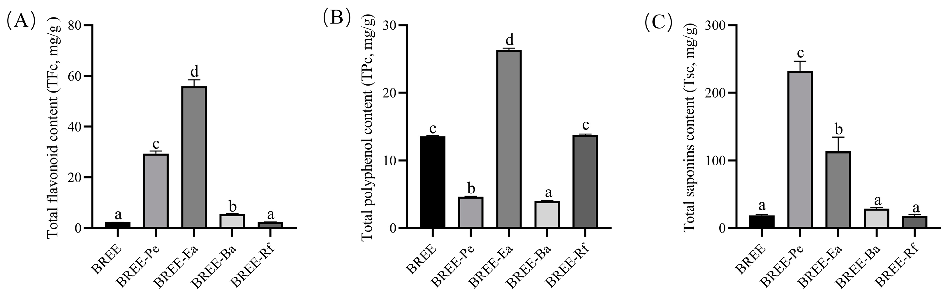
2.3.1. Total Flavonoid Content (TFc)

The TFc was determined according to the aluminum trichloride colorimetric method described in [24]. For the assay, accurately weigh 12 mg of rutin, fill with anhydrous ethanol to 50 mL, shake well, and set aside. The standard solution of 0, 0.1, 0.2, 0.3, 0.4, and 0.5 mL was accurately absorbed with a pipette gun, and anhydrous ethanol was added to a constant volume of 540 μL. After that, 30 μL 5% NaNO2 solution was added and shaken well before being left for 6 min. Then 30 μL 10% AlCl3 was added and shaken well before being left for 6 min. Finally, 400 μL 4% NaOH solution was added and shaken well before being left for 15 min. The absorbance of the reaction mixture was measured at 510 nm. The standard curve was drawn with absorbance (A) as the ordinate and mass concentration of rutin (C, mg/mL) as the abscissa. Samples were determined in the same method, and values of TFc were calculated according to the standard curve. The data were expressed in mg rutin/g dry extract.

2.3.2. Total Polyphenol Content (TPc)

In the experiment, the TPc was determined by the folin phenol colorimetry [25], and gallic acid was used as the standard reference. A total of 1 mg of gallic acid was weighed with precision and dissolved with distilled water to a fixed volume of 10 mL to create a gallic acid reference product working liquid. Separately, 0, 40, 80, 120, 160, 200, 300, and 400 μL of reference solution were moved into 1.5 mL tubes, and distilled water was added to a constant volume of 400μL. After that, 400 μL foline-phenol (1N) was added into each tube and mixed, the reaction was carried out for 5 min at room temperature and away from light. Finally, 200 μL 15% Na2CO3 solution was added and shaken well before being incubated at room temperature for 2 h away from light. The absorbance of the reaction mixture was measured at 760 nm with an enzyme label. The standard curve was drawn with absorbance (A) as the ordinate and mass concentration of gallic acid (C, mg/mL) as the abscissa. Samples were determined via the same method, and values of TPc were calculated according to the standard curve. The data were expressed in mg gallic acid/g dry extract.

2.3.3. Total Saponin Content (TSc)

The TSc in samples was determined by vanillin–ethanol method, and β-sitosterol was used as the standard reference [26]. The standard β-sitosterol 29 mg was accurately weighed and dissolved in 100 mL absolute ethanol to obtain a 0.29 mg/mL reference reserve solution. Separately, 0, 20, 40, 80, 120, 160, and 200 μL of reference solution were moved into 1.5 mL tubes, and absolute ethanol was added to a constant volume of 200 μL. After that, 0.2 mL 5% vanillin–ethanol solution and 0.8 mL 77% sulfuric acid aqueous solution were added into each tube and shaken well before being bathed in water at 60 °C for 20 min. Finally, tubes were put into ice water for 10 min and kept at room temperature for 60 min before being measured at 550 nm. The standard curve was drawn with absorbance (A) as the ordinate and concentration of β-sitosterol (C, mg/mL) as the abscess. Samples were determined via the same method, and values of TPc were calculated according to the standard curve. The data were expressed as mg β-sitosterol/g dry extract.

2.4. Antioxidant Activity

2.4.1. DPPH Method

The DPPH free radical scavenging ability was evaluated via the method described by Cao et al. [27] with small modifications. For this assay, 2 mg DPPH was accurately weighed and fixed to 25 mL with absolute ethanol, which was prepared into 2 × 10⁻⁴ M working solution and stored away from light. The working solution should be used as soon as it is ready. The mixture containing 30 μL sample solution and 210 μL of DPPH working solution was reacted for 30 min away from light in a 96-well plate. The absorbance of the mixture at 517 nm was measured, and Vc was used as a positive control. The scavenging percentage was calculated according to Formulation 1. EC50 values were expressed as mg dry extract/mL.
Scavenging rate (%) = (1 − (Asample − Ablank)/Acontrol) × 100%
where Acontrol, Asample, and Ablank are the absorbances of the control group (DPPH + ethanol), the test group (DPPH + sample), and the blank group (sample + ethanol), respectively.

2.4.2. ABTS Assay

The ABTS free radical scavenging activity was performed according to the method reported by Miao et al. [28]. Vc was used as a positive control, and the results were expressed as EC50 values.

2.4.3. Reducing Power

The reducing power was tested by referring to the ferric-reducing antioxidant power (FRAP) assay [29]. Vc was used as a positive control, and FRAP was expressed as mg Vc/g samples.

2.5. Cell Culture and Treatment

Differentiated rat pheochromocytoma (PC12) cell strain was purchased from the Shanghai Cell Resource Center, Chinese Academy of Sciences (Shanghai, China). PC12 cells were cultured in RPMI-1640 culture medium containing 10% FBS (v/v), penicillin, and streptomycin (100 μg/mL, respectively) in 5% CO2 at 37 °C. Cells were collected every other day, and logarithmic growth phase cells were retained for subsequent series of experiments.
PC12 cells at the logarithmic phase were incubated for 24 h; then, the medium was replaced with the corresponding sample solution for 24 h and incubated with 200 μM H2O2 for another 4 h. The experiment was divided into a control group, the active fraction with a high-dosage group, the active fraction with a middle-dosage group, the active fraction with a low-dosage group, the DMSO group, the H2O2 group, and the Vc group. At last, H2O2 aqueous solution was added to all groups other than the control group during the incubation. The control group was incubated with the same amount of 1640 medium, and the positive group used 200 μM Vc, while different BR active fractions groups were incubated with different concentrations of corresponding drugs for 24 h. BR active fractions were dissolved in DMSO, respectively, and subsequently deliquated in a culture medium with the final concentration of DMSO less than 1% (v/v).

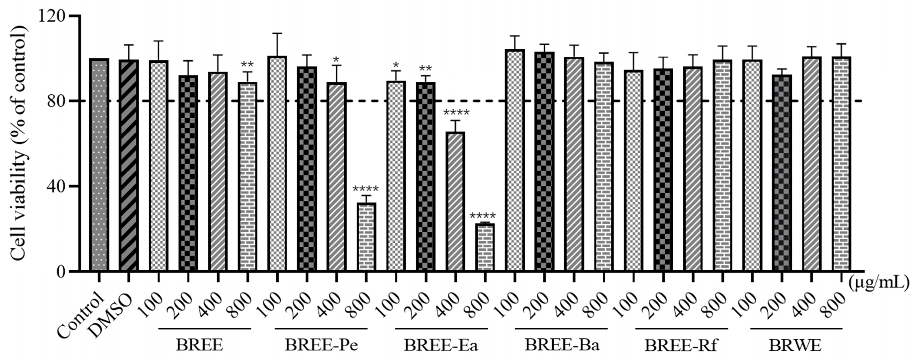
2.6. Cell Viability Assay

Cell viability was measured using an MTT assay. First, PC12 cells in the logarithmic growth phase were incubated for 24 h at 1 × 10⁴ cells per well uniformly spread on 96-well plates. Then, the medium in the wells was replaced with different concentrations of BR active fractions for 24 h. Finally, the medium was removed; 100 µL MTT (0.5 mg/mL) solution was added to each well, the incubation continued for 4 h, and the dark-blue formazan was dissolved with 150 μL DMSO. The absorbance at 490 nm was then measured. Cell viability was expressed as a percentage of control cells.

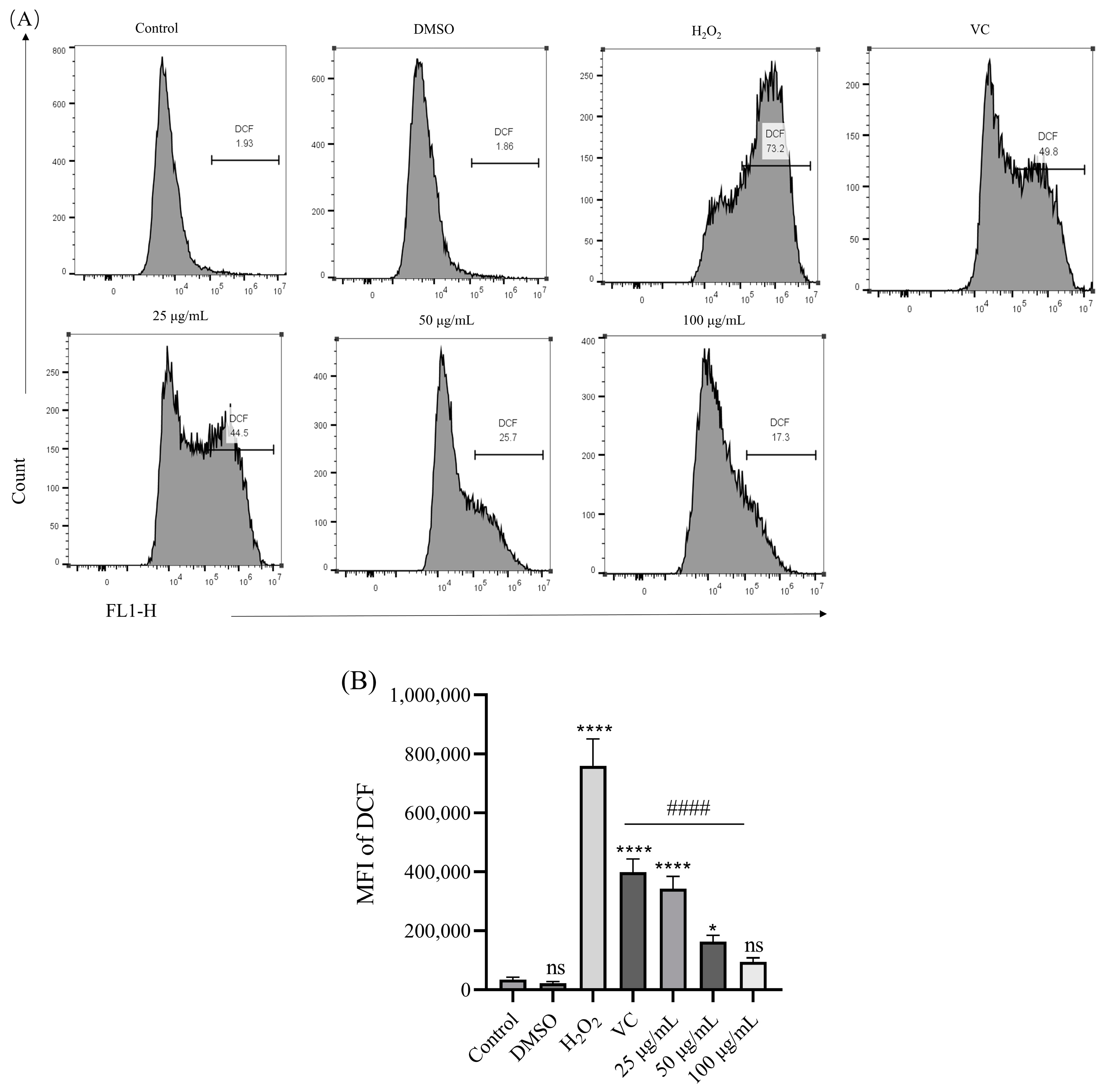
2.7. Detection of Intracellular ROS Accumulation in PC12 Cells

Intracellular ROS levels were detected using the green fluorescent probe 2,7-dichlorodihydrofluorescein diacetate (DCFH-Da). H2O2 can transform green fluorescent DCFH into DCF, which has a higher fluorescence [30]. In this study, ROS levels in PC12 cells were detected by ROS kit (Beyotime, Shanghai, China) according to the methods referred to by Li, R. L. et al. [31].

2.8. Determination of MDA, SOD, GSH-Px, and LDH in H2O2-Induced PC12 Cells

The cells were incubated in 6-well plates with 1 × 10⁵ cells per milliliter and given different concentrations of BREE-Ea and H2O2, as described in 2.5. After treating the cells with H2O2 for 4 h, cell supernatant and cells were collected, and cell supernatant was used to detect LDH release. The collected PC12 cells were washed 3 times with PBS, and the cells were well-broken by Ultrasonic Cell Disruption System to release the cell contents. Furthermore, the activities of SOD, GSH-Px, MDA, and LDH in PC12 cells were detected using the corresponding kit (Nanjing, China) according to the manufacturer’s instructions.

2.9. UPLC-MS Analysis of BREE-Ea

The compounds in the BREE-Ea were analyzed via UPLC-MS system consisting of a UPLC system (Thermo Fisher Scientific, Waltham, MA, USA) with a UPLC HSS T3 column (2.1 mm × 100 mm, 1.8 μm) coupled to Orbitrap Exploris 120 mass spectrometer (Orbitrap MS, Thermo, Waltham, MA, USA). For the separation of chemical compounds, the mobile phase consisted of 5 mM ammonium acetate, 5 mM acetic acid in water (A), and acetonitrile (B). The injection volume was 2 μL. The gradient program was 0–0.7 min with 1% B and the flow of 0.35 mL/min, 0.7–9.5 min with 1–99% B and the flow of 0.35 mL/min, 9.5–11.8 min with 99% B and the flow of 0.35–0.5 mL/min, 11.8–12 min with 99–1% B and flow of 0.5 mL/min, 12–14.6 min with 1% B and the flow of 0.5 mL/min, 14.6–14.8 min with 1% B and the flow of 0.5–0.35 mL/min, and 14.8–15 min with 1% B and the flow of 0.35 mL/min. The column temperature was 35 °C; the sample room temperature was 4 °C, and the auto-sampler temperature was 4 °C. The mass spectra were recorded both with positive and negative ionization modes. The Orbitrap MS was carried out with the following conditions: capillary temperature 320 °C; the spray voltage 3.8 kV for positive mode; and −3.4 kV for negative mode. The compounds were identified according to their MS2 database.

2.10. Statistical Analysis

All data were presented as mean ± SD and were compared with the respective control groups. Statistical and graphical evaluations were conducted via one-way analysis of variance (ANOVA) using GraphPad Prism 5 software and Microsoft Excel 2010.

3. Results and Discussion

3.1. Detection of Active Components in BR Active Fractions

Contents of total flavonoids, total polyphenols, and total saponins in different active fractions of BR, as listed in Figure 1, and the contents of flavonoids (55.92 mg/g) and polyphenols (26.35 mg/g) in BREE-Ea were of the highest significance. This is in agreement with other researchers [32,33]. However, the content of saponins (113.33 mg/g) in BREE-Pe was the highest.

3.2. Antioxidant Activity

Oxidative stress is not only an important mechanism of brain injury but also one of the main causes of neurodegenerative diseases [34]. As age increases, free radicals build up in brain tissue, and the lack of timely and effective methods to remove free radicals poses a challenge. It will lead to serious damage to neurons and neural stem cells and, eventually, result in a series of problems, such as learning and memory disorders [6]. Therefore, scavenging oxygen free radicals can protect cells from oxidative stress damage. In this study, the antioxidant properties of the BR fractions were evaluated using several in vitro antioxidant models. As shown in Figure 2, DPPH and ABTS free radicals scavenging models were used to assess the capacity of the fractions to scavenge the free radicals, and ferric ion-reducing power (FRAP) was used to assess the capacity of the fractions to reduce Fe2+.
In DPPH and ABTS free radicals scavenging assays, BREE-Ea showed the strongest scavenging capacities among all samples prepared from BR, with half-effective concentration (EC50) values of 1.76 and 0.43 mg/mL for DPPH and ABTS free radicals, respectively. Furthermore, BREE-Ea showed a better free radical scavenging effect than BR polysaccharide (2.772 mg/mL of DPPH scavenging rate, 7.112 mg/mL of ABTS scavenging rate) reported previously [27]. Under the same conditions, EC50 values of the reference standard Vc were 0.04 and 0.22 mg/mL, respectively, while BREE-Pe, BREE-Ba, BREE, and BREE-Rf, with EC50 values of 13.87, 12.06, 12.47, and 14.88 mg/mL for DPPH free radical, and 2.64, 1.23, 1.99, and 1.84 mg/mL for ABTS free radical, respectively. When assessed by FRAP assay, BREE-Ea and BREE- Ba showed the highest reducing activity, followed by BREE, BREE-Pe, and BREE-Rf with values of 18.31, 18.79, 16.83, 6.93, and 3.50 mg Vc/g samples, respectively. In general, BREE-Ea has a strong antioxidant activity than other active components of BR. In the former study, ethyl acetate extract of Biscutella raphanifolia (Brassicaceae) showed the best antioxidant effect when compared with dichloromethane and petroleum ether fractions, and our results are consistent with the previous result [35]. In addition, the contents of total polyphenols and flavonoids play an important role in the antioxidant activity of plants [36,37]. Thus, polyphenols and flavonoids with high contents in BREE-Ea may be the main active components exerting antioxidant activity.

3.3. Cytoprotective Activity on PC12 Cell Viability

Cell viability is generally considered to be the number of healthy cells in a sample. Typically, the same analysis used to detect cell viability over a specified period can reflect cell proliferation. Previous research revealed that the PC12 cell, a rat pheochromocytoma cell line, which has been widely used as a cell model in studies of neurodegenerative diseases for the PC12 cell, has similar neuronal characteristics, physiology, and pathology of the nerve cells [38]. PC12 cells (1 × 10⁵ cells/mL) were treated with different concentrations (0–800 μg/mL) of BR fractions for 24 h at 37 °C after normal culture for 24 h to observe toxicity.
As shown in Figure 3, each fraction with different concentrations exhibited different cell viabilities. BREE-Ba and BREE-Rf did not have obvious effects on the viability of PC12 cells at the test concentrations ranging from 100 to 800 μg/mL. However, with the increase in concentrations of BREE, BREE-Pe, and BREE-Ea, cell viability decreased significantly. Therefore, low concentrations of BREE, BREE-Pe, and BREE-Ea would be used in the following study, as referred to in the following part. BREE and BREE-Pe at the concentration ranging from 100 to 400 μg/mL had no obvious effects on the viability of PC12 cells, so the concentration ranges from 100 to 400 μg/mL for BREE and BREE-Pe were used in the subsequent detection.
Cell viability was more than 80% at the concentrations of 100 and 200 μg/mL of BREE-Ea, although cell viability of PC12 cells in the BREE-Ea (100–800 μg/mL) group was much lower than that in the control group. Hence, BREE-Ea was not cytotoxic in low concentrations, and concentration ranges from 50 to 200 μg/mL were finally selected for the subsequent evaluations of BREE-Ea.

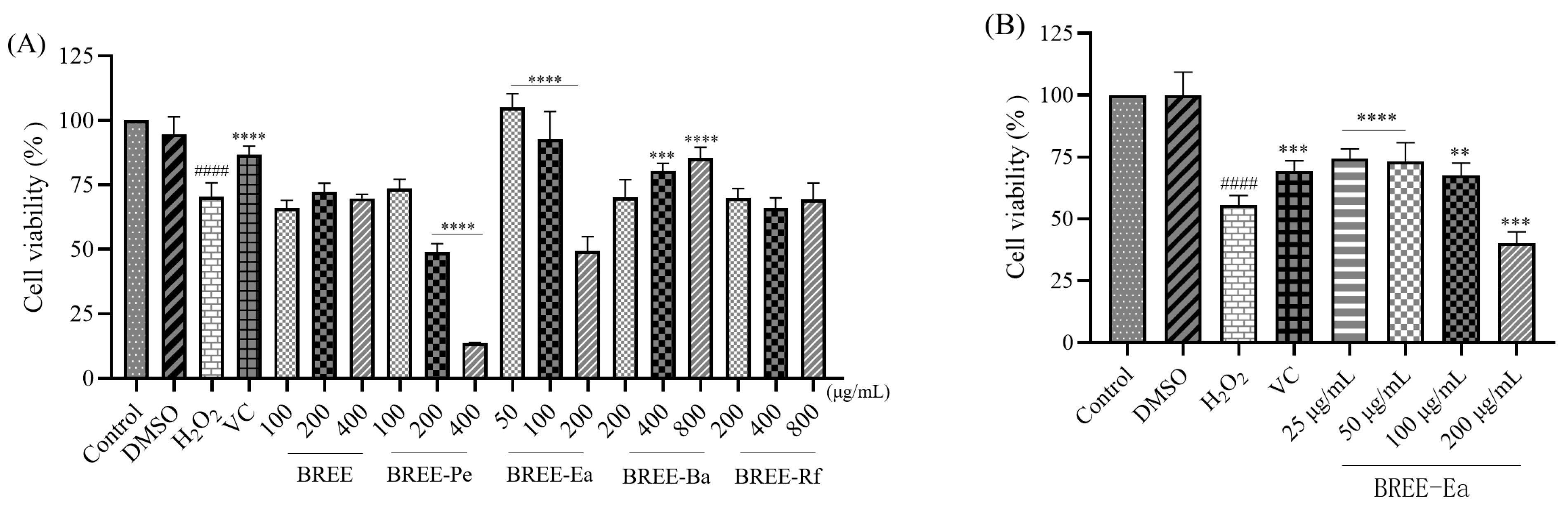
3.4. Cytoprotective Activity on H2O2-Induced PC12 Cell Viability

H2O2 can induce apoptosis in many different cell types, including PC12 cells, by initiating mitochondrial dysfunction [39]. PC12 cells have been widely used in studies of oxidative stress-induced protection models, and in addition, H2O2 possesses a high-cell membrane transmittance [40]. Consequently, H2O2-induced PC12 cells are commonly considered an ideal cell model for studying pathology and screening candidate drugs for neurodegenerative diseases [41,42]. In order to screen the protective active fraction of BR, BR active fractions were preincubated with PC12 cells for 24 h, and then, H2O2 solution was added to replace BR extracts and active fractions, and the new system was incubated for another 4 h. After that, the MTT method was employed to test the cell viability, and higher cell viability means stronger neuroprotective effect.
As shown in Figure 4A, incubation with 200 μM H2O2 for 4 h resulted in a cell viability rate of 70.3% compared with the control, and the difference was statistically significant (p < 0.05). Other researchers damaged PC12 cells for different amounts of time with different concentrations of H2O2, for example, studies have shown the cell viability of 40% at 90 μM H2O2 for 2 h, 38.4% at 300 μM H2O2 for 4 h, and 50% at 100 μM H2O2 for 12 h [31,43,44]. The reasons for the discrepancies may include differences in experimental conditions, manipulation, and reagent quality. Compared with the H2O2 group (70.31%), the viability of PC12 cells increased significantly after being pretreated with different concentrations of BREE-Ea (50 and 100 μg/mL) and BREE-Ba (400 and 800 μg/mL) for 24 h, and the difference was also statistically significant (Figure 4A). The viability of treated cells was found to increase up to 105.1% (50 μg/mL) and 92.6% (100 μg/mL) for BREE-Ea, while BREE-Ba increased cell viability by 80.4% (400 μg/mL) and 85.4% (800 μg/mL), respectively. This may be due to the high contents of flavonoids and polyphenols in BREE-Ea and BREE-Ba fractions, which also show good antioxidant activity and can protect PC12 cells from oxidative damage [45].
These results indicated that BREE-Ea and BREE-Ba could protect PC12 cells from H2O2-induced cell death, with BREE-Ea displaying the maximum protective effect in all the experimental doses. In the subsequent assays, we reduced the concentration gradient of BREE-Ea (Figure 4B). Figure 4B showed that BREE-Ea with different concentrations showed different PC12 cell protective activities. When compared with the H2O2 group (55.6%), BREE-Ea significantly increased cell viability at concentrations of 25, 50, and 100 μg/mL (p < 0.05), with cell viabilities of 74.4%, 73.2%, and 67.6%, respectively, compared with cell viability of the positive control group (Vc group) of 69.3%. However, 200 μg/mL BREE-Ea cannot increase cell viability in this experiment. Therefore, the concentrations of 25 to 100 μg/mL of BREE-Ea for 24 h as optimal concentrations were utilized for the following study. We speculate that BREE-Ea may have a preventive effect on mild to moderate neurodegenerative diseases.

3.5. Effect of BREE-Ea on H2O2-Induced ROS Production

There are more and more studies suggesting that the levels of reactive oxygen species (ROS) were increased as a major cause of cellular damage in a variety of chronic diseases, including neurodegenerative disorders [46,47]. Oxidative stress can cause an excessive generation of ROS, which may result in a loss of mitochondrial function and cell apoptosis [48]. Therefore, to determine the oxidative damage induced by H2O2, intracellular ROS production was measured using the flow cytometry method, and the results were demonstrated in Figure 5. BREE-Ea and Vc were incubated in PC12 cells for 24 h, respectively. After the incubation of PC12 cells with 200 μM H2O2 for 4 h, the level of intracellular ROS was observed. Compared with the control group, the expression of ROS in the H2O2 group significantly increased to approximately 21 folds (p < 0.05). However, ROS contents in three different BREE-Ea groups decreased by 54.9% (25 μg/mL), 78.7% (50 μg/mL), and 87.5% (100 μg/mL) when compared with the H2O2 group, and the difference was statistically significant (p < 0.05). The ROS content of the Vc group also showed a descent of 47.6% compared with the H2O2 group (p < 0.05). The results showed that BREE-Ea could alleviate the abnormal production of H2O2-induced ROS with dose dependence and restore cell vitality to a certain extent. All the above results suggest that BREE-Ea might help to protect PC12 cells from H2O2-induced damage by enhancing the capability of ROS scavenging.

3.6. Effect of BREE-Ea on H2O2-Induced MDA, SOD, LDH, and GSH-Px Activity in PC12 Cells

Malondialdehyde (MDA), as a biomarker of oxidative stress and a product of lipid peroxidation, would significantly increase when cells were exposed to oxidative stimulation, and excessive MDA also could damage the cell membrane [49]. In addition, superoxide dismutase (SOD) and glutathione peroxidase (GSH-Px), as the most important antioxidant enzymes in the body, can remove excess ROS and maintain the balance of ROS levels to protect cell apoptosis. Thus, to elucidate whether BREE-Ea protects PC12 cells from H2O2-stimulated damage through oxidative stress pathway, the effects of BREE-Ea on MDA, SOD, lactate dehydrogenase (LDH), and GSH-Px activities in H2O2-induces PC12 cells were studied.
BREE-Ea and Vc were incubated separately in PC12 cells for 24 h. After the incubation of PC12 cells with 200 μM H2O2 for 4 h, the level of MDA, SOD, LDH, and GSH-Px was observed. As shown in Figure 6, compared with the control group, an extremely significant increase by 6.15 folds in MDA was observed in PC12 cells exposed to 200 μM H2O2 for 4 h (p < 0.01). However, pretreatment with BREE-Ea of three different concentrations significantly reduced MDA contents by 46.31%, 50.15%, and 69.72% when compared to the H2O2-induced group, and the differences between them were statistically significant (p < 0.05) (Figure 6A).
LDH (lactate dehydrogenase) is an enzyme in normal cells, and it is responsible for catalyzing the oxidation of lactate to pyruvate. LDH is released to the outside of cells during cell damage or injury [50]. Thus, the LDH activity of cell supernatant is one of the main tools for the evaluation of cell death. As shown in Figure 6B, when compared with the control group, LDH content increased by 2.78 folds in cell supernatant. However, pretreatment with BREE-Ea of three different concentrations significantly lowered LDH activity by 23.98%, 41.39%, and 69.37% when compared to the H2O2 group (p < 0.05).
Cell death from ROS overproduction is accompanied by decreasing the activity of antioxidant enzymes and an increase in the product of lipid peroxidation. The antioxidant enzymes SOD and GSH-Px play an important role in preventing cell damage and oxidative stress. Thus, enhancing the action of antioxidant enzymes effects can protect cells from oxidative-stimulated damage [50]. Compared with the control group, the activity of SOD and GSH-Px in PC12 cells was reduced by 53.36% and 54.31% in the H2O2 group, respectively, and the difference was statistically significant (p < 0.05). SOD and GSH-Px activity levels in the three BREE-Ea groups were higher by 30.43%, 52.73%, and 66.90% in SOD activity and 37.45%, 70.92%, and 142.27% in GSH-Px activity, when compared with that in the H2O2 group (p < 0.05). The SOD and GSH-Px expression level in the Vc group also showed an improvement of 43.55% and 90.94% compared with the H2O2 group (p < 0.05) (Figure 6C,D).
The results showed that BREE-Ea dampens H2O2-stimulated oxidative damage in PC12 cells by inhibiting oxidative stress. H2O2 exposure would destroy cells, enhance the expression of MDA, increase LDH release, and decrease the expression of SOD and GSH-Px. At the same time, BREE-Ea alleviated the abnormal index changes induced by H2O2, reduced the formation of MDA, reduced LDH release, and increased the activity of SOD and GSH-Px. In previous studies, total phenols and total flavonoids of Rhododendron anthopogonoides showed good antioxidant capacity, and ethyl acetate and n-butanol parts could protect PC12 cells induced by hypoxia, increase the activity of antioxidant enzymes, and reduce LDH and MDA levels, which was consistent with our results [33].

3.7. Chemical Composition of BBRE-Ea

UPLC-MS is a method for essential qualitative analysis of the chemical compounds in the plant. To preliminary identify the compounds contributing to the bioactivity, the BREE-Ea was analyzed by UPLC-MS. In short, 60 compounds were tentatively identified in our experimental conditions (Table S1). The positive ionization mode (POS) contained 46 compounds, and the negative ionization mode (NES) contained 14 compounds. These compounds fall into six major kinds, such as alkaloids, organic acids, terpenoids, flavonoids, polyphenols, and amino acids. There are 12 kinds of organic acids, 10 kinds of alkaloids, 9 kinds of terpenoids, 7 kinds of flavonoids, 5 kinds of polyphenols, 4 kinds of amino acids, and 13 kinds of other compounds, including glycosides and phenylpropanoids. All these 60 compounds were identified by the TCMSP database (https://old.tcmsp-e.com/index.php) (accessed on 15 February 2023) for related diseases and targets, and 12 compounds were associated with neurodegenerative diseases, as shown in Table S1. Furthermore, many papers have reported the antioxidant, anti-inflammatory, or acetylcholinesterase inhibitory properties of these compounds. For example, bergenin and m-coumaric acid have been reported to have strong antioxidant properties [51,52]. Nobiletin and tangeritin belong to the flavonoid compounds, and their effects have been reported on anti-neurodegenerative diseases and other degenerative diseases [53,54,55]. Furthermore, (Z)-Resveratrol has been reported to reduce Aβ aggregation by binding to β amyloid, so it has a potential anti-Alzheimer effect [56]. Thus, all these results suggest that BREE-Ea has good potential to be used in the treatment of neurodegenerative diseases. However, BREE-Ea shows a good protective effect on H2O2-induced oxidative damage in PC12 cells in the present study, but the exact active compound or active compound combinations still need further study.

4. Conclusions

In the present study, the neuroprotective effect of various active fractions prepared from BR was investigated by antioxidant activity and neuroprotective effect on PC12 cells exposed to H2O2-induced oxidative damage in vitro, and these results indicated that BREE-Ea extract has the best antioxidant activity compared with other active fractions. BREE-Ea also can lower oxidative damage of PC12 cells by increasing SOD and GSH-Px activities, decreasing the contents of released LDH, and reducing the formation of ROS and MDA. Therefore, as a good antioxidant, BREE-Ea would be a promising therapeutic agent for neurodegenerative disease and many other psychiatric disorders mainly caused by excessive oxidative stress, and the good performance of BREE-Ea in this study might be ascribed to its abundant active compounds. Overall, as a valuable resource of natural antioxidants, the consumption of BR would be promoted for people with neurodegenerative diseases (including AD and PD) at their early stages.

Supplementary Materials

The following supporting information can be downloaded at: https://www.mdpi.com/2304-8158/12/10/2075/s1, Table S1: Compounds detected by UPLC-MS of BREE-Ea [53,56,57,58,59,60,61,62,63,64,65,66,67,68,69,70,71,72,73,74,75,76,77,78,79,80,81,82,83,84,85,86,87,88,89,90,91,92,93,94,95,96,97,98,99,100,101,102,103,104]; Figure S1. Mass spectrum in (Z)-Resveratrol. RT: 5.24; Figure S2. Mass spectrum in Bergenin. RT: 1.92; Figure S3. Mass spectrum in m-Coumaric acid. RT: 4.30; Figure S4. Mass spectrum in Nobiletin. RT: 7.62; Figure S5. Mass spectrum in Tangeritin. RT: 8.00; Figure S6. Total ion maps of compounds from Brassica rapa L. under positive ion model; Figure S7. Total ion maps of compounds from Brassica rapa L. under negative ion model.

Author Contributions

Conceptualization, J.M.; validation, S.X.; formal analysis, J.W.; investigation, J.W. and S.X.; data curation, J.W. and Q.C.; writing—original draft, J.W.; writing—review and editing, J.M.; visualization, J.W.; supervision, J.M. and J.L.; funding acquisition, J.M. All authors have read and agreed to the published version of the manuscript.

Funding

This research was supported by the Science and Technology Program from Xinjiang Uygur Autonomous Region (No. 2020D01C029) and Major science and technology projects of Xinjiang Uygur Autonomous Region (No. 2022A03017-5).

Data Availability Statement

All the data are included in the article.

Conflicts of Interest

The authors declare no conflict of interest.

Correction Statement

This article has been republished with a minor correction to the Funding statement. This change does not affect the scientific content of the article.

References

  1. Pisoschi, A.M.; Pop, A. The Role of Antioxidants in the Chemistry of Oxidative Stress: A Review. Eur. J. Med. Chem. 2015, 97, 55–74. [Google Scholar] [CrossRef]
  2. Ebert, T.; Neytchev, O.; Witasp, A.; Kublickiene, K.; Stenvinkel, P.; Shiels, P.G. Inflammation and Oxidative Stress in Chronic Kidney Disease and Dialysis Patients. Antioxid. Redox Signal. 2021, 35, 1426–1448. [Google Scholar] [CrossRef] [PubMed]
  3. Liguori, I.; Russo, G.; Curcio, F.; Bulli, G.; Aran, L.; Della-Morte, D.; Gargiulo, G.; Testa, G.; Cacciatore, F.; Bonaduce, D.; et al. Oxidative Stress, Aging; Diseases. Clin. Interv. Aging 2018, 13, 757–772. [Google Scholar] [CrossRef] [PubMed][Green Version]
  4. Uchida, D.; Takaki, A.; Oyama, A.; Adachi, T.; Wada, N.; Onishi, H.; Okada, H. Oxidative Stress Management in Chronic Liver Diseases and Hepatocellular Carcinoma. Nutrients 2020, 12, 1576. [Google Scholar] [CrossRef] [PubMed]
  5. Forman, H.J.; Zhang, H. Author Correction: Targeting Oxidative Stress in Disease: Promise and Limitations of Antioxidant Therapy. Nat. Rev. Drug Discov. 2021, 20, 689–709. [Google Scholar] [CrossRef] [PubMed]
  6. Singh, A.; Kukreti, R.; Saso, L.; Kukreti, S. Oxidative Stress: A Key Modulator in Neurodegenerative Diseases. Molecules 2019, 24, 1583. [Google Scholar] [CrossRef] [PubMed][Green Version]
  7. Tadokoro, K.; Ohta, Y.; Inufusa, H.; Loon, A.F.N.; Abe, K. Prevention of Cognitive Decline in Alzheimer’s Disease by Novel Antioxidative Supplements. Int. J. Mol. Sci. 2020, 21, 1974. [Google Scholar] [CrossRef][Green Version]
  8. Neha, K.; Haider, M.R.; Pathak, A.; Yar, M.S. Medicinal Prospects of Antioxidants: A Review. Eur. J. Med. Chem. 2019, 178, 687–704. [Google Scholar] [CrossRef]
  9. Feng, Y.; Wang, X. Antioxidant Therapies for Alzheimer’s Disease. Oxid. Med. Cell. Longev. 2012, 18, 228–237. [Google Scholar] [CrossRef][Green Version]
  10. Albarracin, S.L.; Stab, B.; Casas, Z.; Sutachan, J.J.; Samudio, I.; Gonzalez, J.; Gonzalo, L.; Capani, F.; Morales, L.; Barreto, G.E. Effects of Natural Antioxidants in Neurodegenerative Disease. Nutr. Neurosci. 2012, 15, 1–9. [Google Scholar] [CrossRef]
  11. Xie, Y.; Jiang, S.; Su, D.; Pi, N.; Ma, C.; Gao, P. Composition Analysis and Anti-Hypoxia Activity of Polysaccharide from Brassica Rapa L. Int. J. Biol. Macromol. 2010, 47, 528–533. [Google Scholar] [CrossRef] [PubMed]
  12. Cao, Q.; Wang, G.; Peng, Y. A Critical Review on Phytochemical Profile and Biological Effects of Turnip (Brassica Rapa L.). Front. Nutr. 2021, 8, 721733. [Google Scholar] [CrossRef] [PubMed]
  13. Zhu, H.; Wang, C.; Cheng, Y.; Guo, Y.; Qian, H.; Liu, Y. Brassica Rapa L. (Tibetan Turnip) Prevents Sleep-Deprivation Induced Cognitive Deficits Via the Inhibition of Neuroinflammation and Mitochondrial Depolarization. Food Funct. 2022, 13, 10610. [Google Scholar] [CrossRef]
  14. Higdon, J.V.; Delage, B.; Williams, D.E.; Dashwood, R.H. Cruciferous Vegetables and Human Cancer Risk: Epidemiologic Evidence and Mechanistic Basis. Pharmacol. Res. 2007, 55, 224–236. [Google Scholar] [CrossRef][Green Version]
  15. Johnson, I.T. Cruciferous Vegetables and Risk of Cancers of the Gastrointestinal Tract. Mol. Nutr. Food Res. 2018, 62, e1701000. [Google Scholar] [CrossRef] [PubMed][Green Version]
  16. Eunyoung, H.; Gun-Hee, K. Anticancer and Antimicrobial Activities of β-Phenylethyl Isothiocyanate in Brassica Rapa L. Food Sci. Technol. Res. 2008, 14, 377–382. [Google Scholar]
  17. Liu, L.; Liu, C.; Hua, H.; Zhao, W.; Zhu, H.; Cheng, Y.; Guo, Y.; Qian, H. Effect of Polysaccharides from Tibetan Turnip (Brassica Rapa L.) on the Gut Microbiome after in Vitro Fermentation and in Vivo Metabolism. Food Funct. 2022, 13, 3063–3076. [Google Scholar] [CrossRef]
  18. Hua, H.; Zhu, H.; Liu, C.; Zhang, W.; Li, J.; Hu, B.; Guo, Y.; Cheng, Y.; Pi, F.; Xie, Y.; et al. Bioactive Compound from the Tibetan Turnip (Brassica Rapa L.) Elicited Anti-Hypoxia Effects in Ogd/R-Injured Ht22 Cells by Activating the Pi3k/Akt Pathway. Food Funct. 2021, 12, 2901–2913. [Google Scholar] [CrossRef] [PubMed]
  19. Jung, U.J.; Baek, N.I.; Chung, H.G.; Bang, M.H.; Jeong, T.S.; Lee, K.T.; Kang, Y.J.; Lee, M.K.; Kim, H.J.; Yeo, J.; et al. Effects of the Ethanol Extract of the Roots of Brassica Rapa on Glucose and Lipid Metabolism in C57bl/Ksj-Db/Db Mice. Clin. Nutr. 2008, 27, 158–167. [Google Scholar] [CrossRef] [PubMed]
  20. Aires, A.; Fernandes, C.; Carvalho, R.; Bennett, R.N.; Saavedra, M.J.; Rosa, E.A. Seasonal Effects on Bioactive Compounds and Antioxidant Capacity of Six Economically Important Brassica Vegetables. Molecules 2011, 16, 6816. [Google Scholar] [CrossRef][Green Version]
  21. Fernandes, F.; Valentão, P.; Sousa, C.; Pereira, J.A.; Seabra, R.M.; Andrade, P.B. Chemical and Antioxidative Assessment of Dietary Turnip ( Brassica Rapa Var. Rapa L.). Food Chem. 2007, 105, 1003–1010. [Google Scholar] [CrossRef]
  22. Chu, B.; Chen, C.; Li, J.; Chen, X.; Li, Y.; Tang, W.; Jin, L.; Zhang, Y. Effects of Tibetan Turnip (Brassica Rapa L.) on Promoting Hypoxia-Tolerance in Healthy Humans. J. Ethnopharmacol. 2017, 195, 246–254. [Google Scholar] [CrossRef] [PubMed]
  23. Li, Y.; Han, J.; Chen, Y.; Chen, C.; Chu, B.; Zhang, Y. P-Coumaric Acid as a Prophylactic Measure against Normobaric Hypoxia Induced Pulmonary Edema in Mice. Life Sci. 2018, 211, 215–223. [Google Scholar] [CrossRef] [PubMed]
  24. Alothman, M.; Bhat, R.; Karim, A.A. Antioxidant Capacity and Phenolic Content of Selected Tropical Fruits from Malaysia, Extracted with Different Solvents. Food Chem. 2008, 115, 785–788. [Google Scholar] [CrossRef]
  25. Ainsworth, E.A.; Gillespie, K.M. Estimation of Total Phenolic Content and Other Oxidation Substrates in Plant Tissues Using Folin-Ciocalteu Reagent. Nat. Protoc. 2007, 2, 875–877. [Google Scholar] [CrossRef]
  26. Hu, T.; Guo, Y.Y.; Zhou, Q.F.; Zhong, X.K.; Zhu, L.; Piao, J.H.; Chen, J.; Jiang, J.G. Optimization of Ultrasonic-Assisted Extraction of Total Saponins from Eclipta Prostrasta L. Using Response Surface Methodology. J. Food Sci. 2012, 77, C975–C982. [Google Scholar] [CrossRef]
  27. Cao, W.; Wang, C.; Mayhesumu, X.; Pan, L.; Dang, Y.; Yili, A.; Abuduwaili, A.; Mansur, S. Isolation, Structural Elucidation, Antioxidant and Hypoglycemic Activity of Polysaccharides of Brassica Rapa L. Molecules 2022, 27, 3002. [Google Scholar] [CrossRef]
  28. Miao, J.; Li, X.; Zhao, C.; Gao, X.; Wang, Y.; Gao, W. Active Compounds, Antioxidant Activity and Alpha-Glucosidase Inhibitory Activity of Different Varieties of Chaenomeles Fruits. Food Chem. 2018, 248, 330–339. [Google Scholar] [CrossRef]
  29. Miao, J.; Zhao, C.; Li, X.; Chen, X.; Mao, X.; Huang, H.; Wang, T.; Gao, W. Chemical Composition and Bioactivities of Two Common Chaenomeles Fruits in China: Chaenomeles Speciosa and Chaenomeles Sinensis. J. Food Sci. 2016, 81, H2049–H2058. [Google Scholar] [CrossRef]
  30. Chen, X.; Zhong, Z.; Xu, Z.; Chen, L.; Wang, Y. 2′,7′-Dichlorodihydrofluorescein as a Fluorescent Probe for Reactive Oxygen Species Measurement: Forty Years of Application and Controversy. Free Radic. Res. 2010, 44, 587–604. [Google Scholar] [CrossRef]
  31. Li, R.L.; Zhang, Q.; Liu, J.; Sun, J.Y.; He, L.Y.; Duan, H.X.; Peng, W.; Wu, C.J. Hydroxy-Alpha-Sanshool Possesses Protective Potentials on H2o2-Stimulated Pc12 Cells by Suppression of Oxidative Stress-Induced Apoptosis through Regulation of Pi3k/Akt Signal Pathway. Oxid. Med. Cell. Longev. 2020, 2020, 3481758. [Google Scholar] [PubMed]
  32. Asraoui, F.; Kounnoun, A.; Cadi, H.E.; Cacciola, F.; Majdoub, Y.O.E.; Alibrando, F.; Mandolfino, F.; Dugo, P.; Mondello, L.; Louajri, A. Phytochemical Investigation and Antioxidant Activity of Globularia Alypum L. Molecules 2021, 26, 759. [Google Scholar] [CrossRef] [PubMed]
  33. Jing, L.; Ma, H.; Fan, P.; Gao, R.; Jia, Z. Antioxidant Potential, Total Phenolic and Total Flavonoid Contents of Rhododendron Anthopogonoides and Its Protective Effect on Hypoxia-Induced Injury in Pc12 Cells. BMC Complement. Altern. Med. 2015, 15, 287–298. [Google Scholar] [CrossRef] [PubMed][Green Version]
  34. Chen, Z.; Zhong, C. Oxidative Stress in Alzheimer’s Disease. Neurosci. Bull. 2014, 30, 271–281. [Google Scholar] [CrossRef][Green Version]
  35. Boudouda, H.B.; Zeghib, A.; Karioti, A.; Bilia, A.R.; Ozturk, M.; Aouni, M.; Kabouche, A.; Kabouche, Z. Antibacterial, Antioxidant, Anti-Cholinesterase Potential and Flavonol Glycosides of Biscutella Raphanifolia (Brassicaceae). Pak. J. Pharm. Sci. 2015, 28, 153–158. [Google Scholar]
  36. Sen, S.; De, B.; Devanna, N.; Chakraborty, R. Total Phenolic, Total Flavonoid Content; Antioxidant Capacity of the Leaves of Meyna Spinosa Roxb., an Indian Medicinal Plant. Chin. J. Nat. Med. 2013, 11, 149–157. [Google Scholar] [CrossRef]
  37. Curcic, M.G.; Stankovic, M.S.; Radojevic, I.D.; Stefanovic, O.D.; Comic, L.R.; Topuzovic, M.D.; Djacic, D.S.; Markovic, S.D. Biological Effects, Total Phenolic Content and Flavonoid Concentrations of Fragrant Yellow Onion (Allium Flavum L.). Med. Chem. 2012, 8, 46–51. [Google Scholar] [CrossRef]
  38. Li, N.; Wen, L.; Li, T.; Yang, H.; Qiao, M.; Wang, T.; Song, L.; Huang, X.; Li, M.; Bukyei, E.; et al. Alleviating Effects of Black Soybean Peptide on Oxidative Stress Injury Induced by Lead in Pc12 Cells Via Keap1/Nrf2/Txnip Signaling Pathway. Nutrients 2022, 14, 3102. [Google Scholar] [CrossRef]
  39. Jang, J.H.; Surh, Y.J. Protective Effects of Resveratrol on Hydrogen Peroxide-Induced Apoptosis in Rat Pheochromocytoma (Pc12) Cells. Mutat. Res. 2001, 496, 181–190. [Google Scholar] [CrossRef]
  40. Duan, L.H.; Li, M.; Wang, C.B.; Wang, Q.M.; Liu, Q.Q.; Shang, W.F.; Shen, Y.J.; Lin, Z.H.; Sun, T.Y.; Wu, Z.Z.; et al. Protective Effects of Organic Extracts of Alpinia Oxyphylla against Hydrogen Peroxide-Induced Cytotoxicity in Pc12 Cells. Neural Regen. Res. 2020, 15, 682–689. [Google Scholar]
  41. Demirci, S.; Kutluhan, S.; Naziroglu, M.; Uguz, A.C.; Yurekli, V.A.; Demirci, K. Effects of Selenium and Topiramate on Cytosolic Ca(2+) Influx and Oxidative Stress in Neuronal Pc12 Cells. Neurochem. Res. 2013, 38, 90–97. [Google Scholar] [CrossRef] [PubMed]
  42. Sai, Y.; Wu, Q.; Le, W.; Ye, F.; Li, Y.; Dong, Z. Rotenone-Induced Pc12 Cell Toxicity Is Caused by Oxidative Stress Resulting from Altered Dopamine Metabolism. Toxicol. Vitro 2008, 22, 1461–1468. [Google Scholar] [CrossRef] [PubMed]
  43. Zou, Y.P.; Lu, Y.H.; Wei, D.Z. Protective Effects of a Flavonoid-Rich Extract of Hypericum Perforatum L. Against Hydrogen Peroxide-Induced Apoptosis in Pc12 Cells. Phytother. Res. 2010, 24 (Suppl. S1), S6–S10. [Google Scholar] [CrossRef] [PubMed]
  44. Zhang, Z.D.; Yang, Y.J.; Liu, X.W.; Qin, Z.; Li, S.H.; Bai, L.X.; Li, J.Y. The Protective Effect of Aspirin Eugenol Ester on Oxidative Stress to Pc12 Cells Stimulated with H(2)O(2) through Regulating Pi3k/Akt Signal Pathway. Oxid. Med. Cell. Longev. 2021, 2021, 5527475. [Google Scholar] [CrossRef]
  45. Mutungi, M.M.; Muema, F.W.; Kimutai, F.; Xu, Y.B.; Zhang, H.; Chen, G.L.; Guo, M.Q. Antioxidant and Antiproliferative Potentials of Ficus Glumosa and Its Bioactive Polyphenol Metabolites. Pharmaceuticals 2021, 14, 266. [Google Scholar] [CrossRef]
  46. Teixeira, J.P.; de Castro, A.A.; Soares, F.V.; Da Cunha, E.F.F.; Ramalho, T.C. Future Therapeutic Perspectives into the Alzheimer’s Disease Targeting the Oxidative Stress Hypothesis. Molecules 2019, 24, 4410. [Google Scholar] [CrossRef][Green Version]
  47. Markesbery, W.R. Oxidative Stress Hypothesis in Alzheimer’s Disease. Free Radic. Biol. Med. 1997, 23, 134–147. [Google Scholar] [CrossRef]
  48. Cheignon, C.; Tomas, M.; Bonnefont-Rousselot, D.; Faller, P.; Hureau, C.; Collin, F. Oxidative Stress and the Amyloid Beta Peptide in Alzheimer’s Disease. Redox Biol. 2018, 14, 450–464. [Google Scholar] [CrossRef]
  49. Valko, M.; Leibfritz, D.; Moncol, J.; Cronin, M.T.; Mazur, M.; Telser, J. Free Radicals and Antioxidants in Normal Physiological Functions and Human Disease. Int. J. Biochem. Cell. Biol. 2007, 39, 44–84. [Google Scholar] [CrossRef]
  50. Olatunji, O.J.; Feng, Y.; Olatunji, O.O.; Tang, J.; Wei, Y.; Ouyang, Z.; Su, Z. Polysaccharides Purified from Cordyceps Cicadae Protects Pc12 Cells against Glutamate-Induced Oxidative Damage. Carbohydr. Polym. 2016, 153, 187–195. [Google Scholar] [CrossRef]
  51. Qiao, S.; Liu, R.; Lv, C.; Miao, Y.; Yue, M.; Tao, Y.; Wei, Z.; Xia, Y.; Dai, Y. Bergenin Impedes the Generation of Extracellular Matrix in Glomerular Mesangial Cells and Ameliorates Diabetic Nephropathy in Mice by Inhibiting Oxidative Stress Via the Mtor/Beta-Trcp/Nrf2 Pathway. Free Radic. Biol. Med. 2019, 145, 118–135. [Google Scholar] [CrossRef] [PubMed]
  52. Sarker, U.; Oba, S. Phenolic Profiles and Antioxidant Activities in Selected Drought-Tolerant Leafy Vegetable Amaranth. Sci. Rep. 2020, 10, 18287. [Google Scholar] [CrossRef]
  53. Nakajima, A.; Ohizumi, Y. Potential Benefits of Nobiletin, a Citrus Flavonoid, against Alzheimer’s Disease and Parkinson’s Disease. Int. J. Mol. Sci. 2019, 20, 3380. [Google Scholar] [CrossRef] [PubMed][Green Version]
  54. Fatima, A.; Siddique, Y.H. Role of Flavonoids in Neurodegenerative Disorders with Special Emphasis on Tangeritin. CNS Neurol. Disord. Drug Targets 2019, 18, 581–597. [Google Scholar] [CrossRef] [PubMed]
  55. Singh, B.; Singh, J.P.; Kaur, A.; Singh, N. Phenolic Composition, Antioxidant Potential and Health Benefits of Citrus Peel. Food Res. Int. 2020, 132, 109114. [Google Scholar] [CrossRef]
  56. Al-Edresi, S.; Alsalahat, I.; Freeman, S.; Aojula, H.; Penny, J. Resveratrol-Mediated Cleavage of Amyloid Beta(1-42) Peptide: Potential Relevance to Alzheimer’s Disease. Neurobiol. Aging 2020, 94, 24–33. [Google Scholar] [CrossRef]
  57. Kim, G.R.; Kim, E.N.; Park, K.J.; Kim, K.H.; Jeong, G.S. Inhibitory Effect of Lgs and Ode Isolated from the Twigs of Syringa Oblata Subsp. Dilatata on Rankl-Induced Osteoclastogenesis in Macrophage Cells. Molecules 2021, 26, 1779. [Google Scholar] [CrossRef]
  58. Ilgisonis, E.V.; Shalina, R.; Kasum-Zade, N.; Burkova, K.G.; Trifonova, O.P.; Maslov, D.L.; Kaysheva, A.L.; Markin, S.S. Metabolomic Markers for Predicting Preeclampsia in the First Trimester of Pregnancy: A Retrospective Study. Molecules 2022, 27, 2475. [Google Scholar] [CrossRef]
  59. Wieczorek, Z.; Zimecki, M.; Trojnar, J. Immunomodulatory Activity of a Potent Thymopentin Analog: Disulphide Bridged Beta-Mercaptopropionyl-Arginyl-Lysyl-Aspartyl-Valyl-Tyrosyl-Cysteine Amide. Pol. J. Pharmacol. 1996, 48, 31–38. [Google Scholar]
  60. Fiore, A.; Murray, P.J. Tryptophan and Indole Metabolism in Immune Regulation. Curr. Opin. Immunol. 2021, 70, 7–14. [Google Scholar] [CrossRef]
  61. Jeong, S.J.; Miyamoto, T.; Inagaki, M.; Kim, Y.C.; Higuchi, R. Rotundines a-C, Three Novel Sesquiterpene Alkaloids from Cyperus Rotundus. J. Nat. Prod. 2000, 63, 673–675. [Google Scholar] [CrossRef] [PubMed]
  62. Macias, F.A.; Torres, A.; Galindo, J.L.; Varela, R.M.; Alvarez, J.A.; Molinillo, J.M. Bioactive Terpenoids from Sunflower Leaves Cv. Peredovick. Phytochemistry 2002, 61, 687–692. [Google Scholar] [CrossRef] [PubMed]
  63. Chen, M.; Chen, C.; Gao, Y.; Li, D.; Huang, D.; Chen, Z.; Zhao, X.; Huang, Q.; Wu, D.; Lai, T.; et al. Bergenin-Activated Sirt1 Inhibits Tnf-Alpha-Induced Proinflammatory Response by Blocking the Nf-Kappab Signaling Pathway. Pulm. Pharmacol. Ther. 2020, 62, 101921. [Google Scholar] [CrossRef] [PubMed]
  64. Huang, J.; Chang, Z.; Lu, Q.; Chen, X.; Najafi, M. Nobiletin as an Inducer of Programmed Cell Death in Cancer: A Review. Apoptosis 2022, 27, 297–310. [Google Scholar] [CrossRef]
  65. Morrow, N.M.; Trzaskalski, N.A.; Hanson, A.A.; Fadzeyeva, E.; Telford, D.E.; Chhoker, S.S.; Sutherland, B.G.; Edwards, J.Y.; Huff, M.W.; Mulvihill, E.E. Nobiletin Prevents High-Fat Diet-Induced Dysregulation of Intestinal Lipid Metabolism and Attenuates Postprandial Lipemia. Arterioscler. Thromb. Vasc. Biol. 2022, 42, 127–144. [Google Scholar] [CrossRef]
  66. Shi, Y.; Chen, J.; Li, S.; Wu, Y.; Yu, C.; Ni, L.; Xiao, J.; Shao, Z.; Zhu, H.; Wang, J.; et al. Tangeretin Suppresses Osteoarthritis Progression Via the Nrf2/Nf-Kappab and Mapk/Nf-Kappab Signaling Pathways. Phytomedicine 2022, 98, 153928. [Google Scholar] [CrossRef]
  67. Bao, J.; Liang, Z.; Gong, X.; Zhao, Y.; Wu, M.; Liu, W.; Tu, C.; Wang, X.; Shu, X. Tangeretin Inhibits Bace1 Activity and Attenuates Cognitive Impairments in Ad Model Mice. J. Agric. Food Chem. 2022, 70, 1536–1546. [Google Scholar] [CrossRef]
  68. Ishida, T.; Nishiumi, S.; Tanahashi, T.; Yamasaki, A.; Yamazaki, A.; Akashi, T.; Miki, I.; Kondo, Y.; Inoue, J.; Kawauchi, S.; et al. Linoleoyl Ethanolamide Reduces Lipopolysaccharide-Induced Inflammation in Macrophages and Ameliorates 2,4-Dinitrofluorobenzene-Induced Contact Dermatitis in Mice. Eur. J. Pharmacol. 2013, 699, 6–13. [Google Scholar] [CrossRef]
  69. Su, Q.; Krai, P.; Goetz, M.; Cassera, M.B.; Kingston, D.G. Antiplasmodial Isoflavanes and Pterocarpans from Apoplanesia Paniculata. Planta Med. 2015, 81, 1128–1132. [Google Scholar] [CrossRef][Green Version]
  70. Zhou, Q.; Jia, X.; Yao, Y.Z.; Wang, B.; Wei, C.Q.; Zhang, M.; Huang, F. Characterization of the Aroma-Active Compounds in Commercial Fragrant Rapeseed Oils Via Monolithic Material Sorptive Extraction. J. Agric. Food Chem. 2019, 67, 11454–11463. [Google Scholar] [CrossRef]
  71. Wang, H.; Wang, F.; Wu, S.; Liu, Z.; Li, T.; Mao, L.; Zhang, J.; Li, C.; Liu, C.; Yang, Y. Traditional Herbal Medicine-Derived Sulforaphene Promotes Mitophagic Cell Death in Lymphoma Cells through Crm1-Mediated P62/Sqstm1 Accumulation and Ampk Activation. Chem. Biol. Interact. 2018, 281, 11–23. [Google Scholar] [CrossRef] [PubMed]
  72. Gong, G.; Guan, Y.Y.; Zhang, Z.L.; Rahman, K.; Wang, S.J.; Zhou, S.; Luan, X.; Zhang, H. Isorhamnetin: A Review of Pharmacological Effects. Biomed. Pharmacother. 2020, 128, 110301. [Google Scholar] [CrossRef] [PubMed]
  73. Ji, Y.; Mao, K.; Gao, J.; Chitrakar, B.; Sadiq, F.A.; Wang, Z.; Wu, J.; Xu, C.; Sang, Y. Pear Pomace Soluble Dietary Fiber Ameliorates the Negative Effects of High-Fat Diet in Mice by Regulating the Gut Microbiota and Associated Metabolites. Front. Nutr. 2022, 9, 1025511. [Google Scholar] [CrossRef] [PubMed]
  74. Yanagita, T.; Han, S.Y.; Wang, Y.M.; Tsuruta, Y.; Anno, T. Cycloalliin, a Cyclic Sulfur Imino Acid, Reduces Serum Triacylglycerol in Rats. Nutrition 2003, 19, 140–143. [Google Scholar] [CrossRef]
  75. Weiss, T.; Bruning, T.; Bolt, H.M. Dephenylation of the Rubber Chemical N-Phenyl-2-Naphthylamine to Carcinogenic 2-Naphthylamine: A Classical Problem Revisited. Crit. Rev. Toxicol. 2007, 37, 553–566. [Google Scholar] [CrossRef]
  76. Lim, H.; Kwon, Y.S.; Kim, D.; Lee, J.; Kim, H.P. Flavonoids from Scutellaria Baicalensis Inhibit Senescence-Associated Secretory Phenotype Production by Interrupting Ikappabzeta/C/Ebpbeta Pathway: Inhibition of Age-Related Inflammation. Phytomedicine 2020, 76, 153255. [Google Scholar] [CrossRef]
  77. Camagna, M.; Ojika, M.; Takemoto, D. Detoxification of the Solanaceous Phytoalexins Rishitin, Lubimin, Oxylubimin and Solavetivone Via a Cytochrome P450 Oxygenase. Plant. Signal. Behav. 2020, 15, 1707348. [Google Scholar] [CrossRef]
  78. Said, H.M. Biotin: Biochemical, Physiological and Clinical Aspects. Subcell. Biochem. 2012, 56, 1–19. [Google Scholar]
  79. Shao, W.; Wang, X.; Liu, Z.; Song, X.; Wang, F.; Liu, X.; Yu, Z. Cyperotundone Combined with Adriamycin Induces Apoptosis in Mcf-7 and Mcf-7/Adr Cancer Cells by Ros Generation and Nrf2/Are Signaling Pathway. Sci. Rep. 2023, 13, 1384–1395. [Google Scholar] [CrossRef]
  80. Mayengbam, S.; Raposo, S.; Aliani, M.; House, J.D. Oral Exposure to the Anti-Pyridoxine Compound 1-Amino D-Proline Further Perturbs Homocysteine Metabolism through the Transsulfuration Pathway in Moderately Vitamin B(6) Deficient Rats. J. Nutr. Biochem. 2015, 26, 241–249. [Google Scholar] [CrossRef]
  81. Muhammad, D.; Lalun, N.; Bobichon, H.; Le Magrex Debar, E.; Gangloff, S.C.; Nour, M.; Voutquenne-Nazabadioko, L. Triterpenoids from the Leaves of Alphitonia Xerocarpus Baill and Their Biological Activity. Phytochemistry 2016, 129, 45–57. [Google Scholar] [CrossRef] [PubMed]
  82. Jalilian, F.; Moieni-Arya, M.; Hosseinzadeh, L.; Shokoohinia, Y. Oxypeucedanin and Isoimperatorin Extracted from Prangos Ferulacea (L.) Lindl Protect Pc12 Pheochromocytoma Cells from Oxidative Stress and Apoptosis Induced by Doxorubicin. Res. Pharm. Sci. 2022, 17, 12–21. [Google Scholar] [PubMed]
  83. Li, N.; Yang, L.; Zuo, H. Arborinine Suppresses Ovarian Cancer Development through Inhibition of Lsd1. Life Sci. 2022, 291, 120275. [Google Scholar] [CrossRef] [PubMed]
  84. Hwang, D.; Lim, Y.H. Resveratrol Antibacterial Activity against Escherichia Coli Is Mediated by Z-Ring Formation Inhibition Via Suppression of Ftsz Expression. Sci. Rep. 2015, 5, 10029–10038. [Google Scholar] [CrossRef][Green Version]
  85. Mendez-Lopez, L.F.; Caboni, P.; Arredondo-Espinoza, E.; Carrizales-Castillo, J.J.J.; Balderas-Renteria, I.; Camacho-Corona, M.D.R. Bioassay-Guided Identification of the Antiproliferative Compounds of Cissus Trifoliata and the Transcriptomic Effect of Resveratrol in Prostate Cancer Pc3 Cells. Molecules 2021, 26, 2200. [Google Scholar] [CrossRef]
  86. Kowalska, E.; Kozik, A. The Genes and Enzymes Involved in the Biosynthesis of Thiamin and Thiamin Diphosphate in Yeasts. Cell. Mol. Biol. Lett. 2008, 13, 271–282. [Google Scholar] [CrossRef]
  87. Feng, P.; Li, Q.; Liu, L.; Wang, S.; Wu, Z.; Tao, Y.; Huang, P.; Wang, P. Crocetin Prolongs Recovery Period of Dss-Induced Colitis Via Altering Intestinal Microbiome and Increasing Intestinal Permeability. Int. J. Mol. Sci. 2022, 23, 3832. [Google Scholar] [CrossRef]
  88. Tan, R.X.; Wolfender, J.L.; Ma, W.G.; Zhang, L.X.; Hostettmann, K. Secoiridoids and Antifungal Aromatic Acids from Gentiana Algida. Phytochemistry 1996, 41, 111–116. [Google Scholar] [CrossRef]
  89. Martin, S.A.; Brash, A.R.; Murphy, R.C. The Discovery and Early Structural Studies of Arachidonic Acid. J. Lipid Res. 2016, 57, 1126–1132. [Google Scholar] [CrossRef][Green Version]
  90. Gorica, E.; Calderone, V. Arachidonic Acid Derivatives and Neuroinflammation. CNS Neurol. Disord. Drug Targets 2022, 21, 118–129. [Google Scholar] [CrossRef]
  91. Ito, H.; Sun, X.L.; Watanabe, M.; Okamoto, M.; Hatano, T. Chlorogenic Acid and Its Metabolite M-Coumaric Acid Evoke Neurite Outgrowth in Hippocampal Neuronal Cells. Biosci. Biotechnol. Biochem. 2008, 72, 885–888. [Google Scholar] [CrossRef] [PubMed][Green Version]
  92. Widmeier, E.; Airik, M.; Hugo, H.; Schapiro, D.; Wedel, J.; Ghosh, C.C.; Nakayama, M.; Schneider, R.; Awad, A.M.; Nag, A.; et al. Treatment with 2,4-Dihydroxybenzoic Acid Prevents Fsgs Progression and Renal Fibrosis in Podocyte-Specific Coq6 Knockout Mice. J. Am. Soc. Nephrol. 2019, 30, 393–405. [Google Scholar] [CrossRef][Green Version]
  93. Lang, A.; Heckl, C.; Vogeser, M.; Stauch, T.; Homann, C.; Hennig, G.; Sroka, R.; Stepp, H. Rapid Spectrophotometric Quantification of Urinary Porphyrins and Porphobilinogen as Screening Tool for Attacks of Acute Porphyria. J. Biomed. Opt. 2018, 23, 1–12. [Google Scholar] [CrossRef] [PubMed]
  94. Weng, J.R.; Omar, H.A.; Kulp, S.K.; Chen, C.S. Pharmacological Exploitation of Indole-3-Carbinol to Develop Potent Antitumor Agents. Mini Rev. Med. Chem. 2010, 10, 398–404. [Google Scholar] [CrossRef]
  95. Fang, J.G.; Lu, M.; Chen, Z.H.; Zhu, H.H.; Li, Y.; Yang, L.; Wu, L.M.; Liu, Z.L. Antioxidant Effects of Resveratrol and Its Analogues against the Free-Radical-Induced Peroxidation of Linoleic Acid in Micelles. Chemistry 2002, 8, 4191–4198. [Google Scholar] [CrossRef] [PubMed]
  96. Day, Z.I.; Mayfosh, A.J.; Giel, M.C.; Hong, Y.; Williams, S.A.; Santavanond, J.P.; Rau, T.F.; Poon, I.K.; Hulett, M.D. Novel Formulation of Undecylenic Acid Induces Tumor Cell Apoptosis. Int. J. Mol. Sci. 2022, 23, 14170. [Google Scholar] [CrossRef]
  97. Masyita, A.; Salim, E.; Asri, R.M.; Nainu, F.; Hori, A.; Yulianty, R.; Hatta, M.; Rifai, Y.; Kuraishi, T. Molecular Modeling and Phenoloxidase Inhibitory Activity of Arbutin and Arbutin Undecylenic Acid Ester. Biochem. Biophys. Res. Commun. 2021, 547, 75–81. [Google Scholar] [CrossRef]
  98. Mostafa, H.H.A.; Wang, H.; Song, J.; Li, X. Effects of Genotypes and Explants on Garlic Callus Production and Endogenous Hormones. Sci. Rep. 2020, 10, 4867–4877. [Google Scholar] [CrossRef][Green Version]
  99. Chen, C.Y.; Chiu, C.C.; Wu, C.P.; Chou, Y.T.; Wang, H.M. Enhancements of Skin Cell Proliferations and Migrations Via 6-Dehydrogingerdione. J. Agric. Food Chem. 2013, 61, 1349–1356. [Google Scholar] [CrossRef]
  100. Huang, S.H.; Lee, C.H.; Wang, H.M.; Chang, Y.W.; Lin, C.Y.; Chen, C.Y.; Chen, Y.H. 6-Dehydrogingerdione Restrains Lipopolysaccharide-Induced Inflammatory Responses in Raw 264.7 Macrophages. J. Agric. Food Chem. 2014, 62, 9171–9179. [Google Scholar] [CrossRef]
  101. Yao, J.; Ge, C.; Duan, D.; Zhang, B.; Cui, X.; Peng, S.; Liu, Y.; Fang, J. Activation of the Phase Ii Enzymes for Neuroprotection by Ginger Active Constituent 6-Dehydrogingerdione in Pc12 Cells. J. Agric. Food Chem. 2014, 62, 5507–5518. [Google Scholar] [CrossRef] [PubMed]
  102. Kwon, J.O.; Jin, W.J.; Kim, B.; Kim, H.H.; Lee, Z.H. Myristoleic Acid Inhibits Osteoclast Formation and Bone Resorption by Suppressing the Rankl Activation of Src and Pyk2. Eur. J. Pharmacol. 2015, 768, 189–198. [Google Scholar] [CrossRef] [PubMed]
  103. Quan, L.H.; Zhang, C.; Dong, M.; Jiang, J.; Xu, H.; Yan, C.; Liu, X.; Zhou, H.; Zhang, H.; Chen, L.; et al. Myristoleic Acid Produced by Enterococci Reduces Obesity through Brown Adipose Tissue Activation. Gut 2020, 69, 1239–1247. [Google Scholar] [CrossRef] [PubMed]
  104. Liang, S.S.; Liao, W.T.; Kuo, C.J.; Chou, C.H.; Wu, C.J.; Wang, H.M. Phthalic Acid Chemical Probes Synthesized for Protein-Protein Interaction Analysis. Int. J. Mol. Sci. 2013, 14, 12914. [Google Scholar] [CrossRef] [PubMed][Green Version]
Figure 1. Active components of the fractions from BR. (A) Content of total flavonoids. (B) Content of total polyphenol. (C) Content of total saponins. The same letters denoted no significant difference (p > 0.05); different letters denoted significant difference (p < 0.05).
Foods 12 02075 g001
Figure 2. Antioxidant activity. (A) DPPH free radical scavenging assay. (B) ABTS free radical scavenging assay. (C) FRAP assay. The same letter means that there is no significant difference, and different letters mean that there is a significant difference (p < 0.05).
Foods 12 02075 g002
Figure 3. Effect of different concentrations of the BR active fraction on PC12 cell viability. * for p < 0.05, ** for p < 0.01, **** for p < 0.0001 when compared with control group.
Foods 12 02075 g003
Figure 4. Protective effects of BR active fraction on the cell viability of H2O2-stimulated PC12 cells. (A) BR fractions. (B) BREE-Ea. #### for p < 0.0001 when compared with control group. ** for p < 0.01, *** for p < 0.001, **** for p < 0.0001, when compared with H2O2 group.
Foods 12 02075 g004
Figure 5. Effects of BREE-Ea on ROS levels of H2O2-induced PC12 cells. * for p < 0.05, **** for p < 0.0001, ns for no significance when compared with the Control group. #### for p < 0.0001, when compared with H2O2 group.
Foods 12 02075 g005
Figure 6. Effects of BREE-Ea on MDA, LDH, SOD, and GSH-Px activity of H2O2-induced PC12 cells. (A) MDA content. (B) SOD activity. (C) LDH activity. (D) GSH-Px activity. * for p < 0.05, ** for p < 0.01, **** for p < 0.0001, ns for no significance when compared with the control group. ## for p < 0.01, ### for p < 0.001, #### for p < 0.0001, compared with the H2O2 group.
Foods 12 02075 g006
Disclaimer/Publisher’s Note: The statements, opinions and data contained in all publications are solely those of the individual author(s) and contributor(s) and not of MDPI and/or the editor(s). MDPI and/or the editor(s) disclaim responsibility for any injury to people or property resulting from any ideas, methods, instructions or products referred to in the content.

