Article

Selenium-Chelating Peptide Derived from Wheat Gluten: In Vitro Functional Properties

1
Food and Pharmacy College, Xuchang University, Xuchang 461000, China
2
College of Food and Biological Engineering, Henan University of Animal Husbandry and Economy, Zhengzhou 450044, China
3
Collaborative Innovation Center of Functional Food by Green Manufacturing, Xuchang 461000, China
*
Author to whom correspondence should be addressed.
These authors contributed equally to this work.
Foods 2024, 13(12), 1819; https://doi.org/10.3390/foods13121819
Submission received: 22 April 2024 / Revised: 20 May 2024 / Accepted: 28 May 2024 / Published: 10 June 2024

Abstract

:
The efficacy of selenium-chelating polypeptides derived from wheat protein hydrolysate (WPH-Se) includes enhancing antioxidant capacity, increasing bioavailability, promoting nutrient absorption, and improving overall health. This study aimed to enhance the bioavailability and functional benefits of exogenous selenium by chelating with wheat gluten protein peptides, thereby creating bioactive peptides with potentially higher antioxidant capabilities. In this study, WPH-Se was prepared with wheat peptide and selenium at a mass ratio of 2:1, under a reaction system at pH 8.0 and 80 °C. The in vitro antioxidant activity of WPH-Se was evaluated by determining the DPPH, OH, and ABTS radical scavenging rate and reducing capacity under different conditions, and the composition of free amino acids and bioavailability were also investigated at various digestion stages. The results showed that WPH-Se possessed significant antioxidant activities under different conditions, and DPPH, OH, and ABTS radical scavenging rates and reducing capacity remained high at different temperatures and pH values. During gastrointestinal digestion in vitro, both the individual digestate and the final digestate maintained high DPPH, OH, and ABTS radical scavenging rates and reducing capacity, indicating that WPH-Se was able to withstand gastrointestinal digestion and exert antioxidant effects. Post-digestion, there was a marked elevation in tryptophan, cysteine, and essential amino acids, along with the maintenance of high selenium content in the gastrointestinal tract. These findings indicate that WPH-Se, with its enhanced selenium and amino acid profile, serves as a promising ingredient for dietary selenium and antioxidant supplementation, potentially enhancing the nutritional value and functional benefits of wheat gluten peptides.

1. Introduction

Gluten is a natural protein extracted from wheat. It is light yellow, with a protein content of up to 80%, and is a nutritious plant protein resource. Peptide is a new type of protein hydrolysis product, derived from edible protein as a raw material, through enzymatic digestion (commercial and fermentation enzymes), followed by separation and purification [1]. In recent years, both domestic and international scholars have conducted numerous studies on the structural characterization and functional activities of peptides derived from animal, microbiological, and plant proteins [2,3]. They are responsible for signal transmission, nutrient provision, and enhancing metabolic function, playing significant roles from the cellular level to organs, systems, and overall organism behavior [4,5,6]. It has been demonstrated that peptides can be absorbed and utilized more completely than proteins. Moreover, the specific functions of peptides can vary according to the sequence and composition of amino acids. It has been observed that peptides exhibit activities such as antimicrobial, antifreeze, antioxidant, antiobesity, antidiabetic, and hypotensive properties [7,8,9].
It was the Swedish chemist Jungs Jacob Betzelius who discovered selenium in 1817 [10]. Selenium is an essential trace element involved in several important metabolic pathways in the body [11]. These pathways include thyroid hormone metabolism, the antioxidant defense system (with active centers proximate to glutathione), and immune function [12]. Selenium deficiency can lead to a number of health disorders, including Creutzfeldt–Jakob disease, cancer, heart disease, and hyperthyroidism [13,14]. Selenium has insulin-like effects in promoting cellular glucose transport, glucose metabolism, and signal transduction [15]. Selenium supplementation can improve the antioxidant capacity of brain tissue in diabetic mice, which is important for the protection of brain tissue from oxidative damage and the maintenance of normal physiological function [16]. Xu et al. [17] reported that selenium-enriched rice protein hydrolysate could dose-dependently protect against Pb2+-induced cytotoxicity in RAW264.7 and PC12 cells. In addition, selenium can affect the active centers of proteins and enhance their antioxidant capacity. Specifically, organic selenium can even be directly involved in scavenging free radicals [18]. Since selenium cannot be synthesized by the human body and can only be supplemented through food, developing effective food-based selenium supplementation products is highly beneficial. Selenium exists in inorganic salts which are difficult for the body to absorb [19,20]. It also has certain toxic properties, which can damage mitochondria and induce apoptosis. Inorganic selenium is frequently converted into organic selenium through chelation to ensure safe supplementation [21,22].
Bioactive peptides, characterized by potent physiological effects, high bioavailability, and diverse origins, serve as promising candidates for selenium delivery [23]. A novel product incorporating selenium and bioactive peptides synergistically provides a suitable medium for selenium supplementation, offering combined benefits of selenium and peptide activities. Currently, there is more research on chelating peptides with Fe, Zn, Ca, and other minerals, yet fewer investigations on chelating peptides of selenium, despite its considerable potential for development [24,25,26,27]. The preliminary study in our laboratory has shown that wheat protein peptides could be chelated with selenium to form chelates (WPH-Se), and it was proved that peptide–selenium chelate was formed by the N-terminal amino group, C-terminal carboxyl group, amino acid side chains, and carbon and imino groups in the peptide chain [28]. Understanding the impact of processing methods on the antioxidant activity of WPH-Se is crucial for its potential application in functional foods or nutritional health products. Gallego et al. [29] reported that the scavenging ability of Ser-Asn-Ala-Ala-Cys (SNAAC) to peroxidize free radicals and ABTS and the bleaching ability of β-carotene decreased significantly after digestion in vitro. Wong et al. [30] confirmed that the peptide Met-Tyr-Pro-Gly-Leu-Ala maintained excellent activity in the simulated intestinal tract. Udenigwe and Fogliano [31] suggested that many peptides easily had chemical reactions on their nucleophilic amino, carboxyl, imine, and sulfhydryl groups, which reduced the bioavailability of bioactive peptides. Therefore, further research is needed to evaluate the stability of WPH-Se under different processing conditions and gastrointestinal proteolytic enzymes to determine its potential as a selenium supplement [32]. Inorganic selenium exhibits a low absorption rate and presents poor safety of intake, whereas organic selenium possesses low toxicity and can be transported and absorbed through peptide transport channels, making it currently a hot research topic [33].
Both theoretical predictions and practical experiments are necessary to comprehensively understand the behavior of WPH-Se in the intestinal environment and its potential impact on bioavailability and efficacy. This study aimed to evaluate its antioxidant and bioavailability under various conditions, including different processing parameters such as temperature and pH. The investigation also involved analyzing its antioxidant activity, free amino acid profile, and changes in Se content during in vitro digestion. Therefore, this study can serve as a theoretical foundation and development efforts in utilizing WPH-Se in various functional food and nutritional formulations.

2. Materials and Methods

2.1. Materials

Porcine pancreatin (4× USP) was obtained from Sigma-Aldrich Co., Ltd. (St. Louis, MI, USA). Alkaline protease (200 U/mg) was obtained from Beijing Solarbio Science & Technology Co., Ltd. (Beijing, China) DPPH (1,1-diphenyl-2-picrylhydrazyl), ABTS (2,2′-azino-bis-3-ethylbenzothiazoline-6-sulfonic acid) diammonium salt, EDTA (ethylene diamine tetra-acetic acid), Na2SeO3, and H2O2 (v/v, 30%) were obtained from Shanghai Maclin Biochemical Technology Co., Ltd. (Shanghai, China) The wheat protein hydrolysate, with a degree of hydrolysis amounting to 38.72%, was prepared in the laboratory. All other chemicals were analytical grade.

2.2. Preparation of WPH-Se

The preparation of wheat protein hydrolysate (WPH) followed the method by Yang et al. [28]. Briefly, wheat gluten was suspended in deionized water (m/v, 1:9), mixed, and ultrasonicated for 20 min (55 °C, 40 kHz) (Jiangda Wukesong Biotechnology Co., Ltd., Zhenjiang, China). The mixture was extracted, adjusted to pH 8, and the enzymatic conditions were as follows: 55 °C, 2.5 h, and alkaline protease (dosage: 8000 U/g), maintained at pH 7.0~9.0 with Hcl or NaOH (1.0 M). The enzymes were subsequently inactivated by heating at 100 °C for 15 min. After cooling to room temperature, the sample was centrifuged (4000 rpm, 10 min), and the supernatant was collected and dried to WPH. WPH was suspended in a solution containing 1 mg/mL Na2SeO3 (m/m, 2:1). The chelation reaction between WPH and Se was conducted under specific conditions: 80 °C, pH 8.0, 120 rpm, and 60 min. Subsequently, the solution was supplemented with anhydrous ethanol at 5:1 (v/v) and allowed to stand for 60 min. the precipitate was dried to constant weight to obtain the WPH-Se [34].

2.3. Analysis of Free Amino Acids after In Vitro Digestion of WPH-Se

Free amino acids in the sample were detected following the method of Fen et al. [35] with minor modification. In brief, WPH-Se (10 mL) before and after digestion was mixed with sulfosalicylic acid (10%, w/v, 2.5 mL), then filtered through a 0.45 μm membrane and left at 2~8 °C for 1 h, followed by centrifugation at 4000 rpm for 15 min. The supernatant was centrifuged again at 4000 rpm for 5 min. The sample dilutions were diluted at 1:4~1:199 and filtered again through the 0.45 μm membrane for the automatic amino acid analyzer (Membra Pure A300, Hennigsdorf, Germany).

2.4. Effect of Temperature and pH on In Vitro Antioxidant Activity

The radical scavenging capacity of ABTS, OH (Hydroxyl), DPPH, and the reducing capacity of WPH-Se (5 mg/mL) were assessed under different temperature (25, 40, 60, 80, 100 °C) and pH levels (2, 4, 6, 8, 10).

2.4.1. The DPPH Radical Scavenging Activity

The determination method of DPPH radical scavenging activity was modified according to that described by Ren et al. [36]. The scavenging activity of DPPH was calculated using the following equation.
DPPH   scavenging   activity = 1   -     A sample   -   A blank A control
Asample is the absorbance value of the sample group; Ablank is the absorbance value of the blank sample; Acontrol is the absorbance value of the control group. All absorbances were measured at 517 nm.

2.4.2. The OH Radical Scavenging Activity

The OH radical scavenging activity was evaluated by the method of Homayouni-Tabrizi et al. [37] with slight modifications. The OH radical scavenging activity of the samples was evaluated by the following equation.
OH   radical   scavenging   activity ( % ) =   A s   -   A i A 0   -   A i × 100
As is the absorbance of the sample group; Ai is the absorbance of the control containing 1,10-phenanthroline, FeSO4, and H2O2; A0 is the absorbance of the blank containing 1,10-phenanthroline and FeSO4. Ultimately, the absorbance was evaluated at 536 nm.

2.4.3. The ABTS Radical Scavenging Activity

The ABTS radical scavenging activity was measured based on the method of described by Yuan et al. [38] with some slight modifications. ABTS radical scavenging activity was calculated using the following equation.
  ABTS   radical   scavenging   activity ( % ) =   1   -   A sample A blank × 100  
Asample is the absorbance of the sample group; Ablank is the absorbance of the blank group. Ultimately, the absorbance was determined at 734 nm.

2.4.4. Reducing Capacity

The reducing capacity for Fe3+ was measured according to the method by Siddhuraju et al. [39]. Then, the absorbance was measured at 700 nm.

2.5. Determination of Antioxidant Activity of WPH-Se In Vitro Digestion

The simulation utilized gastric and intestinal fluids formulated based on the TNO digestion model (Netherlands), which simulates the physiological environment of the human gastric and intestine [40,41,42]. The preparation of gastric and intestine solutions was performed using the method of Minekus et al. [43] with some modifications. Porcine gastric pepsin (0.55 g, 3000 U/mg), 0.28 g of NaCl, 0.052 g of KCl, 0.21 g of NaHCO3, 0.012 g of KH2PO4, 0.002 g of MgCl2(H2O)6, and 0.005 g of (NH4)2CO3 were dispersed in a 100 mL volumetric flask using deionized water; subsequently, 7.7 mL of 2 mmol/L CaCl2 was added. After magnetic stirring for 5 min at room temperature. Porcine pancreatin (0.25 g; 4XUSP, Sigma-Aldrich, St. Louis, MI, USA), 1 g of Pulvis fellis suis, 0.007 g of MgCl2 (H2O)6, 0.714 g of NaHCO3, 0.22 g of NaCl, 0.051 g of KCl, and 0.011 g of KH2PO4 were fixed in a 100 mL volumetric flask with deionized water, after which 7.30 mL of 9 mmol/L CaCl2 was added. After stirring magnetically for 10 min at room temperature, the solution was placed in the refrigerator for later use. WPH-Se solution (5 mg/mL, 20 mL) and artificial gastric fluid (20 mL) were added into the centrifuge tube, and the pH was adjusted to 2.0 with 1 M HCl, mixed well, and then oscillated in a water bath at 37 °C for 2 h. Artificial intestinal fluid (10 mL) was added, and the pH was adjusted to 6.8 with 2 M NaOH; the solution was then oscillated in a water bath at 37 °C for 0, 30, 60, 90, and 120 min, and then removed from the centrifuge tube. After that, the enzyme was inactivated by a water bath at 100 °C for 10 min and centrifuged at 4000 rpm for 10 min. Then, the ABTS, OH, and DPPH radical scavenging activities and reducing capacity were measured.

2.6. Statistical Analysis

Results were expressed as the mean ± standard deviation of the information obtained through triplicating calculations. Analysis of variance (ANOVA) was conducted with a p-value < 0.05. Multiple comparison was compared with Duncan’s test.

3. Results and discussion

3.1. Fabrication of WPH-SE

3.1.1. Changes in Free Amino Acids of WPH-Se In Vitro Digestion

The role of bioactive peptides in the body is closely linked to their amino acid composition [44,45], which underscores the importance of studying the amino acid profile of WPH-Se at various stages of digestion, including the gastric and intestinal phases. Amino acids can be categorized based on their side chains’ chemical properties, including whether they are acidic, basic, or hydrophobic. The content of alkaline, acidic, and hydrophobic amino acids in WPH-Se accounted for 60.4%, 27.1%, and 8.5% of the total amino acid (Table 1), respectively, while essential amino acids constituted only 4.4%. Following digestion of WPH-Se in the gastric environment, an increase in alanine (Ala), cysteine (Cys), valine (Val), and leucine (Leu) was observed. Particularly, the solution exhibited a high concentration of hydrophobic amino acids. Comparative analysis of the free amino acid composition between gastric-digested and intestinal-digested WPH-Se, as well as undigested samples, revealed that the digested samples had a higher proportion of essential amino acids, especially noting the increase in tryptophan (Try), leucine (Leu), phenylalanine (Phe), and threonine (Thr) content. Zhang et al. [46] purified and identified a variety of antioxidant peptides from walnut protein hydrolysate containing specific amino acid residues such as tyrosine, cysteine, and histidine, and the amino acid composition of these antioxidant peptides plays a significant role in their antioxidant activity. Pea peptides, which contain amino acids such as tyrosine, lysine, and cysteine, have also been shown to have a strong antioxidant capacity [47]. Moreover, the nutritional value of WPH-Se was improved by a significant increase in its essential amino acid content.

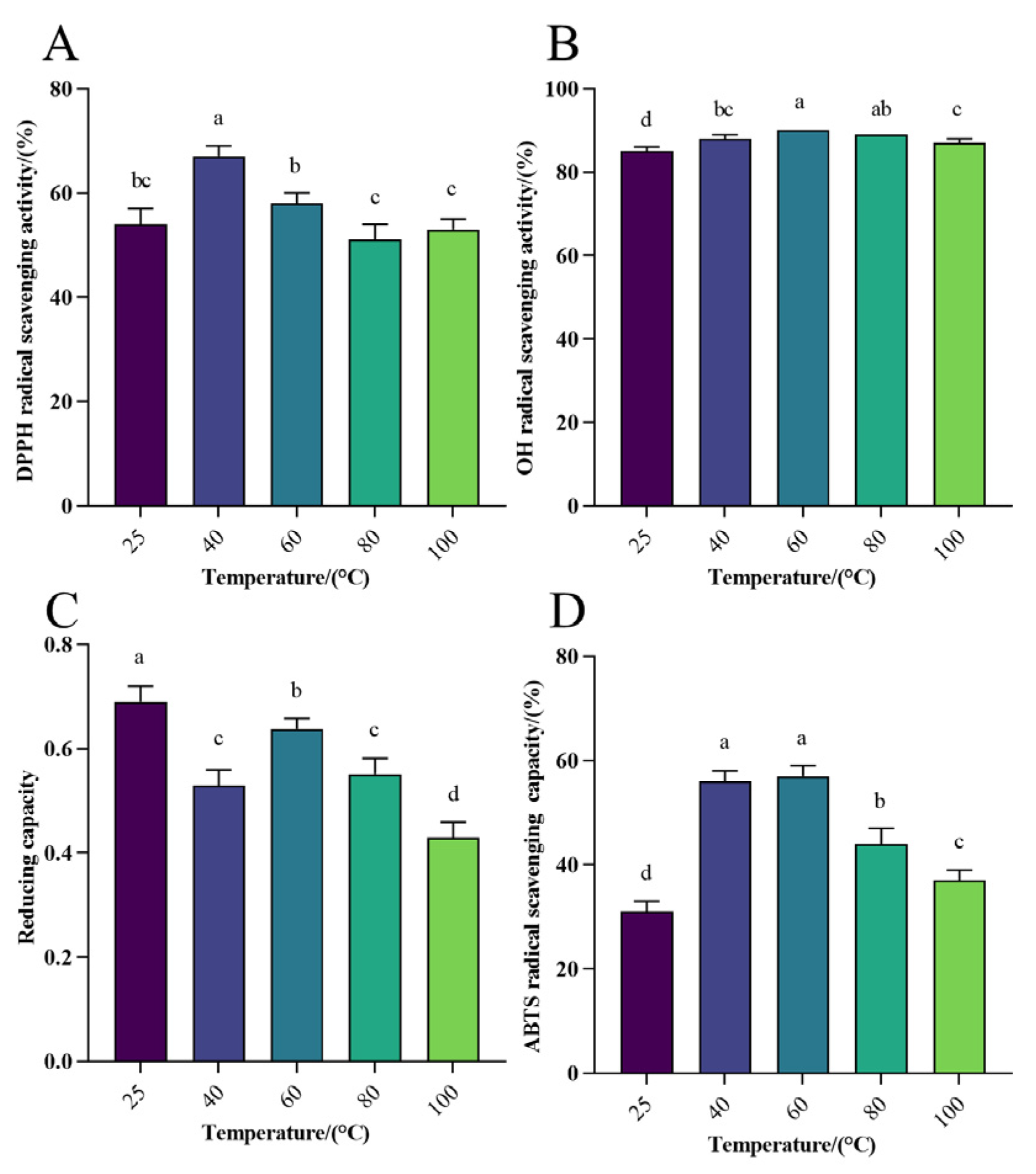
3.1.2. Antioxidant Activity Analysis of WHP-SE at Different Temperatures

Thermal processing remains a common processing method widely used in the production of general foods, nutraceuticals, and pharmaceuticals, and it is essential to investigate the thermal processing characteristics of selenium-chelated peptides as a future research direction [48,49,50]. The investigation was conducted to assess the impact of varying temperatures on the antioxidant properties of WPH-Se, with 25 °C serving as the control group. The rate of DPPH radical scavenging at different temperatures was approximately 50~70% (Figure 1A). The peak scavenging rate occurred at approximately 40 °C, showing a notable increase in scavenging between 25 °C and 40 °C (p < 0.05). The OH scavenging rate of WPH-Se varied from 85% to 95% across different temperatures (Figure 1B). The maximum OH scavenging rate occurred at around 60 °C, evidenced by a linear increase between 25 °C and 60 °C (p < 0.05). Therefore, the change in temperature had little effect on the OH radical scavenging rate, highlighting the potentially efficient capacity of WPH-Se for OH radical scavenging. Furthermore, the reducing capacity of WPH-Se varied from 0.4 to 0.7 at different temperatures (Figure 1C). The highest scavenging rate was observed at approximately 25 °C, followed by a decline between 25 °C and 40 °C (p < 0.05), indicating that temperature variance had a significant impact on the reducing capacity of WPH-Se. Finally, the ABTS scavenging rate of WPH-Se at various temperatures ranged from 20% to 60% (Figure 1D). The scavenging rate of WPH-Se demonstrated an initial rise followed by a decline as temperature levels increased (p < 0.05), peaking at a rate between 40% to 60%. Thus, this indicates that temperature fluctuation has a significant effect on the ABTS scavenging rate of WPH-Se. The free radical scavenging was slightly reduced under high temperature conditions, probably due to high temperatures not causing irreversible denaturation of the peptides [51]. Instead, they might have altered the secondary structure, or prolonged exposure to high temperatures may have led to denaturation and aggregation of the antioxidant peptides, affecting their antioxidant activity and stability. This aspect warrants further exploration in future studies [52]. However, the WPH-Se prepared in this study retained a certain level of antioxidant activity at higher temperatures, indicating its potential application in thermally processed foods while maintaining good nutritional properties.

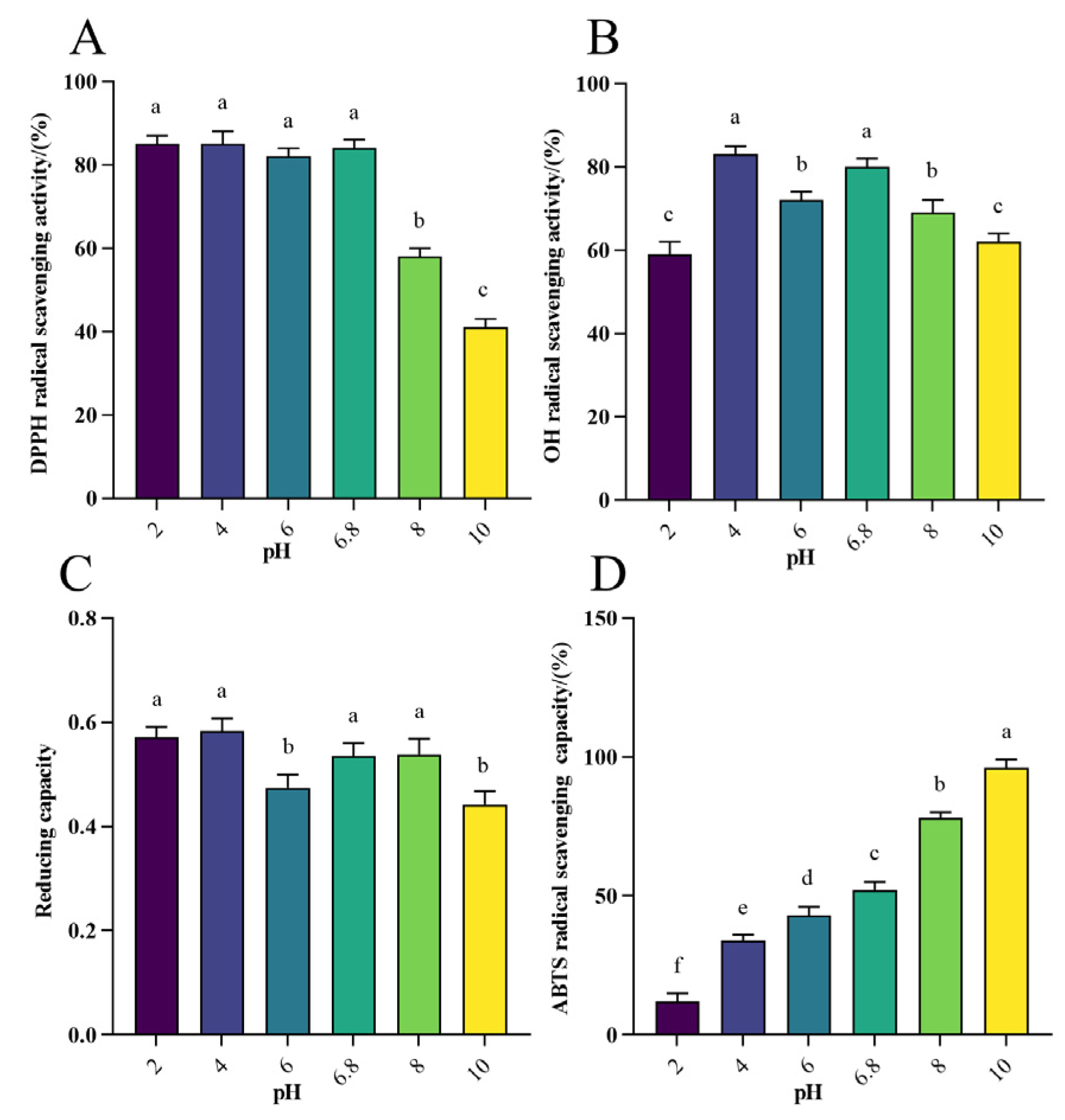
3.1.3. Antioxidant Activity Analysis of WHP-Se at Different pH

The aim of this study was to evaluate the effect of different pH on the antioxidant properties of WPH-Se, using pH 6.8 as the control. The DPPH radical scavenging rate of WPH-Se at different pH ranged from approximately 40% to 90% (Figure 2A), with the scavenging rate exceeding 80% between pH 2 and 6.8. The DPPH scavenging rate gradually decreased as the pH continued to increase (p < 0.05). Notably, a higher DPPH radical scavenging rate was preserved in both the gastric environment at pH 2 and the intestinal environment at pH 6.8. This observation underscores that WPH-Se exhibits noteworthy effects on the DPPH radical scavenging rate. The OH radical scavenging rate of WPH-Se ranged from approximately 50% to 80% across different pH levels (Figure 2B), with the highest rates occurring at pH 4 and pH 6.8 (p < 0.05). As alkalinity increased, the OH scavenging rate gradually decreased, indicating that WPH-Se possesses significant OH scavenging capacity, particularly in the intestinal environment. The reducing capacity of WPH-Se across different pH ranged from 0.4 to 0.6, with the peak observed at pH 4. This capacity remained strong at pH 2 and pH 6.8, suggesting that WPH-Se maintains effective reducing capacity in the gastrointestinal environment (Figure 2C). The ABTS radical scavenging rate of WPH-Se was varied from approximately 10% to 99% across different pH levels (Figure 2D), and the ABTS scavenging rate increased correspondingly with the increase in pH from 2 to 10 (p < 0.05). The ABTS radical scavenging rate was 52% at pH 6.8 and decreased to 12% at pH 2; this indicates that pH has a significant effect on the ABTS radical scavenging rate of WPH-Se. However, considering other data, WPH-Se maintains high antioxidant activity under different pH conditions. Changes in pH can alter electrostatic interactions between charged amino acids, affecting the shape of peptide molecules and leading to changes in chemical properties [53]. Acidic conditions reduce the solubility of the peptide and reduce the effectiveness of the hydroxyl group, whereas alkaline conditions reduce the DPPH radical scavenging rate of the active peptide by a wider range of factors, including racemization, deamidation, and hydrogen depletion on the hydrogen donor, and these changes may be irreversible [54,55,56]. However, ABTS radicals are mainly scavenged by antioxidant electron transfer or hydrogen protonation, and acidic groups (e.g., carboxyl groups) in peptides may be more readily protonated to their negatively charged forms under alkaline conditions [57,58]. This negatively charged state may enhance the interaction of the peptide with free radicals, thereby improving antioxidant activity.

3.2. Physicochemical Property Analysis of WPH-Se

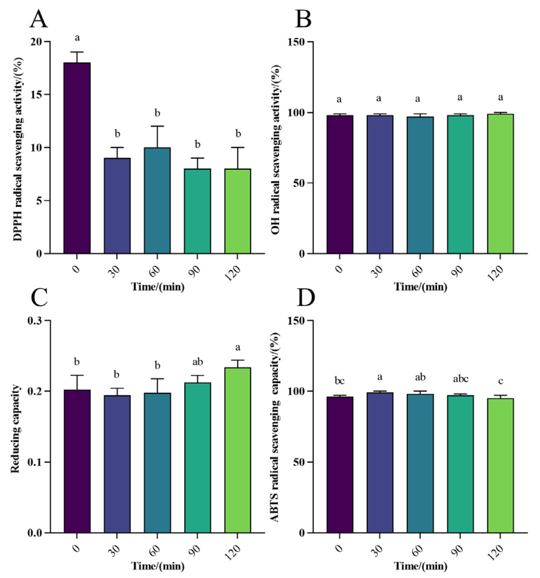
3.2.1. Antioxidant Activity Analysis of WPH-Se In Vitro Digestion

The DPPH radical scavenging rate ranged from 3% to 20% in gastric digestion in vitro (Figure 3A), and for intestinal digestion, it ranged from 30% to 40%, with the peak reaching 40% (Figure 4A), indicating a moderate scavenging rate of WPH-Se in the intestine compared to a lower rate in the gastric environment. However, the overall scavenging level was marginally higher than that in the gastric. With increasing digestion time (Figure 5A), the DPPH radical scavenging rate in the gastric initially decreased, reaching a maximum at 30 min. Conversely, the DPPH radical scavenging rate initially decreased and then increased over time in the intestine. Although the scavenging rate could reach 40% after 2 h, it declined with further digestion time following intestinal treatment. Overall, DPPH scavenging of WPH-Se in the intestinal tract showed a decline with prolonged digestion time. The OH scavenging of WPH-Se was approximately 100% (Figure 3B, Figure 4B, and Figure 5B), demonstrating consistent efficacy in both the gastric and intestinal digestion stages. This high performance indicates that our screened WPH-Se possesses a stable structure. Consequently, it has potential applications in treating health problems associated with hydroxyl radicals, including oxidative stress, inflammation, and atherosclerosis [59,60,61]. However, these applications will require validation through further cellular and animal experiments. The reducing capacity of WPH-Se initially increased in the gastric with time, reaching its peak at 60 min, and then decreased over time (Figure 3C). Conversely, the reducing capacity in intestinal digestion increased more slowly with time (Figure 4C); the reducing capacity of WPH-Se that underwent gastrointestinal digestion exceeded the reducing capacity of WPH-Se that was digested only in the gastric (Figure 5C). It is evident that the reducing capacity of WPH-Se is compromised after exposure to the gastric, leading to an overall reducing capacity of WPH-Se in the intestinal tract remaining at a moderate level. The ABTS radical scavenging rate was initially increased in gastric digestion and then decreased over time, reaching its peak at 60 min, and ranging from 59% to 69% (Figure 3D). Similarly, the scavenging rate was also increased initially in the intestine, reaching its maximum at 30 min, and ranging from 81% to 90% (Figure 4D). Subsequently, the ABTS radical scavenging rate of WPH-Se after gastric and intestinal digestion exceeded the ABTS radical scavenging rate of single gastric and intestinal fluids (Figure 5D), suggesting that the ABTS radical scavenging rate was significantly improved during the intestinal digestion phase, with ABTS scavenging rates ranging from 95% to 99%. The observed increase in the ABTS radical scavenging rate of WPH-Se during the digestion process is primarily attributed to enzymatic degradation by enzymes such as pepsin, trypsin, and others. Through this degradation, more antioxidant active sites are exposed, facilitating the formation of small-molecular-weight peptides and free amino acids [62]. Consequently, these compounds exhibit an enhanced ability to react with ABTS radicals, leading to a significant boost in antioxidant activity. The observed low DPPH radical scavenging rate may be attributed to pH-induced alterations in the amino acid bioactivity of the enzymatically treated WPH-Se. Interestingly, under non-enzyme conditions, it demonstrated sustained high radical scavenging rates.
With further investigation into the targeted delivery of active peptides, researchers have found that these peptides can maintain their intact structures for targeted intestinal release [63,64,65]. Therefore, we tested the antioxidant activity of WPH-Se encapsulated in carriers such as lipid particles, chitosan, and cyclodextrins, focusing specifically on its release in intestinal fluid. This encapsulation method allowed for targeted release in the intestines, where WPH-Se demonstrated effective antioxidant properties. In conclusion, it was demonstrated that WPH-Se resists gastrointestinal digestion, thereby maintaining its structural integrity. Additionally, WPH-Se exhibited notable adaptability to varying pH levels. Specifically, the ABTS radical scavenging rate, which measured the ability to neutralize free radicals, was initially lower during the gastric fluid digestion stage but increased significantly during the intestinal fluid digestion phase.

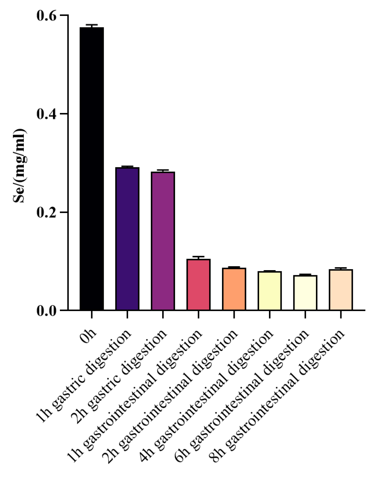
3.2.2. Changes in Selenium Content before and after WPH-Se In Vitro Digestion

The data showed that the selenium content in a solution (5 mg/mL) of WPH-Se was 0.577 mg/mL, corresponding to 11.54% of the total solution concentration (Figure 6). After gastric digestion, nearly half of the selenium in WPH-Se remains free, suggesting some degree of precipitation. The slightly acidic environment of the intestine, with pH 6.8, facilitates the dissolution and absorption of WPH-Se. Once precipitated in the gastric, WPH-Se is resolubilized in the intestine, facilitating the release and absorption of free selenium. Previous studies indicated that organic forms of selenium, typically bound to natural proteins or amino acids, are better absorbed and utilized by the human body compared to inorganic forms [66,67]. This suggests that WPH-Se may be beneficial for enhancing selenium absorption in the human body, potentially contributing to the development of effective selenium supplements.

4. Conclusions

This study rigorously investigated the antioxidant properties of WPH-Se under varying environmental conditions, including pH, temperature, and different stages of gastrointestinal digestion. It was found that WPH-Se retains high antioxidant activity over a broad temperature range, suggesting its potential for thermally processed foods without compromising their nutritional value. Furthermore, WPH-Se demonstrated high adaptability of its antioxidant capacity across various pH levels. Different stages of in vitro digestion revealed that WPH-Se maintained considerable stability during gastrointestinal digestion. Although there was a reduction in the DPPH scavenging rate, the rates for OH and ABTS scavenging remained elevated. Importantly, the maximum free radical scavenging rate of WPH-Se was higher post-intestinal fluid digestion compared to post-gastric fluid digestion, indicating not only preservation but also enhancement of its antioxidant function under intestinal conditions. This enhancement is essential for the effective antioxidant impact of WPH-Se in vivo. Additionally, a significant increase in the proportion of essential amino acids, particularly tryptophan (Try) and threonine (Thr), was observed during gastrointestinal digestion. The ability of WPH-Se to efficiently maintain and release bioavailable selenium after digestion further underscores its potential as a beneficial dietary supplement, enhancing selenium absorption in the human body. This comprehensive analysis confirms the versatility and effectiveness of WPH-Se as a robust antioxidant agent, suitable for various applications aimed at improving health through enhanced dietary antioxidant intake.

Author Contributions

Y.H. and X.C.: methodology and writing—original draft preparation. S.Y., M.Z., L.P. and X.S.: conceptualization and investigation. Z.W., A.L. and X.Y.: data curation and writing—review and editing. J.H.: supervision and project administration. All authors have read and agreed to the published version of the manuscript.

Funding

This research was supported by The Open Competition Research Projects of Xuchang University (2022JBGS05); Major Science and Technology Projects for Public Welfare of Henan Province: 201300110300; Zhongyuan Scholars of Henan Province in China: 19210151000; Zhongyuan Scholar Workstation Funded Project: ZYGZZ2021056, 224400510026; Central Government Guides the Local Science and Technology Development Special Fund: Z20221341069; Henan Provincial Key Science & Technology Special Project: 232102111060; Key Research and Development Project of Henan Province: 231111310700.

Data Availability Statement

The original contributions presented in the study are included in the article material, further inquiries can be directed to the corresponding author.

Acknowledgments

We are deeply grateful for the support and assistance provided by Lotus Holdings Co., Ltd., Shenlin Mei, Guoqing Zhang, and Jinfeng Jing in refining our Process Methodology.

Conflicts of Interest

The authors declare no conflicts of interest.

References

  1. Hong, H.; Fan, H.; Chalamaiah, M.; Wu, J. Preparation of low-molecular-weight, collagen hydrolysates (peptides): Current progress, challenges, and future perspectives. Food Chem. 2019, 301, 125222. [Google Scholar] [CrossRef] [PubMed]
  2. Fang, Y.; Pan, X.; Zhao, E.; Shi, Y.; Shen, X.; Wu, J.; Pei, F.; Hu, Q.; Qiu, W. Isolation and identification of immunomodulatory selenium-containing peptides from selenium-enriched rice protein hydrolysates. Food Chem. 2019, 275, 696–702. [Google Scholar] [CrossRef] [PubMed]
  3. Sasaki, C.; Tamura, S.; Tohse, R.; Fujita, S.; Kikuchi, M.; Asada, C.; Nakamura, Y. Isolation and identification of an angiotensin I-converting enzyme inhibitory peptide from pearl oyster (Pinctada fucata) shell protein hydrolysate. Process Biochem. 2019, 77, 137–142. [Google Scholar] [CrossRef]
  4. Stührwohldt, N.; Schaller, A. Regulation of plant peptide hormones and growth factors by post-translational modification. Plant Biol. 2019, 21 (Suppl. S1), 49–63. [Google Scholar] [CrossRef] [PubMed]
  5. de Veer, S.J.; Kan, M.W.; Craik, D.J. Cyclotides: From Structure to Function. Chem. Rev. 2019, 119, 12375–12421. [Google Scholar] [CrossRef] [PubMed]
  6. Tang, S.Y.; Wei, H.; Yu, C.Y. Peptide-functionalized delivery vehicles for enhanced cancer therapy. Int. J. Pharm. 2021, 593, 120141. [Google Scholar] [CrossRef] [PubMed]
  7. Wang, S.; Zhao, J.; Chen, L.; Zhou, Y.; Wu, J. Preparation, isolation and hypothermia protection activity of antifreeze peptides from shark skin collagen. LWT 2014, 55, 210–217. [Google Scholar] [CrossRef]
  8. Patil, S.P.; Goswami, A.; Kalia, K.; Kate, A.S. Plant-Derived Bioactive Peptides: A Treatment to Cure Diabetes. Int. J. Pept. Res. Ther. 2020, 26, 955–968. [Google Scholar] [CrossRef]
  9. Escudero, E.; Mora, L.; Toldra, F. Stability of ACE inhibitory ham peptides against heat treatment and in vitro digestion. Food Chem. 2014, 161, 305–311. [Google Scholar] [CrossRef]
  10. Reich, H.J.; Hondal, R.J. Why Nature Chose Selenium. ACS Chem. Biol. 2016, 11, 821–841. [Google Scholar] [CrossRef]
  11. Zhang, J.; Zhou, H.; Li, H.; Ying, Z.; Liu, X. Research progress on separation of selenoproteins/Se-enriched peptides and their physiological activities. Food Funct. 2021, 12, 1390–1401. [Google Scholar] [CrossRef] [PubMed]
  12. Yin, H.; Qi, Z.; Li, M.; Ahammed, G.J.; Chu, X.; Zhou, J. Selenium forms and methods of application differentially modulate plant growth, photosynthesis, stress tolerance, selenium content and speciation in Oryza sativa L. Ecotoxicol. Environ. Saf. 2019, 169, 911–917. [Google Scholar] [CrossRef] [PubMed]
  13. Zhou, H.; Wang, T.; Li, Q.; Li, D. Prevention of Keshan Disease by Selenium Supplementation: A Systematic Review and Meta-analysis. Biol. Trace Elem. Res. 2018, 186, 98–105. [Google Scholar] [CrossRef] [PubMed]
  14. Khanam, A.; Platel, K. Bioaccessibility of selenium, selenomethionine and selenocysteine from foods and influence of heat processing on the same. Food Chem. 2016, 194, 1293–1299. [Google Scholar] [CrossRef] [PubMed]
  15. Becker, D.J.; Reul, B.; Ozcelikay, A.T.; Buchet, J.P.; Henquin, J.C.; Brichard, S.M. Oral selenate improves glucose homeostasis and partly reverses abnormal expression of liver glycolytic and gluconeogenic enzymes in diabetic rats. Diabetologia 1996, 39, 3–11. [Google Scholar] [CrossRef] [PubMed]
  16. Sheng, X.Q.; Huang, K.X.; Xu, H.B. New experimental observation on the relationship of selenium and diabetes mellitus. Biol. Trace Elem. Res. 2004, 99, 241–253. [Google Scholar] [CrossRef] [PubMed]
  17. Xu, Z.; Fang, Y.; Chen, Y.; Yang, W.; Ma, N.; Pei, F.; Kimatu, B.M.; Hu, Q.; Qiu, W. Protective effects of Se-containing protein hydrolysates from Se-enriched rice against Pb2+-induced cytotoxicity in PC12 and RAW264.7 cells. Food Chem. 2016, 202, 396–403. [Google Scholar] [CrossRef] [PubMed]
  18. Rodríguez-Meizoso, I.; Jaime, L.; Santoyo, S.; Cifuentes, A.; García-Blairsy Reina, G.; Señoráns, F.J.; Ibáñez, E. Pressurized fluid extraction of bioactive compounds from Phormidium species. J. Agric. Food Chem. 2008, 28, 3517–3523. [Google Scholar] [CrossRef] [PubMed]
  19. Fukumoto, Y.; Matsuhashi, K.; Tanaka, Y.-K.; Suzuki, N.; Ogra, Y. Band 3/anion exchanger 1/solute carrier family 4 member 1 expression as determinant of cellular sensitivity to selenite exposure. Biochem. Biophys. Rep. 2022, 29, 101223. [Google Scholar] [CrossRef]
  20. Spallholz, J.E.; Hoffman, D.J. Selenium toxicity: Cause and effects in aquatic birds. Aquat. Toxicol. 2002, 57, 27–37. [Google Scholar] [CrossRef]
  21. Chen, N.; Yao, P.; Zhang, W.; Zhang, Y.T.; Xin, N.C.; Wei, H.D.; Zhang, T.H.; Zhao, C.H. Selenium nanoparticles: Enhanced nutrition and beyond. Crit. Rev. Food Sci. Nutr. 2023, 63, 12360–12371. [Google Scholar] [CrossRef] [PubMed]
  22. Xiong, Y.; Chen, Z.H.; Zhang, F.L.; Yu, Z.Y.; Liu, B.; Zhang, C.; Zhao, L.N. A specific selenium-chelating peptide isolated from the protein hydrolysate of Grifola frondosa. RSC Adv. 2021, 11, 10272–10284. [Google Scholar] [CrossRef] [PubMed]
  23. Li, X.; Wang, X.; Liu, G.; Xu, Y.; Wu, X.; Yi, R.; Jin, F.; Sa, C.; Su, X. Antioxidant stress and anticancer activity of peptide-chelated selenium in vitro. Int. J. Mol. Med. 2021, 48, 153. [Google Scholar] [CrossRef] [PubMed]
  24. Zhu, S.; Zheng, Y.; He, S.; Su, D.; Nag, A.; Zeng, Q.; Yuan, Y. Novel Zn-Binding Peptide Isolated from Soy Protein Hydrolysates: Purification, Structure, and Digestion. J. Agric. Food Chem. 2021, 69, 483–490. [Google Scholar] [CrossRef] [PubMed]
  25. Jiang, S.; Dong, W.; Zhang, Z.; Xu, J.; Li, H.; Zhang, J.; Dai, L.; Wang, S. A new iron supplement: The chelate of pig skin collagen peptide and Fe2+ can treat iron-deficiency anemia by modulating intestinal flora. Front. Nutr. 2022, 9, 1055725. [Google Scholar] [CrossRef] [PubMed]
  26. Lee, V.J.; Janisse, S.E.; Heffern, M.C. Plant-derived chelators and ionophores as potential therapeutics for metabolic diseases. Chem. Soc. Rev. 2023, 52, 3927–3945. [Google Scholar] [CrossRef] [PubMed]
  27. Udechukwu, M.C.; Collins, S.A.; Udenigwe, C.C. Prospects of enhancing dietary zinc bioavailability with food-derived zinc-chelating peptides. Food Funct. 2016, 7, 4137–4144. [Google Scholar] [CrossRef] [PubMed]
  28. Yang, S.R.; Hou, Y.C.; Huang, J.H.; Zhang, Q.S.; Liao, A.M.; Shen, X.L.; Yuan, X.Q.; Lu, M.X. Preparation, structural characterization and functional properties of a novel selenium chelating peptide derived from the hydrolyzate of wheat protein. Int. J. Food Prop. 2023, 26, 3241–3260. [Google Scholar] [CrossRef]
  29. Gallego, M.; Mora, L.; Reig, M.; Toldrá, F. Stability of the potent antioxidant peptide SNAAC identified from Spanish dry-cured ham. Food Res. Int. 2018, 105, 873–879. [Google Scholar] [CrossRef]
  30. Wong, F.C.; Xiao, J.B.; Ong, M.G.L.; Pang, M.J.; Wong, S.J.; Teh, L.K.; Chai, T.T. Identification and characterization of antioxidant peptides from hydrolysate of blue-spotted stingray and their stability against thermal, pH and simulated gastrointestinal digestion treatments. Food Chem. 2019, 271, 614–622. [Google Scholar] [CrossRef]
  31. Udenigwe, C.C.; Fogliano, V. Food matrix interaction and bioavailability of bioactive peptides: Two faces of the same coin? J. Funct. Foods 2017, 35, 9–12. [Google Scholar] [CrossRef]
  32. Liu, K.; Du, R.; Chen, F. Stability of the antioxidant peptide SeMet-Pro-Ser identified from selenized brown rice protein hydrolysates. Food Chem. 2020, 319, 126540. [Google Scholar] [CrossRef] [PubMed]
  33. Roman, M.; Jitaru, P.; Barbante, C. Selenium biochemistry and its role for human health. Metallomics 2014, 6, 25–54. [Google Scholar] [CrossRef]
  34. Saidi, S.; Deratani, A.; Belleville, M.P.; Amar, R.B. Production and fractionation of tuna by-product protein hydrolysate by ultrafiltration and nanofiltration: Impact on interesting peptides fractions and nutritional properties. Food Res. Int. 2014, 65, 453–461. [Google Scholar] [CrossRef]
  35. Feng, Y.Z.; Wu, W.Y.; Chen, T.; Huang, M.T.; Zhao, M.M. Exploring the core functional microbiota related with flavor compounds in fermented soy sauce from different sources. Food Res. Int. 2023, 173, 113456. [Google Scholar] [CrossRef]
  36. Ren, Y.; Wu, H.; Li, X.; Lai, F.; Zhao, G.; Xiao, X. A Two-Step, One-Pot Enzymatic Method for Preparation of Duck Egg White Protein Hydrolysates with High Antioxidant Activity. Appl. Biochem. Biotechnol. 2014, 172, 1227–1240. [Google Scholar] [CrossRef] [PubMed]
  37. Homayouni-Tabrizi, M.; Asoodeh, A.; Soltani, M. Cytotoxic and antioxidant capacity of camel milk peptides: Effects of isolated peptide on superoxide dismutase and catalase gene expression. J. Food Drug Anal. 2017, 25, 567. [Google Scholar] [CrossRef] [PubMed]
  38. Yuan, B.; Yang, X.Q.; Kou, M.; Lu, C.Y.; Wang, Y.Y.; Peng, J.; Chen, P.; Jiang, J.H. Selenylation of Polysaccharide from the Sweet Potato and Evaluation of Antioxidant, Antitumor, and Antidiabetic Activities. J. Agric. Food Chem. 2017, 65, 605. [Google Scholar] [CrossRef] [PubMed]
  39. Siddhuraju, P.; Mohan, P.S.; Becker, K. Studies on the antioxidant activity of Indian Laburnum (Cassia fistula L.): A preliminary assessment of crude extracts from stem bark, leaves, flowers and fruit pulp. Food Chem. 2002, 79, 61–67. [Google Scholar] [CrossRef]
  40. Minekus, M.; Smeets-Peeters, M.; Bernalier, A.; Marol-Bonnin, S.; Havenaar, R.; Marteau, P.; Alric, M.; Fonty, G.; Huis in’t Veld, J.H. A computer-controlled system to simulate conditions of the large intestine with peristaltic mixing, water absorption and absorption of fermentation products. Appl. Biochem. Biotechnol. 1999, 53, 108–114. [Google Scholar] [CrossRef]
  41. Minekus, M.; Marteau, P.; Havenaar, R.; Huis in ‘t Veld, J.H.H. multicompartmental dynamic computer-controlled model simulating the stomach and small intestine. Altern. Lab. Anim. 1995, 23, 197–209. [Google Scholar] [CrossRef]
  42. Oded, S.; Amit, Y. Collagen Structures and Method of Fabricating the Same. EP2640877A2, 25 September 2013. [Google Scholar]
  43. Minekus, M.; Alminger, M.; Alvito, P.; Ballance, S.; Brodkorb, A. A standardised static in-vitro digestion method suitable for food—An international consensus. Food Funct. 2014, 5, 1113–1124. [Google Scholar] [CrossRef] [PubMed]
  44. Kou, X.; Gao, J.; Xue, Z.; Zhang, Z.; Wang, H.; Wang, X. Purification and identification of antioxidant peptides from chickpea (Cicer arietinum L.) albumin hydrolysates. LWT Food Sci. Technol. 2013, 50, 591–598. [Google Scholar] [CrossRef]
  45. Nongonierma, A.B.; FitzGerald, R.J. Bioactive properties of milk proteins in humans: A review. Peptides 2015, 73, 20–34. [Google Scholar] [CrossRef] [PubMed]
  46. Fan, L.; Mao, X.; Wu, Q. Purification, Identification and Molecular Docking of Novel Antioxidant Peptides from Walnut (Juglans regia L.) Protein Hydrolysates. Molecules 2022, 27, 8423. [Google Scholar] [CrossRef]
  47. Li, H.; Aluko, R.E. Identification and inhibitory properties of multifunctional peptides from pea protein hydrolysate. J. Agric. Food Chem. 2010, 58, 11471–11476. [Google Scholar] [CrossRef] [PubMed]
  48. Ng, Z.X.; Kuppusamy, U.R. Effects of different heat treatments on the antioxidant activity and ascorbic acid content of bitter melon, Momordica charantia. Braz. J. Food Technol. 2019, 22, e2018283. [Google Scholar] [CrossRef]
  49. Arnal, M.; Gallego, M.; Talens, P.; Mora, L. Impact of thermal treatments and simulated gastrointestinal digestion on the α-amylase inhibitory activity of different legumes. Food Chem. 2023, 418, 135884. [Google Scholar] [CrossRef]
  50. Sun, X.M.; Wang, L.; Wang, S.; Tan, Y.X.; Zhu, Y.J.; Wang, Q.Y.; Tao, W.B.; Qin, L.X. Effects of thermal treatments on physicochemical properties and peptide profiles of infant formula protein model based on peptidomics. Int. J. Dairy Technol. 2024; Epub ahead of printing. [Google Scholar] [CrossRef]
  51. Singh, B.P.; Vij, S. In vitro stability of bioactive peptides derived from fermented soy milk against heat treatment, pH and gastrointestinal enzymes. LWT Food Sci. Technol. 2018, 91, 303–307. [Google Scholar] [CrossRef]
  52. Asaduzzaman, A.K.M.; Chun, B.S. Recovery of functional materials with thermally stable antioxidative properties in squid muscle hydrolyzates by subcritical water. J. Food Sci. Technol. 2015, 52, 793–802. [Google Scholar] [CrossRef]
  53. Jang, H.L.; Liceaga, A.M.; Yoon, K.Y. Purification, characterisation and stability of an antioxidant peptide derived from sandfish (Arctoscopus japonicus) protein hydrolysates. J. Funct. Foods 2016, 20, 433–442. [Google Scholar] [CrossRef]
  54. Lin, S.Y.; Wang, C.C.; Lu, Y.L.; Wu, W.C.; Hou, W.C. Antioxidant, anti-semicarbazide-sensitive amine oxidase, and anti-hypertensive activities of geraniin isolated from Phyllanthus urinaria. Food Chem. Toxicol. 2008, 46, 2485–2492. [Google Scholar] [CrossRef] [PubMed]
  55. Liu, Y.Y.; Wan, S.Y.; Liu, J.; Zou, Y.X.; Liao, S.T. Antioxidant Activity and Stability Study of Peptides from Enzymatically Hydrolyzed Male Silkmoth. J. Food Process. Preserv. 2017, 41, e13081. [Google Scholar] [CrossRef]
  56. Zheng, L.; Lin, L.Z.; Su, G.W.; Zhao, Q.Z.; Zhao, M.M. Pitfalls of using 1,1-diphenyl-2-picrylhydrazyl (DPPH) assay to assess the radical scavenging activity of peptides: Its susceptibility to interference and low reactivity towards peptides. Food Res. Int. 2015, 76, 359–365. [Google Scholar] [CrossRef] [PubMed]
  57. Xu, Y.-L.; Ye, J.; Yu, H.; Su, L.-J. Optimization of the Enzymatic Hydrolysis Process and Analysis of the Antioxidant Activity of Polypeptide from Apostichopus japonicus Gonads. Sci. Technol. Food Ind. 2018, 39, 181–187. [Google Scholar]
  58. Zheng, L.; Zhao, M.M.; Xiao, C.Q.; Zhao, Q.Z.; Su, G.W. Practical problems when using ABTS assay to assess the radical-scavenging activity of peptides: Importance of controlling reaction pH and time. Food Chem. 2016, 192, 288–294. [Google Scholar] [CrossRef] [PubMed]
  59. Pham-Huy, L.A.; He, H.; Pham-Huy, C. Free radicals, antioxidants in disease and health. Int. J. Biomed. Sci. 2008, 4, 89–96. [Google Scholar] [CrossRef]
  60. Ma, J.; Zeng, X.; Zhou, M.; Cheng, L.; Ren, D. Inhibitory effect of low-molecular-weight peptides (0–3 kDa) from Spirulina platensis on H2O2-induced oxidative damage in L02 human liver cells. Bioresour. Bioprocess. 2021, 8, 36. [Google Scholar] [CrossRef]
  61. Zhang, J.; Wu, S.; Wang, Q.; Yuan, Q.; Li, Y.; Reboredo-Rodríguez, P.; Varela-López, A.; He, Z.; Wu, F.; Hu, H.; et al. Oxidative Stress Amelioration of Novel Peptides Extracted from Enzymatic Hydrolysates of Chinese Pecan Cake. Int. J. Mol. Sci. 2022, 23, 12086. [Google Scholar] [CrossRef]
  62. Moran; Edwin, T. Gastric digestion of protein through pancreozyme action optimizes intestinal forms for absorption, mucin formation and villus integrity. Anim. Feed. Sci. Technol. 2016, 221, 284–303. [Google Scholar] [CrossRef]
  63. Huang, Q.; Yu, H.; Ru, Q. Bioavailability and Delivery of Nutraceuticals Using Nanotechnology. J. Food Sci. 2010, 75, R50–R57. [Google Scholar] [CrossRef]
  64. Goodwin, D. Lipid and Carbohydrate-Based Systems to Enhance the Bioavailability and Immunogenicity of Therapeutic Peptides. Ph.D. Thesis, School of Chemistry and Molecular Biosciences, The University of Queensland, St Lucia, Australia, 2013. [Google Scholar]
  65. Aguilar-Toalá, J.E.; Quintanar-Guerrero, D.; Liceaga, A.M.; Zambrano-Zaragoza, M.L. Encapsulation of bioactive peptides: A strategy to improve the stability, protect the nutraceutical bioactivity and support their food applications. RSC Adv. 2022, 12, 6449–6458. [Google Scholar] [CrossRef] [PubMed]
  66. Takahashi, K.; Suzuki, N.; Ogra, Y. Bioavailability Comparison of Nine Bioselenocompounds In Vitro and In Vivo. Int. J. Mol. Sci. 2017, 18, 506. [Google Scholar] [CrossRef] [PubMed]
  67. Fairweather-Tait, S.J.; Collings, R.; Hurst, R. Selenium bioavailability: Current knowledge and future research requirements. Am. J. Clin. Nutr. 2010, 91, 1484S–1491S. [Google Scholar] [CrossRef] [PubMed]
Figure 1. Effect of different temperatures on antioxidant activity of WPH-Se. (A) Effect of temperature on the scavenging rate of DPPH radicals. (B) Effect of temperature on OH radical scavenging rate. (C) Effect of temperature on the reducing capacity of Fe3+. (D) Effect of temperature on the scavenging rate of ABTS radicals. Note: Different letters indicate a significant difference (p < 0.05).
Foods 13 01819 g001
Figure 2. Effect of different pH on antioxidant activity of WPH-Se. (A) Effect of pH on the scavenging rate of DPPH radicals. (B) Effect of pH on OH radical scavenging rate. (C) Effect of pH on the reducing capacity of Fe3+. (D) Effect of pH on the scavenging rate of ABTS radicals. Note: Different letters indicate a significant difference (p < 0.05).
Foods 13 01819 g002
Figure 3. Effect of gastric digestion on antioxidant activity of WPH-SE in vitro. (A) Changes in DPPH radical scavenging activity in simulated gastric digestion of samples. (B) Changes in OH radical scavenging activity in simulated gastric digestion of samples. (C) Changes in reducing capacity of Fe3+ in simulated gastric digestion of samples. (D) Changes in ABTS radical scavenging activity in simulated gastric digestion of samples. Note: Different letters indicate a significant difference (p < 0.05).
Foods 13 01819 g003
Figure 4. Effect of intestinal digestion on antioxidant activity of WPH-SE in vitro. (A) Changes in DPPH radical scavenging activity in simulated intestinal digestion of samples. (B) Changes in OH radical scavenging activity in simulated intestinal digestion of samples. (C) Changes in reducing capacity of Fe3+ in simulated intestinal digestion of samples. (D) Changes in ABTS radical scavenging activity in simulated intestinal digestion of samples. Note: Different letters indicate a significant difference (p < 0.05).
Foods 13 01819 g004
Figure 5. Effect of gastrointestinal digestion on the antioxidant activity of WPH-SE in vitro. (A) Changes in DPPH radical scavenging activity in simulated gastrointestinal digestion of samples. (B) Changes in OH radical scavenging activity in simulated gastrointestinal digestion of samples. (C) Changes in reducing capacity of Fe3+ in simulated gastrointestinal digestion of samples. (D) Changes in ABTS radical scavenging activity in simulated gastrointestinal digestion of samples. Note: Different letters indicate a significant difference (p < 0.05).
Foods 13 01819 g005
Figure 6. Changes in free selenium during gastrointestinal digestion in vitro.
Foods 13 01819 g006
Table 1. Changes in free amino acids after intestinal digestion of WPH-Se.
Amino Acid NameWPH-Se (%)Gastric WPH-Se (%)Intestinal WPH-Se (%)
Aspartate6.34.93.4
Threonine1.22.52.5
Serine2.75.53.2
Glutamate20.812.57.0
Alanine 2.4
Glycine8.54.78.7
Cysteine 4.53.6
Valine 7.65.8
Leucine 4.9
Phenylalanine 5.1
Histidine42.341.442.5
Tryptophan 1.2
Lysine3.23.16.7
Arginine14.95.910.4
Acidic amino acids27.117.410.4
Alkaline amino acids60.450.459.6
Essential amino acids46.759.563.8
Hydrophobic amino acids8.519.620.8
Aromatic amino acids006.3
Disclaimer/Publisher’s Note: The statements, opinions and data contained in all publications are solely those of the individual author(s) and contributor(s) and not of MDPI and/or the editor(s). MDPI and/or the editor(s) disclaim responsibility for any injury to people or property resulting from any ideas, methods, instructions or products referred to in the content.

