Article

Screening Probiotics for Anti-Helicobacter pylori and Investigating the Effect of Probiotics on Patients with Helicobacter pylori Infection

1
School of Pharmacy, Lanzhou University, Lanzhou 730000, China
2
School of Life Sciences, Lanzhou University, Lanzhou 730000, China
3
Key Laboratory of Cell Activities and Stress Adaptations, Ministry of Education, Lanzhou University, Lanzhou 730000, China
4
Gansu Key Laboratory of Biomonitoring and Bioremediation for Environmental Pollution, Lanzhou University, Lanzhou 730000, China
*
Author to whom correspondence should be addressed.
Foods 2024, 13(12), 1851; https://doi.org/10.3390/foods13121851
Submission received: 25 April 2024 / Revised: 21 May 2024 / Accepted: 29 May 2024 / Published: 13 June 2024
(This article belongs to the Section Food Nutrition)

Abstract

:
Probiotics are natural microbial agents with beneficial properties such as bacteriostatic and anti-infective properties. Lactobacillus plantarum Q21, Q25 and QA85, were isolated from the Chinese specialty fermented food “Jiangshui” and proved to be highly resistant to Helicobacter pylori (p < 0.0001). In vitro results showed that Q21, Q25 and QA85 strongly inhibited H. pylori and could specifically co-aggregate H. pylori in vitro (more than 56%). Strains have the potential to adhere to cells and hinder H. pylori colonization (p < 0.0001). To assess the anti-H. pylori efficacy of strains in vivo, volunteers were recruited and a self-controlled study of probiotic intervention was conducted. Compared to pre-probiotics, volunteers who took Q21, Q25 and QA85 for 1 month showed significant improvement in discomfort, a significant reduction in GSRS scores (p < 0.05), and modulation of inflammatory response (p < 0.05). Q21, Q25 and QA85 resulted in a decreasing trend of H. pylori load in volunteers (454.30 ± 327.00 vs. 328.35 ± 237.19, p = 0.06). However, the strains were not significantly effective in modulating the imbalance of the gut microbiota caused by H. pylori infection. In addition, strains affect metabolic pathways by increasing the levels of O-Phosphoethanolamine and other related metabolites, which may ameliorate associated symptoms. Therefore, Lactobacillus plantarum Q21, Q25 and QA85 can be regarded as a candidate probiotic preparation that exerts direct or indirect anti-H. pylori effects by inhibiting H. pylori activity and colonization, reducing inflammation and discomfort, maintaining homeostasis in the internal environment, affecting the metabolic pathways and repairing the body barrier. They can play a role in relieving H. pylori infection.

Graphical Abstract

1. Introduction

As a common human pathogen, Helicobacter pylori infects over 50% of the global population [1]. In some regions of Latin America, Africa, and Asia, the adult infection rate even exceeds 75% [2,3]. Despite lower infection rates in children, it still reaches as high as 32.3% globally, showing a significant negative correlation with income levels among countries or regions [4]. Given the potential health risks associated with H. pylori infection, including chronic progressive gastritis, peptic ulcer disease, gastric atrophy, and gastric epithelial dysplasia, as well as the eventual progression to mucosa-associated lymphoid tissue lymphoma or gastric cancer [5], timely detection and intervention for H. pylori are necessary. Currently, standard triple or quadruple therapy is commonly employed clinically for H. pylori infection, which includes two antibiotics and a proton pump inhibitor (PPI), with quadruple therapy adding bismuth salts to this regimen. Although the above methods are effective in eradicating H. pylori, they still face many challenges and shortcomings, such as the problem of antibiotic resistance rate [6], the safety of bismuth agents [7], the limitations of some of the PPI formulations, and patient compliance [8], among others. Therefore, there is still a need for further expansion of treatment methods for H. pylori infection.
Probiotics are living microorganisms that can play a beneficial role in the host’s health by ingesting a certain amount [9], primarily including lactobacilli, bifidobacteria, certain streptococci and yeasts [10]. Probiotics have the ability to inhibit the growth of pathogenic bacteria, regulate the microecological balance within the body, and treat diseases caused by pathogenic bacteria, such as acute diarrhea infections and colitis [11]. Additionally, probiotics contribute to enhanced digestion and absorption, as well as the regulation of the immune system, all without adverse effects [12]. Currently, a variety of probiotic preparations have been clinically applied in the treatment of diseases caused by gastrointestinal pathogenic bacterial infections [13,14]. Several studies have demonstrated the effectiveness of various probiotics in the treatment of diseases resulting from H. pylori infections, such as L. acidophilus [15,16], L. rhamnosus GG [17], L. casei [18,19], L. gasseri OLL2716 [20], L. reuteri [21], L. plantarum [22,23], Bifidobacterium [24,25], and others. Previous studies have shown that probiotics inhibit H. pylori by producing substances that inhibit H. pylori urease [26,27], blocking the adhesion of H. pylori to gastrointestinal epithelial cells, and inhibiting the inflammatory response after H. pylori infection [28], among other mechanisms.
Probiotic supplementation in the treatment of H. pylori may increase eradication rates and may reduce the incidence of adverse effects [29,30]. However, there is still no definitive conclusion on whether probiotics can reduce the load of bacteria. Some studies have shown that taking probiotics can reduce the bacterial load of H. pylori and lessen the severity of gastritis [31]. Probiotics are also used to treat diseases that can lead to disturbances in the gut microbiota [32]. However, the regulatory effects of probiotics on the gut microbiota [33] and their influence on fecal metabolites remain unclear.
One of the main sources of isolated Lactobacillus is fermented foods, and Jiangshui is one of the specialty fermented foods in Northwest China, which is very popular among the public. Additionally, some studies have found that Lactobacillus is the dominant species in the microbial composition of Jiangshui [34]. Thus, Jiangshui can be used as an important source for isolation of Lactobacillus. Early studies related to probiotics (e.g., Bifidobacterium, L. acidophilus, and L. casei) on H. pylori focused on the inhibition of H. pylori by probiotics [15,18,19,24], or the effect of probiotics combined with standard therapy on patient eradication rates. Therefore, in this study, we proposed to screen several strains of Lactobacillus plantarum isolated from Jiangshui, a specialty fermented food in Northwest China, with strong anti-H. pylori ability. While focusing on the inhibitory effect of probiotics on H. pylori, the efficacy of probiotics in alleviating the symptoms associated with H. pylori infection in volunteers by administration of probiotics alone was explored, with attention to their role in influencing the gut microbiota and fecal metabolites.

2. Materials and Methods

2.1. Bacterial Strains and Culture Conditions

From more than 100 strains independently isolated in the laboratory, seven strains with strong bacterial inhibitory ability were screened: ZCJ, TSL-6, QX(A)-4, GL-5, Q21, QA85, and Q25. These seven strains were identified as Lactobacillus plantarum by sequencing, and the conservation information is shown in Table S1. Seven strains and one control strain Lactobacillus rhamnosus GG (LGG) were stored at −80 °C in MRS broth containing 20% glycerol (v/v) until tested. LGG is a widely used commercial probiotic strain and hence was used in this study. LGG was stored in a lab at Lanzhou University (China). The strains were cultured in MRS broth at 37 °C for 24 h to activate Lactobacillus, repeated twice and then subjected to each experiment. H. pylori J99 was stored in the Lanzhou University Microbiological Culture Collection and stored at −80 °C in H. Pylori Medium (fluids) containing 20% glycerol (v/v) until tested. H. pylori was grown on Columbia blood agar plates containing 5% (v/v) off fiber sheep blood (Qingdao Hope Bio-Technology Co., Ltd., Qingdao, China) at 37 °C in microaerophilic conditions (5% O2, 10% CO2, and 85% N2).

2.2. Cell Culture

AGS cells (human gastric adenocarcinoma epithelial cells, ATCC CRL 1739) were purchased from Procell Life Science and Technology Co., Ltd. (Wuhan, China). AGS cells were maintained in RPMI 1640 medium supplemented with 15% fetal bovine serum (FBS) (Gemini Fetal Bovine Serum, Gemini Biotechnology Company, West Sacramento, CA, USA), penicillin (100 IU/mL), and streptomycin (100 μg/mL) at 37 °C with 5% CO2.

2.3. Screening for Strains with H. pylori Inhibition Ability

2.3.1. Acid Tolerance of Lactobacillus

After activation of Lactobacillus, the supernatant was discarded by centrifugation. The bacterial precipitate was washed three times with phosphate-buffered saline (PBS) and resuspended to a viable bacterial count of 108 CFU/mL. Lactobacillus suspension with a viable count of 108 CFU/mL was inoculated into simulated gastric juice (0.2% NaCl, 0.35% pepsin, dissolved in distilled water and adjusted to pH 3.0 with HCl; filtered through a 0.22 μm membrane), and sampled at 0 and 3 h of the test, respectively. Counts of viable bacteria were performed using the MRS agar plates method and the survival rate of several Lactobacillus strains in simulated gastric juice after 3 h was calculated separately [35].

2.3.2. Hydrophobic Properties of Lactobacillus

The hydrophobicity of the bacterial surface was determined according to the method of Kos et al. [36]. After the activation of Lactobacillus, the bacteria were washed and resuspended with PBS to give a viable bacterial count of 108 CFU/mL. Then, 3 mL of Lactobacillus suspension and 1 mL of xylene were thoroughly mixed, shaken thoroughly for 5 min, allowed to stand at room temperature, and the absorbance values of the aqueous phase were measured after 0 h and 1 h to calculate the hydrophobicity of Lactobacillus.

2.3.3. Aggregation Properties of Lactobacillus

Autoaggregation properties of Lactobacillus: After activation, the Lactobacillus was washed and resuspended with PBS, so that the number of viable bacteria was 108 CFU/mL. The mixture was incubated at 37 °C, and the absorbance value at 600 nm of the upper layer liquid was measured within 24 h to calculate the autoaggregation rate of Lactobacillus [36,37].
Coaggregation properties of Lactobacillus with H. pylori: After the activation of Lactobacillus, it was washed and resuspended with PBS, so that the number of viable bacteria was 108 CFU/mL. The treatment of H. pylori was the same as the operation described above. Then, 5 mL of Lactobacillus suspension and 5 mL of H. pylori suspension were thoroughly mixed, and we set the mixture at 37 °C for static incubation. Test the absorbance value at 600 nm in the upper layer liquid within 24 h. Calculate the coaggregation rate of the interaction between the Lactobacillus and H. pylori [36,37].

2.3.4. Anti-H. pylori Activity of Lactobacillus

Anti-H. pylori activity of Lactobacillus was determined by the Oxford cup diffusion method. First, 200 μL of H. pylori suspension was evenly spread on Columbia blood agar plates without antibiotics. After spreading, 4 sterilized Oxford cups were placed on the plates, and we added 200 μL of Lactobacillus suspension, Lactobacillus supernatant, amoxicillin solution at a concentration of 0.06 μg/mL (MIC), and a blank MRS broth to Oxford cups, respectively. The plates were incubated at 37 °C under a microaerobic environment for 72 h. At the end of incubation, the diameter of the inhibition circle was measured by vernier caliper [18,38].

2.3.5. Urease Activity Assay

To analyze the inhibitory effects of Lactobacillus against H. pylori urease, 40 μL of H. pylori suspension was mixed with 10 μL of Lactobacillus supernatant, and 10 μL of sterile H. pylori liquid medium was used as control. The mixture was added to a clean and sterile 96-well plate and incubated at 37 °C in a microaerobic environment for 48 h. The incubated mixture was taken out, and 150 μL of urease reagent (20% urea, 0.012% phenol red, dissolved in PBS and adjusted to pH 6.5 with HCl) was added into each well, and then the color change was observed and the OD550nm value was measured [18].

2.4. Co-Culture Assay of Lactobacillus with H. pylori

Activated H. pylori were resuspended in fresh H. pylori liquid medium, and we added 10% Lactobacillus suspension (108 CFU/mL) or supernatant. It was co-cultured for a certain period of time and H. pylori viable cells were counted using their selective medium. At the same time, urease activity was measured.

2.5. Anti-H. pylori Effects of Lactobacillus at the Cell Level

2.5.1. Adhesion Assay of Lactobacillus to AGS Cells

To determine the adhesion capacity of Lactobacillus, Lactobacillus were centrifuged to collect the bacterial bodies and washed with PBS, followed were resuspended with antibiotic-free RPMI 1640 culture medium and adjusting the concentration of Lactobacillus to 106, 107, 108, 109, and 1010 CFU/mL, respectively, and set aside. Cell spreading was performed by adding 1 mL of 100 × 104/mL cell solution per well in a six-well plate, and the cells were cultured to form a monolayer by adhering to the wall. Subsequently, 1 mL Lactobacillus suspension of different concentrations was added to each well, and the experiments were carried out by co-culturing for 4 h at 37 °C. In the end, unadhered bacteria were washed away and the mixture was collected in a centrifuge tube, diluted stepwise and spread on MRS agar plates for counting. The percentage of bacterial adherence to cells was calculated by the following formula: adherence rate (%) = (adherent bacteria)/(total bacteria) × 100. Cell adhesion assay was conducted in biological triplicate to ensure reproducibility.
In order to determine the effect of co-culture duration on adhesion, Lactobacillus was adjusted to 108 CFU/mL, and co-cultured with AGS cells (100 × 104 cells) for 1, 2, 4, and 6 h, respectively. Other operations were the same as above.

2.5.2. Inhibition Assay of H. pylori Adhesion to AGS Cells by Lactobacillus

Lactobacillus at a concentration of 108 CFU/mL was resuspended in an antibiotic-free RPMI 1640 culture medium and set aside. H. pylori was similarly manipulated. Six-well plates were used for AGS cell spreading (100 × 104 cells), and cells were cultured to form monolayers for experiments. To determine the inhibitory effect of Lactobacillus on H. pylori adhesion to AGS cells, it was validated in four sets of experiments. Cells were infected with H. pylori (MOI = 100) and incubated at 37 °C for 4 h as an infected control. After co-culturing the cells with H. pylori (MOI = 100) for 2 h, unadhered bacteria were washed away with PBS, and then Lactobacillus (MOI = 100) was added to co-culture with the cells for 2 h as a displacement experimental group. After co-culturing the cells with Lactobacillus (MOI = 100) for 2 h, unadhered bacteria were washed away with PBS, and then H. pylori (MOI = 100) was added and co-cultured with the cells for 2 h as the exclusion experimental group. The cells were co-cultured with Lactobacillus (MOI = 100), H. pylori (MOI = 100) for 4 h as a competition experimental group. In the end, unadhered bacteria were washed away and the mixture was collected in a centrifuge tube, and H. pylori selective medium was used for counting. The relative percentage of H. pylori adhesion to cells was calculated according to the following formula: relative adhesion (%) = (number of adherent bacteria in the experimental group)/(number of adherent bacteria in the control group) × 100. The cell adhesion assay was performed using the biological triplex method to ensure reproducibility.

2.5.3. Total RNA Extraction and Reverse-Transcription Quantitative PCR (RT-qPCR)

To study the anti-inflammatory effect of Lactobacillus, it was verified in four groups of experiments. AGS cells (100 × 104 cells) were cultured normally for 4 h as a blank control group. AGS cells were treated with H. pylori (MOI = 100) for 4 h as the H. pylori-infected group. After co-culturing the cells with Lactobacillus (MOI = 100) for 2 h, the unadhered bacteria were washed away with PBS, and then H. pylori (MOI = 100) was added and co-cultured with the cells for 2 h as the exclusion experimental group. The cells were co-cultured with Lactobacillus (MOI = 100) and H. pylori (MOI = 100) for 4 h as the competition experimental group.
Total mRNA was extracted with SevenFast® Total RNA Extraction Kit for Cells (Seven Innovation (Beijing) Biotech Co., Beijing, China). cDNA synthesis was performed with SevenClever ™ First Strand cDNA Synthesis Kit (with dsDNase) (Seven Innovation (Beijing) Biotech Co., Beijing, China). RT-qPCR was performed in qPCR 96-well plates on an Mx3000/Mx3005P system (Agilent, Santa Clara, CA, USA) and 2× SYBR Green qPCR MasterMix II (Universal) (Seven Innovation (Beijing) Biotech Co., Beijing, China) to analyze the TNF-α, IL-6, IL-8 and IL-10 mRNA expressions. The oligonucleotide sequence of primers used for RT-qPCR [39,40,41] is listed in Table 1.

2.5.4. Cytotoxicity Assay on AGS Cells

The cytotoxicity effect of Lactobacillus on AGS cells was determined by MTT assay. Firstly, AGS cells (104 cells) were grown and allowed to adhere to a 96-well plate at 37 °C for 12–24 h to approximately 80% confluence and then cells were treated at 37 °C in a 5% CO2 atmosphere with Lactobacillus (MOI = 100) for 6 h. Then 20 μL of MTT solution (5 mg/mL) was added to each well, and the cells were incubated for 4 h at 37 °C. Finally, 150 μL of DMSO was added to each well and we allowed the color to develop. The absorbance was measured at 490 nm. Cell viability (%) = (Asample/Acontrol) × 100, where Asample is the absorbance of the cells that were incubated with the medium containing Lactobacillus, and Acontrol is the absorbance of the cells alone.

2.6. Antibiotic Susceptibility Assay

The susceptibility of Lactobacillus to antibiotics was determined by the disc diffusion method [42]. The concentration of Lactobacillus was adjusted to 108 CFU/mL, and the bacterial suspension was evenly spread on MRS agar plates and left to dry for 5 min. Use tweezers to stick the antibiotic discs (Microbial Reagent Co., Ltd., Hangzhou, China) onto the surface of the inoculated plates, and incubate in an inverted at 37 °C for 24 h, with the quality control bacteria as a control. The diameter of the inhibition circle was measured and recorded by vernier caliper. Three replicates were set up for each antibiotic and the results were averaged. The standard strains of Staphylococcus aureus ATCC 25923 (S. aureus) and Escherichia coli ATCC 25922 (E. coli) were used as the control bacteria, operating as above. The inhibition zone after incubation was measured and interpreted as susceptible, intermediate, or resistant according to the instructions for the antibiotic discs (Microbial Reagent Co., Ltd., Hangzhou, China).

2.7. Effects of Lactobacillus on Patients with H. pylori Infection

2.7.1. Participating Volunteers

Volunteer selection such as inclusion and exclusion criteria were referred to the experimental design of Chenghai Yang [43]. Recruited volunteers should be tested with a 14C urea breath test (14C-UBT) before starting the trial and should be included in the trial if they are positive for H. pylori. A total of 94 volunteers were recruited for the trial, among whom 49 tested positive for H. pylori by 14C-UBT and were eligible for the trial. Volunteers underwent a one-month intervention study using probiotics. In total, 37 volunteers participated in the entirety of the trial.
This study was reviewed by the Ethics Committee of the School of Life Sciences, Lanzhou University, China (No. EAF2023052, 26/9/2023) and adhered to the Declaration of Helsinki. All volunteers were fully aware of the possible benefits and potential risks of participating in the trial and voluntarily signed a written informed consent form.

2.7.2. Study Design

This intervention study was designed as a self-control test before and after taking probiotics volunteers. The dietary intervention used probiotic preparations, and the intervention study lasted for 1 month. The volunteers before and after taking probiotics were named as M0 and M1 groups, respectively. The study used probiotic powder (solid drink) as a way to give probiotics to volunteers. Volunteers were given probiotic powder (1 × 10¹¹ CFU/packet) with warm water, twice a day, one packet each time before breakfast and dinner, for a total of one month. The probiotic product contained 1 × 10¹¹ CFU of Q21, Q25 and QA85 in each packet, prepared as a solid powder (WECARE-BIO Biological Technology Co., Ltd., Suzhou, China) for oral application.

2.7.3. Volunteer Basic Information Statistics and 14C-UBT Detection

Before and after the test, volunteers were tested for 14C-UBT. This non-invasive method is frequently used in the clinical diagnosis of H. pylori infection, and the sensitivity and specificity of this test usually exceeds 95% [44]. A 14C-UBT detection value ≥ 100 is clinically diagnosed as positive for H. pylori infection, and using this diagnostic principle, volunteers were registered for the study. Basic information such as demographic data and medical history was also collected.

2.7.4. Gastrointestinal Symptom Assessment

Referring to the questionnaire designed by Svedlund et al. [45,46], volunteers were asked to recall the history of gastrointestinal symptoms at the beginning of the trial, to observe changes in symptoms during the treatment period, and to fill in the Gastrointestinal Symptom Rating Scale (GSRS) after 1 month. The scale’s symptom scores from mild to severe corresponded to a score of 1–7, and the level of the score was directly proportional to the severity of the gastrointestinal symptoms. Gastrointestinal symptoms were assessed according to the GSRS before (M0) and after the probiotics diet (M1). Pay special attention to symptoms such as acid regurgitation, nausea, abdominal distension, diarrhea, and constipation.

2.7.5. Measurement of Cytokines

We collected serum from volunteers before and after taking probiotics. The levels of inflammatory cytokines (TNF-α, IFN-γ, IL-1β, IL-6, IL-8, IL-10, IL-17) in serum were analyzed by ELISA (Shanghai Kexing Trading Co., Ltd., Shanghai, China) kits according to the instructions.

2.7.6. Analysis of Gut Microbiota

Fecal samples were collected from volunteers before and after the administration of probiotics. Volunteers were instructed to collect their fecal samples at the testing sites using sterile disposable fecal collection tubes. Samples were promptly stored at −80 °C upon collection. The fecal samples were sent to Suzhou PANOMIX Biomedical Tech Co., Ltd., Suzhou, China for 16S rRNA gene sequencing. Genomic DNA was extracted from fecal samples using the DNA Strong Extraction Kit (Omega, Tarzana, CA, USA), following the instructions. DNA was also quantified by Nanodrop (Thermo Fisher Scientific, Waltham, MA, USA) and the quality of DNA extraction was checked by 1.2% agarose gel electrophoresis. According to the concentration, DNA was diluted to 20 ng/μL using sterile water. PCR primers containing a barcode (338F 5′-ACTCCTACGGGAGGCAGCA-3′, 806R 5′-GGACTACHVGGGTWTCTAAT-3′) were used to amplify the bacterial 16S rRNA variable region V3–V4. Amplification conditions: initial denaturation 98 °C 2 min; denaturation 98 °C 15 s; annealing 55 °C 30 s; extension 72 °C 30 s; final extension 72 °C 5 min; 10 °C holding; 25–30 cycles. Subsequently, gel excision was performed targeting the desired bands to obtain purified samples (AxyPrep DNA Gel Extraction Kit, Axygen, Corning, NY, USA). Individual samples were also quantified using a BioTek enzyme labeler (BioTek Flx800 enzyme labeler; Quant-iT PicoGreen dsDNA Assay Kit, Invitrogen, Waltham, MA, USA). Finally, the standard Illumina TruSeq DNA library preparation procedure (Illumina TruSeq DNA Sample Preparation Guide) was used to construct the required onboard libraries. After the libraries passed the test, they were sequenced using NovaSeq6000. Sequencing and data processing were performed by Suzhou PANOMIX Biomedical Tech Co., Ltd., Suzhou, China.

2.7.7. Metabolomics Testing and Analysis

The fecal sample was weighed accurately in a 2 mL centrifuge tube, and 600 µL of methanol containing 2-chloro-L-phenylalanine (4 ppm) was added and vortexed for 30 s. The samples with steel beads were added to a tissue grinder and ground at 50 Hz for 120 s. The samples were ultrasonicated for 10 min at room temperature and centrifuged at 12,000 rpm, 4 °C for 10 min. The supernatant was filtered through a 0.22 μm membrane and the filtrate was added to the detection vial for LC-MS detection. Sequencing and data processing were performed by Suzhou PANOMIX Biomedical Tech Co., Ltd., Suzhou, China.

2.8. Statistical Analysis

The data were expressed as the mean ± standard deviation (SD). All the experiments were conducted at least in triplicate. Differences were determined by One-way analysis of variance (ANOVA) and Dunnett’s test, using GraphPad Prism 9 (GraphPad Software, Inc., San Diego, CA, USA). Differences between the two groups were determined by Independent-Samples T-Test, using IBM SPSS Statistics 26 (IBM SPSS Inc., Chicago, IL, USA). A p-value less than 0.05 was considered as a significant difference.

3. Results

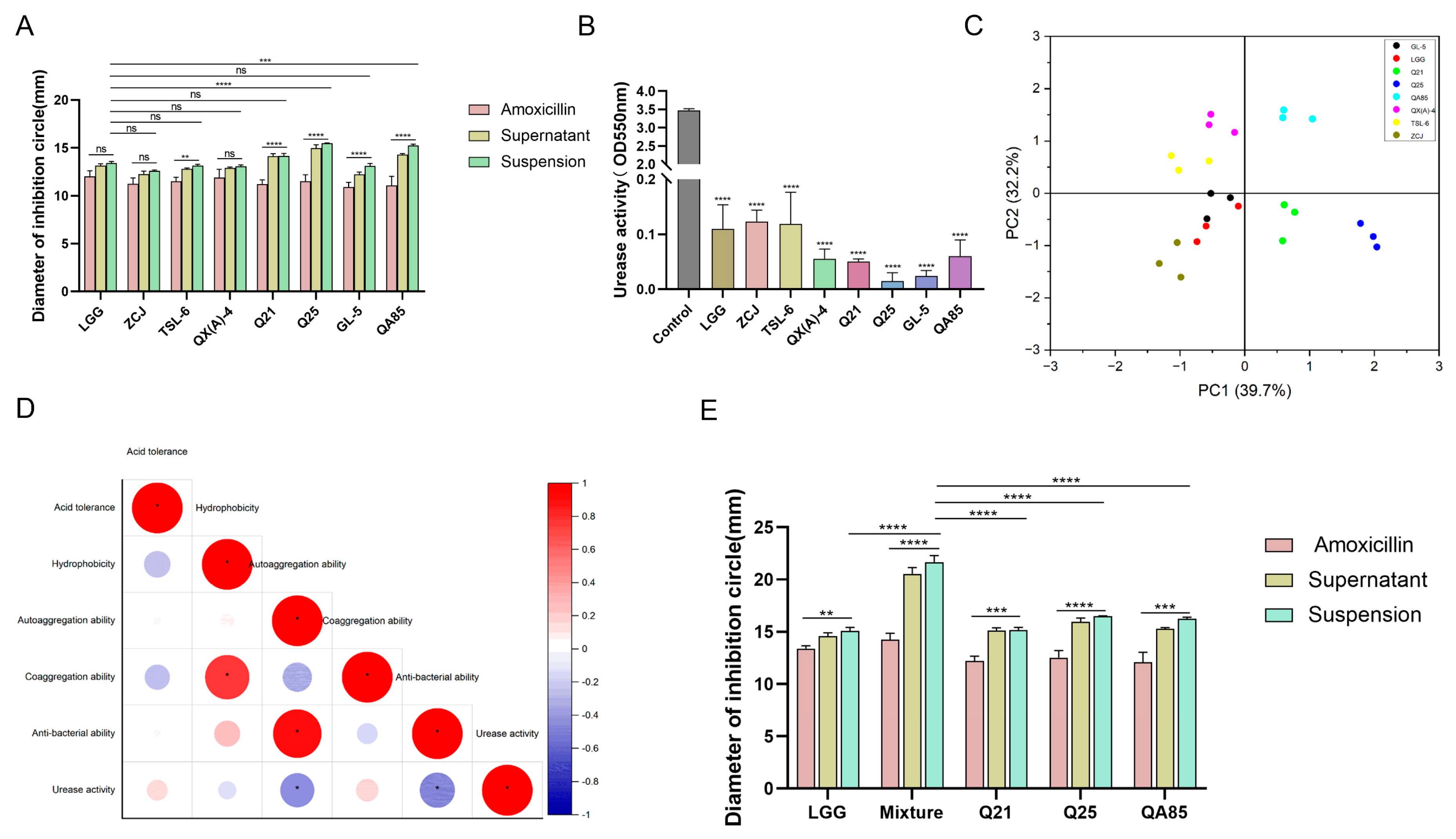
3.1. Three Strains of Lactobacillus with Strongly Inhibitory Effect on H. pylori

The results showed that eight strains could survive for 3 h under artificial gastric juice at pH 3.0, with four strains surviving more than 20% (Figure S1A). The hydrophobicity of these eight strains of Lactobacillus with acid resistance was analyzed (Figure S1B). The results showed that the hydrophobicity of the Lactobacillus ranged from 12–31%, and QA85 was the most hydrophobic.
The aggregation of bacterial strains is divided into two forms: autoaggregation and coaggregation. From the results, most of the Lactobacillus showed strong autoaggregation ability, and the autoaggregation increased with the increase in incubation time. When the incubation time was 24 h, Q25 showed the strongest autoaggregation at 69%, while autoaggregation rates of QA85, Q21, and LGG were also high (above 60%) (Figure S1C). Coaggregation results showed that Lactobacillus and H. pylori coaggregation also increased with time. The maximum coaggregation rate of QA85 was 60% at an incubation time of 24 h (Figure S1D). The aggregation of pathogenic bacteria by Lactobacillus can make it easier for pathogenic bacteria to be eliminated from the intestine.
From the bacterial inhibition assay, it can be seen that the amoxicillin inhibited the growth of H. pylori and the circle of inhibition was around 10–12 mm (Figure 1A). The inhibition circle of Lactobacillus suspensions was in the range of 12–16 mm, and the inhibition circle of Lactobacillus supernatants was in the range of 12–15 mm. The strongest ability to inhibit H. pylori was Q25. The expression level and activity of urease are critical for the survival of H. pylori in an acidic environment. It was demonstrated that the supernatants of Lactobacillus significantly inhibited the urease activity of H. pylori, with Q25 having the strongest inhibitory ability (Figure 1B).
It was found through the factor scoring plot (Figure 1C) that the first component accounted for 39.7% and the second component accounted for 32.2%, with the bacteriostatic ability playing a major contributing role. From the correlation analysis (Figure 1D), there was a close association between the nature of Lactobacillus and its inhibition of H. pylori growth. Therefore, based on the principal component analysis and the properties of H. pylori growth inhibition, Q21, Q25, and QA85 were screened as potential anti-H. pylori probiotics and were used in subsequent studies. The three strains were compounded to verify the antibacterial effect of the mixture (1:1:1). It was found that the mixture also possessed better antibacterial ability (p < 0.0001) (Figure 1E).

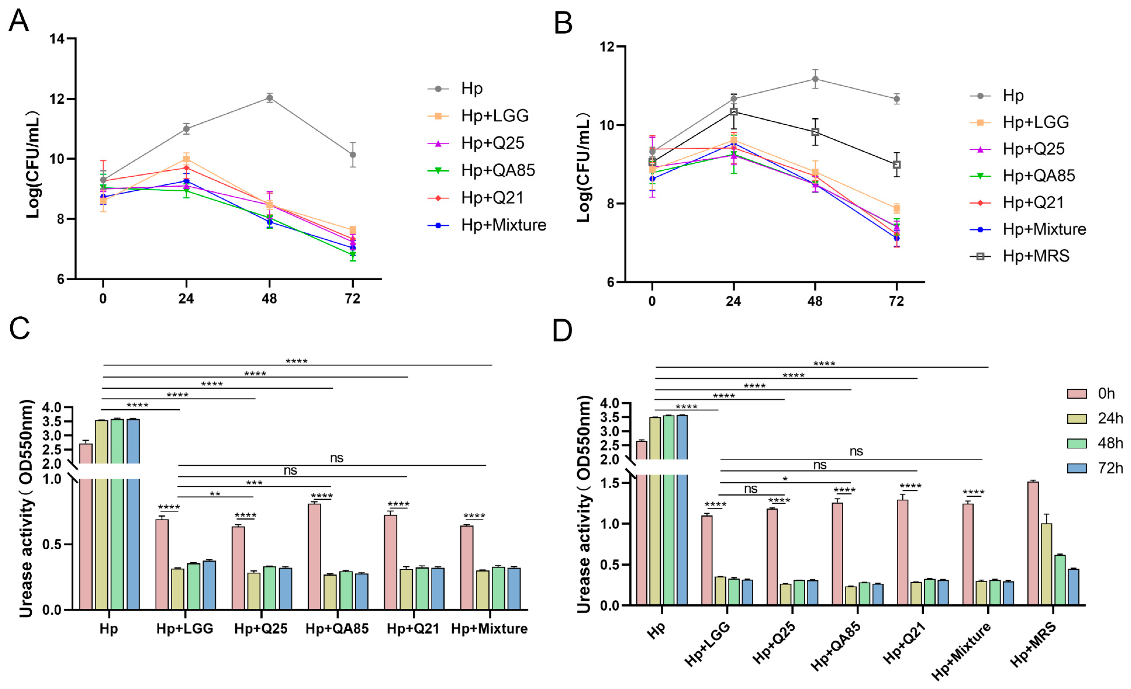
3.2. Co-Culture Validation Results of Lactobacillus and H. pylori

Q21, Q25, and QA85 were screened as potential strains of anti-H. pylori. To further confirm the ability of Lactobacillus to antagonize H. pylori, with LGG as a commercial control strain, we analyzed the effect of Lactobacillus suspension and supernatant on the inhibition of H. pylori growth under co-culture conditions. From the effect of Lactobacillus on the growth and urease activity of H. pylori (Figure 2), the number of viable bacteria of H. pylori decreased with the extension of time. At 24 h, all three strains inhibited H. pylori growth and urease activity, with Q25 being the most potent strain in inhibiting H. pylori. At 48 h and 72 h, the strains also inhibited H. pylori growth and urease activity.

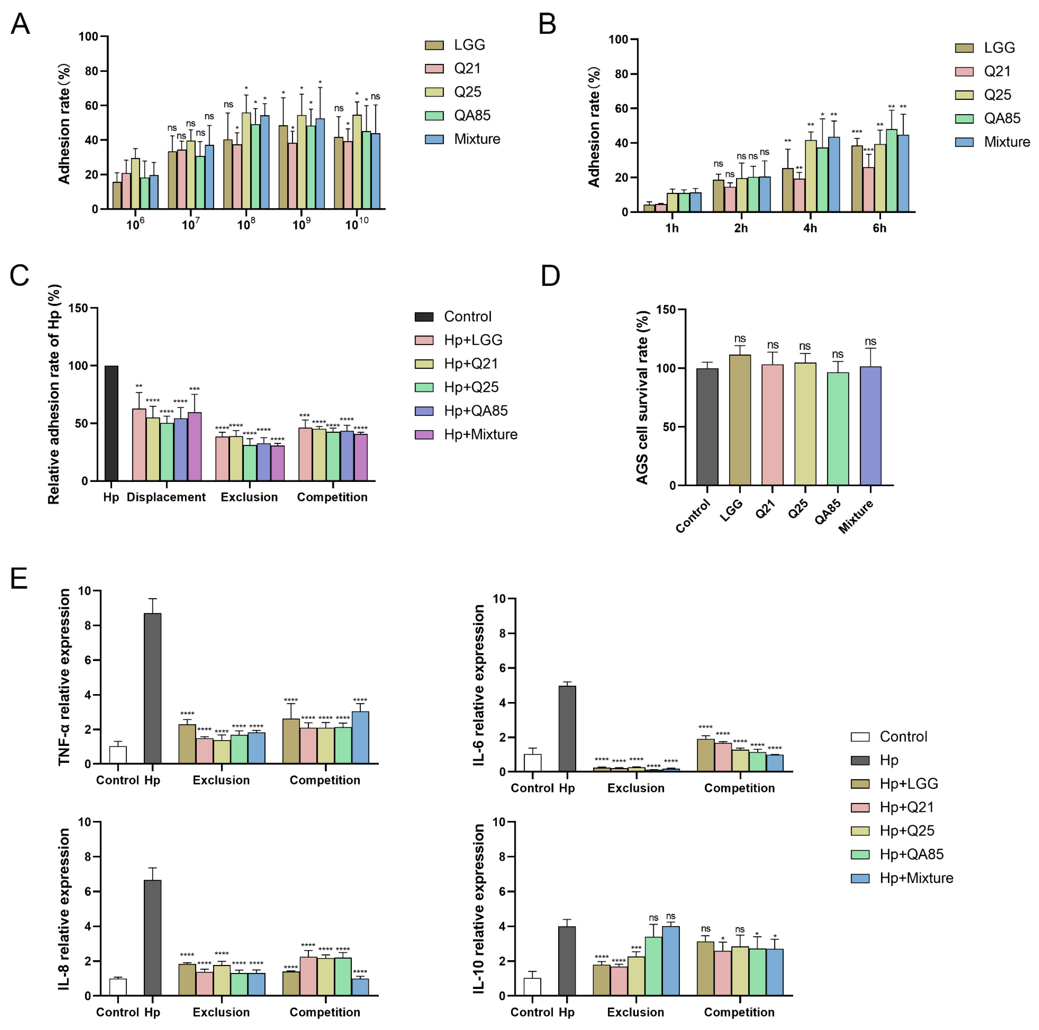
3.3. Adhesion of Lactobacillus to AGS Cells and Inhibition of H. pylori Adhesion

To confirm the ability of several strains to adhere to gastric epithelial cells, their adhesion rates to AGS cells were explored. Firstly, the adhesion rate of Lactobacillus on AGS cells at different concentrations was explored (Figure 3A), and it was found that the adhesion rate of Lactobacillus at a concentration of 108 CFU/mL was significantly different compared with 106 CFU/mL (p < 0.05). Additionally, the adhesion rate of Lactobacillus on AGS cells was explored at different incubation times (Figure 3B), and there was a significant difference in the adhesion rate at 4 h or 6 h compared with 1 h of incubation (p < 0.01, p < 0.001).
Since the screened strains had a strong ability to adhere to AGS cells, we established three cellular models, displacement, exclusion and competition, to explore the potential of the strains to hinder H. pylori colonization. The results showed that these strains inhibited H. pylori adhesion to AGS cells (Figure 3C). Specifically, the relative adhesion rates of H. pylori containing Q21, Q25, QA85, and their mixture were 55.04%, 50.39%, 54.26%, and 59.69%, respectively, compared with H. pylori adhesion to AGS cells alone during displacement adhesion group (p < 0.0001, p < 0.001). Their relative adhesion rates were 38.76%, 31.16%, 32.79%, and 30.85%, respectively, during the exclusion adhesion group (p < 0.0001), and the mixture inhibited adhesion at the highest rate. Their relative adhesion rates were 45.12%, 42.56%, 43.49%, and 40.85% (p < 0.0001), respectively, during the competition adhesion group, and the mixture still had the highest inhibition rate of adhesion. Taken together, the relative adhesion rate of H. pylori in the exclusion group was lower than the other two modes, indicating that the strains had a high ability to hinder H. pylori adhesion under the exclusion conditions, with an inhibition adhesion rate of more than 61%, and the inhibition rate of the mixture was the highest. In the competition group, the inhibition adhesion rate of each strain could reach more than 54%, demonstrating the hindrance of strains to H. pylori.
H. pylori infection induces the production of inflammatory factors and contributes to inflammation in gastric. The exclusion and competition modes were found to be superior to the displacement mode by H. pylori adhesion inhibition with Lactobacillus. Therefore, these two modes were chosen for the study of cytokine levels. From the results, the relative expression levels of TNF-α, IL-6, and IL-8 were significantly increased in H. pylori-treated AGS cells compared to the control group, and after treatment with Q21, Q25, QA85, and mixture, their relative expression levels were significantly decreased compared to the H. pylori-treated group (p < 0.0001) (Figure 3E). The results also revealed that compared with the H. pylori-infected group, after treatment with Lactobacillus, there was a significant change in the relative expression level of IL-10 in the exclusion group and an insignificant change in the competition group. Taken together, the effect of probiotics on the expression of pro-inflammatory factors was much greater than anti-inflammatory factors.
To test the safety of Lactobacillus in future applications, we performed the cell viability assay to determine the cytotoxicity of three Lactobacillus strains and their mixture to AGS cells (Figure 3D). The results showed that the co-culture of Q21, Q25, and QA85, and their mixture with cells for 6 h had no significant cytotoxic effects on cells.

3.4. Antibiotic Susceptibility Analysis of Lactobacillus

Antibiotic susceptibility was an important index for the safety of Lactobacillus before application. Therefore, we tested the susceptibility of these Lactobacillus to 22 antibiotics (Figure S2). As shown in Table 2, Q21, Q25, QA85, and LGG showed varying degrees of drug sensitivity to 22 antibiotics, with sensitivity to penicillin (e.g., ampicillin), tetracyclines (e.g., doxycycline), cephalosporins (e.g., imipenem), and chloramphenicol; and showed intermediate sensitivity to aminoglycosides and macrolides (e.g., gentamicin and erythromycin). In contrast, these strains were resistant to quinolones (e.g., ciprofloxacin and levofloxacin).

3.5. The Effect Analysis of Lactobacillus on Patients with H. pylori Infection

3.5.1. Trial Profile and Volunteer Characteristics

In total, 49 volunteers tested positive for H. pylori by 14C-UBT and were eligible for the trial. Volunteers before and after taking probiotics were used as M0 and M1 groups of the trial, respectively. Since 12 volunteers dropped the probiotics halfway through the program, a total of 37 volunteers were eligible for the protocol analysis. The average age of the volunteers was around 55 years old, and the ratio of men to women was close to 1:1.8. The specific characteristics of the volunteers are shown in Table S2.

3.5.2. Effect of Lactobacillus on H. pylori Load and Gastrointestinal Symptoms in Volunteers

The results of 14C-UBT can reflect the density of H. pylori in H. pylori-infected patients. As can be seen from the test results, there was a decreasing trend of 14C-UBT values after the volunteers took the probiotics (454.30 ± 327.00 vs. 328.35 ± 237.19, p = 0.06) (Figure 4B); 27 volunteers showed a significant decrease in the test value; 4 volunteers showed negative results with a test value below 100.
After 1 month, there was a significant difference in mean GSRS scores between the M0 and M1 groups (24.30 ± 9.05 vs. 20.49 ± 6.73, p < 0.05) (Figure 4A). Specifically, a comparison of gastrointestinal symptoms before and after taking probiotics showed that significantly fewer patients in the M1 group experienced throat discomfort (18% vs. 27%), halitosis (27% vs. 35%), chest pain (8% vs. 16%), acid regurgitation (13% vs. 21%), abdominal distension (32% vs. 59%), and diarrhea (21% vs. 43%) than in the M0 group (Table 3).

3.5.3. Effect of Lactobacillus on Immunity-Related Indicators in Volunteers

As can be seen from the results, the pro-inflammatory factors TNF-α, IFN-γ, IL-1β, IL-6, IL-8, and IL-17 in the serum of the volunteers in the M1 group were significantly reduced compared to the M0 group (p < 0.05). The change of anti-inflammatory factor IL-10 in the serum was not very obvious (Figure 4C). Therefore, probiotics can play a role in regulating the immune level of H. pylori-infected patients, especially in regulating the level of pro-inflammatory factors.

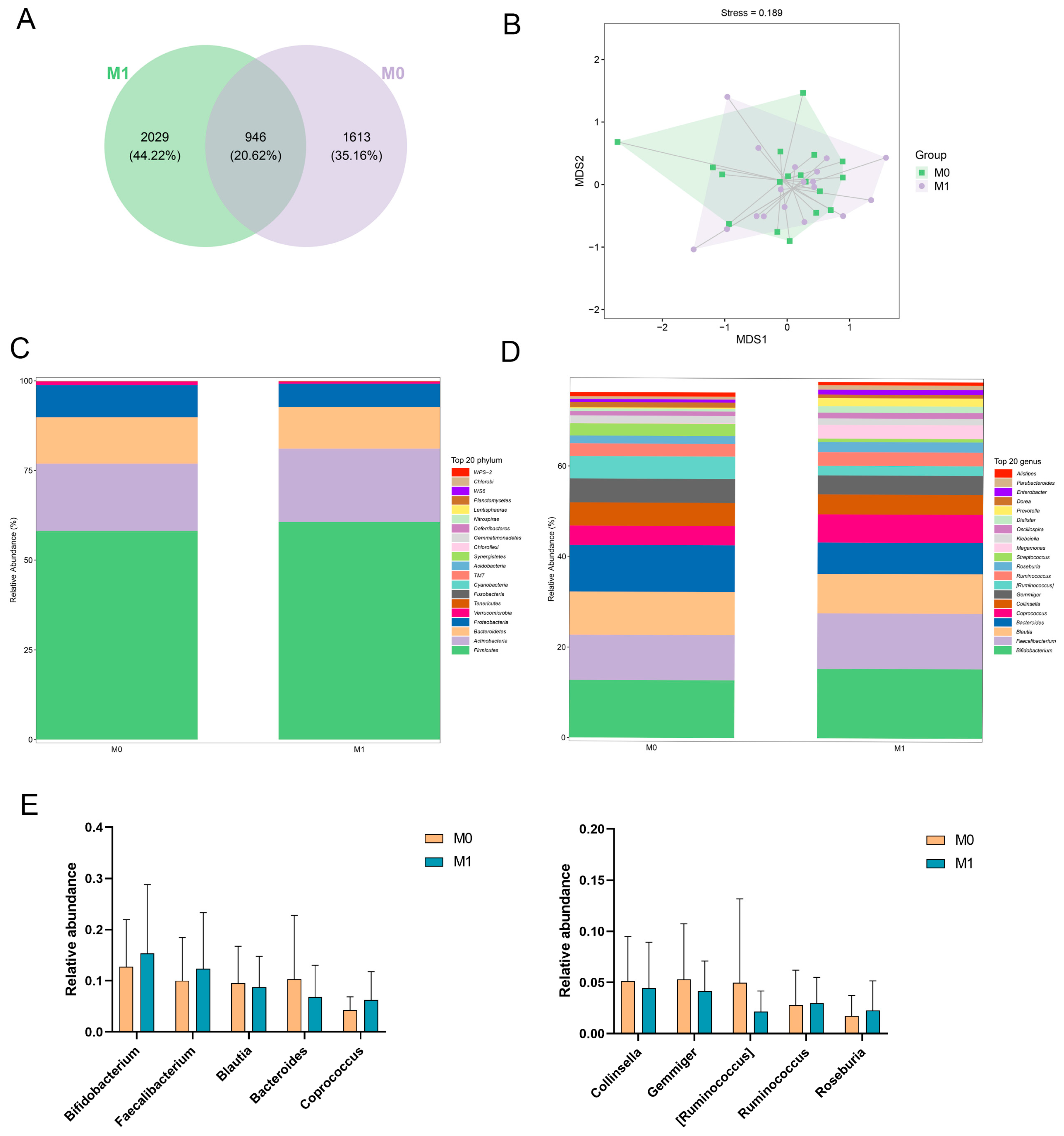
3.5.4. Gut Microbiota Analysis

There was a trend towards increased alpha-diversity (as detected at the species level by Observed species, Chao-1 and Shannon’s index) in the gut microbiota of the M1 group compared to M0 (Table 4). The mutual and unique ASVs among each group were expressed by the Venn plot in Figure 5A. In total, 946 ASVs were mutual by two groups, with the M1 group having the largest count of specific ASVs (2029). Meanwhile, 1613 unique ASVs were observed in the M0 group. These results agreed with the richness index and diversity index in Table 4. Beta-diversity analyses were performed to assess differences in diversity between groups. Bray–Curtis-based NMDS analyses showed no significant segregation of gut microbial structure between groups, and changes were not significant (Figure 5B).
To identify differences in the gut microbiota between the two groups, we analyzed the top 20 most abundant microorganisms at the phylum level. It was found that the dominant phylum in both groups was Firmicutes and Actinobacteria (relative abundance >18%), and their relative abundance was slightly higher in the M1 group compared to the M0 group (Figure 5C). Meanwhile, the relative abundance of Proteobacteria and Verrucomicrobia was reduced in the M1 group compared to the M0 group. It has been shown that the intestinal microecology is disturbed when the host is infected with H. pylori and that the administration of probiotics improves gastrointestinal symptoms and alters the ratio of Firmicutes, among others [47].
When analyzing the community structure at the genus level in detail, it was found that the proportions of Bifidobacterium and Faecalibacterium were higher in the M1 group than in the M0 group (Figure 5D,E). Additionally, the proportion of Bacteroides, [Ruminococcus] was reduced in group M1 compared to group M0. Although there was a change in the community structure in the M1 group compared to the M0 group, there was no significant change, which may be caused by the large individual differences. Taking the dominant group Bifidobacterium as an example, it enables other bacterial populations to stably colonize the intestines, and this strong bacterial correlation shapes the establishment, stability, and evolution of the intestinal microbiota and maintains intestinal microecological health [48]. Thus, probiotics have a positive regulatory effect on the gut microbiota.

3.5.5. Differential Metabolite Analysis and Related Metabolic Pathway Prediction

Differential metabolite analysis and metabolic pathway prediction were performed to investigate the changes in relevant metabolites before and after the probiotic intervention. A total of 19 differential metabolites with significant changes were screened in MS secondary characterization mode (Figure 6A). The metabolic contents of Ureidopropionic acid and O-Phosphoethanolamine were significantly increased in the M1 group compared to the M0 group, and the increase in O-Phosphoethanolamine was particularly significant. The relative contents of other metabolites, such as Indolin-2-one, Undecanoic acid, Phytanic Acid and others showed a certain degree of increase. Significantly different metabolites were introduced into KEGG (Kyoto Encyclopedia of Genes and Genomes) for metabolic pathway analysis to identify the major signaling pathways and biometabolic pathways in which they are involved. The metabolic pathways predominantly involved with O-Phosphoethanolamine encompass the Sphingolipid signaling pathway, Glycosylphosphatidylinositol (GPI)-anchor biosynthesis, Sphingolipid metabolism, and Glycerophospholipid metabolism. The most influential pathway is the Sphingolipid signaling pathway (Figure 6B). An analysis of the KEGG metabolic pathways involved in differential metabolites before and after the intervention with probiotics—the Sphingolipid signaling pathway—is shown in Figure S3.

4. Discussion

The increase in H. pylori antibiotic resistance has led to a decline in the eradication rate of standard triple therapy [49]. Some studies suggest that probiotics are considered alternative or adjunctive therapeutic approaches to improve the eradication rate of H. pylori and reduce drug-related side effects [50,51].
In this study, firstly, three strains of Lactobacillus plantarum, Q21, Q25, and QA85, were screened by utilizing the probiotic properties of Lactobacillus as well as the inhibitory properties of H. pylori growth. Our results indicated that they have the potential to withstand the acidic environment of the stomach. Moreover, these strains exhibit strong hydrophobicity. Strains that are highly hydrophobic may have protein-like substances on their cell surfaces, which may have a better self-protective effect on the cells [52]. Zuo et al. reported that autoaggregation of probiotic strains may be one of the mechanisms to maintain the viability of the strains and increase the resistance of the human upper gastrointestinal tract to pathogenic bacteria. Autoaggregation allows probiotics to form spatial site barriers to impede the colonization of the gastrointestinal tract by pathogenic bacteria such as H. pylori [53]. Certainly, the aggregation of pathogenic bacteria by Lactobacillus can make it easier to expel pathogenic bacteria from the intestine [54]. All three strains screened in this study possessed strong autoaggregation and coaggregation abilities, enabling them to hinder the colonization of H. pylori.
Zheng et al. reported that lactobacilli have anti-H. pylori effects, which depend on their secreted components and lactic acid-mediated bactericidal activity [55,56,57]. Urease activity is an important factor in the colonization of H. pylori, altering the properties of mucus by neutralizing acidic pH [58]. Our results showed that Q21, Q25 and QA85 had high anti-H. pylori activity, which may also be related to their secretions.
Previous studies have found that probiotics can repair the mucosal permeability of the gastric mucosa, form a mucosal barrier, and inhibit the adhesion of pathogenic bacteria such as H. pylori [59]. Do et al. reported that L. rhamnosus JB3 has demonstrated the ability to inhibit the binding of H. pylori to cells [60]. Our research found that Q21, Q25, and QA85 exhibit strong colonization ability in gastric epithelial AGS cells. Additionally, they inhibit the adhesion of H. pylori to AGS cells, further confirming the ability of Lactobacillus to impede the colonization and adhesion of H. pylori on gastric mucosa. Lactobacillus achieve their anti-adhesive effects on H. pylori through the following mechanisms: competing for epithelial cell binding sites, releasing antimicrobial compounds to kill H. pylori, and reducing the expression of adhesin genes [61].
The manifestation of H. pylori-induced gastritis has been associated with the release of a wide variety of inflammatory mediators, such as chemokines and cytokines [62]. H. pylori can release CagA directly into host cells, leading to the production of IL-8 as well as other inflammatory factors by epithelial cells [63,64,65]. Studies have also demonstrated that probiotics inhibit the production of pro-inflammatory factors, such as IL-8, leading to a reduction in neutrophil infiltration in the gastric mucosa. Moreover, they promote the generation of anti-inflammatory factors like IL-10, eliciting an immune response in Th2 cells [66]. Therefore, probiotics can modulate the immune response of the host by adhering to epithelial cells, and in particular, they can reduce the inflammatory response of the stomach by modulating the balance of inflammatory factors [67]. In this study, we found that Q21, Q25 and QA85 were able to reduce the expression of pro-inflammatory factor genes, such as IL-8, with potential immunomodulatory activity. Probiotics are capable of generating a wide variety of immune responses due to strain specificity, and the immune status of the host affects the immune response [68]. Therefore, it is necessary to further investigate the mechanisms by which the strains regulate inflammatory signaling.
In vitro studies revealed that Q21, Q25, and QA85 have anti-H. pylori potential, and we further validated their effects through in vivo studies. In this own-control trial of volunteers taking probiotics, the 14C-UBT assay showed a decreasing trend in the load of H. pylori after one month of taking probiotics (454.30 ± 327.00 vs. 328.35 ± 237.19, p = 0.06). However, probiotics alone cannot eradicate H. pylori. Meanwhile, the use of probiotics can significantly improve the gastrointestinal symptoms of patients, especially for symptoms such as abdominal distension and diarrhea. Some studies reported that Lactobacillus, such as L. acidophilus, L. casei Shirota, and L. johnsonii La1, may reduce the bacterial load of H. pylori [69]. Chen et al. [70] observed that although probiotics were effective in reducing the bacterial load of H. pylori, the beneficial effect disappeared after a week of discontinuing probiotics. Therefore, the use of probiotics alone is not recommended for H. pylori eradication. However, probiotics in combination with triple or quadruple therapy may be a better option.
Some studies have shown that probiotics can reduce the severity of H. pylori-related illnesses [71,72]. Some in vitro studies have shown that L. casei, L. acidophilus and B. lactis strains have inhibitory and bactericidal activity against H. pylori [71]. Probiotics are thought to reduce gastritis in the sinuses and body of the stomach [72]. Probiotics not only inhibit H. pylori, but also alter the host’s immune response to reduce H. pylori-related gastric inflammation by inactivating the SMAD7 and NF-kB pathways [73]. Our findings also revealed that the levels of pro-inflammatory factor proteins such as TNF-α, IL-6, and IL-8 were suppressed in the patients, confirming that probiotics can modulate the immune balance of the host.
Probiotics have shown a role in preventing or reducing H. pylori infection-associated diarrhea. Emara et al. and Chotivitayatarakorn et al. found that probiotics significantly reduced the incidence of diarrhea in a 14-day triple therapy study in combination with probiotics [74,75]. In our study using only probiotics alone, we found that significantly fewer patients experienced diarrhea after taking probiotics than before taking probiotics (21% vs. 43%). This result suggests that probiotics have a positive moderating effect on patients’ discomfort reactions such as diarrhea. Hauser et al. found that a 14-day triple therapy combining L. rhamnosus GG and Bifidobacterium significantly reduced the frequency of abdominal distension [76]. Our conclusions were similar, but unlike their study, we studied the probiotic combination of Lactobacillus plantarum Q21, Q25, and QA85 and did not combine it with triple therapy.
Dysbiosis of the gut microbiota has been reported to be associated with an increased risk of a number of diseases [77]. Probiotics have been suggested to have the potential to treat these diseases by modulating the gut microbiota [78]. However, there are still no clear conclusions as to whether or not probiotics alter the diversity or composition of the gut microbiota [33]. Most studies have shown that probiotics did not significantly affect the diversity of the gut microbiota [79,80]. Rahayu et al. found that giving volunteers Lactobacillus plantarum Dad-13 powder for 90 days resulted in a significant increase in alpha diversity and bacterial abundance of the gut microbiota [81]. It has also been found that taking Lactobacillus paracasei for 4 weeks can change the composition of the fecal microbiota [82]. Our study found that alpha diversity changed but not significantly after taking probiotics, and beta diversity was similar, and that individual differences may be an important factor affecting diversity. It was also found that there was no significant change in relative abundance at the genus level before and after taking probiotics. Our results are consistent with the results of several studies using only probiotics. For example, Chen et al. [70] found that although the use of a combination of Lactobacillus acidophilus and Lactobacillus rhamnosus for 4 weeks reduced the bacterial load of H. pylori, no significant changes in the diversity and composition of the intestinal microbiota, although fluctuations in the diversity and composition of the intestinal microbiota, were observed. Different results may be due to different treatment regimens, average age of patients, probiotic types, dosages, and length of use cycles.
Probiotics cause certain effects on fecal metabolites. These changes in metabolites have a great degree of correlation with various physiological and pathological processes of the organism. We screened the fecal differential metabolites of volunteers before and after taking probiotics and found that there were 19 differential metabolites with significant changes, especially the content of O-Phosphoethanolamine increased significantly after probiotic intervention. A search for its associated metabolic pathways revealed that the main metabolic pathway involved is the Sphingolipid signaling pathway. O-phosphoethanolamine is a phospholipid molecule. This compound plays an important role in living organisms, especially in the construction of cell membranes and metabolic pathways [83,84,85]. The Sphingolipid signaling pathway has a potentially important role in regulating cellular physiological processes, maintaining tissue homeostasis, and in the prevention and treatment of disease [85]. The study of this metabolic pathway has important implications for disease development and the search for therapeutic approaches.
This study has some strengths. Firstly, we recruited volunteers who were allowed to take probiotics as part of their daily routine, which assessed the effectiveness of probiotics in people’s normal life diets that do not exclude outside influences. Secondly, most of the volunteers in this study were around 55 years old, making it more applicable to an older population. Furthermore, probiotics are natural and harmless microbial agents that do not pose drug-like hazards when used on a daily basis. Probiotics can also be supplemented in routine treatment plans for H. pylori. However, there are some limitations to this study. This study was a self-controlled trial before and after taking probiotics and had a small sample size. In addition, we only recorded the bacterial load within 1 month of taking the probiotic, so we cannot know whether the inhibitory effect of the probiotic will persist beyond 1 month. Finally, fecal samples were not collected at additional time points, thus preventing comprehensive monitoring of the dynamics of the gut microbiota.

5. Conclusions

In this study, we screened candidate probiotic strains Lactobacillus plantarum Q21, Q25, and QA85 with strong anti-H. pylori ability and investigated their efficacy in alleviating symptoms associated with H. pylori infection. These strains, given to volunteers in the form of a probiotic powder, have been shown to result in a downward trend in the bacterial load of H. pylori, lower GSRS scores, and improve the severity and frequency of uncomfortable reactions such as abdominal distension and diarrhea, and reduce the inflammatory response. However, their role in regulating the structure and function of the gut microbiota was not significant, while their effect on fecal metabolites was more pronounced. The use of probiotics appears to have emerged as a low-cost and safer adjunctive therapy compared with standard triple or quadruple therapy, which may be one of the ideal strategies for alleviating H. pylori infection.

Supplementary Materials

The following supporting information can be downloaded at: https://www.mdpi.com/article/10.3390/foods13121851/s1, Figure S1: Studies on the properties of Lactobacillus; Figure S2: Typical diagram of Lactobacillus antibiotic susceptibility; Figure S3: Prediction of the metabolic pathway before and after intervention with probiotics; Table S1: Strains conservation information; Table S2: Demographic characteristics of volunteers.

Author Contributions

Conceptualization, H.Y., Y.L., Y.M., J.L. (Jiaru Li), J.L. (Junxiang Li), Z.H. and P.Y.; Methodology, H.Y., Y.L., Y.M., J.L. (Jiaru Li), J.L. (Junxiang Li) and Z.H.; Data curation, H.Y.; Formal analysis, H.Y. and Y.L.; Writing—original draft, H.Y.; Writing—review & editing, Y.L., J.L. (Junxiang Li) and P.Y.; Supervision, C.Z.; Project administration, C.Z.; Funding acquisition, C.Z. All authors have read and agreed to the published version of the manuscript.

Funding

This work was supported by the Science and Technology Major Project of Gansu Province (Grant no. 22ZD1FA001), the Science and Technology Major Project of Gansu Province-Enterprise Innovation Consortium Project (Grant no. 23ZDFA013-3), the Guangzhou Science and Technology Planning Project (Grant no. 202206010165), and the Key Talent Project of Gansu Province (Grant no. 2022RCXM027).

Institutional Review Board Statement

This study was reviewed by the Ethics Committee of the School of Life Sciences, Lanzhou University, China (No. EAF2023052, 26 September 2023) and adhered to the Declaration of Helsinki.

Informed Consent Statement

Informed consent was obtained from all volunteers involved in the study.

Data Availability Statement

The original contributions presented in the study are included in the article/Supplementary Materials, further inquiries can be directed to the corresponding author.

Acknowledgments

The authors sincerely thank the volunteers who participated in this work. Thanks to the Huikang Medical Examination Center for their help in this study. We also would like to express our gratitude to the Core Facility of the School of Life Sciences, Lanzhou University for their support.

Conflicts of Interest

The authors declare no conflict of interest.

References

  1. Hooi, J.K.Y.; Lai, W.Y.; Ng, W.K.; Suen, M.M.Y.; Underwood, F.E.; Tanyingoh, D.; Malfertheiner, P.; Graham, D.Y.; Wong, V.W.S.; Wu, J.C.Y.; et al. Global Prevalence of Helicobacter pylori Infection: Systematic Review and Meta-Analysis. Gastroenterology 2017, 153, 420–429. [Google Scholar] [CrossRef] [PubMed]
  2. Benajah, D.A.; Lahbabi, M.; Alaoui, S.; El Rhazi, K.; El Abkari, M.; Nejjari, C.; Amarti, A.; Bennani, B.; Mahmoud, M.; Ibrahimi, S.A. Prevalence of Helicobacter pylori and Its Recurrence after Successful Eradication in a Developing Nation (Morocco). Clin. Res. Hepatol. Gastroenterol. 2013, 37, 519–526. [Google Scholar] [CrossRef] [PubMed]
  3. Porras, C.; Nodora, J.; Sexton, R.; Ferreccio, C.; Jimenez, S.; Dominguez, R.L.; Cook, P.; Anderson, G.; Morgan, D.R.; Baker, L.H.; et al. Epidemiology of Helicobacter pylori Infection in Six Latin American Countries (SWOG Trial S0701). Cancer Causes Control 2013, 24, 209–215. [Google Scholar] [CrossRef] [PubMed]
  4. Yuan, C.; Adeloye, D.; Luk, T.T.; Huang, L.; He, Y.; Xu, Y.; Ye, X.; Yi, Q.; Song, P.; Rudan, I.; et al. The Global Prevalence of and Factors Associated with Helicobacter pylori Infection in Children: A Systematic Review and Meta-Analysis. Lancet Child. Adolesc. Health 2022, 6, 185–194. [Google Scholar] [CrossRef] [PubMed]
  5. Malfertheiner, P.; Link, A.; Selgrad, M. Helicobacter pylori: Perspectives and Time Trends. Nat. Rev. Gastroenterol. Hepatol. 2014, 11, 628–638. [Google Scholar] [CrossRef] [PubMed]
  6. Savoldi, A.; Carrara, E.; Graham, D.Y.; Conti, M.; Tacconelli, E. Prevalence of Antibiotic Resistance in Helicobacter pylori: A Systematic Review and Meta-Analysis in World Health Organization Regions. Gastroenterology 2018, 155, 1372–1382.e17. [Google Scholar] [CrossRef] [PubMed]
  7. Pelepenko, L.E.; Janini, A.C.P.; Gomes, B.P.F.A.; de-Jesus-Soares, A.; Marciano, M.A. Effects of Bismuth Exposure on the Human Kidney-A Systematic Review. Antibiotics 2022, 11, 1741. [Google Scholar] [CrossRef] [PubMed]
  8. Gebeyehu, E.; Nigatu, D.; Engidawork, E. Helicobacter pylori Eradication Rate of Standard Triple Therapy and Factors Affecting Eradication Rate at Bahir Dar City Administration, Northwest Ethiopia: A Prospective Follow up Study. PLoS ONE 2019, 14, e0217645. [Google Scholar] [CrossRef]
  9. Phumkhachorn, P.; Rattanachaikunsopon, P. Probiotics: Sources, Selection and Health Benefits. Res. J. Biotech. 2023, 18, 102–113. [Google Scholar] [CrossRef]
  10. Wilkins, T.; Sequoia, J. Probiotics for Gastrointestinal Conditions: A Summary of the Evidence. Am. Fam. Physician 2017, 96, 170–178. [Google Scholar]
  11. Sang, L.-X. Remission Induction and Maintenance Effect of Probiotics on Ulcerative Colitis: A Meta-Analysis. WJG 2010, 16, 1908. [Google Scholar] [CrossRef] [PubMed]
  12. Wang, X.; Zhang, P.; Zhang, X. Probiotics Regulate Gut Microbiota: An Effective Method to Improve Immunity. Molecules 2021, 26, 6076. [Google Scholar] [CrossRef] [PubMed]
  13. He, C.; Xie, Y.; Zhu, Y.; Zhuang, K.; Huo, L.; Yu, Y.; Guo, Q.; Shu, X.; Xiong, Z.; Zhang, Z.; et al. Probiotics Modulate Gastrointestinal Microbiota after Helicobacter pylori Eradication: A Multicenter Randomized Double-Blind Placebo-Controlled Trial. Front. Immunol. 2022, 13, 1033063. [Google Scholar] [CrossRef] [PubMed]
  14. Pernica, J.M.; Steenhoff, A.P.; Mokomane, M.; Moorad, B.; Lechiile, K.; Smieja, M.; Mazhani, L.; Cheng, J.; Kelly, M.S.; Loeb, M.; et al. Rapid Enteric Testing to Permit Targeted Antimicrobial Therapy, with and without Lactobacillus reuteri Probiotics, for Paediatric Acute Diarrhoeal Disease in Botswana: A Pilot, Randomized, Factorial, Controlled Trial. PLoS ONE 2017, 12, e0185177. [Google Scholar] [CrossRef] [PubMed]
  15. Lee, C.-Y.; Shih, H.-C.; Yu, M.-C.; Lee, M.-Y.; Chang, Y.-L.; Lai, Y.-Y.; Lee, Y.-C.; Kuan, Y.-H.; Lin, C.-C. Evaluation of the Potential Inhibitory Activity of a Combination of L. acidophilus, L. rhamnosus and L. sporogenes on Helicobacter pylori: A Randomized Double-Blind Placebo-Controlled Clinical Trial. Chin. J. Integr. Med. 2017, 23, 176–182. [Google Scholar] [CrossRef] [PubMed]
  16. Wang, Y.; Huang, Y. Effect of Lactobacillus acidophilus and Bifidobacterium bifidum Supplementation to Standard Triple Therapy on Helicobacter pylori Eradication and Dynamic Changes in Intestinal Flora. World J. Microbiol. Biotechnol. 2014, 30, 847–853. [Google Scholar] [CrossRef] [PubMed]
  17. Myllyluoma, E.; Veijola, L.; Ahlroos, T.; Tynkkynen, S.; Kankuri, E.; Vapaatalo, H.; Rautelin, H.; Korpela, R. Probiotic Supplementation Improves Tolerance to Helicobacter pylori Eradication Therapy--a Placebo-Controlled, Double-Blind Randomized Pilot Study. Aliment. Pharmacol. Ther. 2005, 21, 1263–1272. [Google Scholar] [CrossRef] [PubMed]
  18. Sgouras, D.; Maragkoudakis, P.; Petraki, K.; Martinez-Gonzalez, B.; Eriotou, E.; Michopoulos, S.; Kalantzopoulos, G.; Tsakalidou, E.; Mentis, A. In Vitro and in Vivo Inhibition of Helicobacter pylori by Lactobacillus casei Strain Shirota. Appl. Environ. Microbiol. 2004, 70, 518–526. [Google Scholar] [CrossRef] [PubMed]
  19. Sahagún-Flores, J.E.; López-Peña, L.S.; de la Cruz-Ramírez Jaimes, J.; García-Bravo, M.S.; Peregrina-Gómez, R.; de Alba-García, J.E.G. Eradication of Helicobacter pylori: Triple treatment scheme plus Lactobacillus vs. triple treatment alone. Cir. Y Cir. 2007, 75, 333–336. [Google Scholar]
  20. Deguchi, R.; Nakaminami, H.; Rimbara, E.; Noguchi, N.; Sasatsu, M.; Suzuki, T.; Matsushima, M.; Koike, J.; Igarashi, M.; Ozawa, H.; et al. Effect of Pretreatment with Lactobacillus gasseri OLL2716 on First-line Helicobacter pylori Eradication Therapy. J. Gastro Hepatol. 2012, 27, 888–892. [Google Scholar] [CrossRef]
  21. Ismail, N.I.; Nawawi, K.N.M.; Hsin, D.C.C.; Hao, K.W.; Mahmood, N.R.K.N.; Chearn, G.L.C.; Wong, Z.; Tamil, A.M.; Joseph, H.; Raja Ali, R.A. Probiotic Containing Lactobacillus reuteri DSM 17648 as an Adjunct Treatment for Helicobacter pylori Infection: A Randomized, Double-blind, Placebo-controlled Trial. Helicobacter 2023, 28, e13017. [Google Scholar] [CrossRef] [PubMed]
  22. Del Piano, M.; Pagliarulo, M.; Tari, R.; Carmagnola, S.; Balzarini, M.; Lorenzini, P.; Pane, M. Correlation between Chronic Treatment with Proton Pump Inhibitors and Bacterial Overgrowth in the Stomach: Any Possible Beneficial Role for Selected Lactobacilli? J. Clin. Gastroenterol. 2014, 48 (Suppl. S1), S40–S46. [Google Scholar] [CrossRef] [PubMed]
  23. Wu, S.; Xu, Y.; Chen, Z.; Chen, Y.; Wei, F.; Xia, C.; Zhou, Q.; Li, P.; Gu, Q. Lactiplantibacillus plantarum ZJ316 Reduces Helicobacter pylori Adhesion and Inflammation by Inhibiting the Expression of Adhesin and Urease Genes. Mol. Nutr. Food Res. 2023, 67, 2300241. [Google Scholar] [CrossRef] [PubMed]
  24. Yuan, Z.; Xiao, S.; Li, S.; Suo, B.; Wang, Y.; Meng, L.; Liu, Z.; Yin, Z.; Xue, Y.; Zhou, L. The Impact of Helicobacter pylori Infection, Eradication Therapy, and Probiotics Intervention on Gastric Microbiota in Young Adults. Helicobacter 2021, 26, e12848. [Google Scholar] [CrossRef] [PubMed]
  25. Yakovenko, E.P.; Strokova, T.V.; Iakovenko, A.V.; Ivanov, A.N.; Soluyanova, I.P.; Vasilyev, N.N. A Prospective Randomized Comparative Study of the Efficacy and Safety of a Two-Week Bismuth-Based Quadrotherapy of Helicobacter pylori Infection with the Inclusion of the Probiotic Containing Bifidobacterium longum BB-46 and Enterococcus faecium ENCfa-68. Ter. Arkhiv. 2021, 93, 916–922. [Google Scholar] [CrossRef] [PubMed]
  26. Song, H.-Y.; Zhou, L.; Liu, D.-Y.; Yao, X.-J.; Li, Y. What Roles Do Probiotics Play in the Eradication of Helicobacter pylori? Current Knowledge and Ongoing Research. Gastroenterol. Res. Pract. 2018, 2018, 9379480. [Google Scholar] [CrossRef] [PubMed]
  27. Buckley, M.; Lacey, S.; Doolan, A.; Goodbody, E.; Seamans, K. The Effect of Lactobacillus reuteri Supplementation in Helicobacter pylori Infection: A Placebo-Controlled, Single-Blind Study. BMC Nutr. 2018, 4, 48. [Google Scholar] [CrossRef] [PubMed]
  28. Wang, Q.; Li, J. Intelligent Algorithm-Based Gastrointestinal X-Ray Examination in Evaluating the Therapeutic Effect of Probiotics Combined with Triple Therapy on Children with Helicobacter Infection. Contrast Media Mol. Imaging 2022, 2022, 8464361. [Google Scholar] [CrossRef]
  29. Yu, M.; Zhang, R.; Ni, P.; Chen, S.; Duan, G. Efficacy of Lactobacillus-Supplemented Triple Therapy for H. Pylori Eradication: A Meta-Analysis of Randomized Controlled Trials. PLoS ONE 2019, 14, e0223309. [Google Scholar] [CrossRef]
  30. Zhang, M.; Zhang, C.; Zhao, J.; Zhang, H.; Zhai, Q.; Chen, W. Meta-Analysis of the Efficacy of Probiotic-Supplemented Therapy on the Eradication of H. Pylori and Incidence of Therapy-Associated Side Effects. Microb. Pathog. 2020, 147, 104403. [Google Scholar] [CrossRef]
  31. Holz, C.; Busjahn, A.; Mehling, H.; Arya, S.; Boettner, M.; Habibi, H.; Lang, C. Significant Reduction in Helicobacter pylori Load in Humans with Non-Viable Lactobacillus reuteri DSM17648: A Pilot Study. Probiotics Antimicro. Prot. 2015, 7, 91–100. [Google Scholar] [CrossRef] [PubMed]
  32. Oh, B.; Kim, B.-S.; Kim, J.W.; Kim, J.S.; Koh, S.-J.; Kim, B.G.; Lee, K.L.; Chun, J. The Effect of Probiotics on Gut Microbiota during the Helicobacter pylori Eradication: Randomized Controlled Trial. Helicobacter 2016, 21, 165–174. [Google Scholar] [CrossRef] [PubMed]
  33. Kristensen, N.B.; Bryrup, T.; Allin, K.H.; Nielsen, T.; Hansen, T.H.; Pedersen, O. Alterations in Fecal Microbiota Composition by Probiotic Supplementation in Healthy Adults: A Systematic Review of Randomized Controlled Trials. Genome Med. 2016, 8, 52. [Google Scholar] [CrossRef] [PubMed]
  34. Yi, Z.; Yu-Li, W.; Xiao-Qian, C.; Ping, Z. Isolation and Initiative Identification of Microorganism from Traditional Fermentative Food-Jiangshui. Food Sci. 2007, 28, 219. [Google Scholar]
  35. Zhou, X.; Pan, Y.; Wang, Y.; Li, W. In Vitro Assessment of Gastrointestinal Viability of Two Photosynthetic Bacteria, Rhodopseudomonas Palustris and Rhodobacter Sphaeroides. J. Zhejiang Univ. Sci. B 2007, 8, 686–692. [Google Scholar] [CrossRef] [PubMed]
  36. Kos, B.; Susković, J.; Vuković, S.; Simpraga, M.; Frece, J.; Matosić, S. Adhesion and Aggregation Ability of Probiotic Strain Lactobacillus acidophilus M92. J. Appl. Microbiol. 2003, 94, 981–987. [Google Scholar] [CrossRef] [PubMed]
  37. Collado, M.C.; Meriluoto, J.; Salminen, S. Adhesion and Aggregation Properties of Probiotic and Pathogen Strains. Eur. Food Res. Technol. 2008, 226, 1065–1073. [Google Scholar] [CrossRef]
  38. Ryan, K.A.; Daly, P.; Li, Y.; Hooton, C.; O’Toole, P.W. Strain-Specific Inhibition of Helicobacter pylori by Lactobacillus salivarius and Other Lactobacilli. J. Antimicrob. Chemother. 2008, 61, 831–834. [Google Scholar] [CrossRef] [PubMed]
  39. Thuy, T.T.D.; Kuo, P.-Y.; Lin, S.-M.; Kao, C.-Y. Anti-Helicobacter pylori Activity of Potential Probiotic Lactiplantibacillus pentosus SLC13. BMC Microbiol. 2022, 22, 277. [Google Scholar] [CrossRef]
  40. Jing, D.; Jin, J.; Mei, Z.; Zhu, Q.; Lu, Y.; Wang, X. Effects of Helicobacter pylori Infection and Interleukin 6 on the Expression of ITIH4 in Human Gastric Cancer Cells. Transl. Cancer Res. 2020, 9, 4656–4665. [Google Scholar] [CrossRef]
  41. Jiang, M.; Zhang, F.; Wan, C.; Xiong, Y.; Shah, N.P.; Wei, H.; Tao, X. Evaluation of Probiotic Properties of Lactobacillus plantarum WLPL04 Isolated from Human Breast Milk. J. Dairy. Sci. 2016, 99, 1736–1746. [Google Scholar] [CrossRef] [PubMed]
  42. Tan, Q.; Xu, H.; Aguilar, Z.P.; Peng, S.; Dong, S.; Wang, B.; Li, P.; Chen, T.; Xu, F.; Wei, H. Safety Assessment and Probiotic Evaluation of Enterococcus Faecium YF5 Isolated from Sourdough. J. Food Sci. 2013, 78, M587–M593. [Google Scholar] [CrossRef] [PubMed]
  43. Yang, C.; Liang, L.; Lv, P.; Liu, L.; Wang, S.; Wang, Z.; Chen, Y. Effects of Non-Viable Lactobacillus reuteri Combining with 14-Day Standard Triple Therapy on Helicobacter pylori Eradication: A Randomized Double-Blind Placebo-Controlled Trial. Helicobacter 2021, 26, e12856. [Google Scholar] [CrossRef]
  44. Crowe, S.E. Helicobacter pylori Infection. N. Engl. J. Med. 2019, 380, 1158–1165. [Google Scholar] [CrossRef]
  45. Svedlund, J.; Sjödin, I.; Dotevall, G. GSRS—A Clinical Rating Scale for Gastrointestinal Symptoms in Patients with Irritable Bowel Syndrome and Peptic Ulcer Disease. Dig. Dis. Sci. 1988, 33, 129–134. [Google Scholar] [CrossRef]
  46. Kulich, K.R.; Madisch, A.; Pacini, F.; Piqué, J.M.; Regula, J.; Van Rensburg, C.J.; Ujszászy, L.; Carlsson, J.; Halling, K.; Wiklund, I.K. Reliability and Validity of the Gastrointestinal Symptom Rating Scale (GSRS) and Quality of Life in Reflux and Dyspepsia (QOLRAD) Questionnaire in Dyspepsia: A Six-Country Study. Health Qual. Life Outcomes 2008, 6, 12. [Google Scholar] [CrossRef] [PubMed]
  47. Chen, L.; Xu, W.; Lee, A.; He, J.; Huang, B.; Zheng, W.; Su, T.; Lai, S.; Long, Y.; Chu, H.; et al. The Impact of Helicobacter pylori Infection, Eradication Therapy and Probiotic Supplementation on Gut Microenvironment Homeostasis: An Open-Label, Randomized Clinical Trial. EBioMedicine 2018, 35, 87–96. [Google Scholar] [CrossRef]
  48. Hidalgo-Cantabrana, C.; Delgado, S.; Ruiz, L.; Ruas-Madiedo, P.; Sánchez, B.; Margolles, A. Bifidobacteria and Their Health-Promoting Effects. Microbiol. Spectr. 2017, 5. [Google Scholar] [CrossRef] [PubMed]
  49. Tshibangu-Kabamba, E.; Yamaoka, Y. Helicobacter pylori Infection and Antibiotic Resistance—From Biology to Clinical Implications. Nat. Rev. Gastroenterol. Hepatol. 2021, 18, 613–629. [Google Scholar] [CrossRef]
  50. Zhu, X.Y.; Liu, F. Probiotics as an Adjuvant Treatment in Helicobacter pylori Eradication Therapy. J. Dig. Dis. 2017, 18, 195–202. [Google Scholar] [CrossRef]
  51. Qureshi, N.; Li, P.; Gu, Q. Probiotic Therapy in Helicobacter pylori Infection: A Potential Strategy against a Serious Pathogen? Appl. Microbiol. Biotechnol. 2019, 103, 1573–1588. [Google Scholar] [CrossRef]
  52. Sophatha, B.; Teanpaisan, R. Factors Relating to Adhesion and Aggregation of Lactobacillus paracasei and Lactobacillus rhamnosus Strains. Microbiology 2021, 90, 793–800. [Google Scholar] [CrossRef]
  53. Zuo, F.; Appaswamy, A.; Gebremariam, H.G.; Jonsson, A.-B. Role of Sortase A in Lactobacillus gasseri Kx110A1 Adhesion to Gastric Epithelial Cells and Competitive Exclusion of Helicobacter pylori. Front. Microbiol. 2019, 10, 2770. [Google Scholar] [CrossRef]
  54. Ekmekci, H.; Aslim, B.; Ozturk, S. Characterization of Vaginal Lactobacilli Coaggregation Ability with Escherichia coli. Microbiol. Immunol. 2009, 53, 59–65. [Google Scholar] [CrossRef] [PubMed]
  55. Zheng, P.-X.; Fang, H.-Y.; Yang, H.-B.; Tien, N.-Y.; Wang, M.-C.; Wu, J.-J. Lactobacillus pentosus Strain LPS16 Produces Lactic Acid, Inhibiting Multidrug-Resistant Helicobacter pylori. J. Microbiol. Immunol. Infect. 2016, 49, 168–174. [Google Scholar] [CrossRef]
  56. Kim, T.-S.; Hur, J.-W.; Yu, M.-A.; Cheigh, C.-I.; Kim, K.-N.; Hwang, J.-K.; Pyun, Y.-R. Antagonism of Helicobacter pylori by Bacteriocins of Lactic Acid Bacteria. J. Food Prot. 2003, 66, 3–12. [Google Scholar] [CrossRef] [PubMed]
  57. Alakomi, H.L.; Skyttä, E.; Saarela, M.; Mattila-Sandholm, T.; Latva-Kala, K.; Helander, I.M. Lactic Acid Permeabilizes Gram-Negative Bacteria by Disrupting the Outer Membrane. Appl. Environ. Microbiol. 2000, 66, 2001–2005. [Google Scholar] [CrossRef]
  58. Bansil, R.; Celli, J.P.; Hardcastle, J.M.; Turner, B.S. The Influence of Mucus Microstructure and Rheology in Helicobacter pylori Infection. Front. Immunol. 2013, 4, 310. [Google Scholar] [CrossRef]
  59. Gotteland, M.; Cruchet, S.; Verbeke, S. Effect of Lactobacillus Ingestion on the Gastrointestinal Mucosal Barrier Alterations Induced by Indometacin in Humans. Aliment. Pharmacol. Ther. 2001, 15, 11–17. [Google Scholar] [CrossRef]
  60. Do, A.D.; Chang, C.-C.; Su, C.-H.; Hsu, Y.-M. Lactobacillus rhamnosus JB3 Inhibits Helicobacter pylori Infection through Multiple Molecular Actions. Helicobacter 2021, 26, e12806. [Google Scholar] [CrossRef]
  61. de Klerk, N.; Maudsdotter, L.; Gebreegziabher, H.; Saroj, S.D.; Eriksson, B.; Eriksson, O.S.; Roos, S.; Lindén, S.; Sjölinder, H.; Jonsson, A.-B. Lactobacilli Reduce Helicobacter pylori Attachment to Host Gastric Epithelial Cells by Inhibiting Adhesion Gene Expression. Infect. Immun. 2016, 84, 1526–1535. [Google Scholar] [CrossRef]
  62. Yang, H.; Hu, B. Immunological Perspective: Helicobacter pylori Infection and Gastritis. Mediat. Inflamm. 2022, 2022, 2944156. [Google Scholar] [CrossRef] [PubMed]
  63. Fazeli, Z.; Alebouyeh, M.; Rezaei Tavirani, M.; Azimirad, M.; Yadegar, A. Helicobacter pylori CagA Induced Interleukin-8 Secretion in Gastric Epithelial Cells. Gastroenterol. Hepatol. Bed Bench 2016, 9, S42–S46. [Google Scholar] [PubMed]
  64. Imanishi, J.; Kita, M.; Yamaoka, Y.; Sawai, N.; Tanahashi, T.; Kodama, T. Role of cytokines in the pathogenesis of gastrointestinal diseases associated with Helicobacter pylori infection. C R Seances Soc. Biol. Fil. 1998, 192, 991–996. [Google Scholar] [PubMed]
  65. Noach, L.A.; Bosma, N.B.; Jansen, J.; Hoek, F.J.; van Deventer, S.J.; Tytgat, G.N. Mucosal Tumor Necrosis Factor-Alpha, Interleukin-1 Beta, and Interleukin-8 Production in Patients with Helicobacter pylori Infection. Scand. J. Gastroenterol. 1994, 29, 425–429. [Google Scholar] [CrossRef] [PubMed]
  66. Garcia-Castillo, V.; Marcial, G.; Albarracín, L.; Tomokiyo, M.; Clua, P.; Takahashi, H.; Kitazawa, H.; Garcia-Cancino, A.; Villena, J. The Exopolysaccharide of Lactobacillus fermentum UCO-979C Is Partially Involved in Its Immunomodulatory Effect and Its Ability to Improve the Resistance against Helicobacter pylori Infection. Microorganisms 2020, 8, 479. [Google Scholar] [CrossRef]
  67. Gill, H.S. Probiotics to Enhance Anti-Infective Defences in the Gastrointestinal Tract. Best. Pract. Res. Clin. Gastroenterol. 2003, 17, 755–773. [Google Scholar] [CrossRef]
  68. Haller, D.; Bode, C.; Hammes, W.P.; Pfeifer, A.M.; Schiffrin, E.J.; Blum, S. Non-Pathogenic Bacteria Elicit a Differential Cytokine Response by Intestinal Epithelial Cell/Leucocyte Co-Cultures. Gut 2000, 47, 79–87. [Google Scholar] [CrossRef]
  69. Patel, A.; Shah, N.; Prajapati, J.B. Clinical Application of Probiotics in the Treatment of Helicobacter pylori Infection—A Brief Review. J. Microbiol. Immunol. Infect. 2014, 47, 429–437. [Google Scholar] [CrossRef]
  70. Chen, M.-J.; Chen, C.-C.; Huang, Y.-C.; Tseng, C.-C.; Hsu, J.-T.; Lin, Y.-F.; Fang, Y.-J.; Wu, M.-S.; Liou, J.-M. Taiwan Gastrointestinal Disease, Helicobacter Consortium The Efficacy of Lactobacillus acidophilus and Rhamnosus in the Reduction of Bacterial Load of Helicobacter pylori and Modification of Gut Microbiota-a Double-Blind, Placebo-Controlled, Randomized Trial. Helicobacter 2021, 26, e12857. [Google Scholar] [CrossRef]
  71. Saracino, I.M.; Pavoni, M.; Saccomanno, L.; Fiorini, G.; Pesci, V.; Foschi, C.; Piccirilli, G.; Bernardini, G.; Holton, J.; Figura, N.; et al. Antimicrobial Efficacy of Five Probiotic Strains Against Helicobacter pylori. Antibiotics 2020, 9, 244. [Google Scholar] [CrossRef] [PubMed]
  72. Emara, M.H.; Elhawari, S.A.; Yousef, S.; Radwan, M.I.; Abdel-Aziz, H.R. Emerging Role of Probiotics in the Management of Helicobacter pylori Infection: Histopathologic Perspectives. Helicobacter 2016, 21, 3–10. [Google Scholar] [CrossRef]
  73. Yang, Y.-J.; Chuang, C.-C.; Yang, H.-B.; Lu, C.-C.; Sheu, B.-S. Lactobacillus acidophilus Ameliorates H. Pylori-Induced Gastric Inflammation by Inactivating the Smad7 and NFκB Pathways. BMC Microbiol. 2012, 12, 38. [Google Scholar] [CrossRef] [PubMed]
  74. Chotivitayatarakorn, P.; Mahachai, V.; Vilaichone, R.-K. Effectiveness of 7-Day and 14-Day Moxifloxacin-Dexlansoprazole Based Triple Therapy and Probiotic Supplement for Helicobacter pylori Eradication in Thai Patients with Non-Ulcer Dyspepsia: A Double-Blind Randomized Placebo-Controlled Study. Asian Pac. J. Cancer Prev. 2017, 18, 2839–2843. [Google Scholar] [CrossRef] [PubMed]
  75. Emara, M.H.; Mohamed, S.Y.; Abdel-Aziz, H.R. Lactobacillus reuteri in Management of Helicobacter pylori Infection in Dyspeptic Patients: A Double-Blind Placebo-Controlled Randomized Clinical Trial. Ther. Adv. Gastroenterol. 2014, 7, 4–13. [Google Scholar] [CrossRef] [PubMed]
  76. Hauser, G.; Salkic, N.; Vukelic, K.; JajacKnez, A.; Stimac, D. Probiotics for Standard Triple Helicobacter pylori Eradication: A Randomized, Double-Blind, Placebo-Controlled Trial. Medicine 2015, 94, e685. [Google Scholar] [CrossRef]
  77. Marchesi, J.R.; Adams, D.H.; Fava, F.; Hermes, G.D.A.; Hirschfield, G.M.; Hold, G.; Quraishi, M.N.; Kinross, J.; Smidt, H.; Tuohy, K.M.; et al. The Gut Microbiota and Host Health: A New Clinical Frontier. Gut 2016, 65, 330–339. [Google Scholar] [CrossRef]
  78. Langdon, A.; Crook, N.; Dantas, G. The Effects of Antibiotics on the Microbiome throughout Development and Alternative Approaches for Therapeutic Modulation. Genome Med. 2016, 8, 39. [Google Scholar] [CrossRef] [PubMed]
  79. Alvarez, A.-S.; Tap, J.; Chambaud, I.; Cools-Portier, S.; Quinquis, L.; Bourlioux, P.; Marteau, P.; Guillemard, E.; Schrezenmeir, J.; Derrien, M. Safety and Functional Enrichment of Gut Microbiome in Healthy Subjects Consuming a Multi-Strain Fermented Milk Product: A Randomised Controlled Trial. Sci. Rep. 2020, 10, 15974. [Google Scholar] [CrossRef]
  80. Park, Y.E.; Kim, M.S.; Shim, K.W.; Kim, Y.-I.; Chu, J.; Kim, B.-K.; Choi, I.S.; Kim, J.Y. Effects of Lactobacillus plantarum Q180 on Postprandial Lipid Levels and Intestinal Environment: A Double-Blind, Randomized, Placebo-Controlled, Parallel Trial. Nutrients 2020, 12, 255. [Google Scholar] [CrossRef]
  81. Rahayu, E.S.; Mariyatun, M.; Putri Manurung, N.E.; Hasan, P.N.; Therdtatha, P.; Mishima, R.; Komalasari, H.; Mahfuzah, N.A.; Pamungkaningtyas, F.H.; Yoga, W.K.; et al. Effect of Probiotic Lactobacillus plantarum Dad-13 Powder Consumption on the Gut Microbiota and Intestinal Health of Overweight Adults. World J. Gastroenterol. 2021, 27, 107–128. [Google Scholar] [CrossRef]
  82. Ferrario, C.; Taverniti, V.; Milani, C.; Fiore, W.; Laureati, M.; De Noni, I.; Stuknyte, M.; Chouaia, B.; Riso, P.; Guglielmetti, S. Modulation of Fecal Clostridiales Bacteria and Butyrate by Probiotic Intervention with Lactobacillus paracasei DG Varies among Healthy Adults. J. Nutr. 2014, 144, 1787–1796. [Google Scholar] [CrossRef]
  83. Sasset, L.; Di Lorenzo, A. Sphingolipid Metabolism and Signaling in Endothelial Cell Functions. Adv. Exp. Med. Biol. 2022, 1372, 87–117. [Google Scholar] [CrossRef]
  84. Sakai, A.; Nishiumi, S.; Shiomi, Y.; Kobayashi, T.; Izumi, Y.; Kutsumi, H.; Hayakumo, T.; Azuma, T.; Yoshida, M. Metabolomic Analysis to Discover Candidate Therapeutic Agents against Acute Pancreatitis. Arch. Biochem. Biophys. 2012, 522, 107–120. [Google Scholar] [CrossRef]
  85. Huwiler, A.; Kolter, T.; Pfeilschifter, J.; Sandhoff, K. Physiology and Pathophysiology of Sphingolipid Metabolism and Signaling. Biochim. Biophys. Acta 2000, 1485, 63–99. [Google Scholar] [CrossRef]
Figure 1. Screening Lactobacillus and testing the effect of the mixture. (A) Determination of the inhibitory ability of Lactobacillus against H. pylori. (B) Determination of urease activity (OD550nm). (C) Factor score plots of influencing factors. (D) Correlation analysis of influencing factors. (E) Determination of the inhibitory capacity of a mixture of Q21, Q25, and QA85 against H. pylori. Error bars represent the standard deviation of biological triplicates. *, p < 0.05; **, p < 0.01; ***, p < 0.001; ****, p < 0.0001; ns, no significant difference.
Foods 13 01851 g001
Figure 2. Co-culture validation experiments of Lactobacillus and H. pylori. Counting of live H. pylori in co-culture of Lactobacillus suspension (A) and supernatant (B) with H. pylori. Measurement of urease activity in co-culture of Lactobacillus suspension (C) and supernatant (D) with H. pylori. Error bars represent the standard deviation of biological triplicates. *, p < 0.05; **, p < 0.01; ***, p < 0.001; ****, p < 0.0001; ns, no significant difference.
Foods 13 01851 g002
Figure 3. Functional validation of probiotics at the cell level. (A) Adhesion at different concentrations of Lactobacillus with AGS cells (106 cells). (B) Adhesion of Lactobacillus (108 CFU/mL) to AGS cells (106 cells) at different time durations. (C) Inhibition of H. pylori (MOI = 100) adhesion to AGS cells by Lactobacillus (MOI = 100). (D) Cell viability was determined by MTT assay after 6 h of co-culture (MOI = 100). (E) TNF-α, IL-6, IL-8 and IL-10 expression levels in AGS cells treated with H. pylori and different Lactobacillus. Error bars represent the standard deviation of biological triplicates. *, p < 0.05; **, p < 0.01; ***, p < 0.001; ****, p < 0.0001; ns, no significant difference.
Foods 13 01851 g003
Figure 4. Probiotics modulate gastrointestinal response and inflammatory cytokine balance in patients with H. pylori infection. (A) Comparison of mean GSRS scores between the two groups before and after taking probiotics. (B) Comparison of mean 14C-UBT test values between the two groups before and after taking probiotics. (C) Comparison of inflammatory cytokines between the two groups before and after taking probiotics: IFN-γ, IL-1β, IL-6, IL-8, TNF-a, IL-10, IL-17 in serum. Error bars represent the standard deviation of biological repetition (n = 37). *, p < 0.05; **, p < 0.01.
Foods 13 01851 g004
Figure 5. Effect of probiotics on microbial diversity and gut microbiota composition. (A) Venn plot that illustrated the observed ASV counts in samples. (B) NMDS analysis based on Bray–Curtis. (C) The gut microbiota composition at the phylum level. (D) The gut microbiota composition at genus level. (E) Relative abundance of the ten most predominant gut microbiota at genus level.
Foods 13 01851 g005
Figure 6. Effect of probiotic intervention on differential metabolites. (A) Clustering heat map of differential metabolites before and after the probiotic intervention. (B) Bubble diagram of metabolic pathway influencing factors.
Foods 13 01851 g006
Table 1. Specific primers used for RT-qPCR.
Target GenePrimer Sequence (5′ to 3′)TmSize of Amplicon (bp)
TNF-αF:TTTGATCCCTGACATCTGGA55.83112
R:GGCCTAAGGTCCACTTGTGT59.60
IL-6F:GACAGCCACTCACCTCTTCA59.32457
R:CGCAGAATGAGATGAGTTGT55.87
IL-8F:ACTGAGAGTGATTGAGAGTGGAC59.49112
R:AACCCTCTGCACCCAGTTTTC60.75
IL-10F:AGGGAGGATGAGTGATTTGC57.27783
R:AACTGGGAGGAACACTGACC59.23
β-actinF:GACCTCTATGCCAACACAGT57.23139
R:AGTACTTGCGCTCAGGAGGA60.61
IL: interleukin; F: forward; R: reverse.
Table 2. Results of the sensitivity of Lactobacillus to 22 antibiotics.
ClassAntibioticContentSensitivity
LGGQ21Q25QA85
PenicillinsPenicillin10 URRRS
Oxacillin1 μgRRRS
Ampicillin10 μgSSSS
AminoglycosideGentamicin10 μgIRRR
Streptomycin10 μgRRRR
Kanamycin30 μgRRRR
TetracyclinesTetracycline30 μgSISI
Doxycycline30 μgSSSS
CephalosporinsImipenem10 μgSSSS
Ceftazidime30 μgSRRR
Cefotaxime30 μgIRRR
Cefuroxime30 μgSSIS
MacrolideErythromycin15 μgIIII
QuinolonesCiprofloxacin5 μgRRRR
Norfloxacin10 μgRRRR
Levofloxacin5 μgRRRR
Folate metabolism pathway inhibitorsSulfafurazole300 μgRRRR
Trimethoprim-sulfamethoxazole23.75/1.25 μgSSSS
GlycopeptideVancomycin30 μgRRRR
ChloramphenicolChloramphenicol30 μgSSSS
RifamycinsRifampicin5 μgSRII
R: resistant; I: intermediate; S: susceptible.
Table 3. Comparison of symptoms in volunteers before and after taking probiotics.
SymptomsBefore (M0)After (M1)
n (%)n (%)
Epigastric pain13 (13.5)13 (13.5)
Chest pain16 (16.2)8 (8.1)
Acid regurgitation21 (21.6)13 (13.5)
Nausea10 (10.8)10 (10.8)
Bowel sound21 (21.6)18 (18.9)
Abdominal distension59 (59.5)32 (32.4)
Throat discomfort27 (27.0)18 (18.9)
Halitosis35 (35.1)27 (27.0)
Constipation37 (37.8)35 (35.1)
Diarrhea43 (43.2)21 (21.6)
Note: Values provided as n (%).
Table 4. The gut microbial diversity indices.
GroupRichness IndexDiversity Index
Observed SpeciesChao1SimpsonShannon
M0262.69 ± 80.34275.99 ± 82.320.93 ± 0.045.12 ± 0.74
M1291.82 ± 119.62305.04 ± 120.590.93 ± 0.055.31 ± 0.87
Disclaimer/Publisher’s Note: The statements, opinions and data contained in all publications are solely those of the individual author(s) and contributor(s) and not of MDPI and/or the editor(s). MDPI and/or the editor(s) disclaim responsibility for any injury to people or property resulting from any ideas, methods, instructions or products referred to in the content.

