Review

Sustainable Production of Ulva Oligosaccharides via Enzymatic Hydrolysis: A Review on Ulvan Lyase

1
School of Life Science and Technology, Henan Institute of Science and Technology, Xinxiang 453000, China
2
College of Biotechnology, Tianjin University of Science & Technology, Tianjin 300457, China
3
Key Laboratory of Industrial Fermentation Microbiology, Ministry of Education, Tianjin Key Laboratory of Industrial Microbiology, Tianjin 300457, China
*
Author to whom correspondence should be addressed.
Foods 2024, 13(17), 2820; https://doi.org/10.3390/foods13172820
Submission received: 7 June 2024 / Revised: 23 August 2024 / Accepted: 25 August 2024 / Published: 5 September 2024
(This article belongs to the Section Food Biotechnology)

Abstract

:
Ulvan is a water-soluble sulfated polysaccharide extracted from the green algae cell wall. Compared with polysaccharides, oligosaccharides have drawn increasing attention in various industries due to their enhanced biocompatibility and solubility. Ulvan lyase degrades polysaccharides into low molecular weight oligosaccharides through the β-elimination mechanism. The elucidation of the structure, catalytic mechanism, and molecular modification of ulvan lyase will be helpful to obtain high value-added products from marine biomass resources, as well as reduce environmental pollution caused by the eutrophication of green algae. This review summarizes the structure and bioactivity of ulvan, the microbial origin of ulvan lyase, as well as its sequence, three-dimensional structure, and enzymatic mechanism. In addition, the molecular modification of ulvan lyase, prospects and challenges in the application of enzymatic methods to prepare oligosaccharides are also discussed. It provides information for the preparation of bioactive Ulva oligosaccharides through enzymatic hydrolysis, the technological bottlenecks, and possible solutions to address these issues within the enzymatic process.

1. Introduction

Marine resources are among the most abundant on the planet. In the face of energy shortages and the depletion of non-renewable resources, macroalgae, with its rapid growth and minimal arable land requirements, is considered a promising renewable carbon source [1,2,3,4]. According to their pigments, macroalgae can be classified into brown (Ochrophyta), red (Rhodophyta), and green (Chlorophyta) [5,6]. Green algae has not been well studied compared with red algae and brown algae. Nevertheless, there has been a growing interest and research in green algae since the early 1990s [6].
Green algae of the genus Ulva grow rapidly in different environments and are the main cause of green tides. Ulvan is a water-soluble sulfated polysaccharide extracted from the cell wall of Ulva sp., accounting for about 8% to 29% of the dry weight of the total algae [7,8,9,10]. In addition to ulvan, cell wall polysaccharides include insoluble cellulose, as well as two minor components: alkali-soluble linear xyloglucan and glucuronan [11]. Ulvan has various biological activities, whereas, the application of ulvan is limited due to its high molecular weight, low water solubility, and biocompatibility. In contrast, low molecular weight (LMW) oligosaccharides exhibit improved solubility and biocompatibility, making them more attractive in food and medical fields. Currently, degradation methods include physicochemical approaches such as acid dissolution, ultrasound-assisted extraction, and microwave degradation [12,13]. In comparison, bio-enzymatic hydrolysis has the advantages of mild operating conditions, high efficiency, and substrate specificity [14]. Ulvan lyases (EC number 4.2.2), belonging to the polysaccharide lyase (PL) family, can degrade ulvan into small-molecule oligosaccharides. The cleavage mechanism is similar to other polysaccharide lyase families, cleaving the β-1,4 glycosidic bonds by the β-elimination mechanism to generate oligosaccharides with unsaturated residues at the non-reducing end [15,16].
In recent years, with advancements in genomics and structural biology, there has been a growing number of research characterizing enzymes with ulvan cleavage activity. Furthermore, the determination of ulvan’s lyase structures has significantly enhanced our understanding of catalytic mechanisms. This review aims to highlight the enzymatic production of bioactive Ulva oligosaccharides as a more environmentally friendly and efficient approach. The source of ulvan lyases, three-dimensional structures, catalytic mechanisms, as well as preparation methods and industrial applications, are briefly discussed.

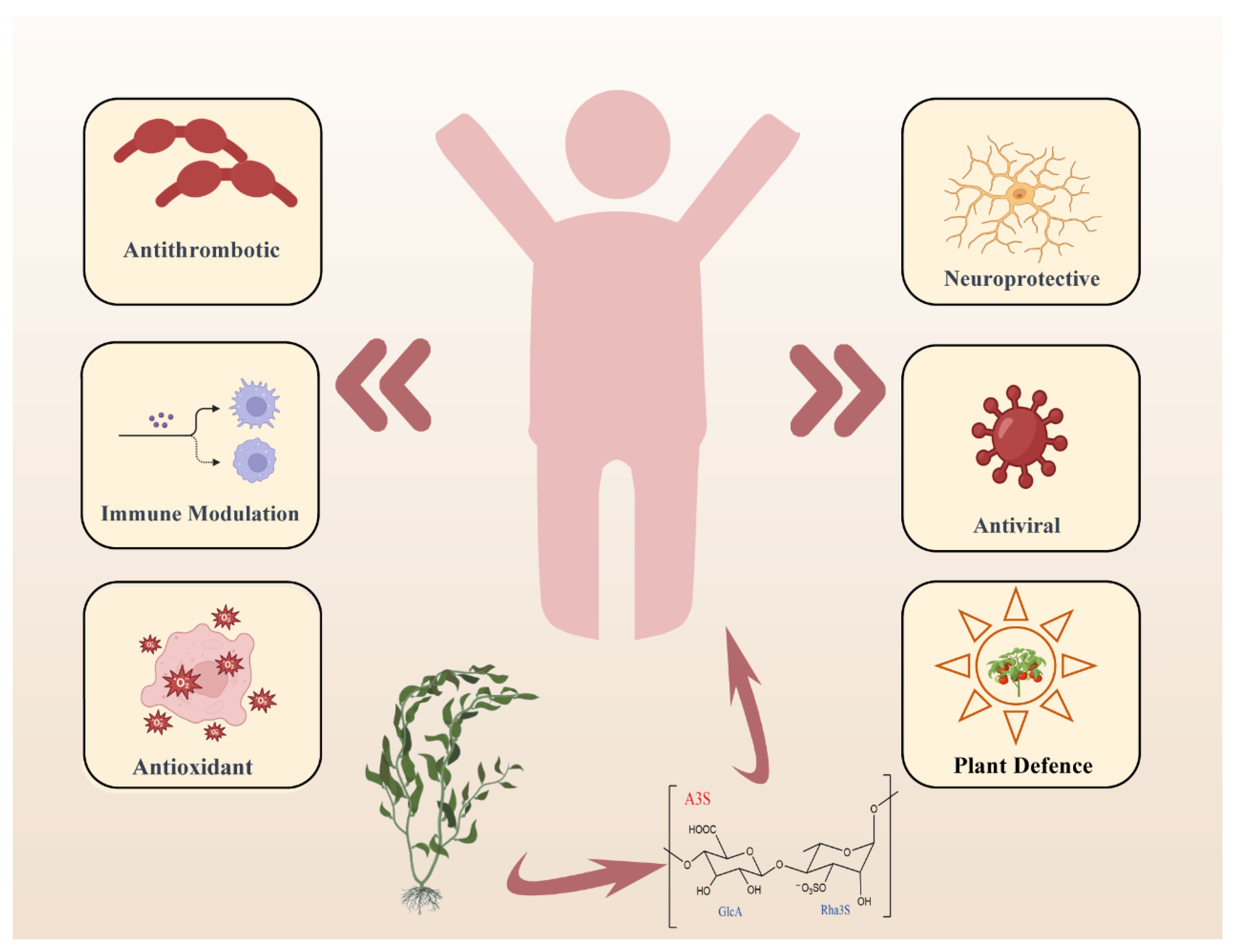
2. The Biological Activity of Ulvan Polysaccharides and Oligosaccharides

The structure of ulvan is mainly composed of sulfated rhamnose (Rha3S), glucuronic acid (GlcA), iduronic acid (IdoA), xylose (Xyl), and xylose-2-sulfate (Xyl2S) [11]. The four disaccharide structural units of the polysaccharide backbone are shown in Figure 1, A3S [→4)-β-D-GlcA-(1→4)-α-L-3-Rha3S-(1→] and B3S [→4)-α-L-IdoA-(1→4)-α-L-3-Rha3S-(1→], U3S: [→4)-β-D-Xyl-(1→4)-α-L-3-Rha3S-(1→], and U2′S3S: [→4)-β-D-sulfated Xyl-(1→4)-α-L-3-Rha3S(1→] [6,13]. The content and composition of these disaccharides vary depending on the cultivation environment and extraction method. The structural components, in terms of dry weight percentage, include rhamnose (16.8–45%), xylose (2.1–12%), glucose (0.5–6.4%), glucuronic acid (6.5–19%), sulfate (16–23.2%), and aedugaric acid (1.1–23.2%).
Traditionally, ulvan has been a customary dietary component (marine lettuce), valued for its nutritional richness and potential health-enhancing attributes [17,18,19,20,21,22]. However, the high molecular weight and low solubility have limited polysaccharide utilization. In contrast, oligosaccharides possess several advantages due to enhanced solubility and absorption, making them more valuable in the field of functional foods and biomedicine [6,8,13,23,24]. Ulva oligosaccharides have various bioactivities (Figure 2), such as antiviral [25,26,27,28], antioxidant activity [27,29,30,31,32,33,34,35], antihyperlipidemic activity [31,36,37,38,39,40], and neuroprotection activity [41,42]. The detailed information of ulvan bioactivities are listed in the Supporting Information (Table S1).
The ulvan showed the anti-virus bioactivity in vitro or in vivo assay. For example, the ulvan from Laminaria angustata algae exhibited inhibitory activity against herpes simplex virus (HSV). It was found that the presence of sulfate groups enhanced the inhibitory ability of polysaccharides against HSV-1 viruses, and the antiviral activity increased with increasing sulfate content [34,43,44]. Chi et al. [28] collected samples of Ulva pertusa from the coast of China, which were cleaved by ulvan lyase to generate ulvan oligosaccharide Ulvan-F1. The experimental results showed that, at a concentration of 100 μg/mL, both polysaccharide and its degradation product Ulvan-F1 exhibited significant inhibitory activity against vesicular stomatitis virus (VSV), with inhibition rates of 40.75% and 40.13%, respectively. There is an increasing number of studies on the conformational relationship of the antiviral effects of sulfated polysaccharides, and these data underline the importance of sulfated polysaccharides in the development of antiviral drugs and drug delivery [26,45].
The antioxidant capacity of ulvan has been reported. The antioxidant activity of ulvan is mainly attributed to its multiple reactive groups, which trap free radicals, inhibit oxidative reactions, and attenuate oxidative stress-induced cellular damage. Hydroxyl radical scavenging activity is proportional to sulfate content [31]. The sulfated polysaccharides extracted using the enzymatic method contained higher levels of glyoxalates, which enhanced their antioxidant potential. Therefore, oligosaccharides extracted using enzymatic methods are an important source for the development of natural antioxidants and are expected to play an important role in the field of functional foods.
Hyperlipidemia is a common metabolic disorder characterized by abnormally high levels of cholesterol and triglycerides in the blood. Qi and co-workers [36] synthesized high sulfate content ulvan (HU) and evaluated its antihyperlipidemic effects in mice. HU exhibited significantly stronger antihyperlipidemic activity compared to natural ulvan. These results emphasize the importance of sulfate content in enhancing ulvan’s antihyperlipidemic potential. The effects of different molecular weights of ulvan on lipid metabolism [37] and the possible antihyperlipidemic mechanisms of ulvan [38] have been further investigated.
Alzheimer’s disease (AD) is a complex neurodegenerative disorder whose pathogenesis involves multiple mechanisms. One of the proposed mechanisms is the abnormal folding and aggregation of amyloid β-protein (Aβ), which is considered to be one of the major factors in the onset and progression of AD. Recent studies have demonstrated that ulvan can inhibit Aβ aggregation in a concentration-dependent manner [41]. This finding provides a new research direction for the use of ulvan as potential functional foods for the treatment of AD.
Ulva lactuca can be added to the feed of weaned piglets as an alternative source of nutrients and bioactive compounds [46]. Given that green algae contain ulvan, which is indigestible to monogastric animals, the addition of ulvan lyase to the feed could enhance the digestibility of ulvan. The results of experiments showed that the addition of 7% Ulva lactuca and 0.01% recombinant ulvan lyase diet to the feed of weaned piglets contributed to an increase in the abundance of intestinal microflora, offering potential applications in the feed industry [47].
Recent studies have reported the role of ulvan and oligosaccharides in plant protection. It has demonstrated that ulvan-treated tomato seedlings and Medicago truncatula exhibit enhanced disease resistance. In tomato seedlings, ulvan treatment led to increased phenylalanine ammonia-lyase (PAL) activity in the leaves [48]. Additionally, the gene expression profiles of ulvan-treated Medicago truncatula were similar to those treated with methyl jasmonate [49]. These findings suggest that ulvan has the potential as a bio-elicitor, enhancing the disease resistance of plants.

3. Source, Properties, Structures, and Catalytic Mechanism of the Ulvan Lyase

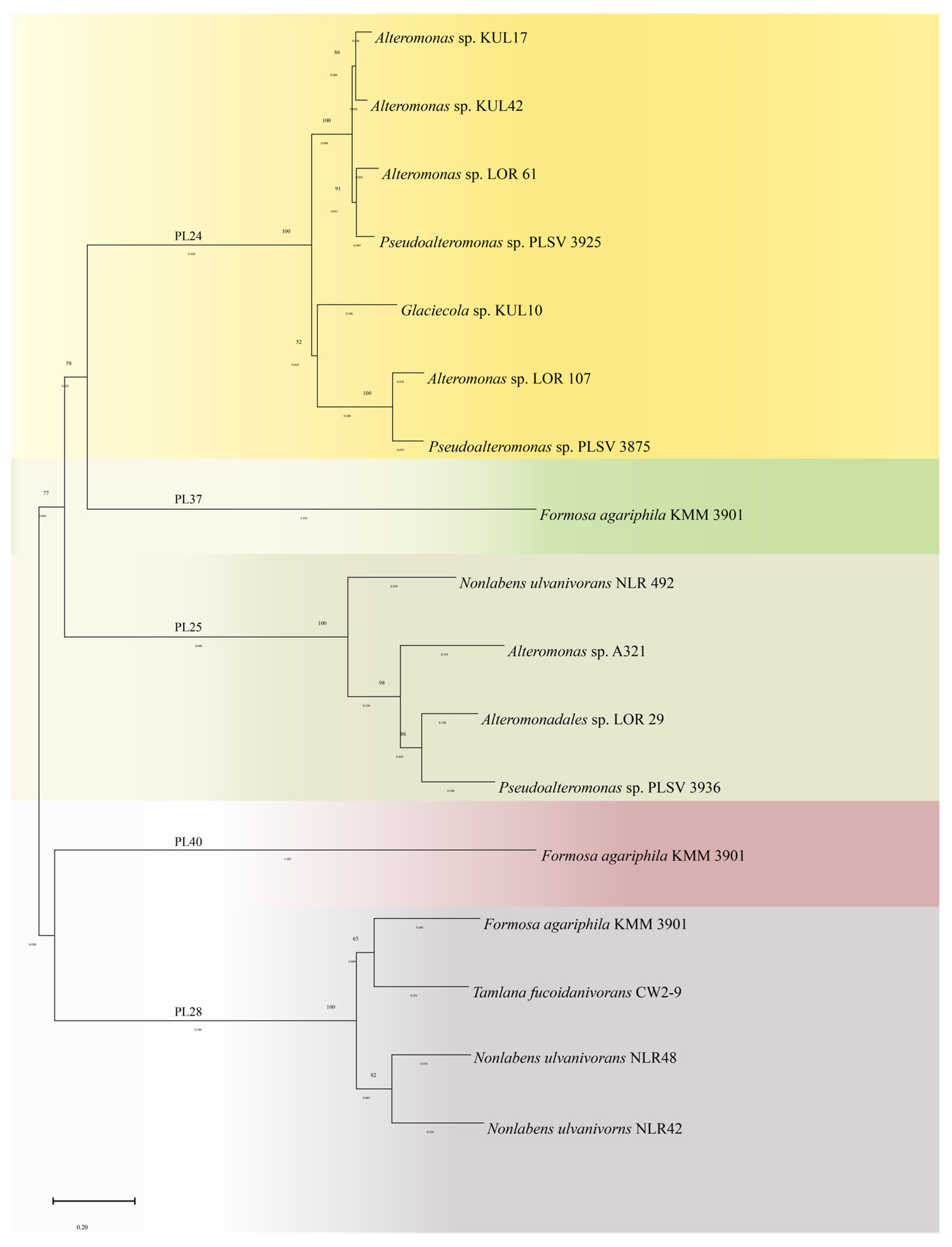
3.1. Sources and Categories of Ulvan Lyase

In 1997, Lahaye et al. [50] first detected the activity of ulvan lyase in marine bacteria. With the development of genomics, many ulvan lyase sequences have been included in the Carbohydrate Active enZYmes (CAZy) database (http://www.cazy.org/, accessed on 24 August 2024) [51]. The filamentous fungus Trichoderma sp. GL2 produced an extracellular glucuronide lyase (GL) that was active toward ulvan when grown under conditions where glucuronic acid was the sole carbon source [52]. The first isolated ulvan lyase was from the flavobacteria Persicivirga ulvanivorans, whose gene encoding the catalytic module had no homologous sequences in the GenBank sequence database and was classified as a new polysaccharide lyase family [53]. This enzyme was identified as an endo-type cleaving the glycosidic bond between glucuronic acid or iduronic acid. Nowadays, the known species that are capable of producing ulvan lyase mainly include Alteromonas, Pseudoalteromonas, Formosa agariphila, and Nonlabens ulvanivorans.
Currently, ulvan lyases are divided into five families, that is PL24, PL25, PL28, PL37, and PL40, based on the amino acid sequence. There are seven members of the PL24 family in the CAZy database, which are derived from Alteromonas sp. KUL17 [54], Alteromonas sp. KUL42 [54], Alteromonas sp. LOR_107 [55], Alteromonas sp. LOR_61 [55], Glaciecola sp. KUL10 [54,56], Pseudoalteromonas sp. PLSV_3875 [57] and Pseudoalteromonas sp. PLSV_3925 [55]. Two PL25 family members in CAZy are derived from Alteromonas sp. A321 [58], and Pseudoalteromonas sp. PLSV_3936 [59]. Although LOR_29 [60] from Alteromonadales sp. and NLR_492 [60] from Nonlabens ulvanivorans were not included in the CAZy database, they have been included in the GenBank under the numbers WP_052010178.1 and WP_036580476.1, respectively. PL28 family members are derived from Formosa agariphila KMM 3901 [61], Nonlabens ulvanivorns NLR48 [62] and Nonlabens ulvanivorns NLR42 [53]. In addition, PL37 [63], as well as PL40 [64], derived from Formosa agariphila KMM 3901 [61], were also included in the CAZy database. Besides, Olivier et al. [10] successfully isolated a microbial species capable of secreting ulvan lyase. Through 16S rRNA sequencing, the microbial species’ gene was identified as GIIUL2 and its affiliation with the genus Alteromonas. However, in comparison to the registered species available in GenBank, the homology of this species was less than 95%. Consequently, the classification of the ulvan lyase produced by this species into a specific PL family remains uncertain.
A phylogenetic tree was constructed using MEGA 11 [65] software to investigate the relationships among five families of ulvan lyase. As shown in Figure 3, the five families were divided into three main branches on the phylogenetic tree. The PL24 and PL25 families were grouped into one major class, the PL28 family formed another independent branch, and the PL37 and PL40 families clustered on the third branch. This is consistent with the structural information that PL24 and PL25 families have a similar 3D structure, which adopts the β-propeller configuration. In contrast, the PL28 family lyase exhibits a β-jelly roll structure, suggesting that there are significant structural differences between the enzymes of different families.

3.2. Enzymatic Properties of Ulvan Lyase

Temperature and pH are two important factors affecting the enzymatic activity of ulvan lyase. The ulvan lyase exhibits the highest activity within the pH range of 7.5 to 9.0. This is attributed to the fact that most identified ulvan lyases are isolated from the marine bacteria and the intestinal environment of marine organisms, which suggests a potential evolutionary adaptation of marine bacteria to the weak alkaline environment [66].
The optimum temperature for the ulvan lyase enzymatic reaction is 30–50 °C, as shown in Table 1. For example, the optimum temperature of PL24 family Alteromonas sp. LOR_107 was 40 °C [55]; for Pseudoalteromonas sp. PLSV_3875, this value was 35 °C [57]. Compared with LOR_107, PLSV_3875 has higher stability and still maintained 100% activity after 24 h at 30 °C, while LOR_107 lost 90% activity under the same conditions. Among the PL25 family, Alteromonadales sp. LOR_29 had the highest enzymatic activity at 45 °C [60]; the optimum temperature of Alteromonas sp. A321 was higher than LOR_29, which was 50 °C [58]. The optimum temperature of ulvan lyase derived from Formosa agariphila KMM 3901 of the PL37 family was 40 °C [63]. The Nonlabens ulvanivorns NLR42 enzyme exhibited the highest activity at 50 °C. However, this enzyme had poor thermal stability at 50 °C and lost more than 75% of enzymatic activity in 5 min. In contrast, when incubated at 30 °C for 2 h, the enzyme activity did not decrease significantly, which indicated that NLR42 was easily inactivated at high temperatures [53].
The enzyme catalytic optimum temperature is higher than the growth temperature of organisms, which is not only found in ulvan lyases. It was reported that many enzymes have optimal catalytic temperatures that are significantly higher or lower than the growth temperature of microorganisms [69]. The differences between microbial optimal growth temperature and enzyme optimal catalytic temperature reflect microorganisms’ adaptive and evolutionary strategies that allow them to maintain physiological function and growth in diverse and fluctuating environments. For instance, the optimal catalytic temperature of PL28 ulvan lyase from Formosa agariphila is 45 °C, but it also shows good catalytic activity from 20–50 °C.
The activity of ulvan lyase was affected by surfactants, metal ions, and chelating agents, and these effects can be divided into activating and inhibitory effects. The enzymatic activity of the PL24 family from Pseudoalteromonas sp. PLSV_3875 was activated by Ca2+ and Co2+, resulting in an increase of 45% and 60%, respectively. The activity was inhibited by Mn2+ and Zn2+, and the presence of diaminetetraacetic acid (EDTA) also reduced the enzymatic activity [66]. The activity of ALT3695 from Alteromonas sp. A321 increased in the presence of Ca2+, Mg2+ and Ba2+. Conversely, low concentrations of Fe2+, Cu2+, and Co2+ resulted in decreased enzyme activity, with complete inactivation occurring at high concentrations. Furthermore, ALT3695 was completely inactivated when exposed to Cd2+, Hg2+, and Fe3+. It is likely attributed to the binding of these ions to the thiol, carboxyl, and amino groups of amino acids, which induces structural alterations and activity loss of the enzyme [58]. Among the PL28 family, the enzyme activity of FaPL28 from Formosa agariphila KMM 3901 increased by 70% in the presence of a medium concentration of NaCl (100–200 mM), while the enzyme activity decreased at a higher concentration of NaCl. Meanwhile, the addition of dithiothreitol (DTT) and EDTA decreased the activity by 36% and 63%, respectively. The impact of DTT on enzyme activity implies the potential significance of the four cysteine residues within FaPL28 [61]. These cysteine residues may play a crucial role in either the enzyme’s activity or stability by participating in the formation of disulfide bonds.

3.3. The Resolved 3D Structure of Ulvan Lyase

Generally, the enzyme 3D structures are essential for understanding catalytic mechanisms. Three families of ulvan lyase have been resolved, that is LOR_107 of the PL24 family (PDB ID: 6BYP) [70], PLSV_3936 of the PL25 family (PDB ID: 5UAM) [58], and NLR48 of the PL28 family (PDB ID: 6D2C) [62], respectively. Recently, a new structure, Uly1 of the PL24 family (PDB ID: 7CZH) [71], has also been determined, which is consistent with the conformation of 6BYP. The protein structures of three families are shown in Figure 4. The conformation of LOR_107 is consistent with that of PLSV_3936, which both adopt β-seven-bladed propeller structure (Figure 4a,b), whereas NLR48 of the PL28 family exhibits a β-sandwich jelly-roll conformation (Figure 4c).
The crystal structure of LOR_107 contains two monomers, and each monomer is folded in the form of a β-seven-bladed propeller comprising four antiparallel β-strands. The Ca2+ metal ions present in the crystal structure are located at the periphery of the propeller, far away from the active site, and play a role in maintaining the stability of the structure.
The PLSV_3936 protein belongs to the PL25 family and also adopts the configuration of β-seven-bladed propeller. Among them, blades 1, 2, 3, 4, 5, and 6 consisted of consecutive residues, while blade 7 consisted of four chains, three of which were C-terminal β-strands, and the other chain was N-terminal residues. In the PL25 family crystal structure, Zn2+ ions are bound between the second and third blades of the propeller, which are closer to the active site. Mutation experiments confirmed that Zn2+ ions play a key role in maintaining structural stability, and its deletion will lead to instability in the region near the active site, resulting in the inactivation of the enzyme.
Different from the PL24 and PL25 family’s crystal structures of ulvan lyase, NLR48 of the PL28 family adopts a β-sandwich jelly-roll fold structure (Figure 4c), with two concave β-folds superimposed on each other. Each β-sheet is composed of seven antiparallel β-strands. The inner β-sheet is composed of β1-β4-β13-β6-β7-β8-β9 strands, while the outer β-sheet contains β2-β3-β14-β5-β10-β11-β12 strands. The inner β-sheet forms a deep cleft in which the substrate-binding site resides. The presence of Ca2+ ions in the crystal structure provides additional stability to the N-terminus, playing a structural stabilizing rather than catalytic role.
The PL24 family of ulvan lyase has catalytic specificity and only cleaves the glycosidic bond between GlcA-Rha3S. In contrast, the PL25 and PL28 family lyases could cleave the glycosidic bonds between GlcA-Rha3S and IdoA-Rha3S. Ulvan lyases exclusively depolymerize polysaccharides into disaccharide units, yielding a predominant presence of the disaccharide ΔUA-Rha3S in the cleavage products. Presently, ulvan lyases are incapable of cleaving the glycosidic bond between Xyl and Rha3S, resulting in the presence of tetrasaccharides containing Xyl in the enzymatic products.

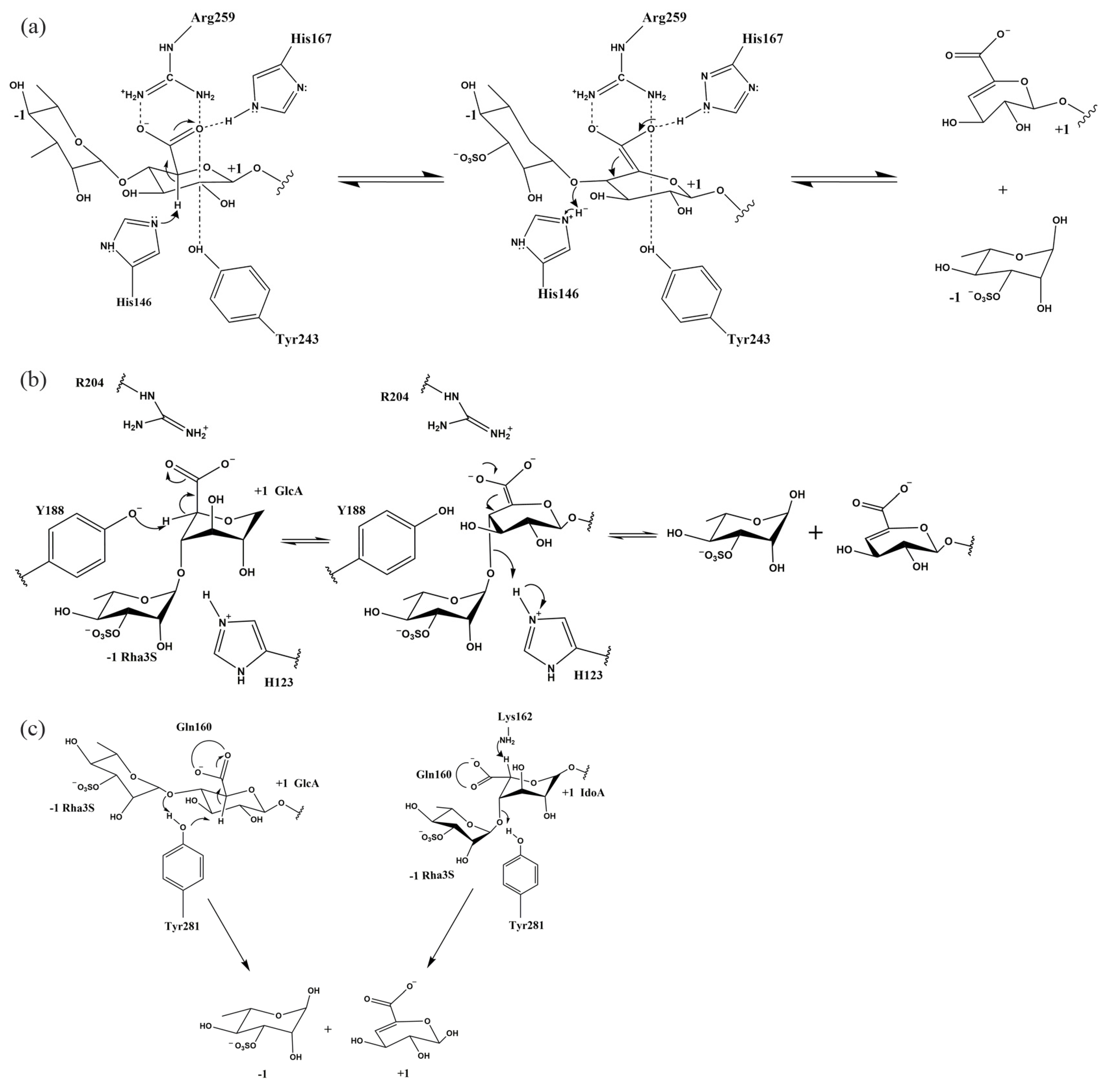
3.4. Catalytic Mechanism of Ulvan Lyase

The biochemical characterization of ulvan lyase indicates its role as an endonuclease cleaving the glycosidic bond between Rha3S and GlcA or IdoA through the β-elimination mechanism. The β-elimination mechanism is proposed by Gacesa and has been observed across various polysaccharide lyases [59,72,73,74]. This mechanism follows three steps: (1) the neutralization of the negative charge on the carboxyl group in the +1 position of the substrate, thereby lowering the pKa of the C5 proton, (2) abstraction of C5 proton by a base, (3) electron transfer takes place within the carboxyl group and causes the cleavage of the C-O4 glycosidic bond, which creates an unsaturated C4-C5 double bond at the non-reducing end [75]. In general, the residue that neutralizes the negative charge of the +1 subsite of the substrate in the first step is asparagine, aspartic acid, glutamine, glutamic acid, or histidine. According to the nomenclature of Davies et al. for polysaccharides, −1 position represents the non-reducing end, +1 position represents the reducing end, and the cleavage site of the glycosidic bond occurs between the −1 and +1 subsites.
The catalytic mechanisms of the PL24, PL25, and PL28 families have been proposed based on the crystallography analysis and mutation experiments. Within the PL24 family, residues Arg259 and His167 form hydrogen bonds with the GlcA carboxyl group at the +1 position, which neutralizes the negative charge on the carboxyl group of the substrate uronic acid (Figure 5a) [70]. In addition, residue Tyr243 also interacts with the carboxyl group of GlcA. Therefore, these three residues achieve neutralization of the charge on the carboxylate, which reduces the pKa of the C5 proton of +1 position monosaccharide. Residue His146 serves as a base to abstract the C5 proton. On the other hand, His146 also acts as an acid to provide proton, thereby facilitating the formation of double bonds between C4 and C5 and breaking of glycosidic bonds. The mutational experiments provided insights into the distinct roles played by three residues in neutralizing the carboxyl group’s negative charge. Inactivation of the R259A and H167A mutants highlighted the crucial roles of Arg259 and His167 in neutralizing the carboxyl group’s negative charge. The Y243F mutant, while showing residual activity, indicated that residue Tyr243 plays a supportive rather than a primary role in carboxyl group neutralization. Furthermore, the loss of enzymatic activity in the R320N mutant suggests that the arginine mutation at position 320 may cause the Arg259 side chain to deviate towards the main chain Gly262-Asn263. This deviation likely triggers subtle rearrangement in the protein structure, which can result in Arg259 being unable to neutralize the acidic group of GlcA in the substrate and hindering the extraction of the C5 proton. This indirectly suggests the importance of Arg259 in charge neutralization.
The crystallographic analysis of the enzyme complexed with a tetrasaccharide substrate containing GlcA, in combination with mutagenesis experiments, was undertaken to identify key residues responsible for catalyzing the cleavage of the GlcA to Rha3S glycosidic bond within the PL25 family. As depicted in Figure 5b [59], the complex structure unveiled the role of Arg204 in neutralizing the acidic group of the aronic acid in the +1 position, with help from His143 and Tyr246. Residue Tyr188 is located near the C5 proton at the +1 position and most likely acts as a general base to the abstract C5 proton. His123, as an acid, will transfer the proton to the bridging oxygen. Breakage of the C4-O4 bond and formation of the C4-C5 double bond will then occur. However, the complex structure of the ΔUA-R3S-IdoA-R3S substrate with the enzyme is currently unresolved, leaving the mechanism for cleavage of the IdoA-containing polysaccharide glycosidic bond not fully elucidated. Despite this, mutation experiments showed that mutations in Arg204, His123, and Tyr188 resulted in enzyme inactivation, strongly suggesting that these residues play an important role in catalysis.
As shown in Figure 5c [62], based on the resolved crystal structure and alignment with other polysaccharide lyases, the mechanism of the PL28 family was deposited. The PL28 targets different cleavage residues containing GlcA and IdoA glycosidic bonds. It is proposed that Gln160 attenuates the pKa of the acidic group by forming two hydrogen bonds with the carboxyl group of GlcA in the +1 position. Arg117 residue also forms a hydrogen bond with the carboxyl group of GlcA, which can further neutralize the negative charge in the carboxyl group of GlcA in the +1 position. From the position of the residues in the crystal structure, the hydroxyl group of Tyr281 points to the proton and could act as a base to abstract the C5 proton. At the same time, this residue acts as an acid, providing a proton to the bridging oxygen, prompting the breaking of the C4-O glycosidic bond. In the IdoA-containing glycosidic linkage case, Tyr281 is located on the other side of the C5 proton and is, therefore, unlikely to extract the proton as a base. Therefore, the hydroxyl group of Lys162 is thought to abstract the proton at the C5 position based on structural alignment with the PL7 and PL13 families. The residues involved in catalysis in the three families are listed in Table 2.

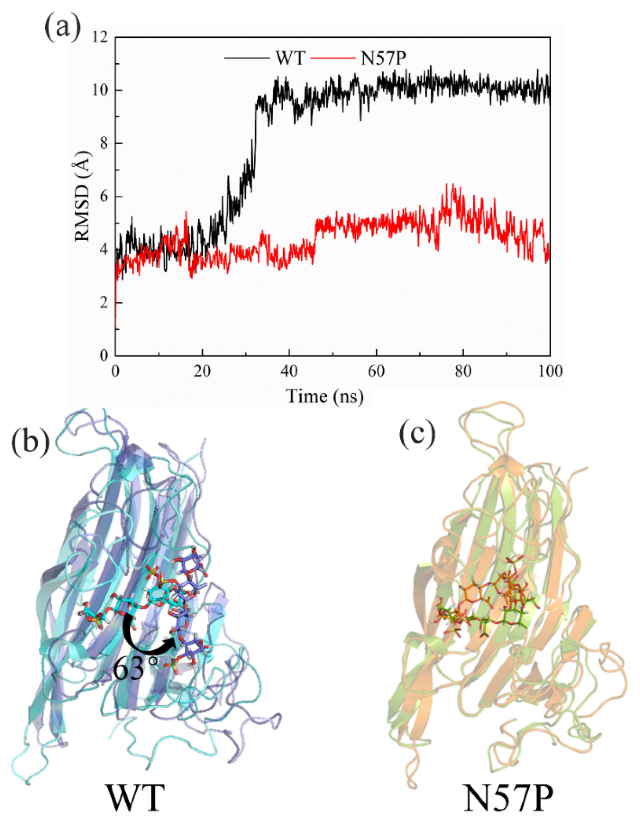
4. Molecular Modification of Ulvan Lyase

Enzyme stability is a critical factor in the industrial production of oligosaccharides. Ulvan lyases from natural resources showed low stability. Therefore, molecular modification of the enzyme is required to improve its stability and activity. The identification of key residues involved in catalysis is essential for enzyme modification. Ulaganathan et al. [59] found that amino acid residues His123, Tyr188, Arg204, and His264 play a key role in substrate catalysis of PLSV_3936. In addition, Qin et al. [66] used a similar approach to construct mutants of ASPL from the genus Alteromonas; it was indicated that mutants H146A, H167A, R259A, R320A, N263F, Y243F, and H384A lost their activity, indicating the importance of these residues in substrate catalysis.
Relative to molecular modification and protein engineering studies of brown algae of alginate lyase [76,77], there is less research on ulvan lyase. Several methods, including disulfide bond engineering [78] and truncation of non-catalytic domains [79,80], have been employed to improve the thermal stability of alginate lyase. An engineered N57P mutant of ulvan lyase from Nonlabens ulvanivorans was recently rationally designed [81]. This mutant demonstrates significantly improved catalytic efficiency and thermostability compared to the wild type (WT). Molecular dynamics (MD) simulations provided insights that N57P reduces protein flexibility through the proline substitution in the flexible loop region E47-N57. This reduction in flexibility indirectly contributed to the stability of the adjacent loop regions, specifically T102-R117 and G209-R216. Consequently, the substrate anchored in the active site, resulting in the enhanced catalytic efficiency observed in N57P. The root-mean-square deviation (RMSD) of the substrate and changes of position from 0 ns to 100 ns in WT and N57P are shown in Figure 6. Nevertheless, the investigation of ulvan lyase protein engineering is still limited; more studies are needed to improve the stability and enzymatic activity. Based on the N57P mutant, N57P/Q229M was further iterated to obtain N57P/Q229M (both enzyme activity and thermal stability were enhanced). MD results showed that the N57P/Q229M mutation improves thermal stability by introducing additional hydrogen bonds [82].

5. Production of Ulvan Lyase

The hydrolysis conditions of different families of enzymes are different, but the hydrolysis pH is weakly alkaline, and the temperature is mostly in the range of 30–50 °C. For instance, 5 mg sulfated polysaccharides extracted from Ulva lactuca were dissolved in 0.5 mL of 20 mM Tris buffer (pH 8.5) containing 150 mM NaCl. After adding 50 μg enzyme, the mixture was incubated at 37 °C for 12 h to obtain the end products [83]. These products were separated by size-exclusion chromatography, yielding oligosaccharides of different molecular structures. Currently, the oligosaccharides obtained from enzymatic hydrolysis of ulvan are complex mixtures of various molecular weights rather than single molecules.
Further structural analysis of the oligosaccharide fragments using 1H NMR and LC-MS revealed that the fragments included disaccharides, trisaccharides, tetrasaccharides, and pentasaccharides. The disaccharide structure consisted of unsaturated uronic acid (ΔUA) linked to Rha3S (ΔUA-Rha3S), the trisaccharide structure was Rha3S-Xyl-Rha3S, the tetrasaccharide had an unsaturated uronic acid at the non-reducing end (ΔUA-Rha3S-Xyl-Rha3S), and the pentasaccharide was Rha3S-Xyl-Rha3S-Xyl-Rha3S, along with some branched oligosaccharides. Experimental results showed that as the enzymatic reaction time increased, the detected oligosaccharide chain length gradually decreased, but only disaccharide units were obtained in the end, with no monosaccharides detected.
Additionally, Qiao et al. [84] explored fermentation conditions in 5 L and 30 L bioreactors and optimized the fermentation process using kinetic models. The optimal conditions were found to be 28–32 °C and an initial pH of 7.0, demonstrating the potential for industrial-scale production of ulvan lyase. After 20 h of fermentation, the enzyme activity reached 1.20 U/mL, which further validated the feasibility of industrial-scale production of ulvan lyase.
The composition of the medium and the culture conditions of strain are the key factors affecting the expression and enzyme activity of ulvan lyase. Luria-Bertani medium with peptone and yeast powder was utilized in most fermentation processes [58,66]. In addition, other researchers utilized ZoBell medium containing aged seawater as fermentation conditions [53].

6. Conclusions and Prospects

Ulva oligosaccharides possess a wide range of biological activities, so they have huge potential for use in food, medicine, and cosmetics. Ulvan lyases are the most effective polysaccharide lyases for the bioconversion of polysaccharides into oligosaccharides, and the production of oligosaccharides using this enzyme is undoubtedly a sustainable and environmental-friendly method. Extensive research has been carried out on the evolutionary relationships between crystal structures and catalytic mechanisms of different families of ulvan lyases. However, compared to polysaccharide lytic enzymes from red and brown algae, such as alginate lyases and carrageenases, ulvan lyase still requires more exploration and research.
The structure of protein is crucial to understanding the catalytic mechanism of enzymes. Therefore, the more high-resolution resolved protein structure will enhance the understanding of structure–function relationships. In addition to structural biology, computational techniques such as Alphafold2, can be used for predicting protein structures. MD simulation and quantum mechanics/molecular mechanics (QM/MM) tools could also be used to guide the design and modification of enzymes to improve their catalytic properties, including activity, substrate specificity, and thermal and pH stability. Specifically, in light of the distinctive catalytic proficiency exhibited by PL24 family lyase, it demonstrates the capability to selectively generate IdoA tetrasaccharides. The PL25 and PL28 family lyases have the potential for large-scale preparation of disaccharides. In addition to improving the catalytic performance of the ulvan lyase, the benefits of enzymatic hydrolysis can be achieved by screening high enzyme-producing strains and genetic engineering of strains to enable them to survive under industrial production conditions. These improvements will provide more possibilities for the industrial enzymatic production of oligosaccharides and marine-derived functional foods and applications in various fields.

Supplementary Materials

The following supporting information can be downloaded at: https://www.mdpi.com/article/10.3390/foods13172820/s1, Table S1: The bioactivities of ulvan and detailed information. Reference [85] is cited in the Supplementary Materials.

Author Contributions

A.H. and X.W., writing and original draft preparation; F.L. (Fuping Lu) supervision; F.L. (Fufeng Liu), reviewing and editing, and funding acquisition. All authors have read and agreed to the published version of the manuscript.

Funding

This work was supported by the National Key R&D Program of China (2021YFC2102700), the National Natural Science Foundation of China (No. 32272269), the Postgraduate Education Reform and the Quality Improvement Project of Henan Province (YJS2024JD19).

Institutional Review Board Statement

Not applicable.

Informed Consent Statement

Not applicable.

Data Availability Statement

No new data were created or analyzed in this study. Data sharing is not applicable to this article.

Acknowledgments

The authors would like to thank Yang Wang and Yang Tianyou for their contributions to the manuscript, particularly in the areas of figure redrawing, tabulation, and language review.

Conflicts of Interest

The authors declare no conflicts of interest.

References

  1. John, R.P.; Anisha, G.S.; Nampoothiri, K.M.; Pandey, A. Micro and macroalgal biomass: A renewable source for bioethanol. Bioresour. Technol. 2011, 102, 186–193. [Google Scholar] [CrossRef] [PubMed]
  2. Brar, A.; Kumar, M.; Soni, T.; Vivekanand, V.; Pareek, N. Insights into the genetic and metabolic engineering approaches to enhance the competence of microalgae as biofuel resource: A review. Bioresour. Technol. 2021, 339, 125597. [Google Scholar] [CrossRef] [PubMed]
  3. Khoo, C.G.; Dasan, Y.K.; Lam, M.K.; Lee, K.T. Algae biorefinery: Review on a broad spectrum of downstream processes and products. Bioresour. Technol. 2019, 292, 121964. [Google Scholar] [CrossRef]
  4. Chapman, R.L. Algae: The world’s most important “plants”—An introduction. Mitig. Adapt. Strateg. Glob. Chang. 2010, 18, 5–12. [Google Scholar] [CrossRef]
  5. Cikoš, A.-M.; Šubarić, D.; Roje, M.; Babić, J.; Jerković, I.; Jokić, S. Recent advances on macroalgal pigments and their biological activities (2016–2021). Algal Res. 2022, 65, 102748. [Google Scholar] [CrossRef]
  6. Alves, A.; Sousa, R.A.; Reis, R.L. A practical perspective on ulvan extracted from green algae. J. Appl. Phycol. 2012, 25, 407–424. [Google Scholar] [CrossRef]
  7. Lahaye, M.; Axelos, M.A.V. Gelling properties of water-soluble polysaccharides from proliferating marine green seaweeds (Ulva spp.). Carbohydr. Polym. 1993, 22, 261–265. [Google Scholar] [CrossRef]
  8. Robic, A.; Gaillard, C.; Sassi, J.F.; Lerat, Y.; Lahaye, M. Ultrastructure of ulvan: A polysaccharide from green seaweeds. Biopolymers 2009, 91, 652–664. [Google Scholar] [CrossRef] [PubMed]
  9. Chi, Y.; Li, H.; Wang, P.; Du, C.; Ye, H.; Zuo, S.; Guan, H.; Wang, P. Structural characterization of ulvan extracted from Ulva clathrata assisted by an ulvan lyase. Carbohydr. Polym. 2020, 229, 115497. [Google Scholar] [CrossRef]
  10. Coste, O.; Malta, E.J.; López, J.C.; Fernández-Díaz, C. Production of sulfated oligosaccharides from the seaweed Ulva sp. using a new ulvan-degrading enzymatic bacterial crude extract. Algal Res. 2015, 10, 224–231. [Google Scholar] [CrossRef]
  11. Lahaye, M.; Robic, A. Structure and functional properties of ulvan, a polysaccharide from green seaweeds. Biomacromolecules 2007, 8, 1765–1774. [Google Scholar] [CrossRef]
  12. Ale, M.T.; Mikkelsen, J.D.; Meyer, A.S. Designed optimization of a single-step extraction of fucose-containing sulfated polysaccharides from Sargassum sp. J. Appl. Phycol. 2012, 24, 715–723. [Google Scholar] [CrossRef]
  13. Kidgell, J.T.; Magnusson, M.; de Nys, R.; Glasson, C.R.K. Ulvan: A systematic review of extraction, composition and function. Algal Res. 2019, 39, 101422. [Google Scholar] [CrossRef]
  14. Kadam, S.U.; Tiwari, B.K.; O’Donnell, C.P. Application of novel extraction technologies for bioactives from marine algae. J. Agric. Food Chem. 2013, 61, 4667–4675. [Google Scholar] [CrossRef]
  15. Li, Q.; Hu, F.; Zhu, B.; Ni, F.; Yao, Z. Insights into ulvan lyase: Review of source, biochemical characteristics, structure and catalytic mechanism. Crit. Rev. Biotechnol. 2020, 40, 432–441. [Google Scholar] [CrossRef] [PubMed]
  16. Tang, T.C.; Cao, S.S.; Zhu, B.W.; Li, Q. Ulvan polysaccharide-degrading enzymes: An updated and comprehensive review of sources category, property, structure, and applications of ulvan lyases. Algal Res. 2021, 60, 102477. [Google Scholar] [CrossRef]
  17. Bocanegra, A.; Bastida, S.; Benedi, J.; Rodenas, S.; Sanchez-Muniz, F.J. Characteristics and nutritional and cardiovascular-health properties of seaweeds. J. Med. Food 2009, 12, 236–258. [Google Scholar] [CrossRef]
  18. Castro-González, M.; Pérez-Gil, F.; Pérez-Estrella, S.; Carrillo-Domínguez, S. Chemical composition of the green alga Ulva lactuca. Cienc. Mar. 1996, 22, 205–213. [Google Scholar] [CrossRef]
  19. Agarwal, S.; Singh, V.; Chauhan, K. Antidiabetic potential of seaweed and their bioactive compounds: A review of developments in last decade. Crit. Rev. Food Sci. Nutr. 2023, 63, 5739–5770. [Google Scholar] [CrossRef]
  20. Nova, P.; Pimenta-Martins, A.; Laranjeira Silva, J.; Silva, A.M.; Gomes, A.M.; Freitas, A.C. Health benefits and bioavailability of marine resources components that contribute to health—what’s new? Crit. Rev. Food Sci. Nutr. 2020, 60, 3680–3692. [Google Scholar] [CrossRef]
  21. Singh, R.P.; Bhaiyya, R.; Khandare, K.; Tingirikari, J.M.R. Macroalgal dietary glycans: Potential source for human gut bacteria and enhancing immune system for better health. Crit. Rev. Food Sci. Nutr. 2022, 62, 1674–1695. [Google Scholar] [CrossRef] [PubMed]
  22. Xie, X.T.; Cheong, K.L. Recent advances in marine algae oligosaccharides: Structure, analysis, and potential prebiotic activities. Crit. Rev. Food Sci. Nutr. 2022, 62, 7703–7717. [Google Scholar] [CrossRef]
  23. Anisha, G.S.; Augustianath, T.; Padmakumari, S.; Singhania, R.R.; Pandey, A.; Patel, A.K. Ulvan from green macroalgae: Bioactive properties advancing tissue engineering, drug delivery systems, food industry, agriculture and water treatment. Bioresour. Technol. Rep. 2023, 22, 101457. [Google Scholar] [CrossRef]
  24. Kaeffer, B.; Benard, C.; Lahaye, M.; Blottiere, H.M.; Cherbut, C. Biological properties of ulvan, a new source of green seaweed sulfated polysaccharides, on cultured normal and cancerous colonic epithelial cells. Planta Med. 1999, 65, 527–531. [Google Scholar] [CrossRef] [PubMed]
  25. Ray, B.; Schutz, M.; Mukherjee, S.; Jana, S.; Ray, S.; Marschall, M. Exploiting the Amazing Diversity of Natural Source-Derived Polysaccharides: Modern Procedures of Isolation, Engineering, and Optimization of Antiviral Activities. Polymers 2020, 13, 136. [Google Scholar] [CrossRef]
  26. Ghosh, T.; Chattopadhyay, K.; Marschall, M.; Karmakar, P.; Mandal, P.; Ray, B. Focus on antivirally active sulfated polysaccharides: From structure-activity analysis to clinical evaluation. Glycobiology 2009, 19, 2–15. [Google Scholar] [CrossRef]
  27. Maray, S.O.; Abdel-Kareem, M.S.M.; Mabrouk, M.E.M.; El-Halmouch, Y.; Makhlof, M.E.M. In Vitro Assessment of Antiviral, Antimicrobial, Antioxidant and Anticancer Activities of Ulvan Extracted from the Green Seaweed. Thalassas 2023, 39, 779–790. [Google Scholar] [CrossRef]
  28. Chi, Y.; Zhang, M.; Wang, X.; Fu, X.; Guan, H.; Wang, P. Ulvan lyase assisted structural characterization of ulvan from Ulva pertusa and its antiviral activity against vesicular stomatitis virus. Int. J. Biol. Macromol. 2020, 157, 75–82. [Google Scholar] [CrossRef]
  29. Qi, H.; Zhao, T.; Zhang, Q.; Li, Z.; Zhao, Z.; Xing, R. Antioxidant activity of different molecular weight sulfated polysaccharides from Ulva pertusa Kjellm (Chlorophyta). J. Appl. Phycol. 2006, 17, 527–534. [Google Scholar] [CrossRef]
  30. Qi, H.; Zhang, Q.; Zhao, T.; Chen, R.; Zhang, H.; Niu, X.; Li, Z. Antioxidant activity of different sulfate content derivatives of polysaccharide extracted from Ulva pertusa (Chlorophyta) in vitro. Int. J. Biol. Macromol. 2005, 37, 195–199. [Google Scholar] [CrossRef]
  31. Qi, H.; Sun, Y. Antioxidant activity of high sulfate content derivative of ulvan in hyperlipidemic rats. Int. J. Biol. Macromol. 2015, 76, 326–329. [Google Scholar] [CrossRef] [PubMed]
  32. Harikrishnan, R.; Devi, G.; Van Doan, H.; Balasundaram, C.; Arockiaraj, J.; Jagruthi, C. Efficacy of ulvan on immune response and immuno-antioxidant gene modulation in Labeo rohita against columnaris disease. Fish. Shellfish. Immunol. 2021, 117, 262–273. [Google Scholar] [CrossRef] [PubMed]
  33. Pradhan, B.; Bhuyan, P.P.; Ki, J.S. Immunomodulatory, Antioxidant, Anticancer, and Pharmacokinetic Activity of Ulvan, a Seaweed-Derived Sulfated Polysaccharide: An Updated Comprehensive Review. Mar. Drugs 2023, 21, 300. [Google Scholar] [CrossRef]
  34. El-Shafei, R.; Hegazy, H.; Acharya, B. A Review of Antiviral and Antioxidant Activity of Bioactive Metabolite of Macroalgae within an Optimized Extraction Method. Energies 2021, 14, 3092. [Google Scholar] [CrossRef]
  35. Abd-Ellatef, G.F.; Ahmed, O.M.; Abdel-Reheim, E.S.; Abdel-Hamid, A.Z. Ulva lactuca polysaccharides prevent Wistar rat breast carcinogenesis through the augmentation of apoptosis, enhancement of antioxidant defense system, and suppression of inflammation. Breast Cancer 2017, 9, 67–83. [Google Scholar]
  36. Qi, H.M.; Huang, L.Y.; Liu, X.L.; Liu, D.M.; Zhang, Q.B.; Liu, S.M. Antihyperlipidemic activity of high sulfate content derivative of polysaccharide extracted from Ulva pertusa (Chlorophyta). Carbohydr. Polym. 2012, 87, 1637–1640. [Google Scholar] [CrossRef]
  37. Pengzhan, Y.; Ning, L.; Xiguang, L.; Gefei, Z.; Quanbin, Z.; Pengcheng, L. Antihyperlipidemic effects of different molecular weight sulfated polysaccharides from Ulva pertusa (Chlorophyta). Pharmacol. Res. 2003, 48, 543–549. [Google Scholar] [CrossRef]
  38. Qi, H.; Sheng, J. The antihyperlipidemic mechanism of high sulfate content ulvan in rats. Mar. Drugs 2015, 13, 3407–3421. [Google Scholar] [CrossRef]
  39. Jiang, N.; Li, B.; Wang, X.; Xu, X.; Liu, X.; Li, W.; Chang, X.; Li, H.; Qi, H. The antioxidant and antihyperlipidemic activities of phosphorylated polysaccharide from Ulva pertusa. Int. J. Biol. Macromol. 2020, 145, 1059–1065. [Google Scholar] [CrossRef]
  40. Qi, H.; Liu, X.; Zhang, J.; Duan, Y.; Wang, X.; Zhang, Q. Synthesis and antihyperlipidemic activity of acetylated derivative of ulvan from Ulva pertusa. Int. J. Biol. Macromol. 2012, 50, 270–272. [Google Scholar] [CrossRef]
  41. Liu, F.; Zhao, W.; Zhao, F.; Dong, Q.; Wang, Y.; Wei, W.; Jia, L.; Li, L.; Lu, F. Dual Effect of the Acidic Polysaccharose Ulvan on the Inhibition of Amyloid-beta Protein Fibrillation and Disintegration of Mature Fibrils. ACS Appl. Mater. Interface 2020, 12, 41167–41176. [Google Scholar] [CrossRef]
  42. Wang, W.; Wang, X.; Gao, W.; Cui, Z.; Zhang, H.; Lu, F.; Liu, F. Ulvan inhibits alpha-synuclein fibrillation and disrupts the mature fibrils: In vitro and in vivo studies. Int. J. Biol. Macromol. 2022, 211, 580–591. [Google Scholar] [CrossRef] [PubMed]
  43. Wang, L.; Wang, X.; Wu, H.; Liu, R. Overview on biological activities and molecular characteristics of sulfated polysaccharides from marine green algae in recent years. Mar. Drugs 2014, 12, 4984–5020. [Google Scholar] [CrossRef] [PubMed]
  44. Zhong, R.T.; Wan, X.Z.; Wang, D.Y.; Zhao, C.; Liu, D.; Gao, L.Y.; Wang, M.F.; Wu, C.J.; Nabavid, S.M.; Daglia, M.; et al. Polysaccharides from Marine: Structure and function. Trends Food Sci. Technol. 2020, 99, 11–20. [Google Scholar] [CrossRef]
  45. Cunha, L.; Grenha, A. Sulfated Seaweed Polysaccharides as Multifunctional Materials in Drug Delivery Applications. Mar. Drugs 2016, 14, 42. [Google Scholar] [CrossRef]
  46. Ribeiro, D.M.; Costa, M.M.; Trevisi, P.; Carvalho, D.F.P.; Correa, F.; Martins, C.F.; Pinho, M.; Mourato, M.; de Almeida, A.M.; Freire, J.P.B.; et al. Piglets performance, nutrient digestibility and gut health in response to feeding Ulva lactuca seaweed supplemented with a recombinant ulvan lyase or a commercial carbohydrase mixture. J. Anim. Physiol. Anim. Nutr. 2024, 17. [Google Scholar] [CrossRef] [PubMed]
  47. Ribeiro, D.M.; Coelho, D.; Costa, M.; Carvalho, D.F.P.; Leclercq, C.C.; Renaut, J.; Freire, J.P.B.; Almeida, A.M.; Prates, J.A.M. Integrated transcriptomics and proteomics analysis reveals muscle metabolism effects of dietary Ulva lactuca and ulvan lyase supplementation in weaned piglets. Sci. Rep. 2024, 14, 15. [Google Scholar] [CrossRef] [PubMed]
  48. El Modafar, C.; Elgadda, M.; El Boutachfaiti, R.; Abouraicha, E.; Zehhar, N.; Petit, E.; El Alaoui-Talibi, Z.; Courtois, B.; Courtois, J. Induction of natural defence accompanied by salicylic acid-dependant systemic acquired resistance in tomato seedlings in response to bioelicitors isolated from green algae. Sci. Hortic. 2012, 138, 55–63. [Google Scholar] [CrossRef]
  49. Jaulneau, V.; Lafitte, C.; Jacquet, C.; Fournier, S.; Salamagne, S.; Briand, X.; Esquerre-Tugaye, M.T.; Dumas, B. Ulvan, a sulfated polysaccharide from green algae, activates plant immunity through the jasmonic acid signaling pathway. J. Biomed. Biotechnol. 2010, 2010, 525291. [Google Scholar] [CrossRef]
  50. Lahaye, M.; Brunel, M.; Bonnin, E. Fine chemical structure analysis of oligosaccharides produced by an ulvan-lyase degradation of the water-soluble cell-wall polysaccharides from Ulva sp. (Ulvales, Chlorophyta). Carbohydr. Res. 1997, 304, 325–333. [Google Scholar] [CrossRef]
  51. Cantarel, B.L.; Coutinho, P.M.; Rancurel, C.; Bernard, T.; Lombard, V.; Henrissat, B. The Carbohydrate-Active EnZymes database (CAZy): An expert resource for Glycogenomics. Nucleic Acids Res. 2009, 37, D233–D238. [Google Scholar] [CrossRef] [PubMed]
  52. Delattre, C.; Michaud, P.; Keller, C.; Elboutachfaiti, R.; Beven, L.; Courtois, B.; Courtois, J. Purification and characterization of a novel glucuronan lyase from Trichoderma sp. GL2. Appl. Microbiol. Biotechnol. 2006, 70, 437–443. [Google Scholar] [CrossRef] [PubMed]
  53. Nyvall Collen, P.; Sassi, J.F.; Rogniaux, H.; Marfaing, H.; Helbert, W. Ulvan lyases isolated from the Flavobacteria Persicivirga ulvanivorans are the first members of a new polysaccharide lyase family. J. Biol. Chem. 2011, 286, 42063–42071. [Google Scholar] [CrossRef] [PubMed]
  54. He, C.; Muramatsu, H.; Kato, S.I.; Ohnishi, K. Characterization of an Alteromonas long-type ulvan lyase involved in the degradation of ulvan extracted from Ulva ohnoi. Biosci. Biotechnol. Biochem. 2017, 81, 2145–2151. [Google Scholar] [CrossRef]
  55. Kopel, M.; Helbert, W.; Belnik, Y.; Buravenkov, V.; Herman, A.; Banin, E. New Family of Ulvan Lyases Identified in Three Isolates from the Alteromonadales Order. J. Biol. Chem. 2016, 291, 5871–5878. [Google Scholar] [CrossRef]
  56. He, C.; Ohnishi, K. Efficient renaturation of inclusion body proteins denatured by SDS. Biochem. Biophys. Res. Commun. 2017, 490, 1250–1253. [Google Scholar] [CrossRef]
  57. Qin, H.M.; Xu, P.P.; Guo, Q.Q.; Cheng, X.T.; Gao, D.K.; Sun, D.Y.; Zhu, Z.L.; Lu, F.P. Biochemical characterization of a novel ulvan lyase from Pseudoalteromonas sp strain PLSV. RSC Adv. 2018, 8, 2610–2615. [Google Scholar] [CrossRef]
  58. Gao, J.; Du, C.; Chi, Y.; Zuo, S.; Ye, H.; Wang, P. Cloning, Expression, and Characterization of a New PL25 Family Ulvan Lyase from Marine Bacterium Alteromonas sp. A321. Mar. Drugs 2019, 17, 568. [Google Scholar] [CrossRef]
  59. Ulaganathan, T.; Boniecki, M.T.; Foran, E.; Buravenkov, V.; Mizrachi, N.; Banin, E.; Helbert, W.; Cygler, M. New Ulvan-Degrading Polysaccharide Lyase Family: Structure and Catalytic Mechanism Suggests Convergent Evolution of Active Site Architecture. ACS Chem. Biol. 2017, 12, 1269–1280. [Google Scholar] [CrossRef]
  60. Foran, E.; Buravenkov, V.; Kopel, M.; Mizrahi, N.; Shoshani, S.; Helbert, W.; Banin, E. Functional characterization of a novel “ulvan utilization loci” found in Alteromonas sp LOR genome. Algal Res. 2017, 25, 39–46. [Google Scholar] [CrossRef]
  61. Reisky, L.; Stanetty, C.; Mihovilovic, M.D.; Schweder, T.; Hehemann, J.H.; Bornscheuer, U.T. Biochemical characterization of an ulvan lyase from the marine flavobacterium Formosa agariphila KMM 3901(T). Appl. Microbiol. Biotechnol. 2018, 102, 6987–6996. [Google Scholar] [CrossRef]
  62. Ulaganathan, T.; Banin, E.; Helbert, W.; Cygler, M. Structural and functional characterization of PL28 family ulvan lyase NLR48 from Nonlabens ulvanivorans. J. Biol. Chem. 2018, 293, 11564–11573. [Google Scholar] [CrossRef] [PubMed]
  63. Konasani, V.R.; Jin, C.; Karlsson, N.G.; Albers, E. A novel ulvan lyase family with broad-spectrum activity from the ulvan utilisation loci of Formosa agariphila KMM 3901. Sci. Rep. 2018, 8, 14713. [Google Scholar] [CrossRef]
  64. Reisky, L.; Prechoux, A.; Zuhlke, M.K.; Baumgen, M.; Robb, C.S.; Gerlach, N.; Roret, T.; Stanetty, C.; Larocque, R.; Michel, G.; et al. A marine bacterial enzymatic cascade degrades the algal polysaccharide ulvan. Nat. Chem. Biol. 2019, 15, 803–812. [Google Scholar] [CrossRef] [PubMed]
  65. Hall, B.G. Building phylogenetic trees from molecular data with MEGA. Mol. Biol. Evol. 2013, 30, 1229–1235. [Google Scholar] [CrossRef] [PubMed]
  66. Qin, H.M.; Gao, D.; Zhu, M.; Li, C.; Zhu, Z.; Wang, H.; Liu, W.; Tanokura, M.; Lu, F. Biochemical characterization and structural analysis of ulvan lyase from marine Alteromonas sp. reveals the basis for its salt tolerance. Int. J. Biol. Macromol. 2020, 147, 1309–1317. [Google Scholar] [CrossRef]
  67. Xu, Y.; Li, J.; An, L.; Qiu, Y.K.; Mao, A.H.; He, Z.X.; Guo, J.L.; Yan, H.B.; Li, H.; Hu, Z. Biochemical Characterization of a Novel Thermostable Ulvan Lyase from Tamlana fucoidanivorans CW2-9. J. Agric. Food Chem. 2024, 72, 11773–11781. [Google Scholar] [CrossRef]
  68. Mann, A.J.; Hahnke, R.L.; Huang, S.; Werner, J.; Xing, P.; Barbeyron, T.; Huettel, B.; Stuber, K.; Reinhardt, R.; Harder, J.; et al. The genome of the alga-associated marine flavobacterium Formosa agariphila KMM 3901T reveals a broad potential for degradation of algal polysaccharides. Appl. Environ. Microbiol. 2013, 79, 6813–6822. [Google Scholar] [CrossRef]
  69. Li, G.; Rabe, K.S.; Nielsen, J.; Engqvist, M.K.M. Machine Learning Applied to Predicting Microorganism Growth Temperatures and Enzyme Catalytic Optima. ACS Synth. Biol. 2019, 8, 1411–1420. [Google Scholar] [CrossRef]
  70. Ulaganathan, T.; Helbert, W.; Kopel, M.; Banin, E.; Cygler, M. Structure-function analyses of a PL24 family ulvan lyase reveal key features and suggest its catalytic mechanism. J. Biol. Chem. 2018, 293, 4026–4036. [Google Scholar] [CrossRef]
  71. Xu, F.; Dong, F.; Sun, X.H.; Cao, H.Y.; Fu, H.H.; Li, C.Y.; Zhang, X.Y.; McMinn, A.; Zhang, Y.Z.; Wang, P.; et al. Mechanistic Insights into Substrate Recognition and Catalysis of a New Ulvan Lyase of Polysaccharide Lyase Family 24. Appl. Environ. Microbiol. 2021, 87, e0041221. [Google Scholar] [CrossRef] [PubMed]
  72. Dong, S.; Wei, T.D.; Chen, X.L.; Li, C.Y.; Wang, P.; Xie, B.B.; Qin, Q.L.; Zhang, X.Y.; Pang, X.H.; Zhou, B.C.; et al. Molecular insight into the role of the N-terminal extension in the maturation, substrate recognition, and catalysis of a bacterial alginate lyase from polysaccharide lyase family 18. J. Biol. Chem. 2014, 289, 29558–29569. [Google Scholar] [CrossRef] [PubMed]
  73. Yip, V.L.; Withers, S.G. Breakdown of oligosaccharides by the process of elimination. Curr. Opin. Chem. Biol. 2006, 10, 147–155. [Google Scholar] [CrossRef]
  74. Li, Q.; Zheng, L.; Guo, Z.; Tang, T.; Zhu, B. Alginate degrading enzymes: An updated comprehensive review of the structure, catalytic mechanism, modification method and applications of alginate lyases. Crit. Rev. Biotechnol. 2021, 41, 953–968. [Google Scholar] [CrossRef] [PubMed]
  75. Garron, M.L.; Cygler, M. Structural and mechanistic classification of uronic acid-containing polysaccharide lyases. Glycobiology 2010, 20, 1547–1573. [Google Scholar] [CrossRef] [PubMed]
  76. Cao, S.; Li, Q.; Xu, Y.; Tang, T.; Ning, L.; Zhu, B. Evolving strategies for marine enzyme engineering: Recent advances on the molecular modification of alginate lyase. Mar. Life Sci. Technol. 2022, 4, 106–116. [Google Scholar] [CrossRef]
  77. Li, L.; Cao, S.; Zhu, B.; Yao, Z.; Zhu, B.; Qin, Y.; Jiang, J. Efficient Degradation of Alginate and Preparation of Alginate Oligosaccharides by a Novel Biofunctional Alginate Lyase with High Activity and Excellent Thermophilic Features. Mar. Drugs 2023, 21, 180. [Google Scholar] [CrossRef]
  78. Yang, M.; Yang, S.X.; Liu, Z.M.; Li, N.N.; Li, L.; Mou, H.J. Rational Design of Alginate Lyase from Microbulbifer sp. Q7 to Improve Thermal Stability. Mar. Drugs 2019, 17, 378. [Google Scholar] [CrossRef]
  79. Hu, F.; Li, Q.; Zhu, B.; Ni, F.; Sun, Y.; Yao, Z. Effects of module truncation on biochemical characteristics and products distribution of a new alginate lyase with two catalytic modules. Glycobiology 2019, 29, 876–884. [Google Scholar] [CrossRef]
  80. Yan, J.; Chen, P.; Zeng, Y.; Men, Y.; Mu, S.; Zhu, Y.; Chen, Y.; Sun, Y. The Characterization and Modification of a Novel Bifunctional and Robust Alginate Lyase Derived from Marinimicrobium sp. H1. Mar. Drugs 2019, 17, 545. [Google Scholar] [CrossRef]
  81. Huang, A.; Chai, C.; Zhang, J.; Zhao, L.; Lu, F.; Liu, F. Engineered N57P Variant of Ulvan Lyase with Improvement of Catalytic Efficiency and Thermostability via Reducing Loop Flexibility and Anchoring Substrate. ACS Sustain. Chem. Eng. 2021, 9, 16415–16423. [Google Scholar] [CrossRef]
  82. Huang, A.; Chen, Z.; Wu, X.; Yan, W.; Lu, F.; Liu, F. Improving the thermal stability and catalytic activity of ulvan lyase by the combination of FoldX and KnowVolution campaign. Int. J. Biol. Macromol. 2024, 257, 128577. [Google Scholar] [CrossRef]
  83. Konasani, V.R.; Jin, C.S.; Karlsson, N.G.; Albers, E. Ulvan lyase from Formosa agariphila and its applicability in depolymerisation of ulvan extracted from three different Ulva species. Algal Res. 2018, 36, 106–114. [Google Scholar] [CrossRef]
  84. Qiao, L.K.; Yang, X.K.; Xie, R.Z.; Du, C.Y.; Chi, Y.Z.; Zhang, J.L.; Wang, P. Efficient production of ulvan lyase from Ulva prolifera by Catenovulum sp. LP based on stage-controlled fermentation strategy. Algal Res. 2020, 46, 8. [Google Scholar] [CrossRef]
  85. Saha, S.; Navid, M.H.; Bandyopadhyay, S.S.; Schnitzler, P.; Ray, B. Sulfated polysaccharides from Laminaria angustata: Structural features and in vitro antiviral activities. Carbohydr. Polym. 2012, 87, 123–130. [Google Scholar] [CrossRef]
Figure 1. The disaccharide unit structures of ulvan.
Foods 13 02820 g001
Figure 2. The various physiological activities of ulvan.
Foods 13 02820 g002
Figure 3. Phylogenetic analysis of ulvan lyase from five families.
Foods 13 02820 g003
Figure 4. Crystal structures of ulvan lyase of PL24, PL25, and PL28 families. (a) LOR_107 of the PL24 family (PDB ID:6BYP) [70], (b) PLSV_3936 of the PL25 family (PDB ID: 5UAM) [58], (c) NLR48 of the PL28 family (PDB ID:6D3U) [62].
Foods 13 02820 g004
Figure 5. The proposed catalytic mechanisms for ulvan lyase. (a) LOR_107 of the PL24 family, (b) PLSV_3936 of the PL25 family, (c) NLR48 of the PL28 family.
Foods 13 02820 g005
Figure 6. The substrate along with the 100 ns MD simulations of WT and mutant N57P. (a) The RMSD of the substrate in WT and N57P from the MD simulations; (b) the protein and substrate structures of WT at 0 ns and 100 ns, the complex structure of 0 ns was shown in cyan, structure of 100 ns was shown in purple; (c) the protein and substrate structures of N57P at 0 ns and 100 ns, the structure of 0 ns was shown in orange, structure of 100 ns was shown in pale green.
Foods 13 02820 g006
Table 1. The information of the characterized ulvan lyase in five families.
FamilyGeneBankBacteriaTopt/°COptimal
pH
MW/kDa 1Ref.
PL24BAY00694.1Alteromonas sp. KUL17--55[54]
BAY00695.1Alteromonas sp. KUL42--111.23[54]
AMA19991.1Alteromonas sp. LOR_107408.059.64[55,66]
WP_032096165.1Alteromonas sp. LOR_61--110.9[55]
BAY00693.1Glaciecola sp. KUL10--112.74[54,56]
AMA19992.1Pseudoalteromonas sp. PLSV_3875358.059[55,57]
WP_033186955.1Pseudoalteromonas sp. PLSV_3925--111.4[55]
PL25QFR04505.1Alteromonas sp. A321508.053[58]
WP_033186995.1Pseudoalteromonas sp. PLSV_3936--54.28[59]
WP_052010178.1Alteromonadales sp. LOR_29457.552[60]
WP_036580476.1Nonlabens ulvanivorans NLR_492--55[60]
WP_129740764.1Alteromonas sp. 76–1459.0
PL28CDF79931.1Formosa agariphila KMM 3901458.554.73[61]
KEZ94336.1Nonlabens ulvanivorans NLR48--30[62]
AEN28574.1Nonlabens ulvanivorns NLR42509.046[53]
WP_139698591Tamlana fucoidanivorans CW2-9409.052[67]
PL37CDF79930.1Formosa agariphila KMM 3901408.069.31[63]
PL40CDF79911.1Formosa agariphila KMM 3901358.030[68]
Uncategorized Alteromonas sp.
GIIUL2
358.0 [10]
1 MW: molecular weight.
Table 2. Residues participated in catalysis in three families of ulvan lyase.
EnzymesMonosaccharides Contained in the SubstrateCatalytic BaseCatalytic AcidNeutralization of Negative Charge in the Substrate
PL24 GlcAHis146His146Arg259 His167 Tyr243
PL25GlcATyr188His123Arg204
PL28 GlcATyr281Tyr281Gln160 Arg117
PL28 IdoALys162Tyr281Gln160 Arg117
Disclaimer/Publisher’s Note: The statements, opinions and data contained in all publications are solely those of the individual author(s) and contributor(s) and not of MDPI and/or the editor(s). MDPI and/or the editor(s) disclaim responsibility for any injury to people or property resulting from any ideas, methods, instructions or products referred to in the content.

